
An Additively Manufactured Sample Holder to Measure the Controlled Release of Vancomycin from Collagen Laminates

 , , , ,
Abstract
:
1. Introduction
2. Materials and Methods
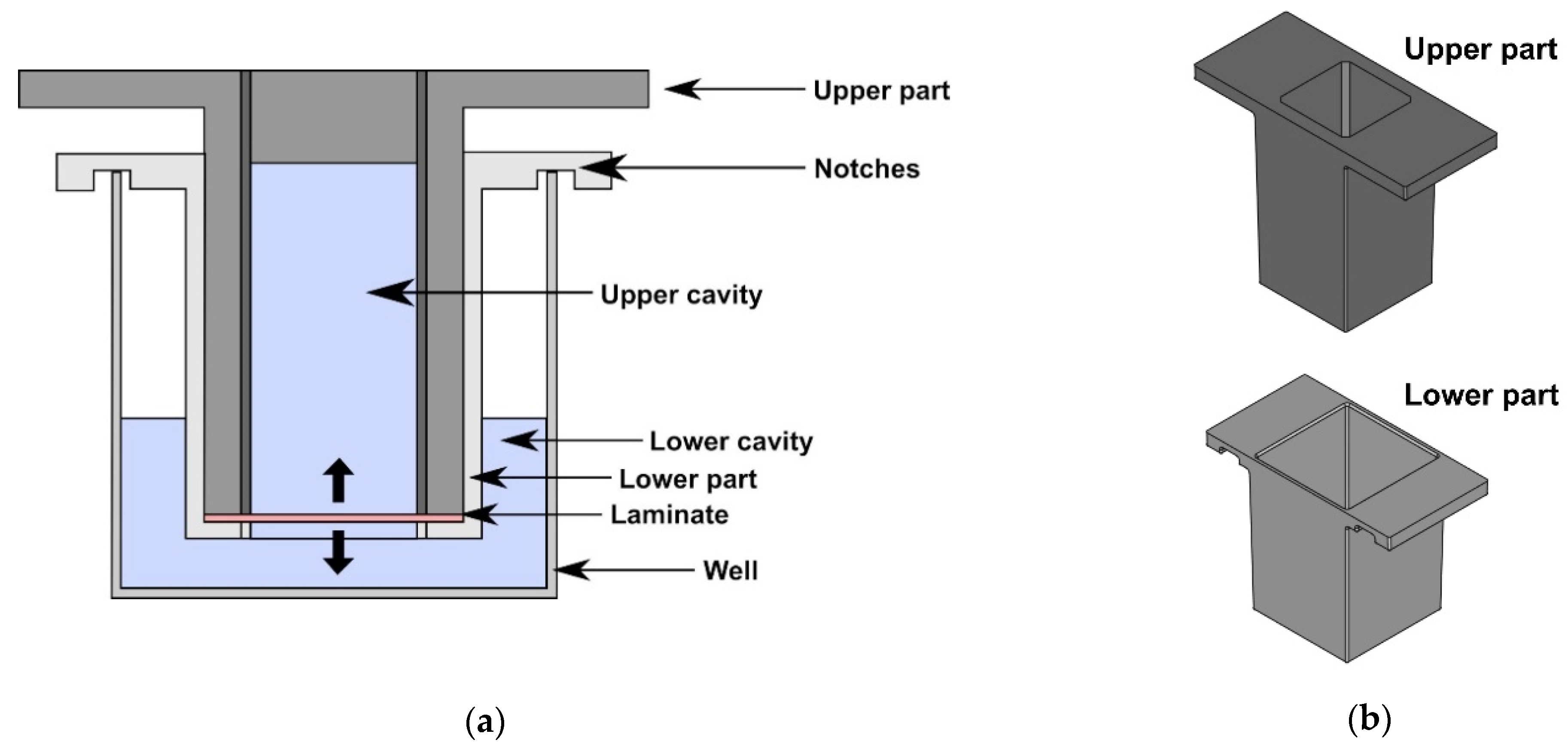
2.1. Fabrication of the Sample Holder
2.2. Fabrication of Collagen Laminates
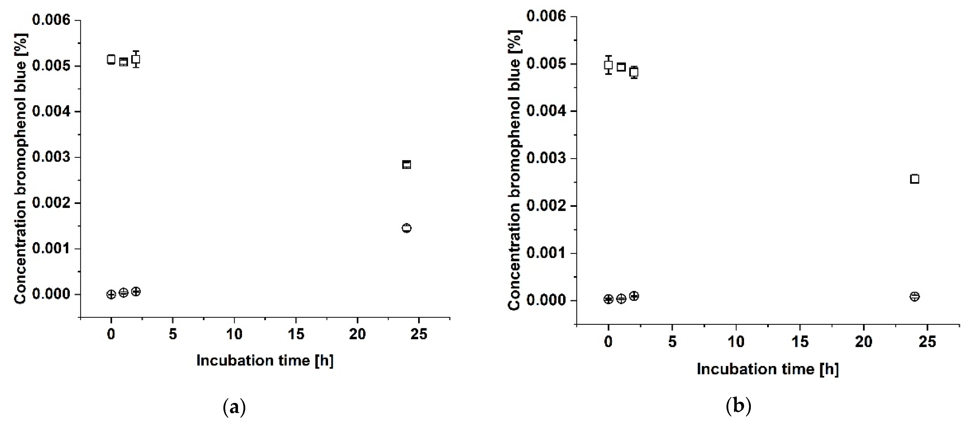
2.3. Proof of Tightness
2.4. Release of Vancomycin
3. Results
3.1. Development of an Additively Manufactured Sample Holder for Release Experiments
3.2. Proof of Tightness
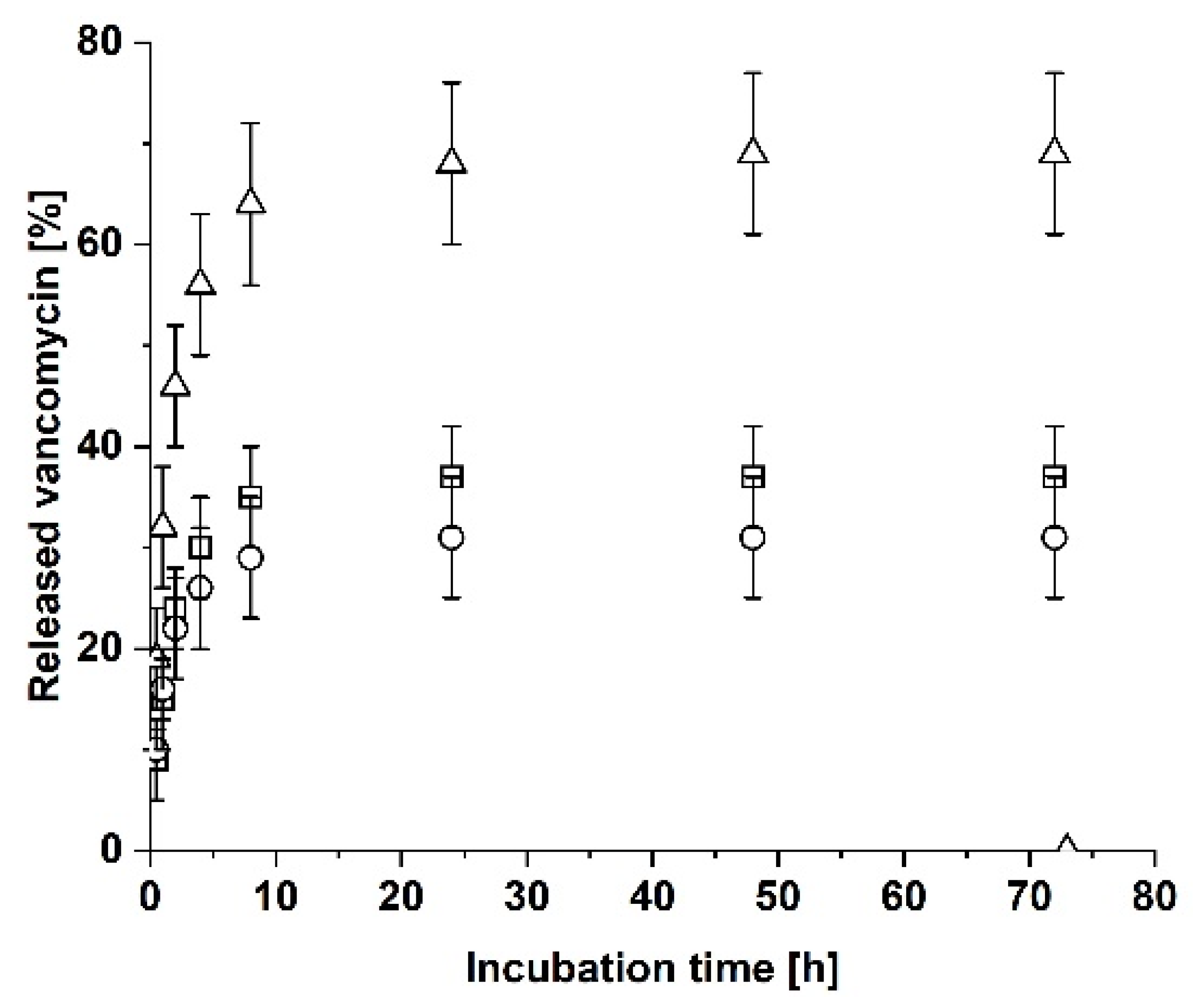
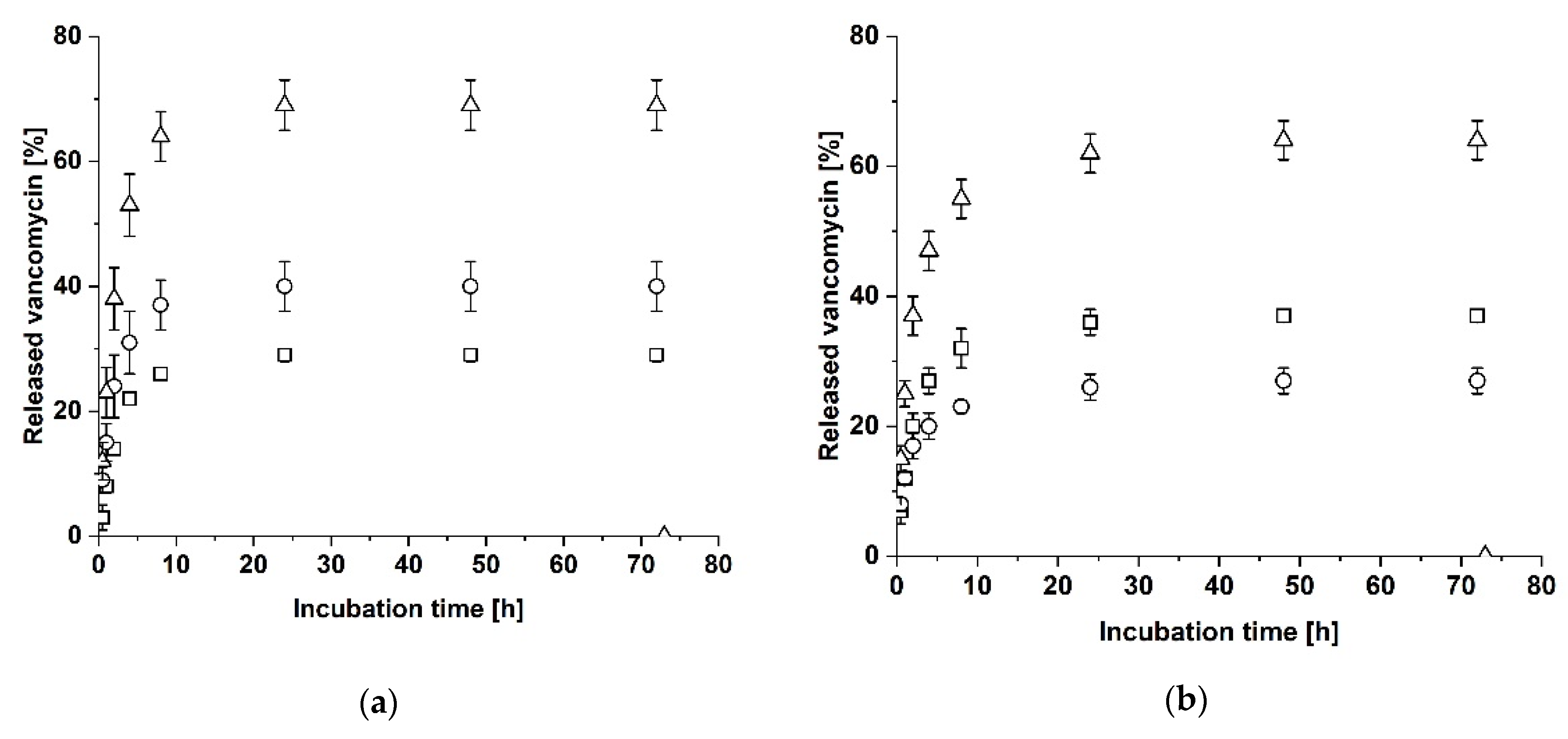
3.3. Release of Vancomycin
4. Discussion
4.1. Proof of Tightness
4.2. Release of Vancomycin
5. Conclusions
6. Patents
Supplementary Materials
Author Contributions
Funding
Institutional Review Board Statement
Informed Consent Statement
Data Availability Statement
Acknowledgments
Conflicts of Interest
References
- Yuan, K.; Chen, H.-L. Obesity and surgical site infections risk in orthopedics: A meta-analysis. Int. J. Surg. 2013, 11, 383–388. [Google Scholar] [CrossRef] [PubMed] [Green Version]
- Horan, T.C.; Gaynes, R.P.; Martone, W.J.; Jarvis, W.R.; Emori, T.G. CDC Definitions of Nosocomial Surgical Site Infections, 1992: A Modification of CDC Definitions of Surgical Wound Infections. Infect. Control Hosp. Epidemiol. 1992, 13, 606–608. [Google Scholar] [CrossRef] [PubMed]
- Abudula, T.; Gauthaman, K.; Mostafavi, A.; Alshahrie, A.; Salah, N.; Morganti, P.; Chianese, A.; Tamayol, A.; Memic, A. Sustainable drug release from polycaprolactone coated chitin-lignin gel fibrous scaffolds. Sci. Rep. 2020, 10, 20428. [Google Scholar] [CrossRef]
- Chen, A.F.; Fleischman, A.; Austin, M.S. Use of Intrawound Antibiotics in Orthopaedic Surgery. J. Am. Acad. Orthop. Surg. 2018, 26, e371–e378. [Google Scholar] [CrossRef]
- Braun, J.; Eckes, S.; Rommens, P.M.; Schmitz, K.; Nickel, D.; Ritz, U. Toxic Effect of Vancomycin on Viability and Functionality of Different Cells Involved in Tissue Regeneration. Antibiotics 2020, 9, 238. [Google Scholar] [CrossRef] [PubMed]
- Kumar, T.S.; Madhumathi, K. Antibiotic delivery by nanobioceramics. Ther. Deliv. 2016, 7, 573–588. [Google Scholar] [CrossRef] [PubMed]
- Snoddy, B.; Jayasuriya, A.C. The use of nanomaterials to treat bone infections. Mater. Sci. Eng. C Mater. Biol. Appl. 2016, 67, 822–833. [Google Scholar] [CrossRef] [PubMed] [Green Version]
- Chang, W.K.; Srinivasa, S.; MacCormick, A.D.; Hill, A.G. Gentamicin-collagen implants to reduce surgical site infection: Systematic review and meta-analysis of randomized trials. Ann. Surg. 2013, 258, 59–65. [Google Scholar] [CrossRef]
- Alt, V. Antimicrobial coated implants in trauma and orthopaedics-A clinical review and risk-benefit analysis. Injury 2017, 48, 599–607. [Google Scholar] [CrossRef] [PubMed]
- Lu, H.; Liu, Y.; Guo, J.; Wu, H.; Wang, J.; Wu, G. Biomaterials with Antibacterial and Osteoinductive Properties to Repair Infected Bone Defects. Int. J. Mol. Sci. 2016, 17, 334. [Google Scholar] [CrossRef]
- Bistolfi, A.; Massazza, G.; Verné, E.; Massè, A.; Deledda, D.; Ferraris, S.; Miola, M.; Galetto, F.; Crova, M. Antibiotic-loaded cement in orthopedic surgery: A review. ISRN Orthop. 2011, 2011, 290851. [Google Scholar] [CrossRef] [PubMed]
- Anagnostakos, K.; Kelm, J. Enhancement of antibiotic elution from acrylic bone cement. J. Biomed. Mater. Res. B Appl. Biomater. 2009, 90, 467–475. [Google Scholar] [CrossRef] [PubMed]
- Chiang, H.-Y.; Herwaldt, L.A.; Blevins, A.E.; Cho, E.; Schweizer, M.L. Effectiveness of local vancomycin powder to decrease surgical site infections: A meta-analysis. Spine J. 2014, 14, 397–407. [Google Scholar] [CrossRef]
- Lopez, N.A.; Luengo, C.V.; Avena, M.J. Uptake/release of vancomycin on/from Mg–Al layered double hydroxides. Adsorption 2019, 25, 1349–1360. [Google Scholar] [CrossRef]
- Hassani Besheli, N.; Mottaghitalab, F.; Eslami, M.; Gholami, M.; Kundu, S.C.; Kaplan, D.L.; Farokhi, M. Sustainable Release of Vancomycin from Silk Fibroin Nanoparticles for Treating Severe Bone Infection in Rat Tibia Osteomyelitis Model. ACS Appl. Mater. Interfaces 2017, 9, 5128–5138. [Google Scholar] [CrossRef] [PubMed]
- Suchý, T.; Šupová, M.; Klapková, E.; Adamková, V.; Závora, J.; Žaloudková, M.; Rýglová, Š.; Ballay, R.; Denk, F.; Pokorný, M.; et al. The release kinetics, antimicrobial activity and cytocompatibility of differently prepared collagen/hydroxyapatite/vancomycin layers: Microstructure vs. nanostructure. Eur. J. Pharm. Sci. 2017, 100, 219–229. [Google Scholar] [CrossRef]
- Hartinger, J.M.; Lukáč, P.; Mitáš, P.; Mlček, M.; Popková, M.; Suchý, T.; Šupová, M.; Závora, J.; Adámková, V.; Benáková, H.; et al. Vancomycin-releasing cross-linked collagen sponges as wound dressings. Bosn. J. Basic Med. Sci. 2021, 21, 61–70. [Google Scholar] [CrossRef] [PubMed] [Green Version]
- Ricard-Blum, S. The collagen family. Cold Spring Harb. Perspect. Biol. 2011, 3, a004978. [Google Scholar] [CrossRef] [PubMed] [Green Version]
- Parenteau-Bareil, R.; Gauvin, R.; Berthod, F. Collagen-Based Biomaterials for Tissue Engineering Applications. Materials 2010, 3, 1863–1887. [Google Scholar] [CrossRef] [Green Version]
- Davison-Kotler, E.; Marshall, W.S.; García-Gareta, E. Sources of Collagen for Biomaterials in Skin Wound Healing. Bioengineering 2019, 6, 56. [Google Scholar] [CrossRef] [PubMed] [Green Version]
- Shoulders, M.D.; Raines, R.T. Collagen structure and stability. Annu. Rev. Biochem. 2009, 78, 929–958. [Google Scholar] [CrossRef] [PubMed] [Green Version]
- Olde Damink, L.H.H.; Dijkstra, P.J.; van Luyn, M.J.A.; van Wachem, P.B.; Nieuwenhuis, P.; Feijen, J. Glutaraldehyde as a crosslinking agent for collagen-based biomaterials. J. Mater. Sci. Mater. Med. 1995, 6, 460–472. [Google Scholar] [CrossRef] [Green Version]
- Rafat, M.; Li, F.; Fagerholm, P.; Lagali, N.S.; Watsky, M.A.; Munger, R.; Matsuura, T.; Griffith, M. PEG-stabilized carbodiimide crosslinked collagen-chitosan hydrogels for corneal tissue engineering. Biomaterials 2008, 29, 3960–3972. [Google Scholar] [CrossRef] [PubMed]
- Weadock, K.S.; Miller, E.J.; Bellincampi, L.D.; Zawadsky, J.P.; Dunn, M.G. Physical crosslinking of collagen fibers: Comparison of ultraviolet irradiation and dehydrothermal treatment. J. Biomed. Mater. Res. 1995, 29, 1373–1379. [Google Scholar] [CrossRef]
- Eckes, S.; Braun, J.; Wack, J.S.; Ritz, U.; Nickel, D.; Schmitz, K. Rose Bengal Crosslinking to Stabilize Collagen Sheets and Generate Modulated Collagen Laminates. Int. J. Mol. Sci. 2020, 21, 7408. [Google Scholar] [CrossRef]
- Bergeron, M.G. Tissue penetration of antibiotics. Clin. Biochem. 1986, 19, 90–100. [Google Scholar] [CrossRef]
- Bracaglia, L.; Sharma, P.; Fisher, J.P. Polymer-Tissue Hybrid Biomaterials and Methods of Making and Using Same. U.S. Patent No. US201514869193 20150929, 29 September 2015. [Google Scholar]
- Campbell, P.G.; Weiss, L.E.; Smith, J. Biocompatible Polymers and Methods of Use. U.S. Patent No. US20060495115 20060728, 28 July 2006. [Google Scholar]
- Drohan, W.N.; Macphee, M.J.; Miekka, S.I.; Singh, M.S.; Elson, C.; Taylor, J.R. Chitin Hydrogels, Methods of Their Production and Use. U.S. Patent No. US19970960555 19971013, 13 October 1997. [Google Scholar]
- Shah, N.J.; Hong, J.; Hyder, N.; Hammond, P.T. Composition and Methods for Coating. U.S. Patent No. US201313746902 20130122, 22 January 2013. [Google Scholar]
- Wen, X.; Kirkwood, K.L. Methods and Compositions for Temporal Release of Agents from a Biodegradable Scaffold. U.S. Patent No. US20100856299 20100813, 13 August 2010. [Google Scholar]
- Vanerio, N.; Stijnen, M.; de Mol, B.A.J.M.; Kock, L.M. Biomedical Applications of Photo- and Sono-Activated Rose Bengal: A Review. Photobiomodul. Photomed. Laser Surg. 2019, 37, 383–394. [Google Scholar] [CrossRef] [PubMed] [Green Version]
- Alexander, W. American society of clinical oncology, 2010 annual meeting and rose bengal: From a wool dye to a cancer therapy. Pharm. Ther. 2010, 35, 469–478. [Google Scholar]
- Davies, G. The Rose Bengal test. Vet. Rec. 1971, 88, 447–449. [Google Scholar] [CrossRef] [PubMed]
- Chen, H.-C. Boyden chamber assay. Methods Mol. Biol. 2005, 294, 15–22. [Google Scholar] [CrossRef]
- Hadjipanayi, E.; Ananta, M.; Binkowski, M.; Streeter, I.; Lu, Z.; Cui, Z.F.; Brown, R.A.; Mudera, V. Mechanisms of structure generation during plastic compression of nanofibrillar collagen hydrogel scaffolds: Towards engineering of collagen. J. Tissue Eng. Regen. Med. 2011, 5, 505–519. [Google Scholar] [CrossRef] [PubMed]
- Kihara, T.; Ito, J.; Miyake, J. Measurement of biomolecular diffusion in extracellular matrix condensed by fibroblasts using fluorescence correlation spectroscopy. PLoS ONE 2013, 8, e82382. [Google Scholar] [CrossRef] [PubMed]
- Leddy, H.A.; Haider, M.A.; Guilak, F. Diffusional anisotropy in collagenous tissues: Fluorescence imaging of continuous point photobleaching. Biophys. J. 2006, 91, 311–316. [Google Scholar] [CrossRef] [Green Version]
- Tsintou, M.; Dalamagkas, K.; Seifalian, A. Injectable Hydrogel versus Plastically Compressed Collagen Scaffold for Central Nervous System Applications. Int. J. Biomater. 2018, 2018, 3514019. [Google Scholar] [CrossRef] [PubMed]

| Laminate or Single Collagen Sheet | Amount of Released Vancomycin | Additional Information | ||
|---|---|---|---|---|
| Name | Property | After 24 h | Half-Maximal Release | |
| A | RGX-modified | 68 ± 8% | 32 ± 6% (1 h) | Equal release, no additional release after incubation without sample holder |
| AC | Vancomycin-loaded A faces upper cavity | 69 ± 4% | 38 ± 5% (2 h) | Unequal release (C > A), no additional release after incubation without sample holder |
| AC | Vancomycin-loaded A faces lower cavity | 62 ± 3% | 37 ± 3% (2 h) | Unequal release (C > A), no additional release after incubation without sample holder |
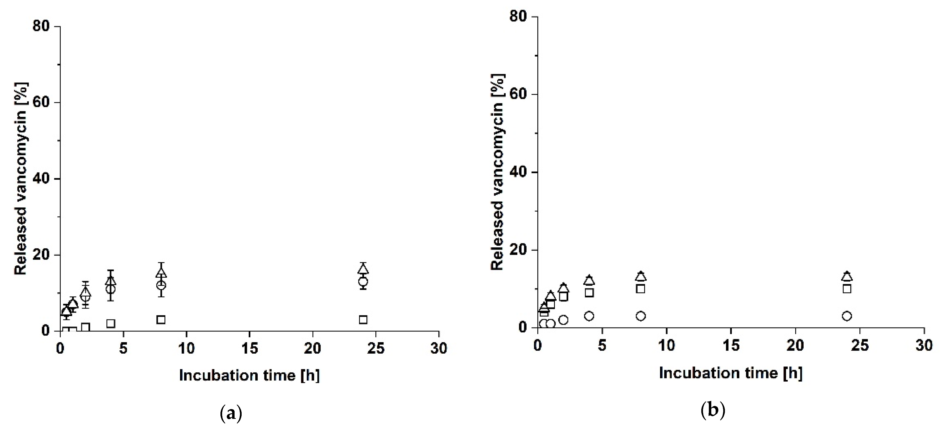
| AC | Vancomycin-loaded C faces lower cavity | 16 ± 2% | 7 ± 2% (1 h) | Unequal release (C > A) |
| AC | Vancomycin-loaded C faces upper cavity | 13 ± 1% | 8 ± 1% (1 h) | Unequal release (C > A) |
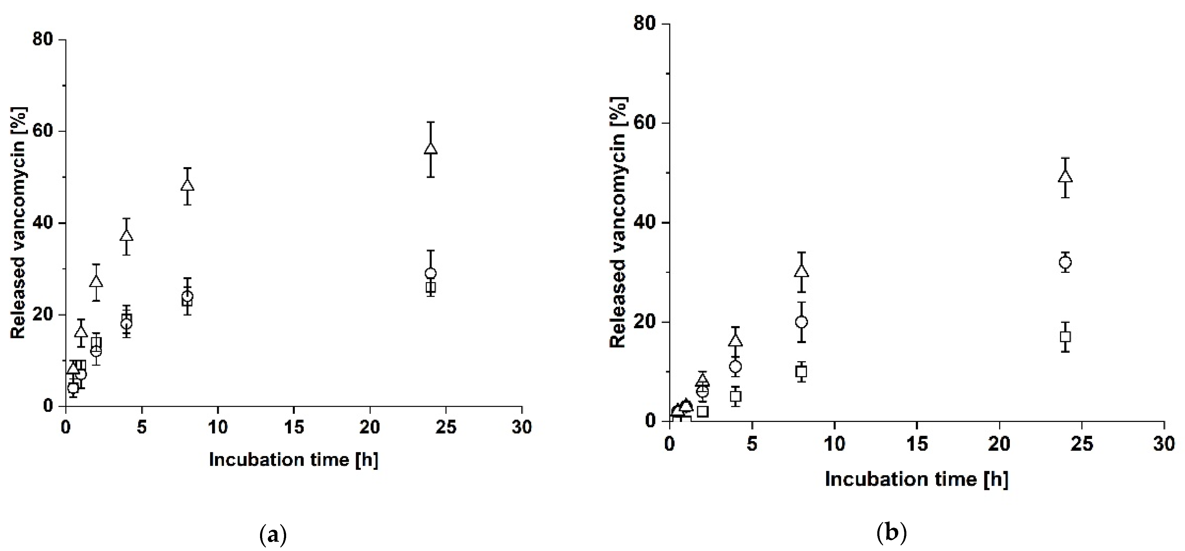
| CAC | Vancomycin-loaded A as central layer | 56 ± 6% | 27 ± 4% (2 h) | Equal release |
| AAA | Vancomycin-loaded A as central layer | 49 ± 4% | 30 ± 4% (8 h) | Unequal release (lower cavity > upper cavity) |
Publisher’s Note: MDPI stays neutral with regard to jurisdictional claims in published maps and institutional affiliations. |
© 2021 by the authors. Licensee MDPI, Basel, Switzerland. This article is an open access article distributed under the terms and conditions of the Creative Commons Attribution (CC BY) license (https://creativecommons.org/licenses/by/4.0/).
Share and Cite
Kilb, M.F.; Moos, Y.; Eckes, S.; Braun, J.; Ritz, U.; Nickel, D.; Schmitz, K. An Additively Manufactured Sample Holder to Measure the Controlled Release of Vancomycin from Collagen Laminates. Biomedicines 2021, 9, 1668. https://doi.org/10.3390/biomedicines9111668
Kilb MF, Moos Y, Eckes S, Braun J, Ritz U, Nickel D, Schmitz K. An Additively Manufactured Sample Holder to Measure the Controlled Release of Vancomycin from Collagen Laminates. Biomedicines. 2021; 9(11):1668. https://doi.org/10.3390/biomedicines9111668
Chicago/Turabian StyleKilb, Michelle Fiona, Yannik Moos, Stefanie Eckes, Joy Braun, Ulrike Ritz, Daniela Nickel, and Katja Schmitz. 2021. "An Additively Manufactured Sample Holder to Measure the Controlled Release of Vancomycin from Collagen Laminates" Biomedicines 9, no. 11: 1668. https://doi.org/10.3390/biomedicines9111668
APA StyleKilb, M. F., Moos, Y., Eckes, S., Braun, J., Ritz, U., Nickel, D., & Schmitz, K. (2021). An Additively Manufactured Sample Holder to Measure the Controlled Release of Vancomycin from Collagen Laminates. Biomedicines, 9(11), 1668. https://doi.org/10.3390/biomedicines9111668