Article

Osteopontin Gene Polymorphisms Are Associated with Cardiovascular Risk Factors in Patients with Premature Coronary Artery Disease

by
Nonanzit Pérez-Hernández
1,
Rosalinda Posadas-Sánchez
2,
Gilberto Vargas-Alarcón
1,
Lizet Paola Hernández-Germán
1,
Verónica Marusa Borgonio-Cuadra
3 and
José Manuel Rodríguez-Pérez
1,*
1
Department of Molecular Biology, Instituto Nacional de Cardiología Ignacio Chávez, Mexico City 14080, Mexico
2
Department of Endocrinology, Instituto Nacional de Cardiología Ignacio Chavez, Mexico City 14080, Mexico
3
Department of Genomic Medicine, Instituto Nacional de Rehabilitación Luis Guillermo Ibarra, Mexico City 14389, Mexico
*
Author to whom correspondence should be addressed.
Biomedicines 2021, 9(11), 1600; https://doi.org/10.3390/biomedicines9111600
Submission received: 23 September 2021 / Revised: 30 October 2021 / Accepted: 31 October 2021 / Published: 2 November 2021
(This article belongs to the Special Issue 30 Years of OPN Milestones and Future Avenues)

Abstract

:
Osteopontin (OPN) is considered a clinical predictor of cardiovascular disease. We aimed to evaluate the association of the OPN gene polymorphisms rs2728127 and rs11730582 with the development of premature coronary artery disease (pCAD), cardiovascular risk factors, and cardiometabolic parameters. We evaluated 1142 patients with pCAD and 1073 controls. Both polymorphisms were determined by Taqman assays. Similar allele and genotype frequencies were observed in both groups; additionally, an association of these polymorphisms with CAD and cardiometabolic parameters was observed in both groups. In patients with pCAD, the rs11730582 was associated with a high risk of hypoadiponectinemia (OR = 1.300, P additive = 0.003), low risk of hypertension (OR = 0.709, P codominant 1 = 0.030), and low risk of having high non-HDL cholesterol (OR = 0.637, P additive = 0.038). In the control group, the rs2728127 was associated with a low risk of fatty liver (OR = 0.766, P additive = 0.038); while the rs11730582 was associated with a low risk of hypoadiponectinemia (OR = 0.728, P dominant = 0.022), and risk of having elevated apolipoprotein B (OR = 1.400, P dominant = 0.031). Our results suggest that in Mexican individuals, the rs11730582 and rs2728127 OPN gene polymorphisms are associated with some abnormal metabolic variables in patients with pCAD and controls.

1. Introduction

Coronary artery disease (CAD) is a complex and multifactorial pathology characterized by chronic vascular inflammation. In this disease, the accumulation of lipid material within the layers of arteries causes endothelium damage, and as a final consequence, the formation of atherosclerotic plaques [1,2,3]. During the progression of the disease, some plaques are calcified mainly by deposits of calcium phosphate [2,4]. Therefore, vascular calcification and coronary artery calcium (CAC) are of clinical relevance as risk markers for CAD [5,6,7]. Several studies have reported the importance of cardiac calcification, and now this process is considered an important marker to early detection of CAD, therefore useful in primary preventive care and treatment of CAD [8,9,10].
Osteopontin (OPN) is an extracellular matrix protein, with diverse functions, including growth factor, structural matrix protein, modulator of matrix-cell interactions, among others. Besides, due to the different post-translational modifications, OPN participate in several biological processes, mainly in the regulation of calcification, biomineralization and proinflammatory response [11,12,13,14]. The human OPN gene is located on chromosome 4 (4q21-23) and is formed by seven exons [13]. This protein has pleiotropic effects, and it is expressed in several cell types such as osteocytes, osteoblasts, pre-osteoblasts, macrophages, T-cells, endothelial cells and fibroblasts, and in different tissues including bone, cartilage, vascular tissue, brain and kidney, with essential physiological and pathophysiological functions [11,13,14,15]. OPN also plays a preponderant role in different pathologies such as autoimmune disorders, chronic inflammatory diseases, distinct types of cancer, diabetes, cardiovascular diseases, among others [15,16]. Under physiological conditions, low circulating OPN levels as well as low OPN expression in vasculature are associated with various pathologies, for instance, diabetes or non-small cell lung cancer [17,18]. Under injury conditions, OPN is over-expressed and upregulated in different vascular cell types including macrophages, vascular smooth muscle cells and endothelial cells [11]. Furthermore, various studies have detected increased circulating serum levels of OPN in cardiovascular diseases, associated particularly with severity of coronary atherosclerosis; high plasma levels of OPN, associated with increased risk for cardiac events; and a high expression of OPN gene in calcified carotid atheroma [10,19,20,21,22]. In the last decade, genetic studies have helped to identify predisposition markers in the development of CAD; the single nucleotide polymorphisms (SNPs) have been reported as genetic markers. Particularly, the OPN gene contains various polymorphic sites in the promoter region, but despite the important participation of the OPN molecule in the physiopathology of cardiovascular diseases, few association studies between this gene and vascular diseases have been performed. Mainly, there have been studies on coronary artery calcification or large artery atherosclerosis; nonetheless, there is only one previous study of OPN gene polymorphisms with CAD [23,24,25].
The human OPN gene is located on chromosome 4 (4q21-23) and is formed by seven exons [13]. This gene contains various polymorphic sites, and despite the important participation of the OPN molecule in the physiopathology of cardiovascular diseases, few association studies between this gene and vascular diseases have been performed. Therefore, more studies in different populations are needed to establish the possible role of this gene in the susceptibility to develop CAD.
Thus, the purpose of this research was to analyze the association of OPN gene polymorphisms (rs2728127 and rs11730582) with the risk of developing premature coronary artery disease (pCAD), cardiovascular risk factors, and cardiometabolic parameters in a representative sample of Mexican mestizo individuals. In addition, we performed bioinformatic analyses to predict the possible functional role of these polymorphisms.

2. Materials and Methods

2.1. Study Individuals

This is a cross-sectional and case-control study of individuals included in the Genetics of Atherosclerotic Disease (GEA) Mexican study. We included 2215 unrelated participants (1142 patients with pCAD and 1073 healthy controls). All participants were consecutively recruited during 2008 to 2013. For the present research, we considered as pCAD the incidence of a clinical coronary event before 55 years of age in men and 65 years in women [26,27]. The patient group was integrated by individuals diagnosed with pCAD established through a previous clinical history of myocardial infarction, angioplasty, revascularization surgery, or through the presence of coronary stenosis >50%, within the age limits indicated above.
In the control group, we included individuals without a personal or family history of CAD. Individuals from the blood bank at the Instituto Nacional de Cardiología Ignacio Chavez (INCICh) in Mexico were invited to participate as controls. Additionally, through brochures posted in social service centers in Mexico City and the INCICh, more individuals were gathered. The exclusion criteria in this group were: thyroid disorders, renal or heart failure, oncological and liver diseases.

2.2. Clinical, Demographic, Biochemical Variables Assessment

Demographic, clinical and biochemical data of participants from the GEA study had been previously reported [28,29,30,31,32,33]. Briefly, body mass index (BMI) was obtained as: weight (kg)/height (m2), waist circumference was obtained in cm, and obesity was defined when the BMI was ≥30 kg/m2. Type 2 diabetes mellitus (T2DM) was defined according to the American Diabetes Association criteria, with a fasting glucose ≥126 mg/dL and was also considered when participants informed the use of hypoglycemic drug treatment. Hypertension was considered when values of diastolic and systolic blood pressures were ≥90 mmHg and ≥140 mmHg, respectively, or when individuals were using antihypertensive drugs. Glucose, apolipoprotein B (ApoB), apolipoprotein A-I, lipoprotein (a) and lipid profiles were quantified using conventional enzymatic colorimetric techniques in a Hitachi model 902 autoanalyzer (Hitachi LTD, Tokio, Japan). Hypoalphalipoproteinemia was considered when HDL-cholesterol <50 mg/dL in women, and <40 mg/dL in men. Smoking habits were determined when the individual self-reported a current use of cigarettes.
The high-sensibility C-reactive protein was measured by immunonephelometry (BN ProSpec Nephelometer, Date Behring, Marburgo, Germany). Levels of adiponectin were quantified with ELISA protocols (Quantikine ELISA, R&D Systems Inc., Minneapolis, MN, USA); specifically, hypoadiponectinemia was defined when adiponectin concentration was ≤25th percentile (5.30 μg/mL in men and 8.67 μg/mL in women). High alkaline phosphatase concentrations were defined when values were ≥75th percentile (83.00 IU/L in men and 90.25 IU/L in women). Percentile values of adiponectin and alkaline phosphatase were acquired from a representative subsample of the GEA participants [34,35].

2.3. Tomographic Evaluation

Visceral abdominal adipose tissue (VAT), liver and spleen attenuation, as well as coronary artery calcium (CAC) were quantified using the computed axial tomography system of 64 channel multidetector (Somaton Sensations, Siemens, Malvern, PA, USA). Expert radiologists measured and interpreted the scans. Scans were read to determine CAC scores using the Agatston method [36]. The specific area of VAT was determined following the description by Kvist et al. [37]. The liver to spleen attenuation ratio was determined in accordance with the report by Longo et al. [38]. Individuals in the control group with evidence of subclinical atherosclerosis were excluded from this study (CAC with a value greater than zero).

2.4. Estimation of Ancestry

All GEA project participants included (cases and controls) were unrelated and were ethnically matched and of self-reported Mexican mestizo ancestry. A Mexican mestizo is defined as someone born in Mexico, who is descendant from the original autochthonous inhabitants of the region and/or from Caucasian and/or African origin, who came to America during the sixteenth century. Further, we determined the genetic background of the GEA participants on the basis of a 265-ancestry markers (AIMs) panel that distinguishes Amerindian, European, and African ancestries [39]. This was assessed with specialized software and the mean global ancestry did not show significant differences in the population analyzed (55.8% vs. 54.0% Amerindian ancestry, 34.3% vs. 35.8% Caucasian ancestry and 9.8% vs. 10.1% African ancestry for patients and controls respectively, with a p > 0.05). Therefore, all GEA participants had a similar genetic background and thus, the population stratification was no genetic bias in the present research [40].

2.5. SNPs Selection and Genetic Characterization

For this research, we chose polymorphic sites with a minor allele frequency (MAF) of at least 5% percent reported in the International HapMap Project. Furthermore, the chosen polymorphisms have been previously associated with some cardiovascular pathologies and are located in the promotor region of the OPN gene.
Genomic DNA was obtained from peripheral blood leukocyte samples using the QIAamp DNA blood extraction kit (Qiagen, Germany). We analyzed two polymorphisms: rs2728127, ID: C___1840806_10, sequence AAATTTTGTTGTTTTTAGAATTTTC[A/G]GACTTCCCTCCACTAAATTGACAAC, and rs11730582, ID: C___1840808_20, sequence GAGTAGTAAAGGACAGAGGCAAGTT[T/C]TCTGAACTCCTTGCAGGCTTGAAC. Both polymorphisms were determined using Taqman genotyping probes on a thermal cycler (ABI Prism 7900-Real Time) according to the manufacture’s conditions (ThermoFisher Scientific, Foster City, CA, USA).

2.6. Bioinformatic Analysis

To determine the possible functional effect of OPN gene polymorphisms, we used the transcription factor affinity prediction (TRAP) software, available as a web server (http://trap.molgen.mpg.de/, accessed on 23 September 2021), specialized in predicting the transcription factor binding affinities to DNA sequences according to the specific polymorphisms [41].

2.7. Statistical Analysis

Both studied polymorphisms were in Hardy-Weinberg equilibrium (p > 0.05). Data are presented as median (interquartile range), mean (standard deviation) or frequencies as required. The analysis of categorical and continuous variables in the population studied was performed using chi-square, Student t, and Mann-Whitney U tests. The genetic associations of both polymorphisms with pCAD were evaluated using logistic regression analysis using the inheritance models: additive, dominant, recessive, heterozygote, codominant 1 and codominant 2. These models were adjusted by age, sex, body mass index, systolic blood pressure, diastolic blood pressure, visceral and abdominal fat, triglycerides, apolipoprotein B, serum calcium, adiponectin, and alkaline phosphatase activity. The associations of the OPN gene polymorphisms with cardiovascular risk factors and metabolic abnormalities were adjusted by sex, age and body mass index. All statistical analyses were performed using the SPSS software, version 24.0. Values of p < 0.05 were considered significant in this study.

3. Results

3.1. Evaluation of Anthropometric, Clinical and Metabolic Parameters and Prevalence of Cardiovascular Risk Factors

Anthropometric, clinical and metabolic variables of the population studied are shown in Table 1. Compared with the control group, patients with pCAD showed higher values for: body mass index, waist circumference, systolic blood pressure, visceral adipose tissue, triglycerides and glucose. In addition, prevalence of hypoalphalipoproteinemia, hypertriglyceridemia, obesity, hypertension, visceral abdominal adipose tissue and hypoadiponectinemia were also higher in patients with pCAD when compared with the control group (Table 2).

3.2. Association of rs2728127 and rs11730582 Polymorphisms with pCAD

A similar distribution of the two polymorphisms was observed in patients with pCAD and controls (Table 3). The inheritance models were adjusted for age, sex, body mass index, systolic and diastolic blood pressure, visceral adipose tissue, concentrations of triglycerides, apolipoprotein B, serum calcium, adiponectin and alkaline phosphatase activity.

3.3. Association of rs2728127 and rs11730582 Polymorphisms with Cardiovascular Risk Factors and Metabolic Parameters

The association of OPN gene polymorphisms with cardiovascular risk factors and metabolic parameters was assessed separately in patients with pCAD and controls. In patients with pCAD and under different inheritance models, we observed that rs11730582 was associated with an increased risk of hypoadiponectinemia (OR = 1.300, 95% CI = 1.096–1.543, P additive = 0.003; OR = 1.462, 95% CI = 1.117–1.912, P dominant = 0.006; OR = 1.375, 95% CI = 1.027–1.840, P recessive = 0.032; OR = 1.371, 95% CI = 1.301–1.824, P codominant 1 = 0.031; OR = 1.678, 95% CI = 1.191–2.365, P codominant 2 = 0.003), with a decreased risk of hypertension (OR = 0.728, 95% CI = 0.543–0.976, P dominant = 0.034; OR = 0.709, 95% CI = 0.519–0.967, P codominant 1 = 0.030), and low risk of having increased levels of non- HDL cholesterol (OR = 0.803, 95% CI = 0.652–0.989, P additive = 0.039; OR = 0.637, 95% CI = 0.415–0.976, P codominant 2 = 0.038). These outcomes are depicted in Figure 1.
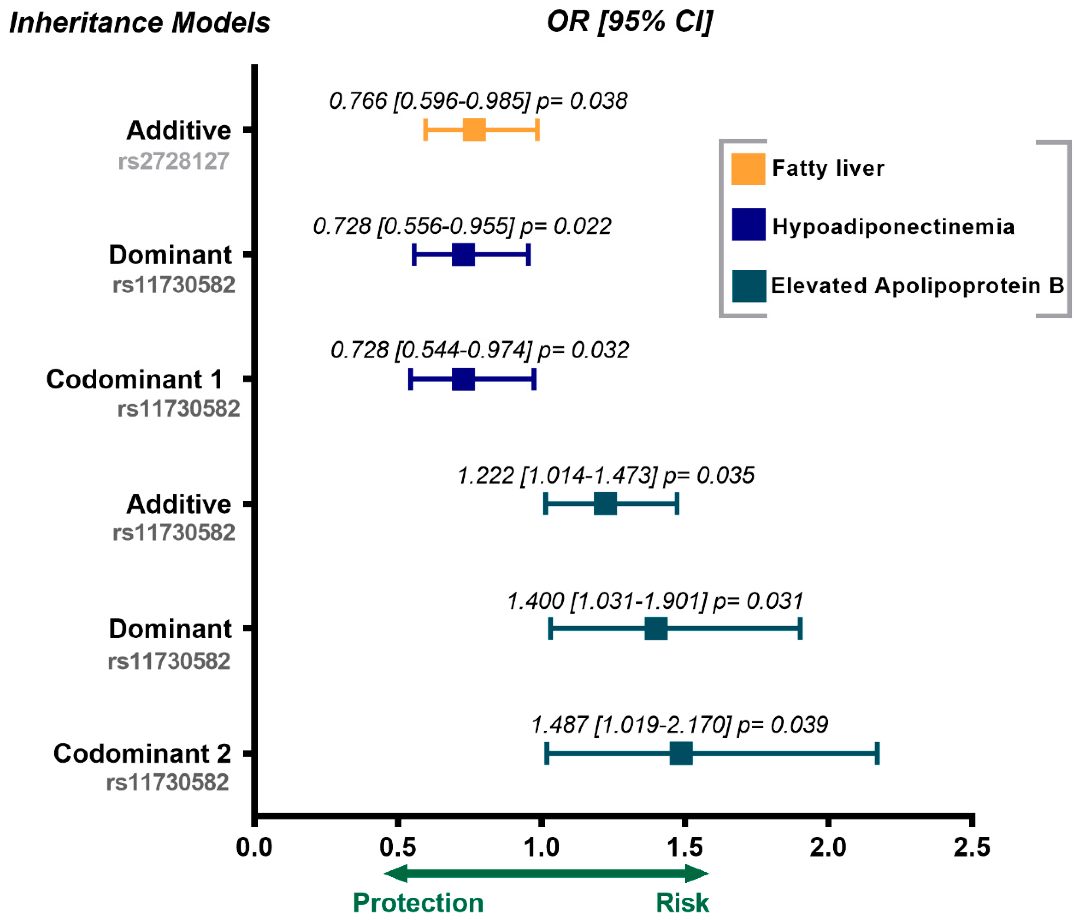
In the control group, the rs2728127 polymorphism was associated with a low risk of fatty liver (OR = 0.766, 95% CI = 0.596–0.985, P additive = 0.038). The rs11730582 polymorphism was associated with a lower risk of hypoadiponectinemia (OR = 0.728, 95% CI = 0.556–9.555, P dominant = 0.022; OR = 0.728, 95% CI = 0.544–0.974, P codominant 1 = 0.032), and high risk of having increased apolipoprotein B levels (OR = 1.222, 95% CI = 1.014–1.473, P additive = 0.035; OR = 1.400, 95% CI = 1.031–1.901, P dominant = 0.031; OR = 1.487, 95% CI = 1.019–2.170, P codominant 2 = 0.039). These results are shown in Figure 2. All genetic models were adjusted for age, sex, and body mass index.

4. Discussion

Despite the importance of OPN as a clinical predictor of cardiovascular disease, to date, few genetic association studies have been performed to determine its involvement as a risk marker for coronary artery disease. This genetic approach is necessary in order to have a better understanding of this complex and multifactorial disease. Thus, in this research, we evaluated two polymorphisms (rs2728127 and rs11730582) of OPN gene, in 1073 healthy individuals and 1142 patients with pCAD, in order to establish their association with the presence of pCAD, cardiovascular risk factors and cardiometabolic parameters in a Mexican population. This study was nested in the GEA Project, a prospective and one of the largest cohorts in Mexican population.
Our results indicate that the studied polymorphisms were not associated with the presence of pCAD. This outcome agrees with the report of Lin et al., who evaluated the rs11730582 polymorphism in 536 patients with CAD, 86 patients with peripheral artery disease (PAD) and 617 controls and did not find a significant association between this polymorphism and the presence of CAD [24]. In the same line, Hou et al., studied the association between four OPN gene polymorphisms (including, rs11730582) and left ventricular hypertrophy (LVH) in 1092 patients with essential hypertension; they did not find significant differences between the groups analyzed [41]. However, Jing et al., reported that rs11730582 polymorphism was associated with an increased risk of ischemic stroke [42]. The discrepancies found between the report by Jing et al. and our study could be due to different aspects: (a) Jing et al., included only 377 patients, while we analyzed 1142 individuals with pCAD; (b) Jing et al., studied patients with ischemic stroke (IS) which is a type of vascular disease, but is not specifically CAD, as in our report; (c) the study by Jing et al., was conducted in an Asian population, while our research was performed in Mexican mestizo population [43,44,45].
The OPN gene polymorphisms have been associated with several pathologies including Crohn’s disease [46], urolithiasis [47,48], knee osteoarthritis [49], breast cancer [50] and diabetic nephropathy [51]. As far as we know, only the study performed by Lin et al., and ours have analyzed the association between OPN gene polymorphisms and CAD. Any other previous research was performed evaluating non-cardiovascular pathologies. Therefore, the association of the OPN genetic variants with susceptibility to cardiovascular diseases remains to a great extent unknown.
We also analyzed the association of rs2728127 and rs11730582 polymorphisms with cardiovascular risk factors and metabolic parameters in both groups. In patients with pCAD, the rs11730582 polymorphism was associated with an increased risk of hypoadiponectinemia. Adiponectin is an essential protein secreted by adipocytes; currently, this adipokine is a marker for several metabolic pathologies and atherosclerotic cardiovascular disease [52,53]. For instance, Hui et al., reported that hypoadiponectinemia is related to the progression of carotid atherosclerosis [54]; while Di Chiara et al., found that hypoadiponectinemia was associated with an increased risk of left ventricular [55].
Additionally, the rs11730582 polymorphism was associated with a lower risk of hypertension, and a decreased risk of having high non-HDL-cholesterol levels. This outcome seems contradictory; nonetheless, it is important to mention that patients with pCAD included in our study received lipid-lowering and antihypertensive therapies, which probably explains these results [56,57,58,59]. Therefore, more studies are needed in this field to elucidate the complete role of this biomolecule.
In our control group, the rs2728127 polymorphism was associated with a decreased risk of having fatty liver. Evidence suggests that OPN plays an essential role in liver diseases [60,61], including fatty liver, which is linked to atherosclerotic cardiovascular pathology [62], as well as subclinical atherosclerosis [63]. Recently, this molecule has also been associated with the presence of cardiac arrhythmias [64]. On the other hand, the rs11730582 polymorphism was associated with a low risk of having hypoadiponectinemia. Some studies have analyzed adiponectin plasma levels in apparently healthy adults and found that hypoadiponectinemia was associated with cardiovascular risk factors leading to a pro-atherogenic condition [65,66,67].
Also in the control group, we detected an association of rs11730582 polymorphisms with a risk of having high apolipoprotein B concentrations. ApoB has been related to dyslipidemia processes and it is considered a significant predictor of cardiovascular diseases such as myocardial infarction [68]. In this sense, our control group was not under a lipid-lowering treatment, this was probably why we found an association between the rs11730582 polymorphism and elevated ApoB levels [69].
It is important to consider that genetic variation reported in other populations may not extrapolate to the Mexican genetic background, i.e., Europeans are closely related, and their genome have had fewer recombination events than Mexicans. Various genetic studies of the Mexican population have reported the proportion of Indian and White genes is 56% and 44%, respectively, in the dihybrid model and 56%, 41% and 4% from Indian, White and Blacks in the trihybrid model [44,70,71,72]. Therefore, the analysis of OPN polymorphisms warrants other studies in populations with different genetic backgrounds.
Another important point to consider in the genetic variability is the Hardy-Weinberg Equilibrium (HWE) in the population analyzed. However, different factors such as a small number of participants can alter the HWE, thus influencing the inappropriate distribution of genotypes in the population. For this reason, we have included an important number of patients that warrants the HWE and supports that the population of cases and controls was ethnically well-matched.
The in-silico analysis showed that variations in the rs2728127 polymorphism could generate binding sites for the heat shock transcription factor 1 (HSF1) and the Kappa-light-chain enhancer of activated B-cells (NFκB). However, both rs2728127 and rs11730582 polymorphisms produced binding sites for the tumor suppressor Trp53 (P53) and all of them were implicated in vascular disorders. HSF1 is a transcriptional factor that interacts with heat shock element (HSE), a specific regulatory element located in promoter regions of the heat shock protein (HSP) genes [73]. The abnormal expression of HSF1 can promote cardiac injury in vascular pathologies; however, HSF1 has an important role in cellular protection from stress conditions [74]. NFκB is a nuclear transcription factor that regulates differential gene expressions affecting a broad spectrum of biological reactions including inflammation states, migration, apoptosis and cell proliferation [75,76,77]. With regards to P53, this transcription factor has a critical regulatory role in several essential genes associated with transactivation and transrepression processes. P53 has been related to angiogenesis, a process with important effects in cardiovascular diseases such as heart failure, myocardial ischemia/reperfusion, and atherosclerosis among others [78].
Some limitations in our research should be taken into consideration. First, we did not measure circulating levels of OPN in the participants; therefore, we did not establish if there were different concentrations of this protein between groups. For this reason, the association between OPN gene polymorphisms and the concentration of OPN was not reported. Second, we could not verify with experimental evidence if the polymorphisms had a functional impact, as we just used an in-silico approach. Third, most of the coronary risk factors were different between patients and controls; even if they were considered to adjust the statistical analyses, the possibility remains of a lower contribution of OPN gene polymorphisms to pCAD than that reported in this study. Nonetheless, our report also has significant strengths: (1) we used a large cohort of Mexican individuals with and without pCAD, with demographic, clinical, tomographic and biochemical variables; (2) our control group only included individuals without subclinical atherosclerosis (CAC score = zero), so there was no bias in selecting the comparison group. As far as we know, this is the first study to report the associations OPN gene with other cardiovascular risk factors.

5. Conclusions

Our results suggest an association of OPN gene polymorphisms with metabolic abnormalities and cardiovascular risk factors in Mexican patients with pCAD and healthy controls. These findings support the participation of OPN polymorphisms as metabolic markers in our population.

Author Contributions

Conceptualization, N.P.-H. and J.M.R.-P.; methodology, L.P.H.-G. and V.M.B.-C.; formal analysis, R.P.-S.; resources, N.P.-H. and J.M.R.-P.; visualization and supervision, N.P.-H. and J.M.R.-P.; project administration, J.M.R.-P.; funding acquisition, J.M.R.-P.; data curation, R.P.-S. and V.M.B.-C.; writing-original draft preparation, N.P.-H., J.M.R.-P., G.V.-A.; writing-review and editing, N.P.-H., J.M.R.-P., G.V.-A. All authors have read and agreed to the published version of the manuscript.

Funding

This study was financed using the budget given to the Genomics Laboratory of the Instituto Nacional de Cardiología Ignacio Chávez (INCICh) for scientific research. Additionally, the INCICh supported this research with an open access publication.

Institutional Review Board Statement

The study was conducted according to the guidelines of the Declaration of Helsinki, and approved by the Ethics and Research Committee of the INCICh Instituto Nacional de Cardiología “Ignacio Chávez” (09-646 and 1 September 2008).

Informed Consent Statement

Informed consent was obtained from all subjects involved in the study.

Data Availability Statement

The data shown in this article are available upon request from the corresponding author.

Acknowledgments

All researchers in this study are grateful to every participant of the GEA project and every work team of the Endocrinology and Molecular Biology Services for the support received in this study. Also, part of these data was obtained by Lizet Paola Hernández-Germán as part of her thesis to obtain a master’s degree in Clinical Biochemistry from the Faculty of Chemistry of the Universidad Nacional Autónoma de México. Finally, all authors are grateful to the technicians Marva Arellano-González and Silvestre Ramírez-Fuentes for the support received.

Conflicts of Interest

The authors declare no conflict of interest.

References

  1. Carresi, C.; Mollace, R.; Macrì, R.; Scicchitano, M.; Bosco, F.; Scarano, F.; Coppoletta, A.R.; Guarnieri, L.; Ruga, S.; Zito, M.C.; et al. Oxidative Stress Triggers Defective Autophagy in Endothelial Cells: Role in Atherothrombosis Development. Antioxidants 2021, 10, 387. [Google Scholar] [CrossRef]
  2. Libby, P.; Buring, J.E.; Badimon, L.; Hansson, G.K.; Deanfield, J.; Bittencourt, M.S.; Tokgözoğlu, L.; Lewis, E.F. Atherosclerosis. Nat. Reviews. Dis. Primers 2019, 5, 56. [Google Scholar] [CrossRef]
  3. Yuvaraj, J.; Cheng, K.; Lin, A.; Psaltis, P.J.; Nicholls, S.J.; Wong, D.T.L. The Emerging Role of CT-Based Imaging in Adipose Tissue and Coronary Inflammation. Cells 2021, 10, 1196. [Google Scholar] [CrossRef]
  4. Hutcheson, J.D.; Maldonado, N.; Aikawa, E. Small entities with large impact: Microcalcifications and atherosclerotic plaque vulnerability. Curr. Opin. Lipidol. 2014, 25, 327–332. [Google Scholar] [CrossRef] [Green Version]
  5. Chua, A.; Blankstein, R.; Ko, B. Coronary artery calcium in primary prevention. Aust. J. Gen. Pract. 2020, 49, 464–469. [Google Scholar] [CrossRef] [PubMed]
  6. Faggiano, A.; Santangelo, G.; Carugo, S.; Pressman, G.; Picano, E.; Faggiano, P. Cardiovascular Calcification as a Marker of Increased Cardiovascular Risk and a Surrogate for Subclinical Atherosclerosis: Role of Echocardiography. J. Clin. Med. 2021, 10, 1668. [Google Scholar] [CrossRef]
  7. Reinhold, S.; Blankesteijn, W.M.; Foulquier, S. The Interplay of WNT and PPARγ Signaling in Vascular Calcification. Cells 2020, 9, 658. [Google Scholar] [CrossRef]
  8. Berezin, A.E.; Kremzer, A.A. Circulating osteopontin as a marker of early coronary vascular calcification in type two diabetes mellitus patients with known asymptomatic coronary artery disease. Atherosclerosis 2013, 229, 475–481. [Google Scholar] [CrossRef]
  9. Kosmopoulos, M.; Paschou, S.A.; Grapsa, J.; Anagnostis, P.; Vryonidou, A.; Goulis, D.G.; Siasos, G. The Emerging Role of Bone Markers in Diagnosis and Risk Stratification of Patients With Coronary Artery Disease. Angiology 2019, 70, 690–700. [Google Scholar] [CrossRef]
  10. Mohamadpour, A.H.; Abdolrahmani, L.; Mirzaei, H.; Sahebkar, A.; Moohebati, M.; Ghorbani, M.; Ferns, G.A.; Ghayour-Mobarhan, M. Serum osteopontin concentrations in relation to coronary artery disease. Arch. Med. Res. 2015, 46, 112–117. [Google Scholar] [CrossRef]
  11. Lok, Z.S.Y.; Lyle, A.N. Osteopontin in Vascular Disease. Arterioscler. Thromb. Vasc. Biol. 2019, 39, 613–622. [Google Scholar] [CrossRef] [Green Version]
  12. Shirakawa, K.; Sano, M. Osteopontin in Cardiovascular Diseases. Biomolecules 2021, 11, 1047. [Google Scholar] [CrossRef]
  13. Subraman, V.; Thiyagarajan, M.; Malathi, N.; Rajan, S.T. OPN -Revisited. J. Clin. Diagn. Res. JCDR 2015, 9, Ze10–Ze13. [Google Scholar] [CrossRef] [PubMed]
  14. Vianello, E.; Kalousová, M.; Dozio, E.; Tacchini, L.; Zima, T.; Corsi Romanelli, M.M. Osteopontin: The Molecular Bridge between Fat and Cardiac-Renal Disorders. Int. J. Mol. Sci. 2020, 21, 5568. [Google Scholar] [CrossRef] [PubMed]
  15. Kahles, F.; Findeisen, H.M.; Bruemmer, D. Osteopontin: A novel regulator at the cross roads of inflammation, obesity and diabetes. Mol. Metab. 2014, 3, 384–393. [Google Scholar] [CrossRef] [PubMed]
  16. Icer, M.A.; Gezmen-Karadag, M. The multiple functions and mechanisms of osteopontin. Clin. Biochem. 2018, 59, 17–24. [Google Scholar] [CrossRef]
  17. Eleftheriadou, I.; Tsilingiris, D.; Tentolouris, A.; Mourouzis, I.; Grigoropoulou, P.; Kapelios, C.; Pantos, C.; Makrilakis, K.; Tentolouris, N. Association of Circulating Osteopontin Levels With Lower Extremity Arterial Disease in Subjects With Type 2 Diabetes Mellitus: A Cross-Sectional Observational Study. Int. J. Low. Extrem. Wounds 2020, 19, 180–189. [Google Scholar] [CrossRef]
  18. Mack, P.C.; Redman, M.W.; Chansky, K.; Williamson, S.K.; Farneth, N.C.; Lara, P.N., Jr.; Franklin, W.A.; Le, Q.T.; Crowley, J.J.; Gandara, D.R. Lower osteopontin plasma levels are associated with superior outcomes in advanced non-small-cell lung cancer patients receiving platinum-based chemotherapy: SWOG Study S0003. J. Clin. Oncol. Off. J. Am. Soc. Clin. Oncol. 2008, 26, 4771–4776. [Google Scholar] [CrossRef] [Green Version]
  19. Carbone, F.; Rigamonti, F.; Burger, F.; Roth, A.; Bertolotto, M.; Spinella, G.; Pane, B.; Palombo, D.; Pende, A.; Bonaventura, A.; et al. Serum levels of osteopontin predict major adverse cardiovascular events in patients with severe carotid artery stenosis. Int. J. Cardiol. 2018, 255, 195–199. [Google Scholar] [CrossRef]
  20. Gürses, K.M.; Yalçın, M.U.; Koçyiğit, D.; Beşler, M.S.; Canpınar, H.; Evranos, B.; Yorgun, H.; Şahiner, M.L.; Kaya, E.B.; Özer, N.; et al. The association between serum angiogenin and osteopontin levels and coronary collateral circulation in patients with chronic total occlusion. Anatol. J. Cardiol. 2019, 22, 77–84. [Google Scholar] [CrossRef]
  21. Higgins, C.L.; Isbilir, S.; Basto, P.; Chen, I.Y.; Vaduganathan, M.; Vaduganathan, P.; Reardon, M.J.; Lawrie, G.; Peterson, L.; Morrisett, J.D. Distribution of alkaline phosphatase, osteopontin, RANK ligand and osteoprotegerin in calcified human carotid atheroma. Protein J. 2015, 34, 315–328. [Google Scholar] [CrossRef]
  22. Wolak, T. Osteopontin—A multi-modal marker and mediator in atherosclerotic vascular disease. Atherosclerosis 2014, 236, 327–337. [Google Scholar] [CrossRef] [PubMed]
  23. Kim, Y.; Lee, C. Haplotype analysis revealed a genetic influence of osteopontin on large artery atherosclerosis. J. Biomed. Sci. 2008, 15, 529–533. [Google Scholar] [CrossRef] [PubMed]
  24. Lin, J.F.; Wu, S.; Juang, J.J.; Chiang, F.T.; Hsu, L.A.; Teng, M.S.; Cheng, S.T.; Huang, H.L.; Ko, Y.L. Osteoprotegerin and osteopontin levels, but not gene polymorphisms, predict mortality in cardiovascular diseases. Biomark. Med. 2019, 13, 751–760. [Google Scholar] [CrossRef]
  25. Taylor, B.C.; Schreiner, P.J.; Doherty, T.M.; Fornage, M.; Carr, J.J.; Sidney, S. Matrix Gla protein and osteopontin genetic associations with coronary artery calcification and bone density: The CARDIA study. Hum. Genet. 2005, 116, 525–528. [Google Scholar] [CrossRef] [PubMed]
  26. Arnett, D.K.; Blumenthal, R.S.; Albert, M.A.; Buroker, A.B.; Goldberger, Z.D.; Hahn, E.J.; Himmelfarb, C.D.; Khera, A.; Lloyd-Jones, D.; Mcevoy, J.W.; et al. 2019 ACC/AHA Guideline on the Primary Prevention of Cardiovascular Disease: A Report of the American College of Cardiology/American Heart Association Task Force on Clinical Practice Guidelines. J. Am. Coll. Cardiol. 2019, 74, e177–e232. [Google Scholar] [CrossRef]
  27. Mahtta, D.; Ramsey, D.J.; Al Rifai, M.; Nasir, K.; Samad, Z.; Aguilar, D.; Jneid, H.; Ballantyne, C.M.; Petersen, L.A.; Virani, S.S. Evaluation of Aspirin and Statin Therapy Use and Adherence in Patients With Premature Atherosclerotic Cardiovascular Disease. JAMA Netw. Open 2020, 3, e2011051. [Google Scholar] [CrossRef]
  28. Acuña-Valerio, J.; Rodas-Díaz, M.A.; Macias-Garrido, E.; Posadas-Sánchez, R.; Juárez-Rojas, J.G.; Medina-Urrutia, A.X.; Cardoso-Saldaña, G.C.; Joge-Galarza, E.; Torres-Tamayo, M.; Vargas-Alarcón, G.; et al. Aortic valve calcification prevalence and association with coronary risk factors and atherosclerosis in Mexican population. Arch. Cardiol. Mex. 2017, 87, 108–115. [Google Scholar] [CrossRef]
  29. Cardoso-Saldaña, G.C.; Medina-Urrutia, A.X.; Posadas-Romero, C.; Juárez-Rojas, J.G.; Jorge-Galarza, E.; Vargas-Alarcón, G.; Posadas-Sánchez, R. Fatty liver and abdominal fat relationships with high C-reactive protein in adults without coronary heart disease. Ann. Hepatol. 2015, 14, 658–665. [Google Scholar] [CrossRef]
  30. López-Bautista, F.; Posadas-Sánchez, R.; Vázquez-Vázquez, C.; Fragoso, J.M.; Rodríguez-Pérez, J.M.; Vargas-Alarcón, G. IL-37 Gene and Cholesterol Metabolism: Association of Polymorphisms with the Presence of Hypercholesterolemia and Cardiovascular Risk Factors. The GEA Mexican Study. Biomolecules 2020, 10, 1409. [Google Scholar] [CrossRef]
  31. Posadas-Romero, C.; López-Bautista, F.; Rodas-Díaz, M.A.; Posadas-Sánchez, R.; Kimura-Hayama, E.; Juárez-Rojas, J.G.; Medina-Urrutia, A.X.; Cardoso-Saldaña, G.C.; Vargas-Alarcón, G.; Jorge-Galarza, E. Prevalence and extent of coronary artery calcification in an asymptomatic cardiovascular Mexican population: Genetics of Atherosclerotic Disease study. Arch. Cardiol. Mex. 2017, 87, 292–301. [Google Scholar] [CrossRef]
  32. Posadas-Sánchez, R.; Pérez-Hernández, N.; Angeles-Martínez, J.; López-Bautista, F.; Villarreal-Molina, T.; Rodríguez-Pérez, J.M.; Fragoso, J.M.; Posadas-Romero, C.; Vargas-Alarcón, G. Interleukin 35 Polymorphisms Are Associated with Decreased Risk of Premature Coronary Artery Disease, Metabolic Parameters, and IL-35 Levels: The Genetics of Atherosclerotic Disease (GEA) Study. Mediat. Inflamm. 2017, 2017, 6012795. [Google Scholar] [CrossRef] [PubMed]
  33. Rodríguez-Pérez, J.M.; Posadas-Sánchez, R.; Blachman-Braun, R.; Vargas-Alarcón, G.; Posadas-Romero, C.; Rodríguez-Cortés, A.A.; López-Bautista, F.; Tovilla-Zárate, C.A.; Rojas-Toledo, E.X.; Borgonio-Cuadra, V.M.; et al. HHIPL-1 (rs2895811) gene polymorphism is associated with cardiovascular risk factors and cardiometabolic parameters in Mexicans patients with myocardial infarction. Gene 2018, 663, 34–40. [Google Scholar] [CrossRef] [PubMed]
  34. López-Bautista, F.; Posadas-Romero, C.; Ruiz-Vargas, L.Y.; Cardoso-Saldaña, G.; Juárez-Rojas, J.G.; Medina-Urrutia, A.; Pérez-Hernández, N.; Rodríguez-Pérez, J.M.; Vargas-Alarcón, G.; Posadas-Sánchez, R. Vitamin D Deficiency is not Associated with Fatty Liver in a Mexican Population. Ann. Hepatol. 2018, 17, 419–425. [Google Scholar] [CrossRef]
  35. Zamarrón-Licona, E.; Rodríguez-Pérez, J.M.; Posadas-Sánchez, R.; Vargas-Alarcón, G.; Baños-González, M.A.; Borgonio-Cuadra, V.M.; Pérez-Hernández, N. Variants of PCSK9 Gene Are Associated with Subclinical Atherosclerosis and Cardiometabolic Parameters in Mexicans. The GEA Project. Diagnostics 2021, 11, 774. [Google Scholar] [CrossRef]
  36. Agatston, A.S.; Janowitz, W.R.; Hildner, F.J.; Zusmer, N.R.; Viamonte, M., Jr.; Detrano,, R. Quantification of coronary artery calcium using ultrafast computed tomography. J. Am. Coll. Cardiol. 1990, 15, 827–832. [Google Scholar] [CrossRef] [Green Version]
  37. Kvist, H.; Chowdhury, B.; Grangård, U.; Tylén, U.; Sjöström, L. Total and visceral adipose-tissue volumes derived from measurements with computed tomography in adult men and women: Predictive equations. Am. J. Clin. Nutr. 1988, 48, 1351–1361. [Google Scholar] [CrossRef]
  38. Longo, R.; Ricci, C.; Masutti, F.; Vidimari, R.; Crocé, L.S.; Bercich, L.; Tiribelli, C.; Dalla Palma, L. Fatty infiltration of the liver. Quantification by 1H localized magnetic resonance spectroscopy and comparison with computed tomography. Investig. Radiol. 1993, 28, 297–302. [Google Scholar] [CrossRef]
  39. Silva-Zolezzi, I.; Hidalgo-Miranda, A.; Estrada-Gil, J.; Fernandez-Lopez, J.C.; Uribe-Figueroa, L.; Contreras, A.; Balam-Ortiz, E.; Del Bosque-Plata, L.; Velazquez-Fernandez, D.; Lara, C.; et al. Analysis of genomic diversity in Mexican Mestizo populations to develop genomic medicine in Mexico. Proc. Natl. Acad. Sci. USA 2009, 106, 8611–8616. [Google Scholar] [CrossRef] [Green Version]
  40. Posadas-Sánchez, R.; Pérez-Hernández, N.; Rodríguez-Pérez, J.M.; Coral-Vázquez, R.M.; Roque-Ramírez, B.; Llorente, L.; Lima, G.; Flores-Dominguez, C.; Villarreal-Molina, T.; Posadas-Romero, C.; et al. Interleukin-27 polymorphisms are associated with premature coronary artery disease and metabolic parameters in the Mexican population: The genetics of atherosclerotic disease (GEA) Mexican study. Oncotarget 2017, 8, 64459–64470. [Google Scholar] [CrossRef] [Green Version]
  41. Hou, X.; Hu, Z.; Huang, X.; Chen, Y.; He, X.; Xu, H.; Wang, N. Serum osteopontin, but not OPN gene polymorphism, is associated with LVH in essential hypertensive patients. J. Mol. Med. 2014, 92, 487–495. [Google Scholar] [CrossRef]
  42. Jing, M.; Li, B.; Hou, X.; Shoba, J.; Li, C.; Liang, H.; Zhang, X.; Liu, E.; Yang, B.; Meng, X. OPN gene polymorphism and the serum OPN levels confer the susceptibility and prognosis of ischemic stroke in Chinese patients. Cell. Physiol. Biochem. Int. J. Exp. Cell. Physiol. Biochem. Pharmacol. 2013, 32, 1798–1807. [Google Scholar] [CrossRef]
  43. Juárez-Cedillo, T.; Zuñiga, J.; Acuña-Alonzo, V.; Pérez-Hernández, N.; Rodríguez-Pérez, J.M.; Barquera, R.; Gallardo, G.J.; Sánchez-Arenas, R.; García-Peña Mdel, C.; Granados, J.; et al. Genetic admixture and diversity estimations in the Mexican Mestizo population from Mexico City using 15 STR polymorphic markers. Forensic Sci. Int. Genet. 2008, 2, e37–e39. [Google Scholar] [CrossRef] [PubMed]
  44. Lisker, R.; Ramírez, E.; Babinsky, V. Genetic structure of autochthonous populations of Meso-America: Mexico. Hum. Biol. 1996, 68, 395–404. [Google Scholar] [PubMed]
  45. Rubi-Castellanos, R.; Martínez-Cortés, G.; Muñoz-Valle, J.F.; González-Martín, A.; Cerda-Flores, R.M.; Anaya-Palafox, M.; Rangel-Villalobos, H. Pre-Hispanic Mesoamerican demography approximates the present-day ancestry of Mestizos throughout the territory of Mexico. Am. J. Phys. Anthropol. 2009, 139, 284–294. [Google Scholar] [CrossRef]
  46. Glas, J.; Seiderer, J.; Bayrle, C.; Wetzke, M.; Fries, C.; Tillack, C.; Olszak, T.; Beigel, F.; Steib, C.; Friedrich, M.; et al. The role of osteopontin (OPN/SPP1) haplotypes in the susceptibility to Crohn’s disease. PLoS ONE 2011, 6, e29309. [Google Scholar] [CrossRef]
  47. Amar, A.; Afzal, A.; Hameed, A.; Ahmad, M.; Khan, A.R.; Najma, H.; Abid, A.; Khaliq, S. Osteopontin promoter polymorphisms and risk of urolithiasis: A candidate gene association and meta-analysis study. BMC Med. Genet. 2020, 21, 172. [Google Scholar] [CrossRef] [PubMed]
  48. Safarinejad, M.R.; Shafiei, N.; Safarinejad, S. Association between polymorphisms in osteopontin gene (SPP1) and first episode calcium oxalate urolithiasis. Urolithiasis 2013, 41, 303–313. [Google Scholar] [CrossRef]
  49. Shang, H.; Hao, Y.; Hu, W.; Hu, X.; Jin, Q. OPN gene locus is associated with the risk of knee osteoarthritis: A case-control study. Biosci. Rep. 2019, 39, BSR20182023. [Google Scholar] [CrossRef] [Green Version]
  50. Liang, L.; Lu, G.; Pan, G.; Deng, Y.; Liang, J.; Liang, L.; Liu, J.; Tang, Y.; Wei, G. A Case-Control Study of the Association Between the SPP1 Gene SNPs and the Susceptibility to Breast Cancer in Guangxi, China. Front. Oncol. 2019, 9, 1415. [Google Scholar] [CrossRef]
  51. Cheema, B.S.; Iyengar, S.; Sharma, R.; Kohli, H.S.; Bhansali, A.; Khullar, M. Association between Osteopontin Promoter Gene Polymorphisms and Haplotypes with Risk of Diabetic Nephropathy. J. Clin. Med. 2015, 4, 1281–1292. [Google Scholar] [CrossRef] [Green Version]
  52. Lim, S.; Quon, M.J.; Koh, K.K. Modulation of adiponectin as a potential therapeutic strategy. Atherosclerosis 2014, 233, 721–728. [Google Scholar] [CrossRef]
  53. Ramakrishnan, N.; Auger, K.; Jialal, I. Biochemistry, Adiponectin; StatPearls Publishing LLC: Treasure Island, FL, USA, 2021. [Google Scholar]
  54. Hui, E.; Xu, A.; Chow, W.S.; Lee, P.C.; Fong, C.H.; Cheung, S.C.; Tse, H.F.; Chau, M.T.; Cheung, B.M.; Lam, K.S. Hypoadiponectinemia as an independent predictor for the progression of carotid atherosclerosis: A 5-year prospective study. Metab. Syndr. Relat. Disord. 2014, 12, 517–522. [Google Scholar] [CrossRef] [Green Version]
  55. Di Chiara, T.; Argano, C.; Scaglione, A.; Duro, G.; Corrao, S.; Scaglione, R.; Licata, G. Hypoadiponectinemia, cardiometabolic comorbidities and left ventricular hypertrophy. Intern. Emerg. Med. 2015, 10, 33–40. [Google Scholar] [CrossRef] [Green Version]
  56. Brea, A.; Hernández-Mijares, A.; Millán, J.; Ascaso, J.F.; Blasco, M.; Díaz, A.; Mantilla, T.; Pedro-Botet, J.C.; Pintó, X. Non-HDL cholesterol as a therapeutic goal. Clínica E Investig. Arterioscler. 31, (Suppl. 2). 28–33. [CrossRef]
  57. Feingold, K.R. Cholesterol Lowering Drugs; Feingold, K.R., Anawalt, B., Boyce, A., Chrousos, G., De Herder, W.W., Dhatariya, K., Grossman, A., Hershman, J.M., Kaltsas, G., Koch, C., et al., Eds.; MDText.com, Inc.: South Dartmouth, MA, USA, 2000. [Google Scholar]
  58. Hsu, H.Y.; Lin, C.J.; Lee, Y.S.; Wu, T.H.; Chien, K.L. Efficacy of more intensive lipid-lowering therapy on cardiovascular diseases: A systematic review and meta-analysis. BMC Cardiovasc. Disord. 2020, 20, 334. [Google Scholar] [CrossRef]
  59. Masson, W.; Lobo, M.; Siniawski, D.; Molinero, G.; Huerín, M.; Nogueira, J.P. Impact of Lipid-Lowering Therapy on Mortality According to the Baseline Non-HDL Cholesterol Level: A Meta-Analysis. High Blood Press. Cardiovasc. Prev. Off. J. Ital. Soc. Hypertens. 2019, 26, 263–272. [Google Scholar] [CrossRef]
  60. Bruha, R.; Vitek, L.; Smid, V. Osteopontin—A potential biomarker of advanced liver disease. Ann. Hepatol. 2020, 19, 344–352. [Google Scholar] [CrossRef]
  61. Wen, Y.; Jeong, S.; Xia, Q.; Kong, X. Role of Osteopontin in Liver Diseases. Int. J. Biol. Sci. 2016, 12, 1121–1128. [Google Scholar] [CrossRef] [Green Version]
  62. Park, J.H.; Koo, B.K.; Kim, W.; Kim, W.H. Histological severity of nonalcoholic fatty liver disease is associated with 10-year risk for atherosclerotic cardiovascular disease. Hepatol. Int. 2021, 15, 1148–1159. [Google Scholar] [CrossRef]
  63. Hsiao, C.C.; Teng, P.H.; Wu, Y.J.; Shen, Y.W.; Mar, G.Y.; Wu, F.Z. Severe, but not mild to moderate, non-alcoholic fatty liver disease associated with increased risk of subclinical coronary atherosclerosis. BMC Cardiovasc. Disord. 2021, 21, 244. [Google Scholar] [CrossRef]
  64. Chen, Z.; Liu, J.; Zhou, F.; Li, H.; Zhang, X.J.; She, Z.G.; Lu, Z.; Cai, J.; Li, H. Nonalcoholic Fatty Liver Disease: An Emerging Driver of Cardiac Arrhythmia. Circ. Res. 2021, 128, 1747–1765. [Google Scholar] [CrossRef]
  65. Im, J.A.; Kim, S.H.; Lee, J.W.; Shim, J.Y.; Lee, H.R.; Lee, D.C. Association between hypoadiponectinemia and cardiovascular risk factors in nonobese healthy adults. Metab. Clin. Exp. 2006, 55, 1546–1550. [Google Scholar] [CrossRef] [PubMed]
  66. Kazumi, T.; Kawaguchi, A.; Hirano, T.; Yoshino, G. Serum adiponectin is associated with high-density lipoprotein cholesterol, triglycerides, and low-density lipoprotein particle size in young healthy men. Metab. Clin. Exp. 2004, 53, 589–593. [Google Scholar] [CrossRef]
  67. Komatsu, M.; Ohfusa, H.; Aizawa, T.; Hashizume, K. Adiponectin inversely correlates with high sensitive C-reactive protein and triglycerides, but not with insulin sensitivity, in apparently healthy Japanese men. Endocr. J. 2007, 54, 553–558. [Google Scholar] [CrossRef] [PubMed] [Green Version]
  68. Vlad, C.E.; Foia, L.; Popescu, R.; Ivanov, I.; Luca, M.C.; Delianu, C.; Toma, V.; Statescu, C.; Rezus, C.; Florea, L. Apolipoproteins A and B and PCSK9: Nontraditional Cardiovascular Risk Factors in Chronic Kidney Disease and in End-Stage Renal Disease. J. Diabetes Res. 2019, 2019, 6906278. [Google Scholar] [CrossRef] [PubMed]
  69. Climent, E.; Benaiges, D.; Pedro-Botet, J. Hydrophilic or Lipophilic Statins? Front. Cardiovasc. Med. 2021, 8, 687585. [Google Scholar] [CrossRef] [PubMed]
  70. Lisker-Yourkowitzky, R.; Ramírez-Arroyo, E.; Pérez-Rendon, G.; Díaz-Barrigo Pardo, R.; Siperstein-Blumovicz, M.; Mutchinick-Baringoltz, O. Genotypes of alcohol-metabolizing enzymes in Mexicans with alcoholic liver cirrhosis. Arch. Med. Res. 1995, 26, S63–S67. [Google Scholar]
  71. Lisker, R.; Pérez-Briceno, R.; Granados, J.; Babinsky, V. Gene frequencies and admixture estimates in the state of Puebla, Mexico. Am. J. Phys. Anthropol. 1988, 76, 331–335. [Google Scholar] [CrossRef]
  72. Rosenberg, N.A.; Huang, L.; Jewett, E.M.; Szpiech, Z.A.; Jankovic, I.; Boehnke, M. Genome-wide association studies in diverse populations. Nat. Rev. Genet. 2010, 11, 356–366. [Google Scholar] [CrossRef] [Green Version]
  73. Metzler, B.; Abia, R.; Ahmad, M.; Wernig, F.; Pachinger, O.; Hu, Y.; Xu, Q. Activation of heat shock transcription factor 1 in atherosclerosis. Am. J. Pathol. 2003, 162, 1669–1676. [Google Scholar] [CrossRef] [Green Version]
  74. Haybar, H.; Shahrabi, S.; Rezaeeyan, H.; Shirzad, R.; Saki, N. Protective role of heat shock transcription factor 1 in heart failure: A diagnostic approach. J. Cell. Physiol. 2019, 234, 7764–7770. [Google Scholar] [CrossRef] [PubMed]
  75. De Luca, F. Regulatory role of NF-κB in growth plate chondrogenesis and its functional interaction with Growth Hormone. Mol. Cell. Endocrinol. 2020, 514, 110916. [Google Scholar] [CrossRef] [PubMed]
  76. Fiordelisi, A.; Iaccarino, G.; Morisco, C.; Coscioni, E.; Sorriento, D. NFkappaB is a Key Player in the Crosstalk between Inflammation and Cardiovascular Diseases. Int. J. Mol. Sci. 2019, 20, 1599. [Google Scholar] [CrossRef] [Green Version]
  77. Zinatizadeh, M.R.; Schock, B.; Chalbatani, G.M.; Zarandi, P.K.; Jalali, S.A.; Miri, S.R. The Nuclear Factor Kappa B (NF-kB) signaling in cancer development and immune diseases. Genes Dis. 2021, 8, 287–297. [Google Scholar] [CrossRef]
  78. Men, H.; Cai, H.; Cheng, Q.; Zhou, W.; Wang, X.; Huang, S.; Zheng, Y.; Cai, L. The regulatory roles of p53 in cardiovascular health and disease. Cell. Mol. Life Sci. CMLS 2021, 78, 2001–2018. [Google Scholar] [CrossRef] [PubMed]
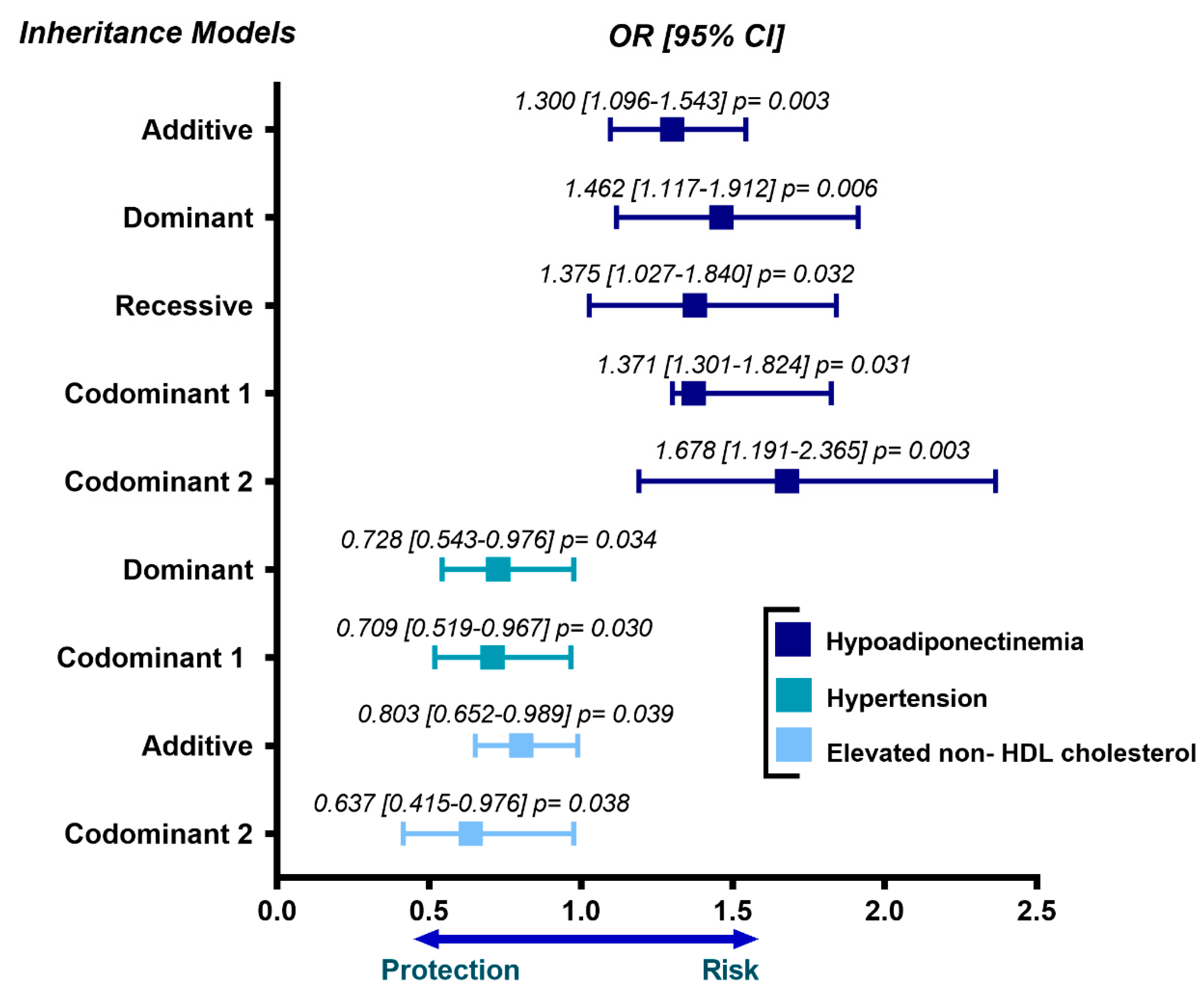
Figure 1. Association of rs11730582 OPN gene polymorphism with metabolic abnormalities in the premature coronary artery disease group. The inheritance models were adjusted for age, sex and body mass index. OR = odds ratio, CI = confidence interval.
Biomedicines 09 01600 g001
Figure 2. Association of OPN gene polymorphism with metabolic abnormalities in the control group. The inheritance models were adjusted for age, sex and body mass index. OR = odds ratio, CI = confidence interval.
Biomedicines 09 01600 g002
Table 1. Clinical and metabolic characteristics of the individuals studied.
CharacteristicsControl
(n = 1073)
pCAD
(n = 1142)
* p
Age (years)51 ± 954 ± 8<0.001
Sex (male %)42.380.8<0.001
Body mass index (kg/m2)27.9 [25.5–30.9]28.3 [25.9–31.1]0.007
Waist circumference (cm)94 ± 1198 ± 10<0.001
Systolic blood pressure (mmHg)113 [104–123]116 [106–127]<0.001
Diastolic blood pressure (mmHg)71 [65–77]71 [66–78]0.046
Visceral abdominal fat (cm2)141 [106–181]168 [129–218]<0.001
High-density lipoprotein cholesterol (mg/dL)45 [36–55]37 [32–44]<0.001
Low-density lipoprotein cholesterol (mg/dL)116 [95–134]91 [68–116]<0.001
Triglycerides (mg/dL)145 [108–202]162 [119–219]<0.001
Apolipoprotein B (mg/dL)94 [76–113]79 [63–102]<0.001
Apolipoprotein A (mg/dL)134 [115–156]120 [101–138]<0.001
Lipoprotein (a) (mg/dL)5.2 [2.3–11.5]4.8 [2.4–14.1]0.478
Glucose (mg/dL)90 [84–97]95 [87–117]<0.001
High-sensitivity C-reactive protein (mg/L)1.5 [0.8–3.1]1.2 [0.6–2.6]<0.001
Adiponectin (µg/mL)8.1 [5.0–12.8]5.2 [3.2–8.1]<0.001
Alkaline phosphatase (IU/L)81 [68–96]76 [63–95]<0.001
Serum calcium (mg/dL)9.7 ± 0.69.7 ± 0.70.278
Data are shown as mean ± standard deviation, median [interquartile range] or percentage. Student’s t-test, Mann Whitney U test or Chi-square test. pCAD = Premature Coronary Artery Disease. * p value.
Table 2. Prevalence of cardiovascular risk factors the individuals studied.
CharacteristicsControl
(n = 1073)
pCAD
(n = 1142)
* p
LDL-cholesterol ≥ 130 mg/dL (%)29.616.1<0.001
Hypoalphalipoproteinemia (%)52.166.9<0.001
Hypertriglyceridemia (%)47.556.2<0.001
Non-HDL cholesterol > 160 mg/dL (%)28.019.6<0.001
Obesity (%)30.535.00.024
Hypertension (%)19.268.0<0.001
High visceral abdominal adipose tissue (%)54.864.5<0.001
Current smoking (%)22.411.6<0.001
Hypoadiponectinemia (%)42.557.5<0.001
Alkaline phosphatase > p75 (%)37.638.90.569
Data are shown as percentages. * Chi-square test. pCAD = Premature Coronary Artery Disease, LDL = low-density lipoprotein, HDL = high-density lipoprotein.
Table 3. Association of OPN gene polymorphisms with premature coronary artery disease.
PolymorphismGenotype FrequencyMAFModelOR [95% CI]p
rs2728127AAAGGG
Additive0.967 [0.804–1.164]0.726
Control (n = 1073)0.6790.2810.0390.160Dominant1.027 [0.829–1.271]0.807
Recessive0.595 [0.333–1.064]0.080
pCAD (n = 1142)0.6810.2930.0250.194Heterozygous1.108 [0.890–1.381]0.359
Co-dominant 11.085 [0.869–1.354]0.471
Co-dominant 20.610 [0.339–1.095]0.098
rs11730582CCCTTT
Additive0.990 [0.863–1.137]0.891
Control (n = 1073)0.3010.4690.2470.464Dominant1.046 [0.841–1.303]0.684
Recessive0.923 [0.729–1.168]0.503
pCAD (n = 1142)0.2820.4860.2320.475Heterozygous1.100 [0.901–1.342]0.350
Co-dominant 11.159 [0.828–1.623]0.389
Co-dominant 21.288 [0.875–1.896]0.199
The inheritance models were adjusted for age, sex, body mass index, systolic blood pressure, diastolic blood pressure, visceral abdominal fat, triglycerides, apolipoprotein B, serum calcium, adiponectin and alkaline phosphatase activity. pCAD = Premature Coronary Artery Disease, OR = Odds Ratio, CI = confidence interval, MAF= Minor Allele Frequency.
Publisher’s Note: MDPI stays neutral with regard to jurisdictional claims in published maps and institutional affiliations.