Article

Genotype-Specific Distribution of High-Risk Human Papillomavirus (HPV) and Microbial Co-Detections in HPV-Positive Women from Southern Croatia

by
Vanja Kaliterna
1,2,*,†,
Tomislav Meštrović
3,4,*,†,
Mirjana Čorić-Mesarić
1 and
Ivana Božičević
5,6
1
Teaching Institute for Public Health of Split and Dalmatia County, 21000 Split, Croatia
2
Faculty of Health Sciences, University of Split, 21000 Split, Croatia
3
University Centre Varaždin, University North, 42000 Varaždin, Croatia
4
Institute for Health Metrics and Evaluation, University of Washington, Seattle, WA 98105, USA
5
WHO Collaborating Centre for HIV Strategic Information, Andrija Štampar School of Public Health, School of Medicine, 10000 Zagreb, Croatia
6
School of Medicine, University of Zagreb, 10000 Zagreb, Croatia
*
Authors to whom correspondence should be addressed.
These authors contributed equally to this work.
Biomedicines 2025, 13(9), 2100; https://doi.org/10.3390/biomedicines13092100
Submission received: 31 July 2025 / Revised: 26 August 2025 / Accepted: 26 August 2025 / Published: 28 August 2025
(This article belongs to the Special Issue Current Perspectives on Human Papillomavirus (HPV))

Abstract

Background/Objectives: High-risk human papillomavirus (HPV) is the principal etiological agent of cervical cancer, with distinct genotype-specific oncogenic potential. While HPV type 16 is most frequently implicated in carcinogenesis, the role of other genotypes and their interaction with sexually transmitted infections and cervico-vaginal dysbiosis is gaining recognition. This study aimed to assess the genotype-specific distribution of high-risk HPV among HPV-positive women from Southern Croatia and examine associations with age and co-infections with selected microbial pathogens. Methods: We conducted a retrospective cross-sectional study on 1211 HPV-positive women (out of 3098 tested) from Split and Dalmatia County between 2023 and 2024. Cervico-vaginal swabs were tested using molecular and culture-based methods for 14 high-risk HPV genotypes and several pathogens, including Chlamydia trachomatis, Mycoplasma genitalium, Mycoplasma hominis, Ureaplasma urealyticum, Gardnerella vaginalis, and other microorganisms. In the analysis, each detected HPV genotype was also treated as a distinct line-level observation. Genotypes were grouped by phylogenetic and carcinogenic profiles, and statistical analyses—including chi-square tests and multinomial logistic regression—were performed to evaluate associations with age and co-infections. Results: Among high-risk HPV-positive women, the most frequently detected high-risk HPV genotypes were HPV 16 (23.3%), HPV 31 (22.4%), and HPV 51 (13.5%). Notably, HPV 18 was less prevalent (8.1%) and occurred at a similar frequency to HPV 58 and 68. Although younger age was significantly associated with high-risk HPV positivity (p < 0.001), no significant differences in HPV genotype group distribution were observed between age groups; however, C. trachomatis and Streptococcus agalactiae were significantly more prevalent in women aged ≤29 years (p < 0.001 and p = 0.029, respectively). Multinomial regression revealed that C. trachomatis was negatively associated with 16-related and lower-risk genotypes, while G. vaginalis showed a positive association with 16-related types. Conclusions: There is a complex interplay between high-risk HPV genotypes and microbial co-infections, which means the broader cervico-vaginal microbiome has to be considered in HPV risk assessment. The findings highlight the need for extended genotyping and microbial screening to inform regional prevention strategies.

1. Introduction

Human papillomavirus (HPV) is a widespread sexually transmitted infection and the principal cause of cervical cancer, detected in 99.7% of all cervical malignancies, and responsible for 10 million disability-adjusted life years (DALYs) per year on a global level [1,2,3]. HPV types 16 and 18 are implicated in approximately 70% of cervical cancer cases worldwide, yet over a dozen other high-risk types are also known to contribute to disease development [4,5,6]. While most HPV infections are transient and cleared by the immune system, persistent infection with high-risk types can lead to high-grade lesions and, ultimately, invasive cancer [7,8]. The distribution of HPV genotypes shows considerable geographic variation [9,10,11], highlighting the need for region-specific data to guide screening strategies and vaccination policies.
Recent studies have emphasized the clinical value of extended HPV genotyping, which provides detailed information on individual high-risk HPV types beyond HPV 16 and 18 [12,13]. This approach supports refined risk stratification, as some genotypes within the commonly used “12 other” pooled category (such as HPV 31, 33, 35, 52 and 58) carry a higher risk for progression to cervical intraepithelial neoplasia grade 3 or worse (≥CIN 3) [14,15]. Consequently, extended genotyping is increasingly advocated in international guidelines to improve patient management and ensure that women at greatest risk receive timely and appropriate care [16]. This is especially relevant for countries like the Republic of Croatia, where data on individual genotype distribution and their clinical implications remain limited.
In addition to virological factors, the composition of the cervico-vaginal microbiota and the presence of co-infections with other sexually transmitted pathogens have been recognized as important modulators of HPV infection dynamics. Pathogens and colonizers, such as Chlamydia trachomatis (C. trachomatis), Neisseria gonorrhoeae (N. gonorrhoeae), Ureaplasma urealyticum (U. urealyticum), Gardnerella vaginalis (G. vaginalis) and Trichomonas vaginalis (T. vaginalis), may impair epithelial integrity or alter local immune responses—potentially promoting viral persistence and disease progression [17,18,19]. Understanding these microbial interactions is, therefore, essential for identifying women at increased risk and for developing comprehensive screening and prevention strategies that account for the broader microbial context of HPV infection [19].
Previous research conducted in Southern Croatia suggested that the presence of specific bacteria—such as those from the genera Ureaplasma, Chlamydia and Gardnerella—may increase the likelihood of HPV infection and accelerate progression to cervical dysplasia [20]. However, such studies often relied on pooled HPV detection or limited genotyping; as a result, they are unable to distinguish the risk profiles of individual HPV types or fully explore how specific microbial patterns may influence different genotype groups.
This study aimed to address this gap by analyzing the distribution of high-risk human HPV genotypes in HPV-positive women from Split and Dalmatia County over a one-year period, and evaluating their associations with age and co-infections with selected sexually transmitted pathogens and indicators of cervico-vaginal dysbiosis. By categorizing HPV genotypes based on their phylogenetic and carcinogenic characteristics, and linking them to microbiological findings and key demographic variables such as age, we aim to contribute to a more nuanced understanding of HPV epidemiology and its microbial context.

2. Materials and Methods

A retrospective study approach was utilized to analyze the distribution of HPV genotypes in HPV-positive samples in a one-year period between 2023 and 2024. The study encompassed a total of 1211 HPV-positive women residing in Split and Dalmatia County, whose samples were collected over the course of one year in an outpatient setting at the Teaching Institute for Public Health of Split and Dalmatia County. Each woman underwent a gynecological examination, during which two cervico-vaginal swabs were collected—one for culture purposes and the other for molecular diagnostic testing targeting genital mycoplasmas, C. trachomatis, T. vaginalis, N. gonorrhoeae and high-risk HPV. Samples were collected using Amies medium (Copan Diagnostics, Brescia, Italy) for bacteriological analysis and eNAT medium (Copan Diagnostics, Brescia, Italy) for molecular diagnostics to ensure proper preservation and transport. The age of participants was noted to support epidemiological analysis and subgroup comparisons. All participants were informed about the procedures and provided verbal consent that was recorded in the patient medical record, agreeing that their results could be used anonymously for the purposes of this study.
Bacterial cultivation was performed using standard microbiological techniques. Vaginal and cervical swabs were inoculated on a range of selective and non-selective culture media suitable for detecting common aerobic and facultative anaerobic bacteria, yeasts, and microorganisms associated with vaginal dysbiosis. Incubation was carried out under appropriate atmospheric conditions, and microbial identification was performed based on colony morphology, biochemical characteristics, and additional confirmatory methods, as needed.
Molecular detection of Mycoplasma genitalium (M. genitalium), Mycoplasma hominis (M. hominis), U. urealyticum, T. vaginalis, N. gonorrhoeae, C. trachomatis and high-risk HPV was carried out on exfoliated cervical cells using a fully automated, CE-IVD-certified real-time PCR platform from Seegene Inc. (Seoul, South Korea), which integrates nucleic acid extraction, amplification and data analysis into a single streamlined workflow. The Allplex™ STI Essential Assay Q is a multiplex real-time PCR assay used in this study to detect key sexually transmitted pathogens, offering both qualitative identification and quantitative data for M. hominis and U. urealyticum to support appropriate diagnosis and management.
For HPV genotyping, the Allplex™ HPV HR Detection assay was employed. This multiplex PCR test simultaneously amplifies and identifies nucleic acids from 14 high-risk HPV types (16, 18, 31, 33, 35, 39, 45, 51, 52, 56, 58, 59, 66, and 68), along with an internal control, in a single reaction. The assay uses multiple fluorescent channels to differentiate individual genotypes and incorporates an internal control to monitor the entire process, from nucleic acid extraction to amplification. HPV genotypes were assigned based on fluorescence signal thresholds.
The study received ethical approval from the Ethics Committee of the Teaching Institute for Public Health of Split and Dalmatia County (Class: 500-01/15-01/6, No: 2181-103-01-15-1). All procedures were conducted in accordance with the principles outlined in the Declaration of Helsinki.

Data Analysis

Data were analyzed using standard statistical approaches. The age variable was defined as a binary categorical variable, grouping women into two categories: ≤29 years and ≥30 years. This categorization was used consistently in the analysis as a nominal variable. The Kolmogorov–Smirnov test was used to assess the distribution of continuous variables. As the normality assumption was not met, non-parametric methods and categorical tests were applied accordingly. Descriptive statistics were calculated and presented for the overall study population.
Since some women were concurrently positive for multiple high-risk HPV genotypes, each genotype detection was treated as an individual line-level observation in the analysis. As a result, the dataset comprised a total of 1833 HPV entries, reflecting the cumulative number of genotype-specific detections rather than the number of individual participants. This approach allowed for a more detailed exploration of genotype-specific patterns and their associations with co-infecting microorganisms and age.
For the purposes of statistical analysis, high-risk HPV genotypes were categorized into four groups based on their established carcinogenic potential as outlined in the IARC Handbook and supported by data from large-scale epidemiological studies [21,22,23,24]. Specifically, HPV-16 was classified as the highest-risk genotype and analyzed as a distinct group due to its predominant role in cervical cancer. HPV-18 and HPV-45 were grouped together as the “18/45 group,” given their intermediate risk and joint classification in the literature. The remaining high-risk genotypes phylogenetically related to HPV-16 (types 31, 33, 35, 52, and 58) were grouped under “16-related” genotypes. Finally, genotypes with relatively lower carcinogenic risk (types 39, 51, 56, 59, 66, and 68) were categorized as the “lower-risk” group. This classification served as the basis for HPV group comparisons in our subsequent analyses.
To examine associations between categorical variables, including age group and specific pathogen detection, chi-square (χ2) tests of independence were conducted. Participants were classified into the aforementioned age groups (≤29 vs. ≥30 years) and appraised with either positive or negative test results for C. trachomatis, M. hominis, M. genitalium, U. urealyticum, T. vaginalis, Candida albicans, as well as a range of Gram-positive (Staphylococcus aureus, Streptococcus agalactiae) and Gram-negative (Escherichia coli, Klebsiella pneumoniae, Morganella morganii, Pseudomonas aeruginosa) pathogens. The same was carried out for high-risk HPV genotypes, which were grouped into four predefined categories: HPV 16 high-risk, HPV 18/45, HPV 16-related and lower-risk types. The strength of the association was evaluated based on the chi-square statistic and a corresponding p-value. Additionally, Spearman’s rank-order correlation was used to examine the association between age (as a continuous variable) and HPV genotype group, since the data did not meet the assumptions for parametric correlation analysis.
To explore whether the presence of selected microorganisms could predict HPV genotype group, a multinomial logistic regression analysis was performed. The dependent variable was the HPV type group, categorized as “HPV 16 high-risk”, “HPV 16-related”, and “lower-risk HPV”, with the “HPV 18/45” group serving as the reference category. The independent variables included the binary presence (positive vs. negative) of C. trachomatis, M. genitalium, M. hominis, U. urealyticum and G. vaginalis, as determined by microbiological and molecular diagnostics. Multinomial logistic regression was selected to model the log-odds of group membership relative to the reference group. Estimates, standard errors, Z-scores and p-values were obtained for each predictor across all outcome categories. Model fit was assessed using the deviance statistic, Akaike Information Criterion (AIC), and McFadden’s pseudo R2. Statistical significance was set at p < 0.05 (two-tailed).
All statistical analyses were performed using R (version 4.4) [25] and Jamovi software (version 2.6) [26]. The analysis incorporated several R packages, including car for applied regression modelling, ppcor for partial and semi-partial correlation analysis, and nnet for multinomial logistic regression. All packages were obtained from the CRAN repository.

3. Results

In this study, in one-year period, a total of 3098 women were tested for high-risk HPV, with 1211 (39.09%) of them being positive. Among these, 39.9% were infected with multiple HPV types (483 out of 1211). When stratified by age, a positive result was found in 372 of 688 women aged ≤29 years (54.1%) and in 839 of 2410 women aged ≥30 years (34.8%). This difference was statistically significant (χ2 = 82.55, p < 0.001), indicating a markedly higher positivity rate in the younger age group. Among 1211 HPV-positive patients, there were a total of 1833 distinct HPV genotypes. The mean age of the study cohort (N = 1211) was 36.9 years, median was 35 years, mode 27 years, while the interquartile range (IQR) was 16.5 years. The age range was 63 years (the youngest age was 16 and the oldest age was 79 years). Out of the total number of HPV-positive women (N = 1211), 30.7% (372/1211) of them were ≤29, while 69.3% (839/1211) were ≥30 years old (Figure 1).
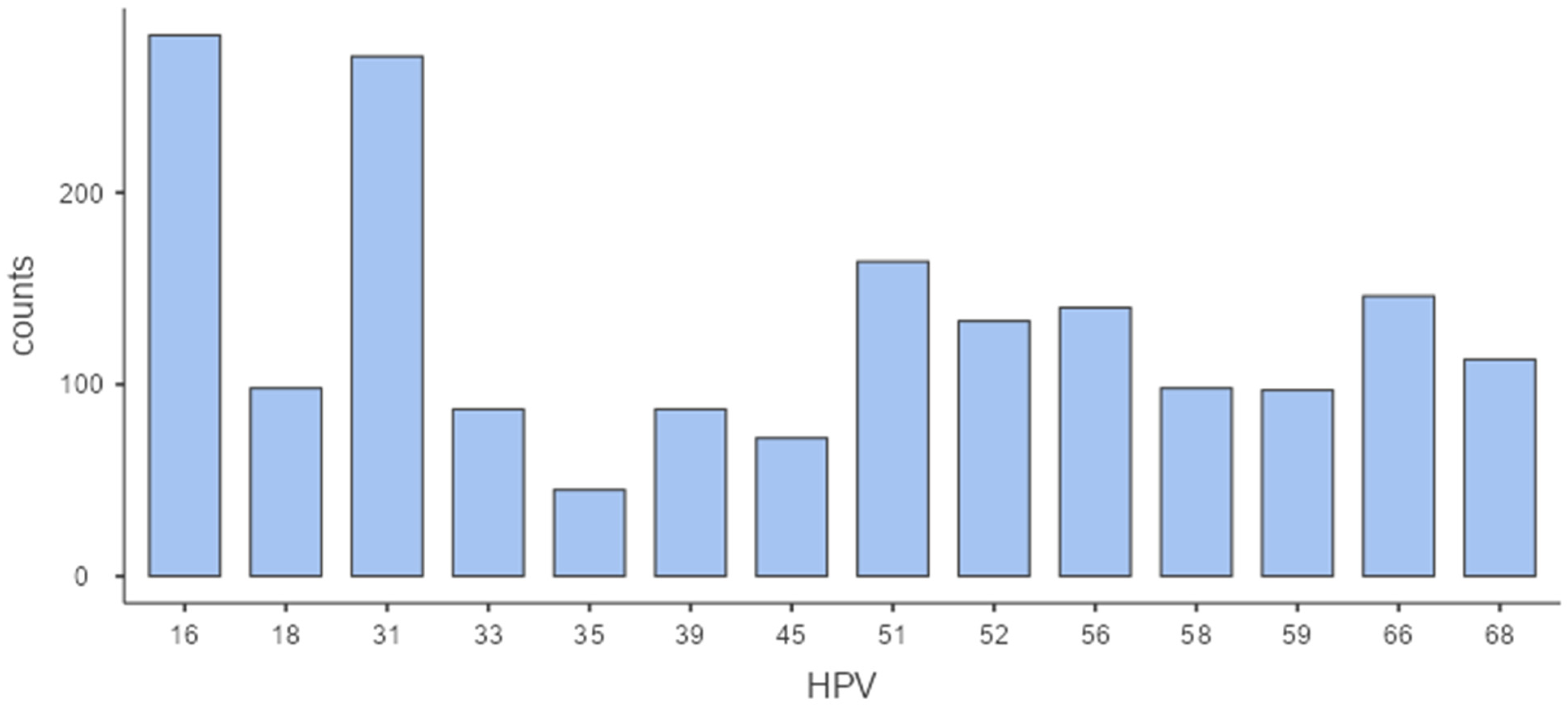
Out of the total number of HPV-positive women (N = 1211), the following genotype distribution was detected: HPV 16 in 282 (23.3%), HPV31 in 271 (22.4%), HPV 51 in 164 (13.5%), HPV 66 in 146 (12.1%), HPV 56 in 140 (11.6%), HPV 52 in 133 (11.0%), HPV 68 in 113 (9.3%) cases, HPV 18, HPV 58 in 98 cases (8.1%), HPV 59 in 97 (8.0%) cases, HPV 33 and HPV 39 each in 87 (7.2%) cases, HPV 45 in 72 (5.9%), and HPV 35 in 45 (3.7%) cases.
Likewise, since a third of HPV-positive women were infected with multiple HPV genotypes, there were a total of 1833 distinct genotypes, among which the most frequently detected were HPV 16 (282 detections, 15.4%), HPV 31 (271 detections, 14.8%) and HPV 51 (164 detections, 8.9%). These were followed by HPV 66 (146; 8.0%), HPV 56 (140; 7.6%), HPV 52 (133; 7.3%) and HPV 68 (113; 6.2%). Less frequently detected types included HPV 18 (98; 5.3%), HPV 58 (98; 5.3%), HPV 59 (97; 5.3%), HPV 33 (87; 4.7%), HPV 39 (87; 4.7%), HPV 45 (72; 3.9%), as well as HPV 35 (45; 2.5%). Collectively, the top three genotypes (HPV 16, 31, and 51) accounted for nearly 40% of all HPV detections in our study. The distribution of all detected HPV genotypes in the study can be seen in Table 1 and Figure 2.
As already mentioned, among all HPV-positive women (N = 1211), 30.7% (372 individuals) were aged 29 or younger, while the remaining 69.3% (839 individuals) were aged 30 or older (χ2 = 82.55, p < 0.001). Statistical analysis revealed no statistically significant association between age group and HPV group (“HPV-16”, “18/45 group”, “16-related”, and “lower-risk” groups) positivity (χ2 = 0.859, p = 0.835), which suggests that the distribution of HPV genotype groups did not differ meaningfully between younger and older women in the sample (Table 2). Likewise, Spearman’s correlation analysis showed no significant association between age and HPV group positivity (Spearman’s rho = 0.014, p = 0.562), indicating that age was not correlated with the type of high-risk HPV infection.
Among the HPV-positive women (N = 1211), a total of 750 of them (234 ≤ 29 and 516 ≥ 30) were also tested for other pathogens in a multiplex panel, while 654 of women (198 ≤ 29 and 456 ≥ 30) were tested for a range of different aerobic/anaerobic cultivable bacteria and fungi—including G. vaginalis. Consequently, C. trachomatis was detected in 3.87% of cases (29/750), while M. genitalium was found in 1.47% (11/750) of them. U. urealyticum had a higher detection rate of 13.07% (98/750), as did M. hominis, detected in 11.46% (86/750) of women. T. vaginalis was detected in 5.33% of cases (4/750), while N. gonorrheae was not detected in our study cohort. G. vaginalis (a marker for cervico-vaginal dysbiosis) was observed in 17.28% (113/654) of the tested individuals. C. albicans was observed in 8.41% (55/654) of tested women, S. agalactiae in 3.06% (20/654), E. coli in 2.75% (18/654), S. aureus in 0.61% (4/654), K. pneumoniae in 0.31% (2/654), while Morganella morganii and Pseudomonas aeruginosa were detected in one woman each (0.15%).
There were statistically significant associations of some other pathogens with age groups; notably, C. trachomatis positivity was substantially higher among women aged 29 or younger (22/234; 9.61%) compared to those aged 30 or older (7/516; 1.40%) (χ2 = 28.07, p < 0.001), indicating that younger age was significantly associated with increased C. trachomatis detection in this cohort. Likewise, S. agalactiae was more frequently isolated in women aged 29 or younger, with 6.06% detection rate (12/198) compared to 1.75% (8/456) in women aged 30 or older (χ2 = 7.06, p = 0.029), suggesting a differing age-related distribution pattern in microbial findings. However, there were no statistically significant differences in relation to age groups for U. urealyticum (39/234 in 29 or younger; 59/516 in 30 or older) (χ2 = 3.56, p = 0.059), M. hominis (34/234 in 29 or younger; 52/516 in 30 or older) (χ 2= 2.92, p = 0.087), M. genitalium (4/234 in 29 or younger; 7/516 in 30 or older) (χ2 = 0.0029, p = 0.957) and G. vaginalis (30/198 in 29 or younger; 83/456 in 30 or older) (χ2 = 0.72, p = 0.397) positivity.
When comparing the HPV 16 high-risk group to the reference category (18/45), none of the predictors reached statistical significance, although G. vaginalis showed a borderline association (p = 0.073), suggesting a potential trend toward increased odds of membership in the HPV 16 group in women with dysbiosis. In contrast, statistically significant associations were found in the 16-related and lower-risk HPV groups. Specifically, C. trachomatis was negatively associated with being in the 16-related group (Estimate = −0.9583, p = 0.045), indicating that women with C. trachomatis had approximately 62% lower odds (OR ≈ 0.38) of belonging to this group compared to the 18/45 reference group. Similarly, G. vaginalis was positively associated with the 16-related group (Estimate = 0.9610, p = 0.029), with an odds ratio of approximately 2.6 (Table 3).
For the lower-risk HPV group, C. trachomatis was again a significant negative predictor (Estimate = −1.2722, p = 0.007), with an estimated 72% reduction in odds (OR ≈ 0.28) of being in the lower-risk group when C. trachomatis was present. G. vaginalis showed a near-significant positive association (Estimate = 0.8316, p = 0.055), suggesting a possible trend toward increased likelihood of lower-risk HPV infection in the presence of cervico-vaginal dysbiosis. Taken together, these findings suggest that C. trachomatis is more frequently detected in women infected with HPV 18/45 than in those with 16-related or lower-risk HPV genotypes. Conversely, G. vaginalis (as a marker of cervico-vaginal dysbiosis) appears more closely associated with non-18/45 HPV groups, particularly the 16-related group. Model fit statistics indicated limited explanatory power (Deviance = 2201, AIC = 2237, McFadden’s R2 = 0.00680) (Table 3).

4. Discussion

This retrospective study provides a detailed overview of the genotype-specific distribution of high-risk HPV in HPV-positive women from Southern Croatia, alongside its associations with selected sexually transmitted pathogens and microbial correlates. The most frequently detected genotypes were HPV 16, 31 and 51, collectively accounting for nearly 40% of all HPV genotype detections (and represented in almost 60% of all high-risk HPV-positive women). These findings are consistent with global and European trends, where HPV 16 remains the most prevalent and oncogenic type, which is also the case for Central and Eastern Europe [27,28]. Genotypes such as HPV 31 and 51 are also commonly reported in certain regional studies [29]. The relatively high prevalence of HPV 31 in our cohort further supports its classification as a high-risk genotype deserving focused clinical attention in screening and vaccination strategies.
The age is a known determinant of HPV acquisition and persistence [30,31], and we also showed how younger age is significantly linked with higher HPV prevalence. Several previous studies conducted in European, Chinese and North American populations also demonstrated that high-risk HPV prevalence consistently peaked in women under the age of 35 and declined with advancing age [29,32,33,34,35]. However, there is a lower body of literature on the interrelation between HPV types and different age categories. Our analysis did not reveal a statistically significant association between age groups and specific HPV genotype clusters, which aligns with some recent studies suggesting that the distribution of individual genotypes is not always age-dependent [36]. Such patterns may be influenced by the natural history of each genotype, with some (e.g., HPV 16) being more persistent and oncogenic, thereby remaining detectable over longer periods regardless of age [37,38]. Furthermore, this stability in genotype distribution across age groups could reflect regional transmission dynamics, vaccine coverage or cohort-specific risk behaviours that shape the local epidemiology of HPV—factors that were beyond the scope of this study.
When other pathogens were concerned, co-infections with C. trachomatis were significantly more frequent in women aged 29 younger in our analysis. This finding goes in line with the common assumption that younger women are more likely to harbour sexually transmitted infections (STIs) due to higher sexual activity and partner turnover [39], and many different studies show that the younger age group is disproportionately affected [40,41]. We also found a significant correlation of S. agalactiae positivity and age, with higher frequency in younger age groups. However, this association is not consistently reported in the literature, as some studies have observed higher colonization rates in older age groups [42]. In any case, our findings highlight the need for broader STI screening approaches that consider not only age, but also microbial co-factors that may act synergistically with HPV in promoting persistence and disease progression.
The present study confirms and expands upon earlier findings regarding the distribution of high-risk HPV genotypes in Southern Croatia. In the 2013 study by Kaliterna et al. [43], conducted on a cohort of 1160 women screened using Hybrid Capture 2 (HC2) with 406 (35%) HPV-positive cases, followed by PCR-based genotyping, HPV 16 emerged as the most prevalent genotype (in 30.8% cases), followed by HPV 18 (22.2%) and HPV 31 (6.7%) among the 406 high-risk HPV positive women. In contrast, our current study utilized a more sensitive multiplex real-time PCR assay and identified different HPV genotype distribution among 1211 HPV-positive women: HPV 16 as the most prevalent genotype as well (in 23.3% of cases), followed by HPV 31 (22.4%) and HPV 51 (13.5%)—showing higher prevalence of certain genotypes compared to the 2013 data. This marked increase in HPV 31 may be attributed to the methodological improvements in genotyping sensitivity and resolution, as well as possible changes in local epidemiological dynamics over the past decade. Additionally, HPV 18, which was the second most common genotype in 2013, was less dominant ten years later in this study.
Present study focused exclusively on high-risk HPV-positive women, enabling more detailed genotype-specific analysis and assessment of co-infection patterns. Likewise, a study conducted in Northern Croatia (Zagreb County) on cervical samples found HPV 16 being the most commonly detected high-risk genotype, present in 33.3% of HPV-positive samples, followed by HPV 31 in 14.0% of HPV-positive samples [44]. This comparison highlights the importance of study design and target population in shaping observed genotype prevalence patterns. In any case, the findings suggest a broader and more diverse HPV genotype landscape in different regions of Croatia today, underscoring the need for ongoing epidemiological surveillance with high-resolution molecular diagnostics approaches.
The findings can also be contextualized by comparing them with a recent study conducted in the same geographic region in 2023 [20], which investigated HPV prevalence and its association with genital microbiota in a general population of asymptomatic women. The mentioned study and our study both aimed to appraise the role of microbial co-infections (most notably with C. trachomatis and G. vaginalis); however, our study further disentangles these associations by employing multinomial regression to demonstrate genotype-specific relationships. For instance, C. trachomatis showed a significant negative association with 16-related and lower-risk HPV groups, whereas G. vaginalis was positively associated with 16-related types. Unlike the 2023 study, which focused primarily on overall HPV positivity, this work aimed to provide insights into how distinct microbial patterns may influence the distribution of specific oncogenic genotypes, contributing, in turn, to a more nuanced understanding of HPV–microbiome interactions in cervical cancer risk stratification.
While HPV 16 remains the most commonly detected high-risk genotype, our findings align with broader global and European trends that highlight the growing relevance of other high-risk HPV types. For example, in the general European population, HPV 31 and HPV 18 are frequently reported following HPV 16 [38]. In our Southern Croatian cohort, after HPV 16 (detected in 23.3% HPV-positive women), HPV 31 and HPV 51 were particularly prevalent (in 22.4% and 13.5% cases, respectively), along with notable detection rates for HPV 66 (12.1%), HPV 56 (11.6%) and HPV 52 (11.0%). Some parallels can also be drawn with a recent Greek study, which identified HPV 31, HPV 66, HPV 56 and HPV 51 as the most common high-risk HPV types after HPV 16 in their study population [39]. In the northern region of Portugal, the most common high-risk HPV genotypes among high-risk HPV positive women were HPV 16 (17.5%), followed closely by HPV 39 (16.7%), HPV 31 (15.0%), HPV 68 (13.2%), HPV 52 (10.7%) and HPV 51 (10.6%) [45]. These aforementioned genotypes—particularly HPV 51, HPV 56 and HPV 66—are not covered by the currently available nine-valent HPV vaccine [46], emphasizing the need for ongoing regional surveillance. Our data also underscore the importance of monitoring shifts in genotype distribution to inform local vaccination strategies and to support the potential development of next-generation multivalent vaccines tailored to regional epidemiological profiles.
A recent systematic review analyzing 55 European studies on high-grade cervical intraepithelial neoplasia (CIN 2+) found that HPV 16 was consistently the most prevalent high-risk genotype both before and after treatment, followed by HPV 18, HPV 31 and HPV 58 [47]. In one study on 216 women that was included in the aforementioned systematic review, HPV 16 was detected in 55.5% of cases pre-treatment and remained in 7.9% six months after conization, while HPV 31 and HPV 52 also showed notable persistence [47]. The systematic review revealed that although overall HPV positivity declined substantially after excisional therapy, certain genotypes such as HPV 16 and HPV 58 demonstrated a greater tendency to persist, contributing to the risk of recurrent disease [47]. The findings emphasize the need for genotype-specific post-treatment monitoring, considering the fact that the persistence of particular high-risk types may influence long-term outcomes and inform future vaccination and screening strategies.
This study has several limitations that should be acknowledged. The retrospective and cross-sectional design limits the ability to infer causality between specific microbial co-infections and HPV genotype distribution. While the study included a relatively large sample of HPV-positive women, it was conducted in a single geographic region, which may limit the generalizability of findings to other populations in Croatia or neighbouring countries. Then, the analysis relied on cervico-vaginal swabs collected during routine gynecological examinations, but lacked accompanying clinical data such as cytological results or specific behavioural risk factors (e.g., number of sexual partners, contraceptive use), which could further contextualize the associations observed. And although molecular testing platforms used were CE-IVD-certified and capable of detecting multiple pathogens, the microbiological component did not assess the broader composition of the cervico-vaginal microbiota, limiting insights into complex microbial community interactions beyond selected pathogens. Furthermore, the study did not include data on participants’ comorbidities, fertility status, prior HPV infection history, HPV vaccination status and/or use of concomitant medications, all of which could influence HPV genotype prevalence, co-infection rates and immune response. These gaps may introduce potential bias in data interpretation and limit the ability to draw definitive conclusions about observed associations. Finally, the grouping of HPV genotypes into four predefined categories may obscure more subtle differences among individual types, and the relatively low pseudo R2 value of the regression model indicates limited explanatory power, suggesting that other unmeasured variables may contribute to the observed genotype distributions.

5. Conclusions

Our study offered a detailed snapshot of the genotype-specific distribution of high-risk HPV and its microbial correlates in HPV-positive women from Southern Croatia, more specifically from Split and Dalmatia County. HPV 16, 31, and 51 emerged as the most prevalent high-risk genotypes, reinforcing global and regional trends and emphasizing the need for ongoing surveillance beyond HPV 16 and 18. Importantly, we identified significant associations between cervico-vaginal microorganisms and HPV genotype groupings, which underscore the value of integrating extended HPV genotyping with microbial screening in routine gynecological care to yield clinically relevant insights into co-infection patterns and understanding potential implications for cervical cancer risk.
Despite limitations related to its retrospective and single-region design, we believe the study contributes to a more nuanced understanding of HPV epidemiology and highlights the importance of context-specific data. From a policy perspective, the results support the expansion of national cervical cancer screening programmes to include extended genotyping for high-risk HPV types beyond those covered by current vaccines. The high prevalence of non-vaccine genotypes underscores the need to monitor shifts in genotype distribution that may impact long-term vaccine effectiveness. This also has important implications for clinical oncology practice, as understanding the patterns of HPV co-infection may inform risk stratification, improve the interpretation of screening results, and also guide the development of more targeted prevention and management strategies for HPV-associated cervical lesions.

Author Contributions

Conceptualization, V.K. and T.M.; Methodology, V.K. and T.M.; Software, T.M.; Validation, V.K., T.M. and M.Č.-M.; Formal Analysis, T.M. and I.B.; Investigation, V.K., T.M., M.Č.-M. and I.B.; Resources, V.K.; Data Curation, V.K. and M.Č.-M.; Writing—Original Draft Preparation, V.K. and T.M.; Writing—Review and Editing, V.K., T.M., M.Č.-M. and I.B.; Visualization, T.M.; Supervision, V.K.; Project Administration, V.K. All authors have read and agreed to the published version of the manuscript.

Funding

This research received no external funding.

Institutional Review Board Statement

The study was conducted in accordance with the Declaration of Helsinki and approved by the Institutional Ethics Committee of the Teaching Institute for Public Health of Split and Dalmatia County, within which the presented work was performed (Class: 500-01/15-01/6; No: 2181-103-01-15-1; 12-05-2015).

Informed Consent Statement

Verbal informed consent was obtained from all subjects involved in the study.

Data Availability Statement

Original dataset available upon reasonable request from the authors.

Conflicts of Interest

The authors declare no conflicts of interest.

Abbreviations

The following abbreviations are used in this manuscript:
AICAkaike Information Criterion
CE-IVDConformité Européenne—In Vitro Diagnostic
CIN 2Cervical Intraepithelial Neoplasia Grade 2
CIN 3Cervical Intraepithelial Neoplasia Grade 3
CTChlamydia trachomatis
DALYDisability-Adjusted Life Year
GVGardnerella vaginalis
HPVHuman Papillomavirus
HRHigh Risk
IQRInterquartile Range
MGMycoplasma genitalium
MHMycoplasma hominis
NGNeisseria gonorrhoeae
OROdds Ratio
PapPapanicolaou
PCRPolymerase Chain Reaction
SEStandard Error
STISexually Transmitted Infection
UUUreaplasma urealyticum
TVTrichomonas vaginalis

References

  1. Ghosh, S.K.; Choudhury, B.; Hansa, J.; Mondal, R.; Singh, M.; Singh, M.; Duttagupta, S.; Das, A.; Kumar, R.; Laskar, R.S.; et al. Human papillomavirus testing for suspected cervical cancer patients from Southern Assam by fast-PCR. Asian Pac. J. Cancer Prev. 2011, 12, 749–751. [Google Scholar]
  2. Okunade, K.S. Human papillomavirus and cervical cancer. J. Obstet. Gynaecol. 2020, 40, 602–608. [Google Scholar] [CrossRef]
  3. Naghavi, M.; Meštrović, T.; Gray, A.; Gershberg Hayoon, A.; Swetschinski, L.R.; Robles Aguilar, G.; Davis Weaver, N.; Ikuta, K.S.; Chung, E.; Wool, E.E.; et al. Global burden associated with 85 pathogens in 2019: A systematic analysis for the Global Burden of Disease Study 2019. Lancet Infect. Dis. 2024, 24, 868–895. [Google Scholar] [CrossRef] [PubMed]
  4. Su, P.; Ma, J.; Yu, L.; Tang, S.; Sun, P. Clinical significance of extended high-risk human papillomavirus genotyping and viral load in cervical cancer and precancerous lesions. Gynaecol. Obstet. Clin. Med. 2023, 3, 22–29. [Google Scholar] [CrossRef]
  5. Baba, S.K.; Alblooshi, S.S.E.; Yaqoob, R.; Behl, S.; Al Saleem, M.; Rakha, E.A.; Malik, F.; Singh, M.; Macha, M.A.; Akhtar, M.K.; et al. Human papilloma virus (HPV) mediated cancers: An insightful update. J. Transl. Med. 2025, 23, 483. [Google Scholar] [CrossRef] [PubMed]
  6. Van Den Borst, E.; Bell, M.; Op De Beeck, K.; Van Camp, G.; Van Keer, S.; Vorsters, A. Lineages and sublineages of high-risk HPV types associated with cervical cancer and precancer: A systematic review and meta-analysis. J. Natl. Cancer Inst. 2025; ahead of print. [Google Scholar] [CrossRef]
  7. Zang, L.; Hu, Y. Risk factors associated with HPV persistence after conization in high-grade squamous intraepithelial lesion. Arch. Gynecol. Obstet. 2021, 304, 1409–1416. [Google Scholar] [CrossRef]
  8. Ntuli, L.; Mtshali, A.; Mzobe, G.; Liebenberg, L.J.; Ngcapu, S. Role of Immunity and Vaginal Microbiome in Clearance and Persistence of Human Papillomavirus Infection. Front. Cell. Infect. Microbiol. 2022, 12, 927131. [Google Scholar] [CrossRef] [PubMed]
  9. Lin, S.; Gao, K.; Gu, S.; You, L.; Qian, S.; Tang, M.; Wang, J.; Chen, K.; Jin, M. Worldwide trends in cervical cancer incidence and mortality, with predictions for the next 15 years. Cancer 2021, 127, 4030–4039. [Google Scholar] [CrossRef]
  10. Du, J.; Ahrlund-Richter, A.; Nasman, A.; Dalianis, T. Human papilloma virus (HPV) prevalence upon HPV vaccination in Swedish youth: A review based on our findings 2008–2018, and perspectives on cancer prevention. Arch. Gynecol. Obstet. 2021, 303, 329–335. [Google Scholar] [CrossRef] [PubMed]
  11. Oyervides-Munoz, M.A.; Perez-Maya, A.A.; Sanchez-Dominguez, C.N.; Berlanga-Garza, A.; Antonio-Macedo, M.; Valdez-Chapa, L.D.; Cerda-Flores, R.M.; Trevino, V.; Barrera-Saldana, H.A.; Garza-Rodriguez, M.L. Multiple HPV Infections and Viral Load Association in Persistent Cervical Lesions in Mexican Women. Viruses 2020, 12, 380. [Google Scholar] [CrossRef]
  12. Tao, X.; Zhang, H.; Zhang, H.; Xiao, Y.; Zhong, F.; Zhou, X.; Cong, Q.; Sui, L.; Zhao, C. The clinical utility of extended high-risk HPV genotyping in risk-stratifying women with L-SIL cytology: A retrospective study of 8726 cases. Cancer Cytopathol. 2022, 130, 542–550. [Google Scholar] [CrossRef]
  13. De Marco, L.; Bisanzi, S.; Ronco, G.; Mancuso, P.; Carozzi, F.; Allia, E.; Rizzolo, R.; Gustinucci, D.; Frayle, H.; Viti, J.; et al. Extended HPV genotyping by the BD Onclarity assay: Concordance with screening HPV-DNA assays, triage biomarkers, and histopathology in women from the NTCC2 study. Microbiol. Spectr. 2025, 13, e0089724. [Google Scholar] [CrossRef] [PubMed]
  14. Ye, Y.; Jones, T.; Wang, T.; Zeng, X.; Liu, Y.; Zhao, C. Comprehensive overview of genotype distribution and prevalence of human papillomavirus in cervical lesions. Gynecol. Obstet. Clin. Med. 2024, 4, e000005. [Google Scholar] [CrossRef]
  15. Lei, J.; Cuschieri, K.; Patel, H.; Lawrence, A.; Deats, K.; Sasieni, P.; Lim, A.W.W. Human papillomavirus genotype and cycle threshold value from self-samples and risk of high-grade cervical lesions: A post hoc analysis of a modified stepped-wedge implementation feasibility trial. PLoS Med. 2024, 21, e1004494. [Google Scholar] [CrossRef]
  16. Massad, L.S.; Clarke, M.A.; Perkins, R.B.; Garcia, F.; Chelmow, D.; Cheung, L.C.; Darragh, T.M.; Egemen, D.; Lorey, T.S.; Nayar, R.; et al. Applying Results of Extended Genotyping to Management of Positive Cervicovaginal Human Papillomavirus Test Results: Enduring Guidelines. J. Low. Genit. Tract. Dis. 2025, 29, 134–143. [Google Scholar] [CrossRef]
  17. Anton, L.; Ferguson, B.; Friedman, E.S.; Gerson, K.D.; Brown, A.G.; Elovitz, M.A. Gardnerella vaginalis alters cervicovaginal epithelial cell function through microbe-specific immune responses. Microbiome 2022, 10, 119. [Google Scholar] [CrossRef]
  18. Gargiulo Isacco, C.; Balzanelli, M.G.; Garzone, S.; Lorusso, M.; Inchingolo, F.; Nguyen, K.C.D.; Santacroce, L.; Mosca, A.; Del Prete, R. Alterations of Vaginal Microbiota and Chlamydia trachomatis as Crucial Co-Causative Factors in Cervical Cancer Genesis Procured by HPV. Microorganisms 2023, 11, 662. [Google Scholar] [CrossRef]
  19. Cui, M.; Wu, Y.; Liu, Z.; Liu, Y.; Fan, L. Advances in the interrelated nature of vaginal microecology, HPV infection, and cervical lesions. Front. Cell. Infect. Microbiol. 2025, 15, 1608195. [Google Scholar] [CrossRef]
  20. Kaliterna, V.; Kaliterna, P.; Pejkovic, L.; Vulic, R.; Zanchi, L.; Cerskov, K. Prevalence of Human Papillomavirus (HPV) among Females in the General Population of the Split and Dalmatia County and Its Association with Genital Microbiota and Infections: A Prospective Study. Viruses 2023, 15, 443. [Google Scholar] [CrossRef] [PubMed]
  21. Demarco, M.; Hyun, N.; Carter-Pokras, O.; Raine-Bennett, T.R.; Cheung, L.; Chen, X.; Hammer, A.; Campos, N.; Kinney, W.; Gage, J.C.; et al. A study of type-specific HPV natural history and implications for contemporary cervical cancer screening programs. EClinicalMedicine 2020, 22, 100293. [Google Scholar] [CrossRef] [PubMed]
  22. de Sanjosé, S.; Serrano, B.; Tous, S.; Alejo, M.; Lloveras, B.; Quirós, B.; Clavero, O.; Vidal, A.; Ferrándiz-Pulido, C.; Pavón, M.Á.; et al. Burden of Human Papillomavirus (HPV)-Related Cancers Attributable to HPVs 6/11/16/18/31/33/45/52 and 58. JNCI Cancer Spectr. 2019, 2, pky045. [Google Scholar] [CrossRef]
  23. Bouvard, V.; Wentzensen, N.; Mackie, A.; Berkhof, J.; Brotherton, J.; Giorgi-Rossi, P.; Kupets, R.; Smith, R.; Arrossi, S.; Bendahhou, K.; et al. The IARC Perspective on Cervical Cancer Screening. N. Engl. J. Med. 2021, 385, 1908–1918. [Google Scholar] [CrossRef] [PubMed]
  24. Suba, E.J.; Ortega, R.E.; Mutch, D.G. The IARC Perspective on Cervical Cancer Screening. N. Engl. J. Med. 2022, 386, 607. [Google Scholar]
  25. R Core Team. R: A language and environment for statistical computing. In R Foundation for Statistical Computing; R Core Team: Vienna, Austria, 2020; Available online: https://www.R-project.org/ (accessed on 15 August 2025).
  26. The jamovi Project. jamovi (Version 2.6) [Computer Software]. 2025. Available online: https://www.jamovi.org (accessed on 15 August 2025).
  27. WHO/ICO Information Centre on HPV and Cervical Cancer. HPV and cervical cancer in the 2007 report. Vaccine 2007, 25 (Suppl. S3), C1–C230. [Google Scholar]
  28. Poljak, M.; Seme, K.; Maver, P.J.; Kocjan, B.J.; Cuschieri, K.S.; Rogovskaya, S.I.; Arbyn, M.; Syrjänen, S. Human Papillomavirus Prevalence and Type-Distribution, Cervical Cancer Screening Practices and Current Status of Vaccination Implementation in Central and Eastern Europe. Vaccine 2013, 31, H59–H70. [Google Scholar] [CrossRef] [PubMed]
  29. Tsakogiannis, D.; Zografos, E.; Tzioga, L.; Zografos, C.G.; Zagouri, F.; Bletsa, G. Prevalence and Genotype Distribution of High-Risk HPV Genotypes Among Women in Greece: A Retrospective Analysis of 3500 Women. Cancers 2025, 17, 1267. [Google Scholar] [CrossRef] [PubMed]
  30. Kang, L.N.; Castle, P.E.; Zhao, F.H.; Jeronimo, J.; Chen, F.; Bansil, P.; Li, J.; Chen, W.; Zhang, X.; Qiao, Y.L. A prospective study of age trends of high-risk human papillomavirus infection in rural China. BMC Infect. Dis. 2014, 14, 96. [Google Scholar] [CrossRef]
  31. Burger, E.A.; Kim, J.J.; Sy, S.; Castle, P.E. Age of Acquiring Causal Human Papillomavirus (HPV) Infections: Leveraging Simulation Models to Explore the Natural History of HPV-induced Cervical Cancer. Clin. Infect. Dis. 2017, 65, 893–899. [Google Scholar] [CrossRef]
  32. Liu, S.; Mei, B.; Ouyang, Y.; Li, C. Prevalence and genotype distribution of human papillomavirus infection among women in Jingzhou, China: A population-based study of 51,720 women. Virol. J. 2023, 20, 297. [Google Scholar] [CrossRef]
  33. Hu, J.P.; Wang, J.L.; Li, Y.; Feng, Y.; Tian, C.Q.; Zhang, G.H.; Chen, X.Q.; Liu, H.X.; Yang, J.S.; Fang, Z.W.; et al. Prevalence and genotype distribution of human papillomavirus infection among 66000 women from 2014 to 2023 in the plateau region of Southwest China. Virol. J. 2024, 21, 176. [Google Scholar] [CrossRef]
  34. Berza, N.; Zodzika, J.; Kivite-Urtane, A.; Baltzer, N.; Curkste, A.; Pole, I.; Nygard, M.; Parna, K.; Stankunas, M.; Tisler, A.; et al. Understanding the high-risk human papillomavirus prevalence and associated factors in the European country with a high incidence of cervical cancer. Eur. J. Public Health 2024, 34, 826–832. [Google Scholar] [CrossRef]
  35. Clarke, M.A.; Risley, C.; Stewart, M.W.; Geisinger, K.R.; Hiser, L.M.; Morgan, J.C.; Owens, K.J.; Ayyalasomayajula, K.; Rives, R.M.; Jannela, A.; et al. Age-specific prevalence of human papillomavirus and abnormal cytology at baseline in a diverse statewide prospective cohort of individuals undergoing cervical cancer screening in Mississippi. Cancer Med. 2021, 10, 8641–8650. [Google Scholar] [CrossRef]
  36. Wang, Y.; Hou, H.; Dong, G.; Zhang, H.; Zhang, X.; Zhou, Y.; Xue, M.; Wang, Z.; Geng, J.; Liu, L. Age-related distribution of human papillomavirus genotypes in women with cervical squamous cell carcinoma from Linyi, China, 2015–2023. Virol. J. 2025, 22, 157. [Google Scholar] [CrossRef]
  37. Koshiol, J.; Lindsay, L.; Pimenta, J.M.; Poole, C.; Jenkins, D.; Smith, J.S. Persistent human papillomavirus infection and cervical neoplasia: A systematic review and meta-analysis. Am. J. Epidemiol. 2008, 168, 123–137. [Google Scholar] [CrossRef] [PubMed]
  38. Bruno, M.T.; Valenti, G.; Ruggeri, Z.; Incognito, G.G.; Coretti, P.; Montana, G.D.; Panella, M.M.; Mereu, L. Correlation of the HPV 16 Genotype Persistence in Women Undergoing LEEP for CIN3 with the Risk of CIN2+ Relapses in the First 18 Months of Follow-Up: A Multicenter Retrospective Study. Diagnostics 2024, 14, 509. [Google Scholar] [CrossRef] [PubMed]
  39. Agwu, A. Sexuality, Sexual Health, and Sexually Transmitted Infections in Adolescents and Young Adults. Top. Antivir. Med. 2020, 28, 459–462. [Google Scholar] [PubMed]
  40. Gupta, K.; Harrison, S.A.; Davis, N.A.; Culp, M.L.; Hand, S.C.; Simpson, T.; Van Der Pol, B.; Galbraith, J.W.; Van Wagoner, N.J.; Morrison, S.G.; et al. Prevalence of Chlamydia trachomatis Infection in Young Women and Associated Predictors. Sex. Transm. Dis. 2021, 48, 529–535. [Google Scholar] [CrossRef]
  41. Li, Y.; Liao, Z.; Wang, Q.; He, W.; Deng, Y.; Liu, C. Prevalence of Chlamydia trachomatis, Neisseria gonorrhoeae, and Ureaplasma urealyticum Infections in Males and Females of Childbearing Age in Chengdu, China. Front. Cell. Infect. Microbiol. 2025, 15, 1566163. [Google Scholar] [CrossRef]
  42. Martins, E.R.; Melo-Cristino, J.; Ramirez, M.; Portuguese Group for the Study of Streptococcal Infections. Dominance of Serotype Ia among Group B Streptococci Causing Invasive Infections in Nonpregnant Adults in Portugal. J. Clin. Microbiol. 2012, 50, 1219–1227. [Google Scholar] [CrossRef]
  43. Kaliterna, V.; Kaliterna, M.; Pejković, L.; Hofman, I.D.; Andelinović, S. Prevalence and genotyping of the human papillomavirus in the cervical specimens among women of Southern Croatia (Dalmatia County). Cent. Eur. J. Public Health 2013, 21, 26–29. [Google Scholar] [CrossRef]
  44. Sabol, I.; Milutin Gašperov, N.; Matovina, M.; Božinović, K.; Grubišić, G.; Fistonić, I.; Belci, D.; Alemany, L.; Džebro, S.; Dominis, M.; et al. Cervical HPV type-specific pre-vaccination prevalence and age distribution in Croatia. PLoS ONE 2017, 12, e0180480. [Google Scholar] [CrossRef] [PubMed]
  45. Sousa, H.; Tavares, A.; Campos, C.; Marinho-Dias, J.; Brito, M.; Medeiros, R.; Baldaque, I.; Lobo, C.; Leča, L.; Monteiro, P.; et al. High-Risk human papillomavirus genotype distribution in the Northern region of Portugal: Data from regional cervical cancer screening program. Papillomavirus Res. 2019, 8, 100179. [Google Scholar] [CrossRef]
  46. Pešut, E.; Šimić, I.; Fureš, R.; Milutin Gašperov, N.; Lež, C.; Feratović, F.; Kukina Žvigač, T.; Grce, M.; Erceg Ivkošić, I.; Sabol, I. Monitoring HPV Prevalence and Risk Cofactors for Abnormal Cytology in the Post-Vaccination Period among Croatian Women. Viruses 2024, 16, 642. [Google Scholar] [CrossRef] [PubMed]
  47. Reuschenbach, M.; Valente, S.; Takyar, J.; Dhawan, A.; Hall, A.; Agrawal, N.; Ghelardi, A.; Del Pino, M.; Nowakowski, A.; Sabale, U. Treatment characteristics, HPV genotype distribution and risk of subsequent disease among women with high-grade cervical intraepithelial neoplasia in Europe: A systematic literature review. Eur. J. Obstet. Gynecol. Reprod. Biol. 2024, 300, 129–140. [Google Scholar] [CrossRef] [PubMed]
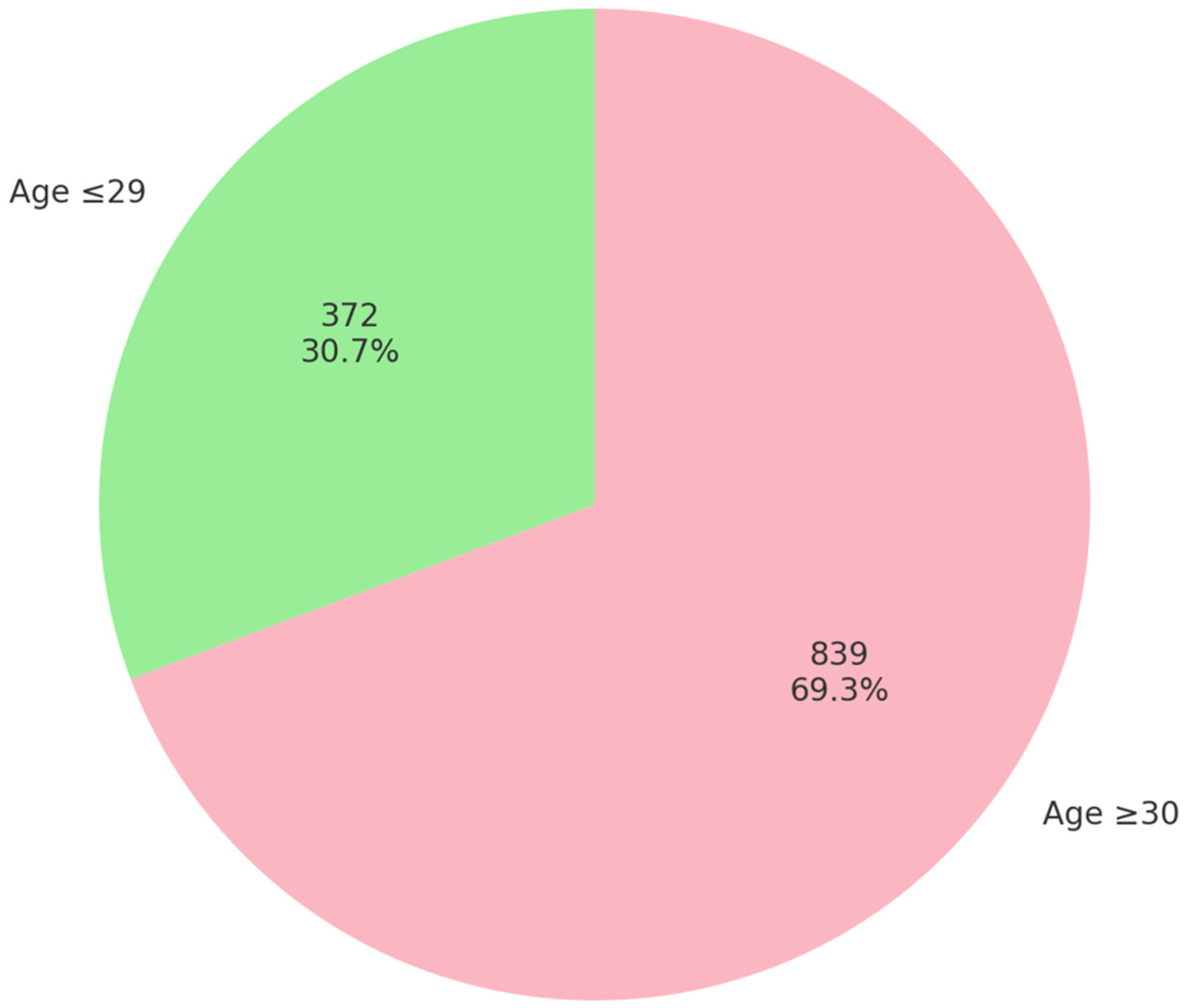
Figure 1. Age distribution of HPV-positive women included in the study (N = 1211).
Biomedicines 13 02100 g001
Figure 2. Bar graph displaying the frequency of each HPV genotype detected, with HPV 16 and HPV 31 being the most prevalent types (N = 1833).
Biomedicines 13 02100 g002
Table 1. Distribution of detected HPV genotypes in the study cohort.
HPV GenotypeCounts% of Total High-Risk HPV-Positive Women (N = 1211)% of Total Genotypes (N = 1833)
1628223.3%15.4%
18988.1%5.3%
3127122.4%14.8%
33877.2%4.7%
35453.7%2.5%
39877.2%4.7%
45725.9%3.9%
5116413.5%8.9%
5213311.0%7.3%
5614011.6%7.6%
58988.1%5.3%
59978.0%5.3%
6614612.1%8.0%
681139.3%6.2%
Table 2. Distribution of high-risk HPV genotype groups across age categories.
High-Risk HPV Group
Age Group18/4516 High Risk16-RelatedLower RiskTotal
29 or under1091833934671152
30 or over6199241280681
Total1702826347471833
p-value 0.835
Table 3. Multinomial logistic regression predicting HPV genotype group membership based on the presence of other pathogens.
HPV GroupsPredictorEstimateSEZp-ValueOR
16 high-risk—18/45Intercept0.56260.1693.336<0.0011.755
CT:
P—N−0.65780.531−1.2390.2150.518
MG:
P—N−0.30400.946−0.3210.7480.738
Mh:
P—N−0.17400.471−0.3690.7120.840
Uu:
P—N−0.07860.407−0.1930.8470.924
GV:
P—N0.85120.4751.7910.0732.343
16-related—18/45Intercept1.38760.1519.195<0.0014.005
CT:
P—N−0.95830.479−2.0000.0450.384
MG:
P—N−1.08220.941−1.1500.2500.339
Mh:
P—N−0.17780.425−0.4180.6760.837
Uu:
P—N−0.25720.371−0.6940.4880.773
GV:
P—N0.96190.4412.1800.0292.617
Lower risk—18/45Intercept1.64030.14711.134<0.0015.157
CT:
P—N−1.27220.474−2.6840.0070.280
MG:
P—N−0.49830.822−0.6060.5440.608
Mh:
P—N0.11080.4080.2710.7861.117
Uu:
P—N−0.11850.357−0.3320.7400.888
GV:
P—N0.83160.4341.9160.0552.297
The table displays the results of a multinomial logistic regression analysis assessing the association between selected microorganisms and HPV genotype group membership. The HPV 18/45 group served as the reference category. For each comparison (16 high-risk, 16-related and lower-risk HPV groups vs. 18/45), regression coefficients (estimates), standard errors (SE), Z-values, p-values and odds ratios (OR) are presented. “P—N” denotes “Positive—Negative”; “CT” denotes “Chlamydia trachomatis”; “MG” denotes “Mycoplasma genitalium”; “MH” denotes “Mycoplasma hominis”; “Uu” denotes “Ureaplasma urealyticum”; “GV” denotes “Gardnerella vaginalis”.
Disclaimer/Publisher’s Note: The statements, opinions and data contained in all publications are solely those of the individual author(s) and contributor(s) and not of MDPI and/or the editor(s). MDPI and/or the editor(s) disclaim responsibility for any injury to people or property resulting from any ideas, methods, instructions or products referred to in the content.