Review

Shed Syndecan-4 and Its Possible Roles in Osteoarthritis

1
State Key Laboratory of Oral & Maxillofacial Reconstruction and Regeneration, National Clinical Research Center for Oral Diseases, Shaanxi International Joint Research Center for Oral Diseases, School of Stomatology, The Fourth Military Medical University, Xi’an 710032, China
2
School/Hospital of Stomatology, Lanzhou University, Lanzhou 730000, China
3
Key Laboratory of Shaanxi Province for Craniofacial Precision Medicine Research, Department of Orthodontics, College of Stomatology, Xi’an Jiaotong University, Xi’an 710049, China
*
Authors to whom correspondence should be addressed.
These authors contributed equally to this work.
Biomedicines 2025, 13(5), 1037; https://doi.org/10.3390/biomedicines13051037
Submission received: 27 February 2025 / Revised: 15 April 2025 / Accepted: 22 April 2025 / Published: 25 April 2025
(This article belongs to the Section Molecular and Translational Medicine)

Abstract

:
The specific pathogenesis of osteoarthritis (OA) remains not fully understood. As a transmembrane heparan sulfate proteoglycan, syndecan-4 (SDC4) has been proven to play an important role in the development of OA. Notably, the extracellular domain of SDC4 can be cleaved by proteolytic enzymes, leading to the release of shed SDC4 (sSDC4), which subsequently regulates various biological processes in an autocrine or paracrine manner. This review analyzed 97 publications (1987–2025) from Pubmed and the Web of Science Core Collection using specific key words (syndecan-4, shed syndecan-4, and osteoarthritis), providing a comprehensive overview of the current research on sSDC4, including its shedding enzymes and specific cleavage sites, as well as the factors and mechanisms that influence SDC4 shedding. Furthermore, it summarizes the functions of both sSDC4 and its remaining membrane-bound domain. Finally, the roles of sSDC4 in OA are discussed to identify potential therapeutic targets and explore new strategies for the treatment of OA.

1. Introduction

Osteoarthritis (OA), a chronic degenerative joint disease, is characterized by pain and impaired joint mobility, significantly reducing quality of life and imposing economic burdens on patients and society [1]. It is estimated that over 500 million individuals worldwide suffer from OA [2]. However, there is no effective disease-modifying therapy for this prevalent and debilitating condition. Current management strategies for OA, including pharmacological interventions, physical therapy, and surgical procedures, provide palliative care but do not halt disease progression [3,4]. A definitive cure for OA remains elusive. Therefore, it is essential to understand and identify the key signaling pathways and molecules that play critical roles in the initiation and progression of OA.
The primary pathological changes in OA encompass the progressive degradation of articular cartilage, abnormal remodeling of subchondral bone, osteophyte formation, and synovial inflammation [5]. Considerable effort has been directed towards understanding the molecular mechanisms of OA and developing a fundamental treatment aimed at preventing the condition or repairing damaged articular cartilage. Articular cartilage is predominantly composed of the extracellular matrix (ECM) and chondrocytes. The interaction between the ECM and chondrocytes is crucial for maintaining articular cartilage homeostasis. The ECM, composed of type II collagen (COL II), proteoglycans (PGs), hyaluronic acid (HA), and chondroitin sulfate (CS), regulates chondrocyte differentiation, metabolism, matrix remodeling, and survival through integrin-mediated signaling. In OA, increased ECM degradation, mediated by enzymes like matrix metalloproteinases (MMPs) and aggrecanases, disrupts ECM components. Chondrocytes respond to these changes by releasing inflammatory cytokines, such as IL-1β and TNF-α, which exacerbate inflammation and ECM breakdown, while also increasing chondrocyte death and furthering cartilage degeneration [6]. The degradation of the ECM and the chondrocyte death are central pathological events in OA, making the interaction between the ECM and chondrocytes a critical factor in maintaining articular cartilage homeostasis [6].
Heparan sulfate proteoglycans (HSPGs), which are type I transmembrane proteoglycans that interact with both the ECM and chondrocytes, contain complex highly sulfated carbohydrate chains covalently attached to their core proteins [7]. HSPGs play essential roles in the interaction between the ECM and chondrocytes. Key members of the HSPG family include the multidomain proteoglycans syndecan (SDC) 1–4 [8]. Among them, SDC4 is the smallest, with a molecular weight of approximately 22 kDa [9]. As a ubiquitous cell surface receptor, SDC4 modulates cell proliferation, migration, mechanotransduction, and endocytosis [10]. Previous studies have demonstrated that SDC4 can modulate the secretion of inflammatory cytokines, which is regulated by low-density lipoprotein receptor-related protein (LRP) in metabolic processes [11] and critically participates in mechanotransduction pathways [12]. As a transmembrane proteoglycan, SDC4 primarily exerts localized effects within the articular microenvironment. In OA, upregulated SDC4 expression promotes disease progression through microRNA-mediated regulation by elevating miR-96-5p to suppress hypoxia-inducible factor 2 alpha (HIF-2α) expression, thereby inhibiting chondrocyte hypertrophy and matrix degradation [13], as well as through the activation of the mitogen-activated protein kinase (MAPK) signaling pathways of extracellular regulated protein kinase (ERK) 1/2 and p38 [14]. Notably, intra-articular injection of SDC4 antibody has been observed to ameliorate cartilage degeneration [14]. These findings collectively suggest that SDC4 participates in multiple pathological pathways in OA, establishing it as a pivotal molecular target with significant therapeutic potential. It is precisely this multifaceted involvement in OA pathogenesis that motivated our investigation of SDC4 as a promising therapeutic strategy. However, its detailed mechanism remains unclear. Interestingly, the extracellular domain (ectodomain) of SDC4, which is normally shed by proteolytic enzymes at low levels under physiological conditions, exhibits increased shedding in response to pathological stimuli such as inflammation or mechanical stress [15]. The soluble shed syndecan-4 (sSDC4) ectodomain, released into the ECM, retains the ability to bind growth factors and can function as an agonist or antagonist in an autocrine or paracrine manner [16]. It can also enter the synovial fluid and blood and be transported to other sites where it may exert its effects [17,18,19,20,21,22]. Thus, sSDC4 may have broader effects than the membrane-anchored form. It not only retains the functions of membrane-anchored SDC4, but also being freely diffusible, mediates broader paracrine actions. This soluble ectodomain can influence distal cells beyond their tissue of origin, potentially propagating OA-related pathological changes through synovial-fluid-mediated dissemination. In this review, we first review the sheddases, cleavage sites, and possible affecting factors of SDC4 shedding. Then, the functional consequences of SDC4 shedding and the potential mechanisms of sSDC4 are discussed. Finally, the possible roles of sSDC4 in OA pathogenesis are reviewed.

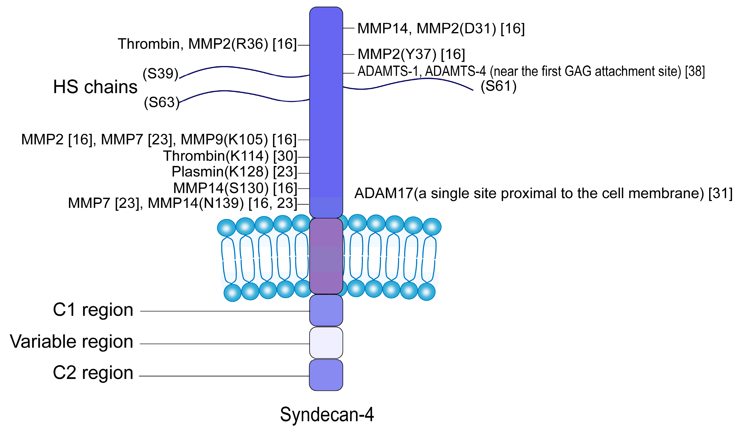
2. Proteolytic Enzymes and Cleavage Sites of SDC4 Shedding

The core protein of SDC4 comprises three distinct domains: an N-terminal ectodomain, a transmembrane domain, and a C-terminal intracellular (cytoplasmic) domain [23]. The intracellular domain of SDC4 interacts with numerous cytosolic binding partners involved in the mechanotransduction and signaling [24,25]. The ectodomain, approximately 120 amino acids in length, extends from the extracellular end to the cell membrane. This ectodomain consists of a heparan sulfate (HS)-chain-binding domain, a cell-binding domain, and proteolytic cleavage site domains [26]. SDC4 undergoes regulated proteolytic cleavage, typically near the plasma membrane, in a process known as shedding [23]. HS chains attached to the core protein inhibit the shedding of syndecan; when these HS chains are enzymatically degraded or absent, syndecan shedding is dramatically enhanced [27]. After shedding, the ectodomain of SDC4, which contains HS-chain-binding domains, can enter the ECM as an intact proteoglycan following proteolytic cleavage [28].
It is currently known that SDC4 can be shed by a variety of proteolytic enzymes, including matrix metalloproteinases (MMPs) [29], plasmin [30], thrombin [30], a disintegrin and metalloproteinases (ADAMs) [31], and a disintegrin and metalloproteinase with thrombospondin motifs (ADAMTS). These enzymes are responsible for shedding the SDC4 ectodomain at specific cleavage sites in Figure 1 [32,33]. The specific shedding enzymes involved can vary among different cell types in Table 1.

3. Factors Affecting SDC4 Shedding

Ectodomain shedding is an important regulatory mechanism. The shedding of SDC4 occurs constitutively to a small degree as part of normal turnover. This process is accelerated under certain conditions, including inflammation, oxidative stress, mechanical stress, and the upregulated expression or activity of chemokines and cytokines, such as growth factors [39], bacterial virulence factors [40], and trypsin [15]. Different extracellular stimuli activate different intracellular signaling pathways to accelerate shedding [41].

3.1. Inflammation

Inflammation, mediated by tumor necrosis factor-α (TNFα) and interleukin-1β (IL-1β), can significantly upregulate the expression and shedding of SDC4. After islet beta-cells are stimulated by IL-1β, SDC4 is upregulated and shed, involving the scleroderma renal crisis-signal transducer and activator of transcription 3 (SRC-STAT3) pathway, with MMP9 potentially participating in this process [9]. Similarly, IL-1β stimulation increases the level of sSDC4 in chondrocyte culture supernatants, with MMP9 acting as the responsible sheddase [17]. Under diabetic conditions, IL-1β, rather than hyperglycemia, enhances the shedding of SDC4 from glomerular endothelial cells in an MMP9-dependent manner [36]. TNFα, IL-1β, and lipopolysaccharides (LPSs) induce SDC4 expression and shedding in cardiac myocytes and fibroblasts through the toll-like receptor 4 (TLR4) and nuclear factor kappa-B (NF-κB) signaling pathways [42]. Strand et al. discovered that IL-1β-induced upregulation of SDC4 expression in cardiomyocytes could be significantly reversed by the IL-1 receptor antagonist (IL-1Ra) in vitro [18]. Oxidized linoleic acid, the principal oxidizable fatty acid in low-density lipoprotein (LDL), and its oxidized products contribute to atherosclerosis and other chronic inflammatory cardiovascular diseases [43]. Houston et al. demonstrated that oxidized linoleic acid regulated the expression and shedding of sSDC4 on airway smooth muscle cells (ASMCs) through the generation of intracellular hydrogen peroxide and activation of the ERK signaling pathway, which might be associated with pathological processes such as atherosclerosis [43]. These findings suggest that the shedding of SDC4 is influenced by multiple inflammatory factors.
After the shedding of SDC4 in various tissues, significant amounts of soluble sSDC4 are released into body fluids, including serum. Currently, the serum levels of sSDC4 in various inflammatory diseases are summarized in Table 2. Studies have reported that serum sSDC4 levels are mainly associated with inflammation, such as pneumonia. In patients with mild pneumonia, serum sSDC4 levels were higher in comparison to those with severe pneumonia. Interestingly, short-term antibiotic therapy further elevated sSDC4 levels, leading to the hypothesis that sSDC4 might possess anti-inflammatory properties [13]. Consistently, Sato et al. observed increased serum sSDC4 levels in patients with idiopathic interstitial pneumonia [44]. Luo et al. also demonstrated that serum sSDC4 levels were associated with severe community-acquired pneumonia, and these elevated serum levels were linked to a higher mortality rate [45]. Additionally, increased serum sSDC4 levels have been reported in patients with atopic dermatitis [46] and acute myocardial infarction [47]. Serum levels of sSDC4 have thus been shown to be associated with various diseases. Therefore, differentiating the cause of elevated sSDC4 levels in different patients to determine the specific disease they relate to remains a challenge. Considering the critical role of the heparan sulfate–galactosylated glycosaminoglycan (HS-GAG) chains in cellular signaling, through their sulfated domains, HS-GAG chains mediate high-affinity binding with growth factors, cytokines, and receptors. These interactions enable direct receptor activation while sulfation patterns critically modulate fibroblast growth factor (FGF)/Wnt pathway activation. Thus, more detailed analyses of the shedding site and/or modification of HS-GAG chains may enhance the sensitivity of sSDC4 as a biomarker for the respective diseases [7]. Thus, more detailed analyses of the shedding site and/or modification of the HS-GAG chains may enhance the sensitivity of sSDC4 as a biomarker for the respective diseases [7].

3.2. Oxidative Stress

Oxidative stress, arising from an imbalance between reactive oxygen species (ROS) production and the antioxidant defense system [48], has been shown to affect SDC4 ectodomain shedding. It has been shown that oxidative stress can trigger the activity of ADAM-17, one of the SDC4 sheddases [49]. Silent information regulator factor 1 (Sirtuin1), a histone deacetylase, exerts significant antioxidative stress effects. In fibrosis-prone endothelial Sirtuin1-deficient (Sirt1endo−/−) mice, SDC4 expression was elevated via enhanced NF-κB signaling in Sirt1-deficient endothelial cells, and oxidative stress induced by Sirt1 deficiency contributed to shedding of SDC4 [50]. Wu et al. found that advanced glycation end products (AGEs) could induce a senescence-associated secretory phenotype (SASP) in late-passage endothelial progenitor cells, and that resveratrol could inhibit oxidative stress and reduce ROS levels by activating Sirtuin1, thereby reducing SDC4 shedding induced by AGEs [51]. Xie et al. reported that AGEs promoted the SDC4 shedding through the receptor for advanced glycation endproducts (RAGE) and ROS pathways. The application of the antioxidant N-acetylcysteine (NAC) can inhibit AGE-induced SDC4 shedding, suggesting that oxidative stress plays a role in this process [52]. Moreover, increased SDC4 shedding in atrial fibrillation patients is associated with higher levels of markers of inflammation and oxidative damage, which may mediate SDC4 shedding via MMPs [53]. Increased ROS levels in OA cartilage can induce inflammation and matrix degradation in chondrocytes [48,54,55]. The relationship between oxidative stress and SDC4 ectodomain shedding in OA cartilage requires further investigation.

3.3. Mechanical Stress

SDC4 serves as a crucial mediator transducing mechanical stress from the ECM to chondrocytes, making mechanical stress a key factor influencing the shedding of the SDC4 ectodomain. In 2020, Jiang et al. revealed that SDC4-dependent mechanotransduction in endothelial cells is principally mediated through a dynamic scissoring mechanism of its transmembrane dimer, whereby the two extracellular domains undergo coordinated anti-parallel displacement. This scissoring motion serves as a key mechanotransduction mechanism in endothelial cells, transmitting shear stress forces from the ectodomain to the intracellular domain. Flexible linker regions enable the motion, facilitating force transfer to the cytoskeleton and downstream signaling. The heparan sulfate chains stabilize the SDC4 core protein during this process, preserving endothelial glycocalyx function [56]. Lee et al. observed exercise-induced changes in SDC4 levels among sedentary men, comparing those with dysglycemia to normoglycemic individuals. The results showed that acute exercise significantly increased serum concentrations of soluble SDC4 in both groups, suggesting that enhanced blood flow and mechanical stress during exercise may promote SDC4 shedding from the endothelial glycocalyx. This finding indicates that the observed elevation in SDC4 levels represents a physiological response to acute exercise rather than being specific to either metabolic state [20]. Abnormal mechanical loading is also a risk factor for OA [57]. The direct relationship between changes in mechanical stress changes and SDC4 shedding during the OA process remains unclear.

3.4. Different Specific Cytokines

Under normal physiological conditions, the shedding of HSPGs from the cell surface was not inhibited by the matrix metalloproteinase inhibitor tissue inhibitor of metalloproteinase-3 (TIMP-3). However, following abnormal stimulation, the shedding of the ectodomain was inhibited by TIMP-3 [29]. This suggests that abnormal stimuli activate different shedding enzymes and intracellular signaling pathways, consequently accelerating shedding, although the specific mechanism remains unclear.
Protein kinase C (PKC), which can be activated by phorbol esters such as phorbol myristate acetate (PMA), is a family of serine/threonine protein kinases that regulate various cellular processes including cell proliferation, differentiation, and apoptosis [58]. The shedding of SDC4 can be enhanced by PKC activation and is sensitive to peptide hydroxamate metalloproteinase inhibitors [29]. Additionally, inhibiting PKC activity prevents the PMA- and cellular-stress-induced shedding of SDC4 but does not affect the shedding activated by thrombin or the epidermal growth factor receptor. This suggests that the shedding of SDC4 mediated by the epidermal growth factor and thrombin receptors does not to involve PKC activation, which is associated with the activation of the ERK/MAPK pathway [29,39].
C-C motif chemokine ligand 5 (CCL5) is a chemokine that plays a critical role in leukocyte recruitment during inflammation. CCL5 accelerates the shedding of SDC1 and SDC4 from HeLa cells expressing C-C chemokine receptor type 5 (CCR5), and this process involves MAPK- and PKC-dependent signaling pathways [59].
Stromal cell-derived factor-1 (SDF-1), now known as C-X-C motif chemokine ligand 12 (CXCL12), is a CXC chemokine that is constitutively expressed in a wide variety of tissues. Recent research has revealed that exogenous SDF-1 demonstrates therapeutic potential in OA through activating the adenosine monophosphate-activated protein kinase (AMPK) signaling pathway in synovial cells to suppress NOD-like receptor family, pyrin domain containing 3 (NLRP3) inflammasome assembly and attenuate pyroptosis [60], while it can also restore mitochondrial homeostasis via regulation of the Sirtuin 3/peroxisome proliferator-activated receptor gamma coactivator 1-alpha (PGC-1α) signaling cascade [61], contributing to OA symptom relief. SDF1 can bind to the seven-transmembrane G protein-coupled receptor (GPCR) C-X-C chemokine receptor type 4 (CXCR4). SDF-1 promotes OA cartilage degeneration through the CXCR4 receptor, inducing chondrocyte apoptosis, necrosis, and autophagic cell death [62,63]. SDF-1 promotes osteoarthritis cartilage degeneration by activating the hypoxia-inducible factor 1 (HIF-1)/interleukin-6 (IL-6) signaling axis to induce chondrocyte ferroptosis. Specifically, SDF-1 downregulates glutathione peroxidase 4 (GPX4)/solute carrier family 7 member 11 (SLC7A11) and upregulates acyl-coA synthetase long chain family member 4 (ACSL4), leading to ROS and iron accumulation, while disrupting the MMP13/collagen type II alpha 1 (COL2A1)/aggrecan (ACAN) expression balance. These pathological changes can be reversed by the ferroptosis inhibitor Fer-1, and inhibition of the HIF-1 pathway significantly alleviates SDF-1-induced chondrocyte damage [64]. Brule et al. demonstrated that SDF-1 strongly accelerated the shedding of SDC4 from HeLa cells and human primary macrophages in a CXCR4-independent manner and that MMP-9 silencing by RNA interference significantly decreased the SDF-1-accelerated shedding of SDC4 [37].
In summary, although the shedding of SDC4 is a physiological activity, it can be accelerated by certain OA-related factors, including inflammation, oxidative stress, and mechanical stress, and regulated by various chemokines or cytokines, such as TIMP3, PMA, CCL5, and SDF1.

4. Functional Changes in the Remaining Membrane Domain After SDC4 Shedding

In pathological states, the activation-induced shedding of the SDC4 ectodomain results in the remaining portion of the membrane-bound receptor losing its ability to bind ligands, thereby impairing SDC4 signaling and leading to associated functional deficits [41]. For instance, in cardiac fibrosis, thrombin cleaves osteopontin (OPN), which stimulates cardiac fibroblasts to synthesize collagen. SDC4 protects against this by inhibiting OPN cleavage. However, once SDC4 is shed, this protective effect is lost, allowing fibrosis to progress [65]. SDC4 is also involved in cell migration. Upon shedding of its ectodomain, intercellular connections are disrupted, resulting in impaired cell migration. In endothelial progenitor cell (EPC) migration and homing, the ectodomain of SDC4 on EPCs can bind to SDF-1, functioning as an SDF-1 receptor and mediating EPC migration. When the ectodomain is shed, the migratory capacity of EPCs is impaired [52]. SDC4 affects the migration of endothelial cells through shedding, which contributes to the obstruction of large blood vessel formation in diabetes [10]. Conversely, the shedding of the SDC4 ectodomain can also serve as a protective mechanism. In islets, treatment with IL-1β upregulates MMP9 expression, leading to increased SDC4 proteolysis and shedding, which compromises the β-cell glycocalyx and protects them from lymphocyte infiltration [9].

5. Functions of sSDC4

5.1. sSDC4 Can Function as a Decoy Receptor, a Molecular Chaperone, and a Signaling Molecule

sSDC4 retains its original binding properties and can continue to exert biological activity as a soluble agonist or antagonist through paracrine or autocrine signaling [66]. It contains intact HS-GAG chains that retain biological activities similar to those of their parent molecule. Therefore, sSDC4 can downregulate signal transduction by competing with membrane-bound SDC4 for extracellular ligand binding and sequestering HS-binding factors in the ECM [67]. In the context of OA, SDC4 may play a critical regulatory role through its HS chains, which sequester key inflammatory mediators including TNF-α and IL-1β within the cartilage ECM. This molecular interaction maintains precise spatial control over cytokine bioavailability and contributes to the preservation of cartilage homeostasis. However, during OA progression, ECM degradation disrupts this regulatory mechanism, resulting in the pathological release of sequestered cytokines. This process establishes a vicious cycle of inflammation and cartilage catabolism, ultimately accelerating disease progression. This competitive binding allows sSDC4 to function as a decoy receptor, exerting an inhibitory effect on signaling pathways [16,41].
The sSDC4 possesses the function of a molecular chaperone. For example, under the influence of insulin, the ectodomain of SDC4 sheds from the fat cell and binds to active dimeric lipoprotein lipase (LPL) through its HS chains, thereby stabilizing LPL activity during its transcytosis from the fat cell to the endothelium [68].
The sSDC4 can act as a molecule signaling to induce changes in downstream molecules and pathways. For example, in 293T cells, ADAMTS1-mediated shedding of SDC4 may downregulate the activity of the small GTPase RhoA, a key regulator of actin fiber formation, leading to a noticeable redistribution of actin fibers to the periphery of the cell and alterations in morphology, adhesion, and migration functions [38]. In endothelial cells, knockdown of ADAMTS-1 leads to increased expression of MMP9, which in turn promotes SDC4 shedding through increased MMP9 activity and enhancing endothelial cell responsiveness to the pro-angiogenic factor vascular endothelial growth factor A (VEGFA)164, activating vascular endothelial growth factor receptor (VEGFR)2, and increasing cell proliferation and migration, thus promoting angiogenesis [69]. Additionally, either the knockdown of ADAMTS-1 or the suppression of SDC4 expression promotes the formation of fibrillar adhesions containing α5 integrin, enhancing cell migration capacity [69]. In tumorigenesis, the role of syndecan shedding has been well established [34,70,71,72,73,74,75]. For example, shedding of SDC4 can influence various biological behaviors of breast cancer cells, including cell adhesion, migration, and invasion [76], which may be associated with the progression and metastasis of breast cancer. In this process, SDC4 can influence cell migration by modulating Wnt signaling molecules. It has been demonstrated that SDC4 knockdown could reduce the expression of Wnt signaling molecules, but the impact of sSDC4 on these signaling molecules still remains to be elucidated through further experimental investigation [77].

5.2. sSDC4 Is Involved in the Pathogenesis of Numerous Diseases

SDC4 shedding can serve as an effector molecule in the innate immune system, participating in the innate immune regulation and chemotaxis of immune cells [19]. The excessive accumulation of SDC4 ectodomain in the ECM of Sirt1endo−/− mice can act as a chemoattractant for monocytes, leading to high levels of macrophages [50]. In Th1 inflammatory conditions, sSDC4 in airway smooth muscle cells (ASMCs) may regulate chemokine activity and mast cell recruitment to the airway smooth muscle (ASM) in asthma, indirectly contributing to airway hyper-responsiveness [78]. sSDC4 can also act as a damage-associated molecular pattern (DAMP) to activate innate immune pathways [19]. It can regulate the expression of ECM molecules associated with collagen synthesis, cross-linking, and turnover in cardiac fibroblasts affecting ECM remodeling [19]. The expression and shedding of SDC4 in the heart can be induced by intraperitoneal injection of LPS in mice. SDC4 knockout mice, unable to produce sSDC4, show impaired myocardial immune cell recruitment and exacerbated cardiac dysfunction [79].
Furthermore, sSDC4 has unique roles in various diseases, but the specific mechanisms are not fully understood. Lipphardt et al. found that kidneys injected with SDC4 ectodomain exhibited greater interstitial fibrosis compared to the control kidneys in vivo, and SDC4 ectodomain stimulation of kidney fibroblasts generated myofibroblasts in vitro [50]. Thus, it is suggested that sSDC4 exerts pro-fibrotic functions in renal disease. Additionally, SDC4 shedding enhances the ability of albumin to pass through the glomerular endothelial cell monolayer [28]. In heart disease, SDC4 ectodomain fragments can increase the vascular permeability in cultured endothelial cells, which may have significant implications for inflammatory responses and repair processes following cardiac injury [80]. Additionally, shedding of the endothelial glycocalyx (EG), of which SDC4 is a structural component, is associated with poor outcomes in various conditions, including sepsis [81]. The presence of SDC4 in the supernatant is consistent with EG damage [81]. In the wound environment, sSDC4 has been detected, suggesting it may affect wound healing and tissue repair, although the specific mechanisms remain unknown [39,82].
Given its widespread expression across various tissues [83], sSDC4 has been implicated in the pathogenesis of numerous diseases (Table 3). Importantly, sSDC4 demonstrates dual functionality, acting as both a decoy receptor and a signaling molecule. These multifunctional roles highlight the complexity of its biological mechanisms. Consequently, further research is necessary to clarify the molecular interactions involving sSDC4 and to elucidate how these interactions contribute to disease progression.

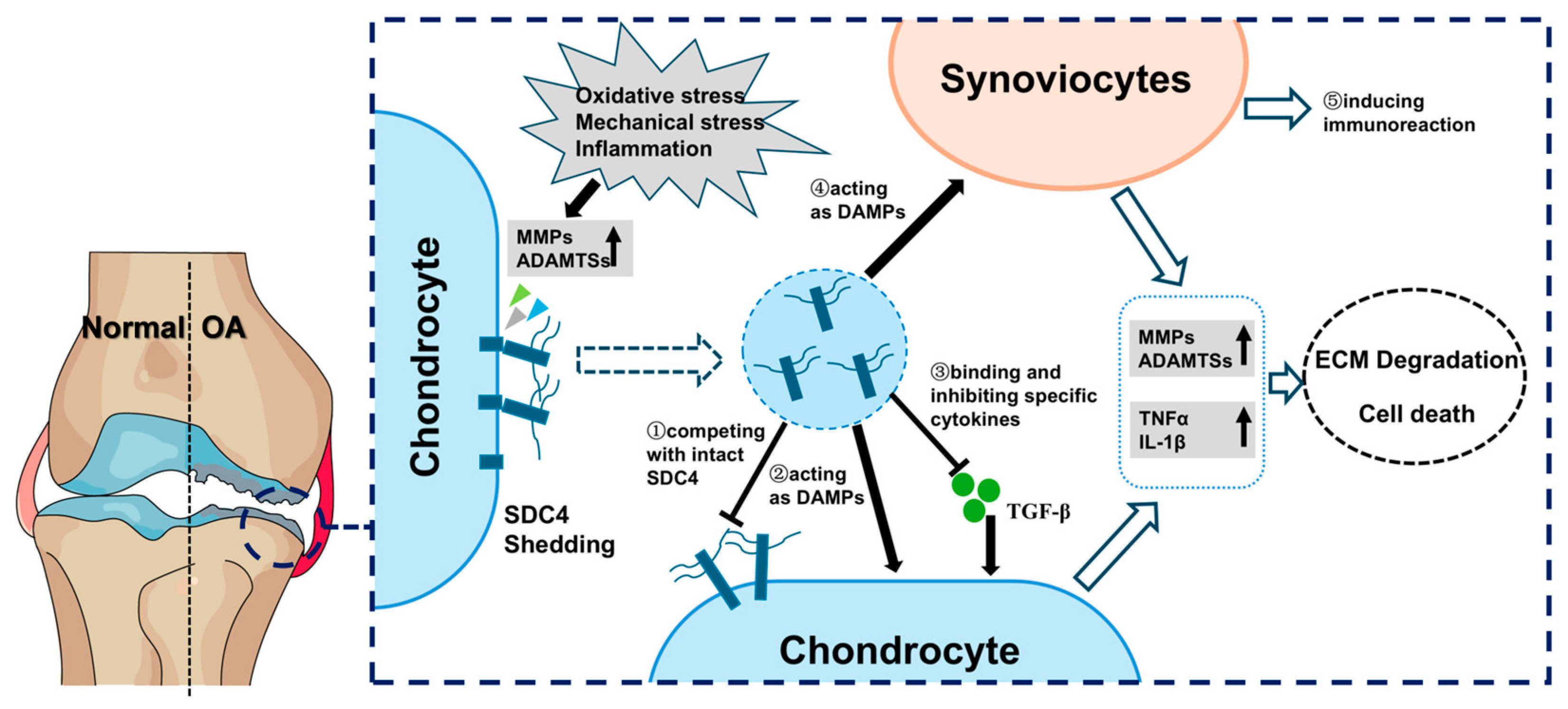
6. The Roles of sSDC4 in OA

6.1. The Level of sSDC4 Is Positively Relative to the Severity of OA

The ectodomain of SDC4 plays a significant role in the progression of OA. Emerging evidence suggests that sSDC4 may serve as a potential biomarker for OA. In 2021, Bollmann et al. observed that the synovial levels of sSDC4 in patients with knee OA significantly increased with the severity of OA, while serum levels of sSDC4 remained unchanged. Using a threshold of 72.9 pg/mL, SDC4 can distinguish between early- and late-stage OA with 67.5% sensitivity and 65.2% specificity. SDC4 shedding positively correlates with MMP-9 levels, which is upregulated in OA, and MMP-9 inhibitors have been shown to reduce SDC4 shedding. Furthermore, SDC4 shedding reduces chondrocyte sensitivity to IL-1β, contributing to disease pathogenesis. These suggest that sSDC4 levels in synovial fluid could serve as a good predictor of OA severity [17]. Similarly, in an animal model of knee osteoarthritis, elevated levels of sSDC4 in the synovial fluid were also observed [85]. However, compared to established OA biomarkers—such as inflammatory markers such as IL-6 and cartilage metabolism markers like cartilage oligomeric matrix protein—the clinical advantages of SDC4 shedding require further investigation to determine its diagnostic and prognostic superiority [86]. Additionally, some studies suggest that SDC4 plays a causal role in the progression of OA [14,87,88].

6.2. sSDC4 Can Affect Cartilage Regeneration

The shedding of SDC4 may play a role in OA through its interaction with transforming growth factor beta (TGF-β). TGF-β binds strongly to pro-inflammatory cytokines such as IL-1β and TNFα, inhibiting their deleterious effects on cartilage [89,90]. It also stimulates the secretion of proteoglycans and type II collagen from chondrocytes, which are essential for cartilage regeneration and repair [91,92]. Activation of the TGF-β/Smad signaling pathway can reduce joint inflammation, promote chondroregeneration, and slow the progression of OA. However, sSDC4, which binds to TGF-β and inhibits the activation of Smad2 and Smad3, suppresses the TGF-β/Smad signaling pathway, potentially promoting inflammation and impeding the regeneration and repair of articular cartilage [93]. Injection of anti-SDC4 antibodies may block the interactions of SDC4 on chondrocytes and sSDC4 in synovial fluid with cytokines such as ADAM metallopeptidase with thrombospondin type 1 motif 5 (ADAMTS-5) and IL-1, thereby inhibiting cartilage degradation and promoting cartilage regeneration [14]. Similarly, another study demonstrated that anti-SDC4 antibody injection upregulated miR-96-5p in chondrocytes, which targeted HIF-2α 3’-UTR sequences and suppressed HIF-2α signaling in murine cartilage tissue and chondrocytes [88]. However, a study in 2023 reported that intra-articular SDC4 injection decreased ADAMTS-5 expression and increased TIMP-3 expression, suggesting that SDC4 treatment inhibits cartilage degeneration in osteoarthritic articular cartilage, potentially enhancing cartilage regeneration [87]. These conflicting findings may be attributed to differences in experimental design, such as the concentration of sSDC4 used in the injections. Anyway, before intra-articular injection of anti-SDC4 antibodies, the precise role of sSDC4 in OA pathogenesis remains to be fully elucidated and warrants further investigation.

6.3. sSDC4 Can Act as a DAMP to Destroy the Cartilage Homeostasis

Necroptosis was the primary form of cell death in the early stage of condylar cartilage degeneration in a rat model of temporomandibular joint osteoarthritis (TMJOA). During necroptosis, SDC4 shedding occurred, and sSDC4 was released into synovial fluid in vivo or the cell supernatant in vitro. Furthermore, the stimulation of chondrocytes with the SDC4 ectodomain led to significantly higher levels of TNFα, ADAMTS5, and MMP13 in both chondrocytes and supernatants in vitro. In addition, blocking SDC4 with a specific antibody downregulated the expression of TNFα, ADAMTS5, and MMP13. Thus, sSDC4 released after chondrocyte necroptosis might independently induce subsequent cartilage degeneration. As expected, blocking SDC4 function significantly attenuated TNFα expression in cartilage and synovium, thereby recovering cartilage thickness and reducing synovial inflammation. Collectively, SDC4 might act as a key DAMP that forms a necroptotic vicious cycle of TNFα-SDC4-TNFα, leading to condylar cartilage degeneration and synovitis [94].

7. Conclusions and Perspectives

In summary, under normal physiological conditions or in response to pathological stimuli such as inflammation, oxidative stress, or mechanical stress, various proteases in cartilage (including MMPs and ADAMTSs) cleave SDC4 at specific sites, leading to the shedding of its ectodomain into the ECM. Upon shedding of the SDC4 ectodomain, the remaining membrane-bound receptor loses its ability to bind ligands, thereby impairing extracellular and intracellular SDC4 signaling and disrupting the normal interaction between chondrocyte and the ECM. On the other hand, soluble sSDC4, which retains its binding properties, can function as a paracrine or autocrine signaling molecule. In local cartilage, sSDC4 can downregulate signal transduction by competing with membrane-bound SDC4 for extracellular ligand binding and sequestering HS-binding factors in the ECM. Additionally, sSDC4 can function as a decoy receptor by competing with intact SDC4 to exert an inhibitory effect. Furthermore, soluble sSDC4 released into synovial fluid or serum can act as a DAMP to stimulate chondrocytes, synovial fibroblasts, macrophages, or other immune cells, leading to degenerative, inflammatory, or immunoreactive changes. The level of sSDC4 in synovial fluid is positively correlated with the severity of OA. Thus, the shedding of SDC4 may play a critical role in the pathogenesis and progression of OA, as shown in Figure 2, although the underlying mechanism remains unclear. Although pain is a frequent manifestation of OA, its mechanisms remain poorly understood, and the potential involvement of SDC4 in pain has received minimal research attention [95]. SDC4 interacts with fibronectin (FN), a crucial extracellular matrix protein, to regulate key signaling molecules including protein kinase C epsilon (PKCε), focal adhesion kinase (FAK), and ERK1/2. This SDC4-FN interaction mediates cell adhesion, mechanotransduction, and cytoskeletal remodeling, ultimately modulating the growth, morphology, and mechanical sensitivity of dorsal root ganglion (DRG) neurons [96]. Given that DRG neurons are essential for transmitting pain signals to the central nervous system, these findings suggest that SDC4 may contribute to OA pain pathogenesis by altering DRG neuron structure and function. However, further investigation on this mechanism, or the effect of sSDC4 on nervous tissues or the expression of pain related cytokines in joint tissues, is warranted to fully elucidate it [97]. To mitigate the effects of the shedding of SDC4 or sSDC4, therapeutic strategies may include the use of specific protease inhibitors to block SDC4 shedding or the intra-articular injection of anti-sSDC4 antibodies. Thus, in future studies, elucidating the specific molecular mechanisms of SDC4 shedding in the OA process and the detailed and main effect of sSDC4 on the OA process will help to identify and validate potential therapeutic targets, providing new strategies for effective OA treatment.

Author Contributions

S.Y., F.L., X.C., and F.H. contributed to the conception and design of the manuscript. K.H. and H.R. contributed to literature collection and drafting the manuscript. H.Z. and Y.Z. contributed to manuscript refinement and editing. All authors have read and agreed to the published version of the manuscript.

Funding

This work was supported by the National Natural Science Foundation of China (Nos. 82271000, 82401153, and 82201089) and the Shaanxi Province Science Foundation (2022JZ-52).

Conflicts of Interest

The authors declare no conflicts of interest.

Abbreviations

The following abbreviations are used in this manuscript:
ADAMsa disintegrin and metalloproteinases
ADAMTSa disintegrin and metalloproteinase with thrombospondin motifs
AGEsadvanced glycation end products
ASMairway smooth muscle
ASMCairway smooth muscle cells
AMPKadenosine monophosphate-activated protein kinase
ACSL4acyl-coA synthetase long chain family member 4
ACANaggrecan
CCL5C-C motif chemokine ligand 5
CCR5C-C chemokine receptor type 5
CXCR4C-X-C chemokine receptor type 4
CXCL12C-X-C motif chemokine ligand 12
COL2A1collagen type II alpha 1
DAMPdamage-associated molecular pattern
DRGdorsal root ganglion
ECMextracellular matrix
EPCendothelial progenitor cell
EGendothelial glycocalyx
ERKextracellular regulated protein kinases
FNfibronectin
FAKfocal adhesion kinase
FGFfibroblast growth factor
VEGFAvascular endothelial growth factor A
VEGFRvascular endothelial growth factor receptor
GAGgalactosylated glycosaminoglycan
GPCRG protein-coupled receptor
GPX4glutathione peroxidase 4
HSPGsheparan sulfate proteoglycans
HSheparan sulfate
HIF-2αhypoxia-inducible factor 2 alpha
HIF-1hypoxia-inducible factor 1
IL-1βinterleukin-1β
IL-1RaIL-1 receptor antagonist
IL-6interleukin-6
LPSlipopolysaccharides
LDLlow-density lipoprotein
LPLlipoprotein lipase
LRPlow-density lipoprotein receptor-related protein
MMPsmatrix metalloproteinases
MAPKmitogen-activated protein kinase
NACN-acetylcysteine
NF-κBnuclear factor kappa-B
NLRP3NOD-like receptor family, pyrin domain containing 3
OAosteoarthritis
OPNosteopontin
PKCprotein kinase C
PMAphorbol myristate acetate
PKCεprotein kinase C epsilon
PGC-1αperoxisome proliferator-activated receptor gamma coactivator 1-alpha
ROSreactive oxygen species
RAGEreceptor for advanced glycation endproducts
SDC4syndecan-4
SDCsyndecan
sSDC4shed syndecan-4
SRCscleroderma renal crisis
STAT3signal transducer and activator of transcription 3
Sirtuin1silent information regulator factor 1
Sirt1endo−/−endothelial sirtuin1-deficient
SASPsenescence-associated secretory phenotype
SDF-1stromal cell-derived factor-1
SLC7A11solute carrier family 7 member 11
TNFαtumor necrosis factor-α
TIMPtissue inhibitor of metalloproteinase
TLR4toll-like receptor 4
TGF-βtransforming growth factor beta
TMJOAtemporomandibular joint osteoarthritis
UUOUnilateral Ureteral Obstruction

References

  1. Hunter, D.J.; Bierma-Zeinstra, S. Osteoarthritis. Lancet 2019, 393, 1745–1759. [Google Scholar] [CrossRef]
  2. Yao, Q.; Wu, X.; Tao, C.; Gong, W.; Chen, M.; Qu, M.; Zhong, Y.; He, T.; Chen, S.; Xiao, G. Osteoarthritis: Pathogenic Signaling Pathways and Therapeutic Targets. Signal Transduct. Target. Ther. 2023, 8, 56. [Google Scholar] [CrossRef]
  3. He, F.; Wu, H.; He, B.; Han, Z.; Chen, J.; Huang, L. Antioxidant Hydrogels for the Treatment of Osteoarthritis: Mechanisms and Recent Advances. Front. Pharmacol. 2024, 15, 1488036. [Google Scholar] [CrossRef]
  4. Matsui, Y.; Hasegawa, M.; Iino, T.; Imanaka-Yoshida, K.; Yoshida, T.; Sudo, A. Tenascin-C Prevents Articular Cartilage Degeneration in Murine Osteoarthritis Models. Cartilage 2018, 9, 80–88. [Google Scholar] [CrossRef]
  5. Hu, Y.; Chen, X.; Wang, S.; Jing, Y.; Su, J. Subchondral Bone Microenvironment in Osteoarthritis and Pain. Bone Res. 2021, 9, 20. [Google Scholar] [CrossRef]
  6. Gao, Y.; Liu, S.; Huang, J.; Guo, W.; Chen, J.; Zhang, L.; Zhao, B.; Peng, J.; Wang, A.; Wang, Y.; et al. The ECM-Cell Interaction of Cartilage Extracellular Matrix on Chondrocytes. Biomed. Res. Int. 2014, 2014, 648459. [Google Scholar] [CrossRef]
  7. Bertrand, J.; Bollmann, M. Soluble Syndecans: Biomarkers for Diseases and Therapeutic Options. Br. J. Pharmacol. 2019, 176, 67–81. [Google Scholar] [CrossRef]
  8. Echtermeyer, F.; Bertrand, J.; Dreier, R.; Meinecke, I.; Neugebauer, K.; Fuerst, M.; Lee, Y.J.; Song, Y.W.; Herzog, C.; Theilmeier, G.; et al. Syndecan-4 Regulates ADAMTS-5 Activation and Cartilage Breakdown in Osteoarthritis. Nat. Med. 2009, 15, 1072–1076. [Google Scholar] [CrossRef]
  9. Brioudes, E.; Alibashe-Ahmed, M.; Lavallard, V.; Berney, T.; Bosco, D. Syndecan-4 Is Regulated by IL-1β in β-Cells and Human Islets. Mol. Cell Endocrinol. 2020, 510, 110815. [Google Scholar] [CrossRef]
  10. Li, R.; Xie, J.; Wu, H.; Li, G.; Chen, J.; Chen, Q.; Wang, L.; Xu, B. Syndecan-4 Shedding Impairs Macrovascular Angiogenesis in Diabetes Mellitus. Biochem. Biophys. Res. Commun. 2016, 474, 15–21. [Google Scholar] [CrossRef]
  11. Cao, C.; Shi, Y.; Zhang, X.; Li, Q.; Zhang, J.; Zhao, F.; Meng, Q.; Dai, W.; Liu, Z.; Yan, W.; et al. Cholesterol-Induced LRP3 Downregulation Promotes Cartilage Degeneration in Osteoarthritis by Targeting Syndecan-4. Nat. Commun. 2022, 13, 7139. [Google Scholar] [CrossRef]
  12. Chronopoulos, A.; Thorpe, S.D.; Cortes, E.; Lachowski, D.; Rice, A.J.; Mykuliak, V.V.; Róg, T.; Lee, D.A.; Hytönen, V.P.; Del Río Hernández, A.E. Syndecan-4 Tunes Cell Mechanics by Activating the Kindlin-Integrin-RhoA Pathway. Nat. Mater. 2020, 19, 669–678. [Google Scholar] [CrossRef]
  13. Nikaido, T.; Tanino, Y.; Wang, X.; Sato, S.; Misa, K.; Fukuhara, N.; Sato, Y.; Fukuhara, A.; Uematsu, M.; Suzuki, Y.; et al. Serum Syndecan-4 as a Possible Biomarker in Patients With Acute Pneumonia. J. Infect. Dis. 2015, 212, 1500–1508. [Google Scholar] [CrossRef]
  14. Chen, X.; He, F.; Zhang, H.; Ma, Y.; Yu, J.; Qin, H.; Wu, F.; Wang, Z.; Zhan, Y.; Zhang, J.; et al. Syndecan-4 Inhibition Attenuates Cartilage Degeneration in Temporomandibular Joint Osteoarthritis. J. Oral. Rehabil. 2024, 51, 2324–2335. [Google Scholar] [CrossRef]
  15. Jalkanen, M.; Rapraeger, A.; Saunders, S.; Bernfield, M. Cell Surface Proteoglycan of Mouse Mammary Epithelial Cells Is Shed by Cleavage of Its Matrix-Binding Ectodomain from Its Membrane-Associated Domain. J. Cell Biol. 1987, 105, 3087–3096. [Google Scholar] [CrossRef]
  16. Manon-Jensen, T.; Multhaupt, H.A.B.; Couchman, J.R. Mapping of Matrix Metalloproteinase Cleavage Sites on Syndecan-1 and Syndecan-4 Ectodomains. FEBS J. 2013, 280, 2320–2331. [Google Scholar] [CrossRef]
  17. Bollmann, M.; Pinno, K.; Ehnold, L.I.; Märtens, N.; Märtson, A.; Pap, T.; Stärke, C.; Lohmann, C.H.; Bertrand, J. MMP-9 Mediated Syndecan-4 Shedding Correlates with Osteoarthritis Severity. Osteoarthr. Cartil. 2021, 29, 280–289. [Google Scholar] [CrossRef]
  18. Strand, M.E.; Vanhaverbeke, M.; Henkens, M.T.H.M.; Sikking, M.A.; Rypdal, K.B.; Braathen, B.; Almaas, V.M.; Tønnessen, T.; Christensen, G.; Heymans, S.; et al. Inflammation and Syndecan-4 Shedding from Cardiac Cells in Ischemic and Non-Ischemic Heart Disease. Biomedicines 2023, 11, 1066. [Google Scholar] [CrossRef]
  19. Strand, M.E.; Aronsen, J.M.; Braathen, B.; Sjaastad, I.; Kvaløy, H.; Tønnessen, T.; Christensen, G.; Lunde, I.G. Shedding of Syndecan-4 Promotes Immune Cell Recruitment and Mitigates Cardiac Dysfunction after Lipopolysaccharide Challenge in Mice. J. Mol. Cell Cardiol. 2015, 88, 133–144. [Google Scholar] [CrossRef]
  20. Lee, S.; Kolset, S.O.; Birkeland, K.I.; Drevon, C.A.; Reine, T.M. Acute Exercise Increases Syndecan-1 and -4 Serum Concentrations. Glycoconj. J. 2019, 36, 113–125. [Google Scholar] [CrossRef]
  21. Solbu, M.D.; Kolset, S.O.; Jenssen, T.G.; Wilsgaard, T.; Løchen, M.-L.; Mathiesen, E.B.; Melsom, T.; Eriksen, B.O.; Reine, T.M. Gender Differences in the Association of Syndecan-4 with Myocardial Infarction: The Population-Based Tromsø Study. Atherosclerosis 2018, 278, 166–173. [Google Scholar] [CrossRef]
  22. Smart, L.; Macdonald, S.P.J.; Burrows, S.; Bosio, E.; Arendts, G.; Fatovich, D.M. Endothelial Glycocalyx Biomarkers Increase in Patients with Infection during Emergency Department Treatment. J. Crit. Care 2017, 42, 304–309. [Google Scholar] [CrossRef]
  23. Gondelaud, F.; Ricard-Blum, S. Structures and Interactions of Syndecans. FEBS J. 2019, 286, 2994–3007. [Google Scholar] [CrossRef]
  24. Iozzo, R.V.; Schaefer, L. Proteoglycan Form and Function: A Comprehensive Nomenclature of Proteoglycans. Matrix Biol. 2015, 42, 11–55. [Google Scholar] [CrossRef]
  25. Qin, Y.; Zhu, Y.; Luo, F.; Chen, C.; Chen, X.; Wu, M. Killing Two Birds with One Stone: Dual Blockade of Integrin and FGF Signaling through Targeting Syndecan-4 in Postoperative Capsular Opacification. Cell Death Dis. 2017, 8, e2920. [Google Scholar] [CrossRef]
  26. McFall, A.J.; Rapraeger, A.C. Identification of an Adhesion Site within the Syndecan-4 Extracellular Protein Domain. J. Biol. Chem. 1997, 272, 12901–12904. [Google Scholar] [CrossRef]
  27. Ramani, V.C.; Pruett, P.S.; Thompson, C.A.; DeLucas, L.D.; Sanderson, R.D. Heparan Sulfate Chains of Syndecan-1 Regulate Ectodomain Shedding. J. Biol. Chem. 2012, 287, 9952–9961. [Google Scholar] [CrossRef]
  28. Ramnath, R.; Foster, R.R.; Qiu, Y.; Cope, G.; Butler, M.J.; Salmon, A.H.; Mathieson, P.W.; Coward, R.J.; Welsh, G.I.; Satchell, S.C. Matrix Metalloproteinase 9-Mediated Shedding of Syndecan 4 in Response to Tumor Necrosis Factor α: A Contributor to Endothelial Cell Glycocalyx Dysfunction. FASEB J. 2014, 28, 4686–4699. [Google Scholar] [CrossRef]
  29. Fitzgerald, M.L.; Wang, Z.; Park, P.W.; Murphy, G.; Bernfield, M. Shedding of Syndecan-1 and -4 Ectodomains Is Regulated by Multiple Signaling Pathways and Mediated by a TIMP-3-Sensitive Metalloproteinase. J. Cell Biol. 2000, 148, 811–824. [Google Scholar] [CrossRef]
  30. Schmidt, A.; Echtermeyer, F.; Alozie, A.; Brands, K.; Buddecke, E. Plasmin- and Thrombin-Accelerated Shedding of Syndecan-4 Ectodomain Generates Cleavage Sites at Lys(114)-Arg(115) and Lys(129)-Val(130) Bonds. J. Biol. Chem. 2005, 280, 34441–34446. [Google Scholar] [CrossRef]
  31. Pruessmeyer, J.; Martin, C.; Hess, F.M.; Schwarz, N.; Schmidt, S.; Kogel, T.; Hoettecke, N.; Schmidt, B.; Sechi, A.; Uhlig, S.; et al. A Disintegrin and Metalloproteinase 17 (ADAM17) Mediates Inflammation-Induced Shedding of Syndecan-1 and -4 by Lung Epithelial Cells. J. Biol. Chem. 2010, 285, 555–564. [Google Scholar] [CrossRef]
  32. Arner, E.C.; Pratta, M.A.; Trzaskos, J.M.; Decicco, C.P.; Tortorella, M.D. Generation and Characterization of Aggrecanase. A Soluble, Cartilage-Derived Aggrecan-Degrading Activity. J. Biol. Chem. 1999, 274, 6594–6601. [Google Scholar] [CrossRef]
  33. Tortorella, M.D.; Pratta, M.; Liu, R.Q.; Austin, J.; Ross, O.H.; Abbaszade, I.; Burn, T.; Arner, E. Sites of Aggrecan Cleavage by Recombinant Human Aggrecanase-1 (ADAMTS-4). J. Biol. Chem. 2000, 275, 18566–18573. [Google Scholar] [CrossRef]
  34. Fears, C.Y.; Gladson, C.L.; Woods, A. Syndecan-2 Is Expressed in the Microvasculature of Gliomas and Regulates Angiogenic Processes in Microvascular Endothelial Cells. J. Biol. Chem. 2006, 281, 14533–14536. [Google Scholar] [CrossRef]
  35. Ali, M.F.; Dasari, H.; Van Keulen, V.P.; Cornec, D.; Vasmatzis, G.; Peikert, T.; Carmona, E.M. Microbial Antigens Stimulate Metalloprotease-7 Secretion in Human B-Lymphocytes Using mTOR-Dependent and Independent Pathways. Sci. Rep. 2017, 7, 3869. [Google Scholar] [CrossRef]
  36. Reine, T.M.; Lanzalaco, F.; Kristiansen, O.; Enget, A.R.; Satchell, S.; Jenssen, T.G.; Kolset, S.O. Matrix Metalloproteinase-9 Mediated Shedding of Syndecan-4 in Glomerular Endothelial Cells. Microcirculation 2019, 26, e12534. [Google Scholar] [CrossRef]
  37. Brule, S.; Charnaux, N.; Sutton, A.; Ledoux, D.; Chaigneau, T.; Saffar, L.; Gattegno, L. The Shedding of Syndecan-4 and Syndecan-1 from HeLa Cells and Human Primary Macrophages Is Accelerated by SDF-1/CXCL12 and Mediated by the Matrix Metalloproteinase-9. Glycobiology 2006, 16, 488–501. [Google Scholar] [CrossRef]
  38. Rodríguez-Manzaneque, J.C.; Carpizo, D.; Plaza-Calonge, M.d.C.; Torres-Collado, A.X.; Thai, S.N.-M.; Simons, M.; Horowitz, A.; Iruela-Arispe, M.L. Cleavage of Syndecan-4 by ADAMTS1 Provokes Defects in Adhesion. Int. J. Biochem. Cell Biol. 2009, 41, 800–810. [Google Scholar] [CrossRef]
  39. Subramanian, S.V.; Fitzgerald, M.L.; Bernfield, M. Regulated Shedding of Syndecan-1 and -4 Ectodomains by Thrombin and Growth Factor Receptor Activation. J. Biol. Chem. 1997, 272, 14713–14720. [Google Scholar] [CrossRef]
  40. Park, P.W.; Foster, T.J.; Nishi, E.; Duncan, S.J.; Klagsbrun, M.; Chen, Y. Activation of Syndecan-1 Ectodomain Shedding by Staphylococcus Aureus Alpha-Toxin and Beta-Toxin. J. Biol. Chem. 2004, 279, 251–258. [Google Scholar] [CrossRef]
  41. Manon-Jensen, T.; Itoh, Y.; Couchman, J.R. Proteoglycans in Health and Disease: The Multiple Roles of Syndecan Shedding. FEBS J. 2010, 277, 3876–3889. [Google Scholar] [CrossRef]
  42. Strand, M.E.; Herum, K.M.; Rana, Z.A.; Skrbic, B.; Askevold, E.T.; Dahl, C.P.; Vistnes, M.; Hasic, A.; Kvaløy, H.; Sjaastad, I.; et al. Innate Immune Signaling Induces Expression and Shedding of the Heparan Sulfate Proteoglycan Syndecan-4 in Cardiac Fibroblasts and Myocytes, Affecting Inflammation in the Pressure-Overloaded Heart. FEBS J. 2013, 280, 2228–2247. [Google Scholar] [CrossRef]
  43. Houston, M.; Julien, M.A.; Parthasarathy, S.; Chaikof, E.L. Oxidized Linoleic Acid Regulates Expression and Shedding of Syndecan-4. Am. J. Physiol. Cell Physiol. 2005, 288, C458–C466. [Google Scholar] [CrossRef]
  44. Sato, Y.; Tanino, Y.; Wang, X.; Nikaido, T.; Sato, S.; Misa, K.; Togawa, R.; Frevert, C.W.; Munakata, M. Baseline Serum Syndecan-4 Predicts Prognosis after the Onset of Acute Exacerbation of Idiopathic Interstitial Pneumonia. PLoS ONE 2017, 12, e0176789. [Google Scholar] [CrossRef]
  45. Luo, Q.; Ning, P.; Zheng, Y.; Shang, Y.; Zhou, B.; Gao, Z. Serum suPAR and Syndecan-4 Levels Predict Severity of Community-Acquired Pneumonia: A Prospective, Multi-Centre Study. Crit. Care 2018, 22, 15. [Google Scholar] [CrossRef]
  46. Nakao, M.; Sugaya, M.; Takahashi, N.; Otobe, S.; Nakajima, R.; Oka, T.; Kabasawa, M.; Suga, H.; Morimura, S.; Miyagaki, T.; et al. Increased Syndecan-4 Expression in Sera and Skin of Patients with Atopic Dermatitis. Arch. Dermatol. Res. 2016, 308, 655–660. [Google Scholar] [CrossRef]
  47. Shaik, F.; Balderstone, M.J.M.; Arokiasamy, S.; Whiteford, J.R. Roles of Syndecan-4 in Cardiac Injury and Repair. Int. J. Biochem. Cell Biol. 2022, 146, 106196. [Google Scholar] [CrossRef]
  48. Ansari, M.Y.; Ahmad, N.; Haqqi, T.M. Oxidative Stress and Inflammation in Osteoarthritis Pathogenesis: Role of Polyphenols. Biomed. Pharmacother. 2020, 129, 110452. [Google Scholar] [CrossRef]
  49. Wang, Y.; Herrera, A.H.; Li, Y.; Belani, K.K.; Walcheck, B. Regulation of Mature ADAM17 by Redox Agents for L-Selectin Shedding. J. Immunol. 2009, 182, 2449–2457. [Google Scholar] [CrossRef]
  50. Lipphardt, M.; Song, J.W.; Ratliff, B.B.; Dihazi, H.; Müller, G.A.; Goligorsky, M.S. Endothelial Dysfunction Is a Superinducer of Syndecan-4: Fibrogenic Role of Its Ectodomain. Am. J. Physiol. Heart Circ. Physiol. 2018, 314, H484–H496. [Google Scholar] [CrossRef]
  51. Wu, H.; Chen, Z.; Chen, J.-Z.; Xie, J.; Xu, B. Resveratrol Improves Tube Formation in AGE-Induced Late Endothelial Progenitor Cells by Suppressing Syndecan-4 Shedding. Oxid. Med. Cell Longev. 2018, 2018, 9045976. [Google Scholar] [CrossRef]
  52. Xie, J.; Li, R.; Wu, H.; Chen, J.; Li, G.; Chen, Q.; Wei, Z.; He, G.; Wang, L.; Ferro, A.; et al. Advanced Glycation Endproducts Impair Endothelial Progenitor Cell Migration and Homing via Syndecan 4 Shedding. Stem Cells 2017, 35, 522–531. [Google Scholar] [CrossRef] [PubMed]
  53. Wu, H.; Zhou, Q.; Xie, J.; Li, G.-N.; Chen, Q.-H.; Kang, L.-N.; Xu, B. Syndecan-4 Shedding Is Involved in the Oxidative Stress and Inflammatory Responses in Left Atrial Tissue with Valvular Atrial Fibrillation. Int. J. Clin. Exp. Pathol. 2015, 8, 6387–6396. [Google Scholar] [PubMed]
  54. Altindag, O.; Erel, O.; Aksoy, N.; Selek, S.; Celik, H.; Karaoglanoglu, M. Increased Oxidative Stress and Its Relation with Collagen Metabolism in Knee Osteoarthritis. Rheumatol. Int. 2007, 27, 339–344. [Google Scholar] [CrossRef] [PubMed]
  55. Bolduc, J.A.; Collins, J.A.; Loeser, R.F. Reactive Oxygen Species, Aging and Articular Cartilage Homeostasis. Free Radic. Biol. Med. 2019, 132, 73–82. [Google Scholar] [CrossRef]
  56. Jiang, X.Z.; Luo, K.H.; Ventikos, Y. Principal Mode of Syndecan-4 Mechanotransduction for the Endothelial Glycocalyx Is a Scissor-like Dimer Motion. Acta Physiol. 2020, 228, e13376. [Google Scholar] [CrossRef]
  57. Sanchez-Lopez, E.; Coras, R.; Torres, A.; Lane, N.E.; Guma, M. Synovial Inflammation in Osteoarthritis Progression. Nat. Rev. Rheumatol. 2022, 18, 258–275. [Google Scholar] [CrossRef]
  58. Ringvold, H.C.; Khalil, R.A. Protein Kinase C as Regulator of Vascular Smooth Muscle Function and Potential Target in Vascular Disorders. Adv. Pharmacol. 2017, 78, 203–301. [Google Scholar] [CrossRef]
  59. Charnaux, N.; Brule, S.; Chaigneau, T.; Saffar, L.; Sutton, A.; Hamon, M.; Prost, C.; Lievre, N.; Vita, C.; Gattegno, L. RANTES (CCL5) Induces a CCR5-Dependent Accelerated Shedding of Syndecan-1 (CD138) and Syndecan-4 from HeLa Cells and Forms Complexes with the Shed Ectodomains of These Proteoglycans as Well as with Those of CD44. Glycobiology 2005, 15, 119–130. [Google Scholar] [CrossRef]
  60. Wang, S.; Mobasheri, A.; Zhang, Y.; Wang, Y.; Dai, T.; Zhang, Z. Exogenous Stromal Cell-Derived Factor-1 (SDF-1) Suppresses the NLRP3 Inflammasome and Inhibits Pyroptosis in Synoviocytes from Osteoarthritic Joints via Activation of the AMPK Signaling Pathway. Inflammopharmacology 2021, 29, 695–704. [Google Scholar] [CrossRef]
  61. Zhao, Y.; Lin, D.; Zhu, X.; Yan, J.; Liang, Y.; Wang, Y.; Dai, T.; Zhang, Z.; Wang, S. SDF-1 Alleviates Osteoarthritis by Resolving Mitochondrial Dysfunction through the Activation of the Sirt3/PGC-1α Signalling Pathway. Arthritis Res. Ther. 2025, 27, 51. [Google Scholar] [CrossRef]
  62. Li, J.; Chen, H.; Cai, L.; Guo, D.; Zhang, D.; Zhou, X.; Xie, J. SDF-1α Promotes Chondrocyte Autophagy through CXCR4/mTOR Signaling Axis. Int. J. Mol. Sci. 2023, 24, 1710. [Google Scholar] [CrossRef] [PubMed]
  63. Li, J.; Chen, H.; Zhang, D.; Xie, J.; Zhou, X. The Role of Stromal Cell-Derived Factor 1 on Cartilage Development and Disease. Osteoarthr. Cartil. 2021, 29, 313–322. [Google Scholar] [CrossRef]
  64. Yang, T.; Yang, X.; Wang, G.; Jia, D.; Li, Y. Unraveling the Crucial Role of SDF-1 in Osteoarthritis Progression: IL6/HIF-1α Positive Feedback and Chondrocyte Ferroptosis. Int. Immunopharmacol. 2025, 152, 114400. [Google Scholar] [CrossRef] [PubMed]
  65. Herum, K.M.; Romaine, A.; Wang, A.; Melleby, A.O.; Strand, M.E.; Pacheco, J.; Braathen, B.; Dunér, P.; Tønnessen, T.; Lunde, I.G.; et al. Syndecan-4 Protects the Heart From the Profibrotic Effects of Thrombin-Cleaved Osteopontin. J. Am. Heart Assoc. 2020, 9, e013518. [Google Scholar] [CrossRef]
  66. Kim, E.Y.; Roshanravan, H.; Dryer, S.E. Syndecan-4 Ectodomain Evokes Mobilization of Podocyte TRPC6 Channels and Their Associated Pathways: An Essential Role for Integrin Signaling. Biochim. Biophys. Acta 2015, 1853, 2610–2620. [Google Scholar] [CrossRef]
  67. Hayashida, K.; Chen, Y.; Bartlett, A.H.; Park, P.W. Syndecan-1 Is an In Vivo Suppressor of Gram-Positive Toxic Shock*. J. Biol. Chem. 2008, 283, 19895–19903. [Google Scholar] [CrossRef] [PubMed]
  68. Reizes, O.; Goldberger, O.; Smith, A.C.; Xu, Z.; Bernfield, M.; Bickel, P.E. Insulin Promotes Shedding of Syndecan Ectodomains from 3T3-L1 Adipocytes: A Proposed Mechanism for Stabilization of Extracellular Lipoprotein Lipase. Biochemistry 2006, 45, 5703–5711. [Google Scholar] [CrossRef]
  69. Lambert, J.; Makin, K.; Akbareian, S.; Johnson, R.; Alghamdi, A.A.A.; Robinson, S.D.; Edwards, D.R. ADAMTS-1 and Syndecan-4 Intersect in the Regulation of Cell Migration and Angiogenesis. J. Cell Sci. 2020, 133, jcs235762. [Google Scholar] [CrossRef]
  70. Endo, K.; Takino, T.; Miyamori, H.; Kinsen, H.; Yoshizaki, T.; Furukawa, M.; Sato, H. Cleavage of Syndecan-1 by Membrane Type Matrix Metalloproteinase-1 Stimulates Cell Migration. J. Biol. Chem. 2003, 278, 40764–40770. [Google Scholar] [CrossRef]
  71. Nikolova, V.; Koo, C.-Y.; Ibrahim, S.A.; Wang, Z.; Spillmann, D.; Dreier, R.; Kelsch, R.; Fischgräbe, J.; Smollich, M.; Rossi, L.H.; et al. Differential Roles for Membrane-Bound and Soluble Syndecan-1 (CD138) in Breast Cancer Progression. Carcinogenesis 2009, 30, 397–407. [Google Scholar] [CrossRef] [PubMed]
  72. Seidel, C.; Sundan, A.; Hjorth, M.; Turesson, I.; Dahl, I.M.; Abildgaard, N.; Waage, A.; Borset, M. Serum Syndecan-1: A New Independent Prognostic Marker in Multiple Myeloma. Blood 2000, 95, 388–392. [Google Scholar] [CrossRef]
  73. Su, G.; Blaine, S.A.; Qiao, D.; Friedl, A. Membrane Type 1 Matrix Metalloproteinase-Mediated Stromal Syndecan-1 Shedding Stimulates Breast Carcinoma Cell Proliferation. Cancer Res. 2008, 68, 9558–9565. [Google Scholar] [CrossRef] [PubMed]
  74. Beauvais, D.M.; Ell, B.J.; McWhorter, A.R.; Rapraeger, A.C. Syndecan-1 Regulates Alphavbeta3 and Alphavbeta5 Integrin Activation during Angiogenesis and Is Blocked by Synstatin, a Novel Peptide Inhibitor. J. Exp. Med. 2009, 206, 691–705. [Google Scholar] [CrossRef]
  75. Pap, T.; Bertrand, J. Syndecans in Cartilage Breakdown and Synovial Inflammation. Nat. Rev. Rheumatol. 2013, 9, 43–55. [Google Scholar] [CrossRef] [PubMed]
  76. Onyeisi, J.O.S.; Lopes, C.C.; Götte, M. Role of Syndecan-4 in Breast Cancer Pathophysiology. Am. J. Physiol. Cell Physiol. 2022, 323, C1345–C1354. [Google Scholar] [CrossRef]
  77. Pham, S.H.; Pratt, K.; Okolicsanyi, R.K.; Oikari, L.E.; Yu, C.; Peall, I.W.; Arif, K.T.; Chalmers, T.-A.; Gyimesi, M.; Griffiths, L.R.; et al. Syndecan-1 and -4 Influence Wnt Signaling and Cell Migration in Human Breast Cancers. Biochimie 2022, 198, 60–75. [Google Scholar] [CrossRef]
  78. Tan, X.; Khalil, N.; Tesarik, C.; Vanapalli, K.; Yaputra, V.; Alkhouri, H.; Oliver, B.G.G.; Armour, C.L.; Hughes, J.M. Th1 Cytokine-Induced Syndecan-4 Shedding by Airway Smooth Muscle Cells Is Dependent on Mitogen-Activated Protein Kinases. Am. J. Physiol. Lung Cell Mol. Physiol. 2012, 302, L700–L710. [Google Scholar] [CrossRef]
  79. Kunnathattil, M.; Rahul, P.; Skaria, T. Soluble Vascular Endothelial Glycocalyx Proteoglycans as Potential Therapeutic Targets in Inflammatory Diseases. Immunol. Cell Biol. 2024, 102, 97–116. [Google Scholar] [CrossRef]
  80. Jannaway, M.; Yang, X.; Meegan, J.E.; Coleman, D.C.; Yuan, S.Y. Thrombin-Cleaved Syndecan-3/-4 Ectodomain Fragments Mediate Endothelial Barrier Dysfunction. PLoS ONE 2019, 14, e0214737. [Google Scholar] [CrossRef]
  81. Milford, E.M.; Meital, L.; Kuballa, A.; Reade, M.C.; Russell, F.D. Fingolimod Does Not Prevent Syndecan-4 Shedding from the Endothelial Glycocalyx in a Cultured Human Umbilical Vein Endothelial Cell Model of Vascular Injury. Intensive Care Med. Exp. 2022, 10, 34. [Google Scholar] [CrossRef] [PubMed]
  82. Brooks, R.; Wei, X.; Lei, M.L.; Cid, F.C.; Roper, J.A.; Williamson, R.C.; Bass, M.D. Inhibition of EphA2 by Syndecan-4 in Wounded Skin Regulates Clustering of Fibroblasts. J. Mol. Cell Biol. 2024, mjae054. [Google Scholar] [CrossRef]
  83. Couchman, J.R.; Gopal, S.; Lim, H.C.; Nørgaard, S.; Multhaupt, H.A.B. Fell-Muir Lecture: Syndecans: From Peripheral Coreceptors to Mainstream Regulators of Cell Behaviour. Int. J. Exp. Pathol. 2015, 96, 1–10. [Google Scholar] [CrossRef]
  84. Scarpellini, A.; Huang, L.; Burhan, I.; Schroeder, N.; Funck, M.; Johnson, T.S.; Verderio, E.A.M. Syndecan-4 Knockout Leads to Reduced Extracellular Transglutaminase-2 and Protects against Tubulointerstitial Fibrosis. J. Am. Soc. Nephrol. 2014, 25, 1013–1027. [Google Scholar] [CrossRef] [PubMed]
  85. Ye, C.; Yang, Z.; Li, P.; Lu, Y.; Lyu, T.; Liu, Y.; Zhou, X.; Zhao, P.; Chen, J. Duhuo Jisheng Decoction in Reduction of Inflammatory Response via Transforming Growth Factor-β/Smad Signaling Pathway for Repairing Rabbit Articular Cartilage Injury: A Randomized Controlled Trial. Int. Immunopharmacol. 2024, 144, 113646. [Google Scholar] [CrossRef] [PubMed]
  86. Hlubek, R.; Kušnierová, P.; Walder, P.; Bystroňová, I.; Douša, P. Biomarkers and Their Role in Understanding Osteoarthritis. Acta Chir. Orthop. Traumatol. Cech. 2025, 92, 36–43. [Google Scholar] [CrossRef]
  87. Hattori, Y.; Hasegawa, M.; Iino, T.; Imanaka-Yoshida, K.; Sudo, A. Role of Syndecan-4 in the Inhibition of Articular Cartilage Degeneration in Osteoarthritis. Biomedicines 2023, 11, 2257. [Google Scholar] [CrossRef]
  88. Zhou, K.; He, S.; Yu, H.; Pei, F.; Zhou, Z. Inhibition of Syndecan-4 Reduces Cartilage Degradation in Murine Models of Osteoarthritis through the Downregulation of HIF-2α by miR-96-5p. Lab. Investig. 2021, 101, 1060–1070. [Google Scholar] [CrossRef]
  89. Wang, L.; He, C. Nrf2-Mediated Anti-Inflammatory Polarization of Macrophages as Therapeutic Targets for Osteoarthritis. Front. Immunol. 2022, 13, 967193. [Google Scholar] [CrossRef]
  90. Zhai, G.; Doré, J.; Rahman, P. TGF-β Signal Transduction Pathways and Osteoarthritis. Rheumatol. Int. 2015, 35, 1283–1292. [Google Scholar] [CrossRef]
  91. Yu, J.-X.; Zhang, X.-T.; Liao, Y.-Q.; Zhang, Q.-Y.; Chen, H.; Lin, M.; Kumar, S. Relationship between Expression of CD105 and Growth Factors in Malignant Tumors of Gastrointestinal Tract and Its Significance. World J. Gastroenterol. 2003, 9, 2866–2869. [Google Scholar] [CrossRef] [PubMed]
  92. Piestrzeniewicz-Ulanska, D.; Brys, M.; Semczuk, A.; Jakowicki, J.A.; Krajewska, W.M. Expression of TGF-β Type I and II Receptors in Normal and Cancerous Human Endometrium. Cancer Lett. 2002, 186, 231–239. [Google Scholar] [CrossRef]
  93. Tanino, Y.; Wang, X.; Nikaido, T.; Misa, K.; Sato, Y.; Togawa, R.; Kawamata, T.; Kikuchi, M.; Frevert, C.W.; Tanino, M.; et al. Syndecan-4 Inhibits the Development of Pulmonary Fibrosis by Attenuating TGF-β Signaling. Int. J. Mol. Sci. 2019, 20, 4989. [Google Scholar] [CrossRef] [PubMed]
  94. He, F.; Ma, Y.; Li, S.; Ren, H.; Liu, Q.; Chen, X.; Miao, H.; Ye, T.; Lu, Q.; Yang, Z.; et al. Necroptotic TNFα-Syndecan 4-TNFα Vicious Cycle as a Therapeutic Target for Preventing Temporomandibular Joint Osteoarthritis. J. Bone Miner. Res. 2022, 37, 1044–1055. [Google Scholar] [CrossRef] [PubMed]
  95. Ma, Z.; Wan, Q.; Qin, W.; Qin, W.; Yan, J.; Zhu, Y.; Wang, Y.; Ma, Y.; Wan, M.; Han, X.; et al. Effect of Regional Crosstalk between Sympathetic Nerves and Sensory Nerves on Temporomandibular Joint Osteoarthritic Pain. Int. J. Oral. Sci. 2025, 17, 3. [Google Scholar] [CrossRef]
  96. Lin, T.-J.; Lu, K.-W.; Chen, W.-H.; Cheng, C.-M.; Lin, Y.-W. Roles of Syndecan-4 and Relative Kinases in Dorsal Root Ganglion Neuron Adhesion and Mechanotransduction. Neurosci. Lett. 2015, 592, 88–93. [Google Scholar] [CrossRef]
  97. Zhu, S.; Zhu, J.; Zhen, G.; Hu, Y.; An, S.; Li, Y.; Zheng, Q.; Chen, Z.; Yang, Y.; Wan, M.; et al. Subchondral Bone Osteoclasts Induce Sensory Innervation and Osteoarthritis Pain. J. Clin. Investig. 2019, 129, 1076–1093. [Google Scholar] [CrossRef]
Figure 1. The localization of cleavage sites and heparan sulfate (HS) side chain attachment sites on the syndecan-4 (SDC4) ectodomain is illustrated. The numbers indicated represent the residue counts between cleavage sites and the galactosylated glycosaminoglycan (GAG) attachment sites. Cleavage sites are predominantly situated near the membrane. The transmembrane domain is represented as a purple bar embedded within the cell membrane. The intracellular domain comprises one variable region flanked by two conserved regions, C1 and C2.
Biomedicines 13 01037 g001
Figure 2. The roles of shed syndecan-4 (sSDC4) in osteoarthritis (OA).
Biomedicines 13 01037 g002
Table 1. Different sheddases’ cleavage sites on SDC4 and the identified cell types.
SheddasesCleavage SitesCell TypesDiseases
MMP2Asp(31)-Leu(32) [16]
Arg(36)-Tyr(37) [16]
Tyr(37)-Phe(38) [16]
Lys(105)-Leu(106) [16]
Mouse brain microvascular endothelial cells Gliomas [34]
MMP7Lys(105)-Leu(106) [23]
Asn(139)-Ile(140) [23]
B-lymphocytes Inflammation caused by infection [35]
MMP9Lys(105)-Leu(106) [16]Chondrocytes,
glomerular endothelial cells,
pancreatic β-cells,
HeLa cells and human primary macrophages,
mouse brain microvascular endothelial cells
Osteoarthritis [17]
Diabetic nephropathy [28,36]
Type 1 diabetes [9]
Tumor [37]
Gliomas [34]
MMP14
(MT1-MMP)
Asp(31)-Leu(32) [16]
Ser(130)-Met(131) [16]
Asn(139)-Ile(140) [16,23]
----
PlasminLys(128)-Val(129) [23]Human umbilical vein endothelial cells Atheriosclerosis [30]
ThrombinArg(36)-Tyr(37) [16]
Lys(114)-Arg(115) [30]
Human umbilical vein endothelial cells Atheriosclerosis [30]
ADAMTS-1 and -4N-terminus near the first GAG attachment site Mouse lung endothelial cells Atheriosclerosis [38]
ADAM17A single site proximal to the cell membrane the bladder carcinoma epithelial cell line ECV304,
the lung epithelial cell line A459, primary alveolar epithelial cells
Acute lung injury [31]
MMP2, matrix metalloproteinase 2; MMP7, matrix metalloproteinase 7; MMP9, matrix metalloproteinase 9; MMP14, matrix metalloproteinase14; MT1-MMP, membrane-type 1 matrix metalloproteinase; ADAMTS-1, a disintegrin and metalloproteinase with thrombospondin-type motifs-1; ADAMTS-4, a disintegrin and metalloproteinase with thrombospondin-type motifs-4; GAG, galactosylated glycosaminoglycan; ADAM17, a disintegrin and metalloprotease 17.
Table 2. Serum levels of sSDC4 in inflammatory diseases.
DiseaseHealth ControlsNumbersAge (Years)Sex (M/F)PatientsNumbersAge (Years)Sex (M/F)Reference
Acute pneumonia15.10 ± 2.60 ng/mL 1150.1 ± 4.86/520.3 ± 8.9 ng/mL3067.1 ± 3.120/10[13]
Idiopathic interstitial pneumonia16.05 ± 0.77 ng/mL4543 ± 229/16Clinically stable idiopathic interstitial pneumonia:
25.22 ± 3.72 ng/mL
6268 ± 147/15[44]
Acute exacerbation of idiopathic interstitial pneumonia: 10.65 ± 0.73 ng/mL5670 ± 147/9
Community-acquired pneumonia14.30 ± 5.34 ng/mL30----Non-severe community-acquired pneumonia:
10.15 ± 4.37 ng/mL
14950
(33–65)
92/57[45]
Severe community-acquired pneumonia:
9.54 ± 5.92 ng/mL
10356.14 ± 17.3273/30
Atopic dermatitis0.71 ± 0.67 ng/mL5633.8 ± 13.1329/271.41 ± 1.06 ng/mL5932.1 ± 12.5632/27[46]
Osteoarthritis0.019 ± 1.36 ng/mL16----19.23 ± 0.92 ng/mL29----[17]
Table 3. Role of sSDC4 in disease pathogenesis.
Disease/ConditionInvolvement of sSDC4Reason for InvolvementOrgan/System AffectedReference
Unilateral Ureteral Obstruction (UUO)Increased sSDC4 levels in the extracellular matrix, contributing to renal fibrosis.1. Enhanced NF-κB signaling leading to increased SDC4 expression.
2. Oxidative-stress-induced shedding of sSDC4.
3. sSDC4 acts as a chemoattractant for monocytes/macrophages.
4. The ectodomain of SDC4 promotes the transition of renal fibroblasts to myofibroblasts and enhances the synthesis of the extracellular matrix.
5. Injection of the SDC4 ectodomain leads to an increase in renal fibrosis.
Kidney[50]
Tubulointerstitial FibrosisSDC4 knockout reduces fibrosis, indicating a pro-fibrotic role of sSDC4.1. SDC4 ectodomain promotes fibroblast-to-myofibroblast transition.
2. SDC4 ectodomain enhances collagen cross-linking.
Kidney[50]
Diabetic NephropathyTNF-α-induced SDC4 shedding leads to increased glomerular permeability and proteinuria.1. TNF-α activates MMP9, leading to SDC4 shedding.
2. Loss of sSDC4 disrupts the glomerular endothelial glycocalyx, increasing protein permeability.
Kidney[28]
Endothelial DysfunctionElevated sSDC4 in the secretome of endothelial cells, contributing to fibrogenesis.1. Sirt1 deficiency leading to increased NF-κB activity and SDC4 expression.
2. Oxidative stress causing increased shedding of SDC4.
Vascular system[50]
Myocardial InfarctionsSDC4 plays a role in wound healing and fibrosis following myocardial infarction.sSDC4 promotes collagen cross-linking and immune cell infiltration.Heart[84]
AsthmaIncreased expression and shedding of SDC4 in airway smooth muscle cells (ASMCs).1. IL-1β and TNF-α enhance SDC4 shedding, which may regulate chemokine activity and mast cell recruitment.
2. sSDC4 binds chemokines like CXCL 10, potentially modulating their activity and contributing to airway hyper-responsiveness and remodeling.
3. Asthmatic ASMC may concentrate more SDC4 on their surface, leading to increased local chemokine accumulation.
Lungs (specifically airway smooth muscle)[78]
Septic Cardiac DysfunctionIncreased expression and shedding of SDC4 in cardiac myocytes and fibroblasts.1. LPS challenge induces NF-κB-dependent upregulation of SDC4 expression and shedding.
2. Shed SDC4 ectodomains promote the expression of adhesion molecules and cytokines, facilitating immune cell recruitment to the myocardium.
3. Shed SDC4 regulates the expression of ECM components, including collagen synthesis and cross-linking enzymes, affecting cardiac stiffness and function.
Heart[19]
Heart FailureElevated circulating levels of sSDC4 in patients.1. Chronic inflammation in heart failure leads to increased SDC4 shedding, which may reflect ongoing cardiac remodeling.
2. sSDC4 levels correlate with cardiac remodeling parameters and could serve as a biomarker for disease severity.
Heart[19]
Acute Myocardial InfarctionIncreased plasma levels of sSDC4;
sSDC4 deficiency exacerbates cardiac dysfunction and impairs immune cell recruitment.
1. SDC4 shedding is part of the acute inflammatory response following myocardial infarction.
2. sSDC4 may play a role in the recruitment of immune cells and ECM remodeling during the healing process.
Heart[19,79]
Thrombotic DiseasesIncreased shedding of SDC4.Elevated thrombin activity during thrombotic events leads to SDC4 cleavage, contributing to endothelial barrier dysfunction.Cardiovascular system[80]
SepsisIncreased shedding of SDC4.Elevated levels of inflammatory mediators lead to endothelial glycocalyx damage and SDC4 shedding.Multiple organs (systemic involvement)[81]
WoundIncreased shedding of SDC4.Unknown.Skin[82]
Disclaimer/Publisher’s Note: The statements, opinions and data contained in all publications are solely those of the individual author(s) and contributor(s) and not of MDPI and/or the editor(s). MDPI and/or the editor(s) disclaim responsibility for any injury to people or property resulting from any ideas, methods, instructions or products referred to in the content.

