Review

Intracellular Calcium Dysregulation: The Hidden Culprit in the Diabetes–Gout Nexus

1
Department of Endocrinology, Zhangjiagang TCM Hospital Affiliated to Nanjing University of Chinese Medicine, Zhangjiagang 215600, China
2
Department of Neurology, Zhangjiagang TCM Hospital Affiliated to Nanjing University of Chinese Medicine, Zhangjiagang 215600, China
*
Authors to whom correspondence should be addressed.
These authors contributed equally to this work.
Biomedicines 2025, 13(11), 2694; https://doi.org/10.3390/biomedicines13112694
Submission received: 18 August 2025 / Revised: 29 October 2025 / Accepted: 29 October 2025 / Published: 2 November 2025
(This article belongs to the Section Endocrinology and Metabolism Research)

Abstract

Type 2 diabetes and gout are both common metabolic disorders that frequently occur together. Research indicates that disturbances in intracellular calcium balance may be a key molecular factor linking the development of these two diseases. Calcium signaling disturbances promote the synergistic progression of both diseases through multiple pathways: In pancreatic β-cells, endoplasmic reticulum (ER) calcium imbalance triggers ER stress, mitochondrial dysfunction, and apoptosis, autophagy, and pyroptosis, leading to impaired insulin secretion. Concurrently, calcium overload exacerbates insulin resistance by disrupting insulin signal transduction in peripheral tissues, while hyperinsulinemia further inhibits uric acid excretion through activation of the renal URAT1 transporter, creating a vicious cycle. Additionally, calcium homeostasis dysregulation activates the NLRP3 inflammasome and promotes the release of pro-inflammatory cytokines, aggravating chronic low-grade inflammation, which further deteriorates β-cell function and peripheral metabolic disorders, collectively driving the pathological link between type 2 diabetes and gout. Although calcium channel modulators show potential in improving β-cell function and reducing inflammation, their clinical application faces challenges such as tissue-specific effects and a lack of high-quality clinical trials. We propose that intracellular calcium dysregulation serves as a central pathological amplifier in the diabetes–gout nexus. Future research on targeted calcium signaling interventions, guided by this integrative concept, may help overcome the therapeutic challenges in managing type 2 diabetes complicated by gout.

1. Introduction

Diabetes mellitus ranks as one of the most common metabolic conditions, presenting a major challenge to global health due to its links to mortality and disability [1,2,3]. According to the latest International Diabetes Federation (IDF) Atlas (11th edition, 2025), the global prevalence of diabetes continues to rise alarmingly, affecting approximately 537 million adults in 2025, with projections indicating this figure may reach 783 million by 2045 [4]. Type 2 diabetes accounts for the vast majority (>90%) of these cases, primarily driven by insulin resistance and dysfunction of β-cells. As type 2 diabetes progresses, it leads to various acute and chronic complications and is closely associated with another metabolic condition, gout. A meta-analysis shows that 16.70% of individuals with gout also have diabetes, with this figure rising to 20.70% in North America [5]. Additionally, a retrospective cohort study in the UK found that those with type 2 diabetes face a 48% greater risk of developing gout compared to non-diabetic individuals of the same age and gender [6]. Thus, type 2 diabetes and gout, both highly prevalent metabolic disorders, exhibit a significant comorbid relationship. This escalating comorbidity underscores the urgent need to elucidate the shared molecular pathways, among which intracellular calcium dysregulation has emerged as a critical but underexplored nexus. This review aims to synthesize the current understanding of calcium homeostasis in type 2 diabetes and gout, bridging fundamental mechanisms with clinical implications to identify novel therapeutic targets.
In this review, we synthesize current evidence to advance the hypothesis that intracellular calcium dysregulation acts as a keystone pathological mechanism and a central amplifier in the comorbidity of type 2 diabetes and gout. We will illustrate how calcium imbalance creates a self-reinforcing vicious cycle by simultaneously driving the core pathologies of both diseases—β-cell failure and insulin resistance in type 2 diabetes, and NLRP3-mediated inflammation in gout—thereby providing a unified perspective on this complex metabolic entanglement.

2. The Pathogenesis of Type 2 Diabetes Coexisting with Gout

Type 2 diabetes can disrupt the metabolism of uric acid through various pathways, contributing to the development of gout. One key factor is the impaired lipid metabolism seen in type 2 diabetes, which leads to higher concentrations of free fatty acids (FFAs). This increase promotes the synthesis of purines and the production of uric acid [7]. Additionally, the early stages of hyperinsulinemia linked to type 2 diabetes stimulate the Na+-H+ exchanger in the kidney’s proximal convoluted tubules. This activation enhances the reabsorption of uric acid by upregulating the urate transporter 1 (URAT1), resulting in elevated uric acid levels and the risk of gout [8,9]. Moreover, type 2 diabetes can trigger the polyol pathway, which boosts fructose production [10]. The significant ATP consumption during the metabolism of fructose increases the availability of purines, further worsening the likelihood of developing gout [11].
Conversely, gout can also play a role in the development and worsening of type 2 diabetes. Initially, the production of uric acid through xanthine oxidoreductase (XOR) is known to stimulate the renin–angiotensin–aldosterone system (RAS), which leads to increased levels of angiotensin II (Ang II) [12]. This rise in Ang II subsequently causes the generation of reactive oxygen species (ROS), resulting in oxidative stress and persistent low-grade inflammation. These detrimental processes directly hinder the functionality of pancreatic β-cells and worsen insulin resistance (IR), thus contributing to the onset of type 2 diabetes [13]. Secondly, during hyperuricemia episodes associated with gout, the activation of peroxisome proliferator-activated receptor-γ (PPAR-γ) in fat cells is inhibited. This suppression decreases the secretion of insulin-sensitizing hormones like adiponectin and increases the expression of monocyte chemoattractant protein-1 (MCP-1), both of which further intensify insulin resistance [14]. Furthermore, uric acid interferes with the insulin signaling pathway by attracting ectonucleotide pyrophosphatase/phosphodiesterase 1 (ENPP1) to the insulin receptor, thereby worsening the progression of type 2 diabetes [15]. In summary, there is a reciprocal relationship between type 2 diabetes and gout, where each condition aggravates the other, leading to a worsening of metabolic disorders.
The regulation of calcium levels within cells is essential for proper cellular operations. The concentration of Ca2+ inside cells is carefully controlled by calcium channels in the plasma membrane, calcium reserves in the endoplasmic reticulum, and calcium cycling in mitochondria showed in Figure 1 and Table 1 [16]. The interaction between STIM1 and Orai1 at the plasma membrane enables store-operated calcium entry (SOCE), allowing calcium ions to flow into the cells. Calcium ions are stored in the endoplasmic reticulum through the action of the sarco/endoplasmic reticulum Ca2+-ATPase (SERCA) and are released via ryanodine receptors (RyR) and inositol 1,4,5-trisphosphate receptors (IP3Rs) [17,18,19,20]. Furthermore, calcium signaling is mediated through mitochondria-associated membranes (MAMs), a functional interface that coordinates signaling between the ER and mitochondria, as well as lysosomal contact sites to uphold calcium homeostasis and cellular functionality [21,22,23,24]. In pancreatic β cells, following the transport of glucose into the cells via glucose transporter 2 (GLUT2), an imbalance in the ATP/ADP ratio results in the closure of ATP-sensitive potassium (K+-ATP) channels, subsequently activating voltage-gated calcium channels (VGCC) to facilitate Ca2+ influx. This influx triggers calcium transients, thereby promoting insulin secretion [25,26,27,28]. Calcium ions function as second messengers, which play a crucial role in several key physiological processes, including lipid synthesis [29], energy metabolism [30], and programmed cell death [31]. Disruption of calcium homeostasis can lead to pancreatic β cell damage [32], activation of inflammatory responses [33], and impairment of insulin receptor signaling [34,35], thereby contributing significantly to the pathogenesis of various metabolic disorders such as obesity [36], diabetes [37], hyperuricemia [38], and liver diseases [39]. Consequently, the dysregulation of calcium homeostasis may represent a key molecular mechanism underlying the comorbidity of type 2 diabetes and gout.

3. Intracellular Calcium Homeostasis and Type 2 Diabetes

Disruption of intracellular calcium homeostasis is a critical factor contributing to β-cell damage and insulin resistance. Under pathological conditions, the imbalance in calcium homeostasis establishes a deleterious cycle with endoplasmic reticulum stress and the accumulation of inflammatory mediators. This cycle directly impairs β-cell function and inhibits insulin secretion while simultaneously exacerbating insulin resistance in peripheral tissues by disrupting insulin signal transduction. This bidirectional regulatory mechanism positions calcium homeostasis as a significant amplifier in the pathogenesis and progression of diabetes [52,53]. Clinical studies have demonstrated that insulin resistance and β-cell dysfunction increase the risk of developing diabetes by 3.2 times and 4.8 times, respectively. When both conditions coexist, the risk escalates to 35.9 times [54]. This non-linear increase in risk indicates a substantial synergistic pathogenic interaction between insulin resistance and β-cell dysfunction. From the perspective of disease progression, early-stage type 2 diabetes is marked by a reduction in insulin sensitivity within peripheral tissues. During this phase, β-cells sustain blood glucose homeostasis through compensatory proliferation. As insulin resistance progressively deteriorates, β-cells experience a prolonged state of overload, leading to successive pathological changes, including oxidative stress, endoplasmic reticulum stress, and chronic inflammation [55,56]. Throughout this process, an imbalance in calcium homeostasis emerges as a critical molecular event, driving multiple terminal damage pathways such as β-cell apoptosis, autophagy dysfunction, and ferroptosis, collectively advancing the progression of diabetes.

3.1. Intracellular Calcium Homeostasis and β-Cell Injury

The programmed death of pancreatic β-cells is orchestrated by a multitude of pathological factors acting in a coordinated manner. Notably, oxidative stress, endoplasmic reticulum stress, and mitochondrial stress exhibit significant molecular interactions with imbalances in calcium homeostasis [57,58,59]. As shown in Table 2, the programmed death of β-cells is characterized by multiple modalities, primarily encompassing apoptosis, regulated necrosis, and autophagy, among others. These modes of cell death form a complex regulatory network: lysosomal calcium release can induce ferroptosis by obstructing autophagic flux; activation of the AMPK-mTOR pathway inhibits autophagy while promoting β-cell apoptosis; and in a high-glucose environment, the inhibition of autophagy in INS-1 cells results in pyroptosis [60,61]. This multi-pathway, cross-interaction mechanism of cell death not only underscores the complexity of β-cell demise but also offers a novel molecular perspective for understanding the progressive functional decline of β-cells in diabetes.

3.1.1. Apoptosis in Pancreatic β-Cells

Endoplasmic Reticulum-Related Apoptosis
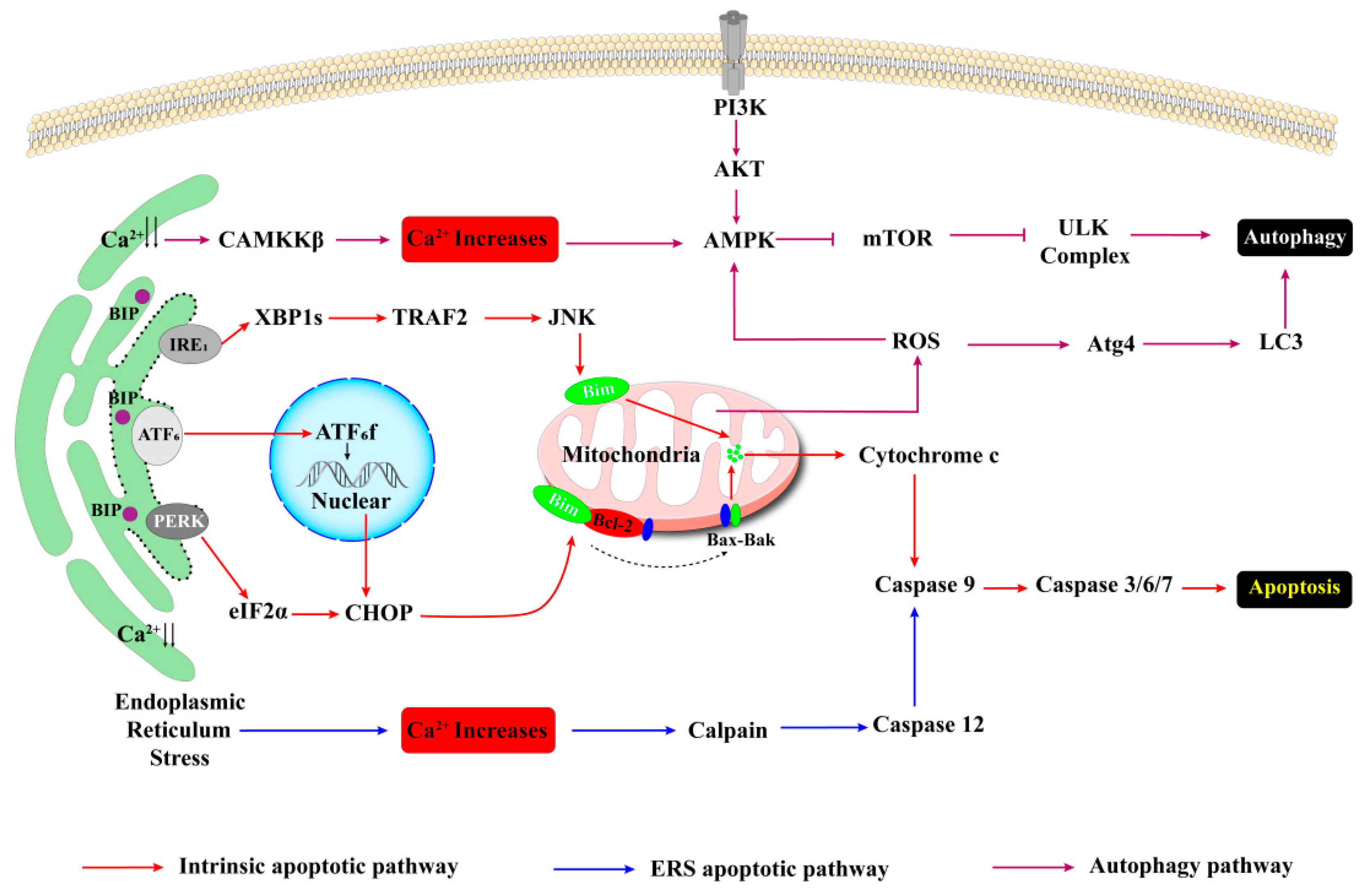
Multiple calcium ion channels play a crucial role in maintaining intracellular calcium homeostasis and the balance of endoplasmic reticulum stress. As highly specialized endocrine cells, β-cells exhibit heightened sensitivity to pathological stimuli such as inflammation, metabolic disturbances, and oxidative stress, which can initiate ER stress responses. In response to these stressors, the BiP/GRP78 chaperone system attempts to restore cellular homeostasis by activating the three branches of the unfolded protein response (UPR): IRE1α, PERK, and ATF6. Nonetheless, chronic ER stress can ultimately lead to β-cell apoptosis showed in Figure 2 [66,67,68,69,70,71,72]. A critical factor in this process is the disruption of intracellular calcium homeostasis due to abnormal SERCA function, which serves as a primary trigger for β-cell apoptosis. Research indicates that in mouse models of obesity induced by a high-fat diet, sustained high glucose levels impair SERCA function in β-cells, leading to ER calcium depletion. This depletion subsequently activates the UPR pathways: PERK/eIF2α/CHOP, ATF6-XBP1, and IRE1α/JNK/XBP1, culminating in β-cell apoptosis [40,41]. Subsequent investigations have demonstrated that intracellular calcium overload, resulting from SERCA dysfunction, primarily facilitates the formation of the DR5/FADD/caspase-8 apoptotic complex through the activation of the NF-κB/TLR-4 signaling pathway, thereby exacerbating β-cell apoptosis [42]. Various calcium release channels exhibit a concerted action in modulating β-cell apoptosis. The calcium homeostasis of the endoplasmic reticulum is sustained by calcium pumps (SERCA) and calcium release channels (IP3Rs/RyRs). Experimental evidence indicates that while the inhibition of RyR or IP3R in isolation has a limited effect on β-cell viability, simultaneous inhibition of both channels can significantly diminish cell death and alleviate endoplasmic reticulum stress [43,44,62]. Notably, the calcium release effects mediated by IP3Rs and RyRs synergize with the SERCA inhibitor thapsigargin, collectively resulting in endoplasmic reticulum calcium depletion and promoting β-cell apoptosis [43]. These findings highlight the crucial function of IP3Rs and RyRs in mediating β-cell apoptosis associated with SERCA dysfunction. Abnormalities in the store-operated calcium entry (SOCE) pathway represent a significant contributor to the exacerbation of calcium homeostasis disorders. Activation of the SOCE pathway occurs upon depletion of calcium reserves in the endoplasmic reticulum. Research has demonstrated that in the presence of SERCA inhibition, the CaMKII/Pyk2 pathway facilitates the tyrosine phosphorylation of STIM1, which subsequently forms a stable complex with Orai1, resulting in sustained calcium influx. The compromised function of SERCA prevents the alleviation of intracellular calcium overload, ultimately leading to β-cell apoptosis via the IRE1-JNK pathway [45]. This mechanism elucidates the detrimental cyclical relationship between disturbances in calcium signaling and endoplasmic reticulum stress.
Beyond the classical caspase-dependent apoptosis pathway, calcium signaling can influence the fate of β-cells by modulating the dynamic equilibrium of apoptotic proteins. A study by Liu revealed that Chlorogenic Acid (CA) significantly downregulates the expression of pro-apoptotic proteins such as Bax and caspase-3/9 by decreasing the cytoplasmic calcium concentration. Simultaneously, it upregulates the level of the anti-apoptotic protein Bcl-2. As a result, this leads to an improvement in glucose-stimulated insulin secretion (GSIS) and a notable inhibition of β-cell apoptosis [73].
Mitochondria-Associated Apoptosis
Mitochondrial dysfunction mediated by calcium homeostasis imbalance is one of the crucial mechanisms underlying β-cell apoptosis [74,75]. Empirical evidence suggests that aberrant calcium signaling can prompt the opening of the mitochondrial permeability transition pore (PTP), resulting in the dissipation of the mitochondrial membrane potential, thereby triggering downstream apoptotic cascades [76,77]. Under conditions of hyperglycemia or hyperlipidemia, calcium depletion in the endoplasmic reticulum occurs. Calcium signals are aberrantly transferred from the endoplasmic reticulum to mitochondria via the IP3Rs-GRP75-VDAC-MCU structural coupling. This process not only initiates the mitochondrial unfolded protein response (UPRmt) but also elicits mitochondrial stress [46,78,79]. This state of mitochondrial stress is manifested as the collapse of the mitochondrial membrane potential, impairments in ATP synthesis, and the explosive generation of reactive oxygen species (ROS). Excessive ROS further disrupts the balance of mitochondrial dynamics, inducing abnormal fission or fusion, thereby establishing a vicious cycle encompassing electron transport chain defects, bioenergetic dysregulation, and calcium homeostasis imbalance [80]. It is worth noting that upon treatment with thapsigargin, SERCA in β-cells is inhibited. This results in a decrease in calcium uptake and an increase in calcium release by the endoplasmic reticulum. Initially, this causes a transient elevation in the mitochondrial membrane potential [43], presumably representing a compensatory response of mitochondria to the sudden surge in cytoplasmic calcium ion concentration. However, over an extended period, marked mitochondrial depolarization becomes evident. When IP3R is concurrently activated at this stage, it accelerates the process of mitochondrial membrane potential collapse. Eventually, this leads to the irreversible opening of the PTP, the release of pro-apoptotic factors such as cytochrome c, and the activation of apoptotic effector proteases like caspase-9, thereby instigating the programmed death of β-cells.

3.1.2. Autophagy in Pancreatic β-Cells

In the pathogenesis of type 2 diabetes, the functional impairment of pancreatic islet β-cells is notably associated with the dynamic regulation of autophagic activity. Moreover, autophagy and apoptosis exhibit a bidirectional regulatory interplay. Moderately activated autophagy can play an anti-apoptotic role by selectively removing damaged organelles, thereby maintaining cellular homeostasis. In contrast, over-activated autophagy may induce autophagic cell death, synergistically facilitating the apoptotic process [81,82].
Intracellular calcium signals intricately regulate the autophagy process via multiple pathways (Figure 2). Primarily, the release of reactive oxygen species (ROS) induced by mitochondrial stress can activate the lysosomal TRPML1 channel, leading to the release of local calcium signals and subsequent nuclear translocation of transcription factor EB (TFEB), thereby upregulating autophagy-related genes [51]. Furthermore, under conditions of ROS accumulation or endoplasmic reticulum stress, the calcium-dependent kinase CaMKKβ is activated. This activation promotes the assembly of the ULK1 complex through the AMPK/mTORC1 signaling axis, enhancing the expression of key autophagy molecules [47,83,84,85,86]. However, it is crucial to note that the effects of calmodulin and calcium homeostasis on autophagy are complex and context-dependent. Persistent disruption of calcium homeostasis leads to mitochondrial Ca2+ overload, which impairs oxidative phosphorylation, generates excessive ROS, and disrupts mitochondrial dynamics, ultimately impairing mitophagy [87]. Additionally, cytoplasmic Ca2+ overload can activate calpains, which may disrupt autophagosome-lysosome fusion or impair lysosomal acidification, thereby hindering autophagic flux.
Under pathological conditions, the abnormal regulation of autophagy can manifest as a paradoxical scenario in which the expression of autophagic proteins is upregulated while the autophagic flux is concurrently downregulated. For example, in palmitate-induced MIN6 cells, cytosolic Ca2+ overload triggers ER stress (via the PERK/eIF2α/CHOP pathway), promoting apoptosis. However, despite MCU knockdown increasing LC3-II expression, it also results in p62 accumulation, indicating that autophagic flux is compromised [88]. This “autophagy blockage” phenomenon may represent a compensatory response of cells to metabolic stress, as corroborated by findings from other studies [89,90]. Experimental evidence demonstrates that calcium channel blockers or mitochondrial calcium uptake regulators can alleviate this impairment, underscoring the critical role of restoring calcium homeostasis in maintaining autophagic function. Notably, in the context of insulin resistance, β cells with high autophagic flux exhibit heightened sensitivity to glucose-stimulated calcium influx. Conversely, knockout of the autophagy regulatory gene Atg7 abolishes this effect, suggesting that precise modulation of autophagic flux governed by calcium homeostasis could enhance β cell function [91].

3.1.3. Ferroptosis in Pancreatic β-Cells

Ferroptosis is an iron-dependent and lipid peroxidation-driven form of programmed cell death. Its characteristic changes encompass iron metabolism disorders, redox imbalance, and peroxidation of polyunsaturated fatty acids in the cell membrane [92]. Research has revealed that calcium homeostasis dysregulation can promote the accumulation of reactive oxygen species (ROS) through multiple mechanisms, including endoplasmic reticulum stress, mitochondrial dysfunction, and lysosomal damage. This, in turn, forms a vicious cycle with the onset and progression of ferroptosis [93,94].
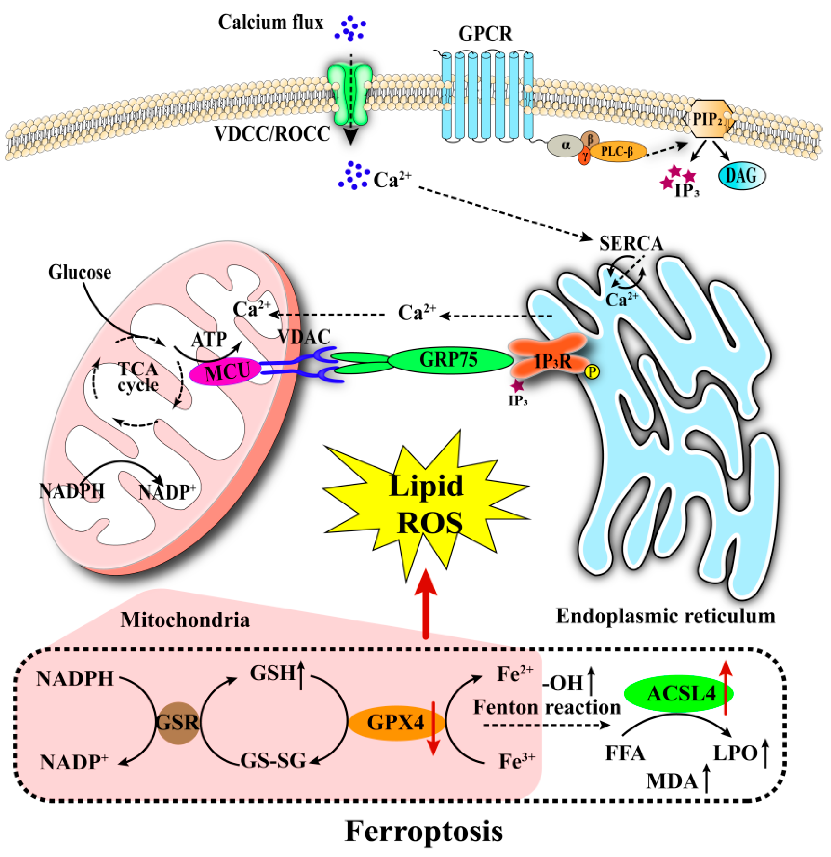
Iron metabolism exerts a dual regulatory function in pancreatic β-cells. On one hand, via the integration of iron ions into Fe-S clusters mediated by mitochondrial iron transporters (DMT-1, Mfrn1/2), it directly participates in the process of glucose-stimulated insulin secretion (GSIS) [95,96,97]. On the other hand, reactive oxygen species (ROS) generated by the Fenton reaction can serve as second messengers to amplify the insulin-secretion signals [98]. As a result of stimulation by elevated glucose concentrations, inflammatory processes, or environmental toxins, mitochondrial membrane depolarization leads to dysfunction within the electron transport chain. This dysfunction not only impedes ATP synthesis but also initiates a substantial increase in reactive oxygen species (ROS) production. Such conditions may further cause dysregulation of β-cell exosome function, iron-ion overload, and inhibition of GPX4 activity, collectively culminating in pathological changes characteristic of ferroptosis [97,99,100,101]. It is important to acknowledge the intricate relationship between ferroptosis and autophagy. The excessive activation of lipofuscin autophagy and ferritin autophagy can enhance lipid peroxidation through the release of free iron ions, while a deficiency in GPX4 markedly intensifies the cytotoxic effects associated with this mechanism.
Calcium signaling and iron metabolism demonstrate a synergistic influence on the regulation of glucose-stimulated insulin secretion (GSIS) [25,26,97]. These interconnected processes collectively contribute to β-cell damage through the endoplasmic reticulum stress-mitochondrial dysfunction axis (Figure 3). Empirical evidence indicates that the activation of the mitochondrial ROS autophagy lysosome pathway, in conjunction with endoplasmic reticulum stress-related signals, can induce ferroptosis in β-cells. This process is marked by the depletion of glutathione (GSH), the accumulation of lipid peroxidation products, and lipotoxic damage [100,101]. Under glucolipotoxic conditions, endoplasmic reticulum stress results in the depletion of calcium stores, leading to the collapse of mitochondrial membrane potential, the accumulation of mitochondrial ROS (mtROS), and the activation of phospholipases, thereby intensifying the lipid peroxidation process [43,102,103]. While dysregulation of calcium homeostasis and aberrations in iron metabolism can both lead to the accumulation of lipid peroxides, the precise mechanisms underlying their concerted interaction in β-cell injury have yet to be fully elucidated. An in-depth dissection of the interactive regulatory mechanisms within the calcium–iron metabolism network may offer novel therapeutic targets for the protection of β-cells in diabetes.

3.1.4. Pyroptosis in Pancreatic β-Cells

Pyroptosis, a form of programmed cell death facilitated by inflammasomes, is distinguished by the perforation of the cell membrane and the subsequent release of pro-inflammatory cytokines. This process is integral to the pathological progression of diabetes and its associated complications. Calcium ions, serving as a critical intracellular second messenger, play a central regulatory role in the pyroptosis of pancreatic β-cells by precisely modulating the activation of the NLRP3 inflammasome [104,105,106,107,108].
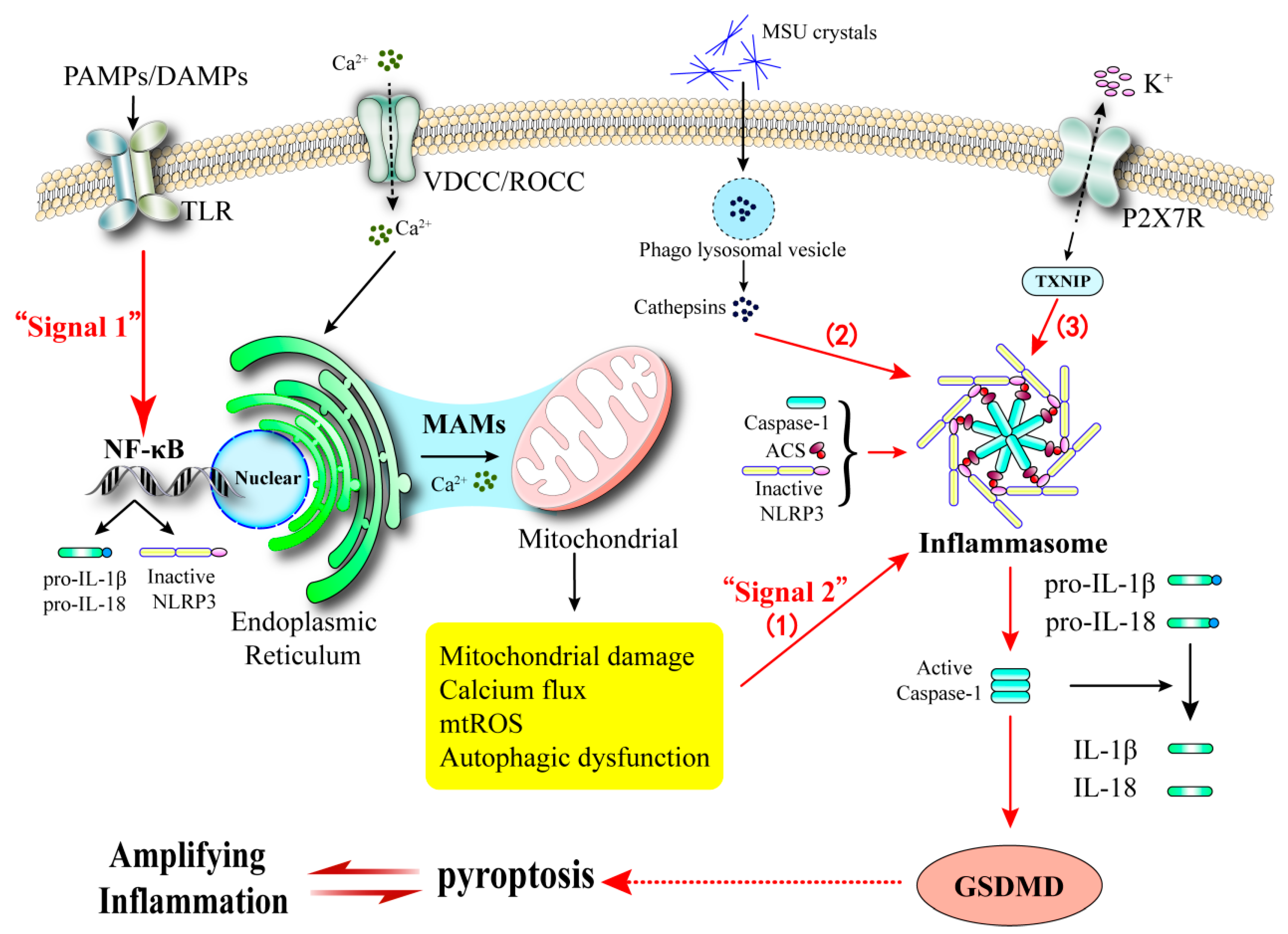
The activation of NLRP3 inflammasome is the core molecular event of pyroptosis, and its triggering factors include multiple stimuli such as ROS accumulation, endoplasmic reticulum stress, lysosomal damage and calcium signal disorder [48,109,110,111]. In the classical pathway, PAMPs/DAMPs promote the assembly of the NLRP3-NEK7-ASC-caspase-1 complex by altering intracellular ion homeostasis (especially K+ efflux and Ca2+ influx), thereby activating caspase-1 and cleaving GSDMD protein, ultimately leading to the formation of cell membrane pores and the release of IL-1β/IL-18 [112,113,114,115,116]. The non-classical pathway is directly activated by LPS to cleave GSDMD through caspase-4/5/11, and indirectly initiates the classical pyroptosis pathway by secreting IL-1β/IL-18 [117,118,119].
Although direct evidence regarding the regulation of β-cell pyroptosis by calcium homeostasis remains to be further substantiated, calcium homeostasis imbalance associated with endoplasmic reticulum stress may serve as a crucial inducer. Research has indicated that IP3R-mediated calcium release can facilitate hepatocyte pyroptosis by activating the CaMKIIγ/Smad3 pathway [48,120,121]. Under diabetic pathological conditions, endoplasmic reticulum stress induced by high glucose and high lipid over-activates IP3R via the IRE1α/PERK pathway. This leads to the depletion of the endoplasmic reticulum calcium store and cytoplasmic/mitochondrial calcium overload [59,122]. Such a state of calcium overload can trigger the collapse of the mitochondrial membrane potential, an outburst of ROS, and the release of mtDNA. Subsequently, this recruits the assembly of the NLRP3 inflammasome, and IL-1β/IL-18 are released through the caspase-1/GSDMD pathway, ultimately precipitating β-cell pyroptosis [50,123,124].
The mechanisms by which calcium-signal-related pathways regulate pyroptosis have been previously reported. PLCγ1 hydrolyzes PIP2 to generate IP3, which activates IP3R to promote calcium release from the endoplasmic reticulum. This, in turn, enhances the membrane translocation of Gsdmd-N and LPS-induced pyroptosis [63]. In a diabetic heart disease model, CaMKII may promote cardiomyocyte pyroptosis via the TLR4/NLRP3 pathway [64]. Notably, calcium channel blockers can mitigate macrophage pyroptosis and the release of IL-1β by inhibiting the TLR4/NF-κB/NLRP3/GSDMD signaling axis [65]. However, the mechanism by which calcium signals regulate pyroptosis in β-cells remains controversial. Currently, research has mainly focused on calcium homeostasis in muscle cells such as cardiomyocytes and skeletal muscle cells. Given the role of calcium homeostasis imbalance in regulating pyroptosis pathways in other cell types, modulating calcium homeostasis to repair β-cell pyroptosis may represent a novel therapeutic strategy.

3.2. Calcium Homeostasis and Insulin Resistance in Diabetes

Insulin resistance is a central pathological aspect in the development and progression of type 2 diabetes. Abundant clinical evidence indicates that it exists prior to the emergence of abnormal β-cell function and can serve as an early predictive marker for diabetes [125,126]. This pathological state is characterized by a reduced sensitivity of peripheral tissues to insulin, concurrently accompanied by compensatory hyperinsulinemia. Significantly, calcium ions, being a crucial second messenger in glucose-stimulated insulin secretion (GSIS), any imbalance in their homeostasis not only impairs β-cell function but also exacerbates peripheral insulin resistance through multiple mechanisms (Table 3) [34,127,128,129].

3.2.1. The Process of Insulin Signaling Transduction

Insulin signaling transduction is a highly intricate cascade reaction. Once insulin binds specifically to the α subunit of the receptor on the surface of target cells, it induces conformational alterations in the β subunit and autophosphorylation of tyrosine residues. This subsequently recruits and phosphorylates insulin receptor substrate (IRS) proteins [131,132]. The phosphorylated IRS activates phosphatidylinositol 3-kinase (PI3K) via its SH2 domain, catalyzing the transformation of phosphatidylinositol 4,5-bisphosphate (PIP2) into phosphatidylinositol 3,4,5 -trisphosphate (PIP3). PIP3 then activates protein kinase B (Akt/PKB) through a phosphoinositide-dependent kinase 1 (PDK1) dependent pathway. The activated Akt, on one hand, facilitates the translocation of the glucose transporter GLUT4 to the cell membrane, thereby enhancing glucose uptake. On the other hand, it inhibits gluconeogenesis and promotes glycogen synthesis by regulating downstream effector molecules such as glycogen synthase kinase-3 (GSK-3).

3.2.2. Insulin Resistance in Target Organs

In hepatic tissues, disruptions in calcium homeostasis substantially impair the PI3K-Akt signaling pathway. Empirical evidence indicates that calcium overload can decrease the phosphorylation level of insulin-stimulated Akt by 50% and reduce glucose uptake efficiency by 60% [127].
In the context of obesity, lipid accumulation in hepatocytes leads to an increased cytoplasmic Ca2+ concentration. The resulting Ca2+-phosphatidylinositol complex competitively inhibits the membrane localization of the PH domain of Akt, thereby directly disrupting insulin signal transduction. Inhibition of SOCE channels has been shown to mitigate these effects [34,128].
In skeletal muscle, the influence of calcium overload on insulin resistance demonstrates a bidirectional regulatory nature. A high-fat diet can induce a burst of mitochondrial reactive oxygen species (ROS), leading to the release of mitochondrial calcium through the opening of the mitochondrial permeability transition pore (mPTP). This release activates calcium-dependent proteases, such as calpain, which disrupt the GLUT4 transport system [133]. Conversely, the CaMKII-AMPK-PKC pathway, activated by calcium influx, can facilitate the translocation of GLUT4 to the membrane, thereby enhancing insulin sensitivity [49]. It is important to note that the overactivation of a different isoform of CaMKII, specifically CaMK2, downregulates the expression of insulin receptors in adipocytes, resulting in impaired insulin signal transduction. This seemingly contradictory phenomenon may be attributed to the specific activation of distinct calcium signaling pathways.
In adipose tissue, the development of insulin resistance is driven by the complex interactions between intracellular and extracellular calcium signaling. Insufficient calcium intake can enhance calcium influx into adipocytes through a vitamin D-dependent mechanism, thereby promoting lipogenesis [134]. In obese models, increased lipid synthesis significantly inhibits the activity of SERCA, leading to a disturbance in endoplasmic reticulum calcium homeostasis. This disruption subsequently induces serine phosphorylation of insulin receptor substrate 1 (IRS1) via the c-Jun N-terminal kinase (JNK)/inhibitor of nuclear factor kappa-B kinase subunit beta (IKKβ) pathway, thereby impairing insulin signal transduction [130,135,136]. Additionally, the activation of calcineurin enhances the expression of lipid-synthesizing enzymes. Lipid metabolites, such as ceramide, produced by these enzymes, inhibit the activation of protein kinase B (Akt) through the protein kinase C (PKC)/protein phosphatase 2A (PP2A) pathway, thereby establishing a self-perpetuating cycle of “calcium imbalance–lipid accumulation–insulin resistance” [137,138].
Although numerous fundamental research studies have demonstrated the substantial impact of calcium homeostasis on insulin resistance, the role of calcium channel blockers in clinical practice remains contentious. Some studies suggest that nifedipine improves insulin resistance by indirectly enhancing the activity of glucose transporter 4 (GLUT-4) through the activation of protein phosphatase 1 (PP-1) [129]. Conversely, a separate randomized double-blind controlled trial found that calcium channel blockers did not significantly affect insulin sensitivity [139]. This inconsistency may arise from the complex pathogenesis of insulin resistance. Calcium signaling constitutes only one element of this intricate network, and other factors, such as post-receptor signaling pathways, may also influence the overall therapeutic outcome.

4. Calcium Homeostasis and Gout

The primary pathological mechanism underlying gout is the inflammatory cascade initiated by the deposition of monosodium urate (MSU) crystals. Recent studies have elucidated the critical regulatory role of disruptions in calcium homeostasis in this process [38,140]. While the role of MSU crystals in triggering inflammation is well-established, aberrant calcium signal transduction also plays a significant role in the pathological progression of gout. This occurs through its impact on immune cell function, activation of the NLRP3 inflammasome, and induction of insulin resistance.

4.1. Abnormal Calcium Signaling in Immune Cells

4.1.1. T Cells and Calcium Homeostasis

The involvement of T-cell-mediated immune dysregulation in gouty arthritis is of considerable importance, with calcium signaling disruption playing a pivotal role [141]. Clinical investigations have demonstrated that patients with early-onset gout exhibit a marked imbalance in the Th17/Treg cell ratio, characterized by an expansion of Th17 cells and a reduction in Treg cells [142]. This immune dysregulation leads to the excessive secretion of pro-inflammatory cytokines, such as interleukin-17 (IL-17), thereby promoting tophi formation and disease progression. Mechanistically, calcium oscillations generated upon T-cell receptor activation play a crucial role in regulating Th1/Th17 cell differentiation and cytokine secretion through the calcineurin-nuclear factor of activated T-cells (NFAT) signaling pathway [143,144]. Importantly, a high-calcium microenvironment can enhance the expression of key transcription factors, including T-box expressed in T cells (T-bet) and retinoid-related orphan receptor gamma t (RORγt), further facilitating the polarization of Th1/Th17 cells [145]. Therefore, targeting calcium-dependent immune regulation may represent a novel therapeutic strategy for gout.

4.1.2. Macrophages and Calcium Homeostasis

The activation of the NLRP3 inflammasome, facilitated by the dysregulation of calcium homeostasis in macrophages, represents a critical component of inflammation associated with gout [146,147]. Throughout the pathogenesis of gout, disruptions in calcium signaling activate the NLRP3 inflammasome via multiple pathways. This activation triggers an inflammatory cascade, culminating in the characteristic clinical symptoms of acute gouty arthritis.
The disturbance of calcium homeostasis is a crucial factor in initiating NLRP3 inflammasome activation. Studies have demonstrated that the deposition of monosodium urate (MSU) crystals impairs mitochondrial function, leading to the accumulation of reactive oxygen species (ROS) and the creation of a pro-inflammatory microenvironment, including hypoxia, which subsequently induces endoplasmic reticulum (ER) stress. In particular, the c-Jun N-terminal kinase (JNK)-mediated hyperphosphorylation of inositol 1,4,5-trisphosphate receptors (IP3Rs) leads to enhanced calcium efflux from the endoplasmic reticulum (ER). Concurrently, mitochondria excessively absorb calcium ions through the voltage-dependent anion channel 1 (VDAC1)-glucose-regulated protein 75 (GRP75)-mitochondrial calcium uniporter (MCU) complex, resulting in the phenomenon known as “calcium signal appropriation.” Therefore, ER stress induced by calcium dysregulation is not exclusive to the metabolic stress in β-cells (explored in Section 3.1.1), ER stress also emerges in immune cells during gouty inflammation, triggered by MSU crystals and oxidative stress, indicating its role as a convergent node in the pathology of both diseases. This disruption of calcium signaling not only compromises the protein-folding capacity of the ER but also activates the nuclear factor-κB (NF-κB) signaling pathway via mitochondrial dysfunction and oxidative stress, thereby facilitating the activation of the NLRP3 inflammasome [148,149,150]. Upon interaction with the macrophage membrane, monosodium urate (MSU) crystals are internalized and transported to lysosomes, leading to lysosomal membrane destabilization and the subsequent release of adenosine triphosphate (ATP). The released adenosine triphosphate (ATP) activates the P2X7 receptor ion channel, thereby facilitating the influx of Ca2+. The inflammatory cascade reaction triggered by the disruption of intracellular calcium homeostasis promotes the assembly of the NLRP3 inflammasome. This, in turn, mediates the maturation and release of key inflammatory cytokines such as IL-1β and IL-8 [151,152,153,154,155]. The ensuing cycle of mitochondrial dysfunction and oxidative stress, precipitated by endoplasmic reticulum (ER) calcium depletion, further intensifies the inflammatory response [156,157]. This calcium-dependent NLRP3 activation in macrophages, central to acute gouty arthritis, is mechanistically paralleled by similar processes in pancreatic β-cells (as discussed in Section 3.1.4) and contributes to the systemic chronic low-grade inflammation.
Additionally, mitochondrial calcium overload in macrophages prompts the opening of the mitochondrial permeability transition pore (mPTP), a process more likely to induce pyroptosis rather than classical apoptosis [158,159]. This preferential shift in cell death modality may constitute a critical feature of acute gout inflammation and provides a novel perspective for understanding the pathological mechanisms underlying gout.

4.2. Insulin Resistance and Imbalance of Calcium Homeostasis in Gout

A bidirectional causal relationship between insulin resistance and gout has been identified. Genetic studies have revealed a significant positive correlation between insulin resistance and an increased risk of developing gout [160]. Clinical observations have corroborated these findings, indicating that the severity of gout is closely associated with markers of insulin resistance [161]. At the molecular level, uric acid has been shown to directly interfere with the insulin signaling pathway by promoting the binding of ENPP1 to the insulin receptor (IR), an effect that occurs independently of the oxidative stress and inflammatory pathways typically activated by uric acid [15]. These findings imply that gout and insulin resistance may form a mutually reinforcing vicious cycle.
The disruption of calcium homeostasis represents a critical pathological link between insulin resistance and gout. Current evidence suggests that the calcium ion signaling system plays a role in the regulation of insulin signal transduction through various mechanisms. Specifically, intracellular calcium overload can hinder IRS tyrosine phosphorylation via endoplasmic reticulum stress. Additionally, abnormal activation of the calcineurin pathway can significantly diminish the efficiency of GLUT4 membrane translocation [49,127,129]. Furthermore, dysregulated expression of endoplasmic reticulum calcium channel genes can exacerbate the endoplasmic reticulum stress response and impair pancreatic β-cell function [162]. Collectively, these mechanisms contribute to a reduction in insulin sensitivity, resulting in insulin resistance and subsequent hyperinsulinemia. Importantly, elevated insulin levels can inhibit uric acid excretion by upregulating the activity of the URAT1 transporter [163]. Conversely, disturbances in uric acid metabolism can further aggravate insulin resistance through oxidative stress and other pathways [15], thereby establishing a positive feedback regulatory loop.
The disruption of calcium homeostasis has been implicated in the onset of gouty inflammation. Studies have demonstrated that calcium influx, triggered by factors such as metabolic abnormalities (including hyperglycemia and hyperlipidemia) and oxidative stress, can directly activate the NLRP3 inflammasome and facilitate the release of IL-1β [117,136,148,164]. This mechanism not only exacerbates insulin resistance but also precipitates acute gouty arthritis. Notably, the overexpression of RIPK3 has been shown to increase cytoplasmic Ca2+ and reactive oxygen species (ROS) levels through the endoplasmic reticulum stress pathway. This, in turn, upregulates the expression of xanthine oxidase (XO), thereby accelerating uric acid production [165]. These findings elucidate the pivotal role of aberrant calcium signaling in the molecular interplay between insulin resistance and gout.

5. The Association Between Intracellular Calcium Homeostasis and Comorbid Gout in Type 2 Diabetes

A significant comorbidity association exists between gout and type 2 diabetes [5]. Epidemiological evidence suggests that the prevalence of gout among individuals with type 2 diabetes is 3.8 times higher than in the general population [166]. Multivariate adjustment analyses reveal that, even after controlling for confounding factors such as obesity and hypertension, gout is associated with a 17% to 47% increased risk of developing type 2 diabetes [167,168]. This notable comorbidity phenomenon involves multiple pathological mechanisms, including insulin resistance, chronic low-grade inflammation, dyslipidemia, and oxidative stress. Importantly, dysregulation of calcium homeostasis may serve as a crucial regulatory nexus, contributing not only to the initiation of the disease but also to the amplification of metabolic disruption signals, thereby facilitating the co-progression of type 2 diabetes and gout.
Beyond the parallel pathophysiological roles of calcium dysregulation in type 2 diabetes and gout individually, its function as a central amplifier in their comorbidity can be conceptualized through three interconnected themes: a self-reinforcing calcium cycle, a central inflammatory nexus, and a novel perspective on β-cell death. First, a self-reinforcing calcium cycle is established: calcium-triggered β-cell dysfunction and peripheral insulin resistance (as detailed in Section 3) lead to hyperinsulinemia and hyperglycemia, which promote hyperuricemia (as outlined in Section 2). The resulting inflammatory milieu, in turn, further disrupts calcium homeostasis in immune cells and metabolic tissues (as explored in Section 4), creating a feed-forward loop that amplifies the entire pathology. Second, calcium acts as the central inflammatory nexus, serving as a common upstream signal that translates diverse metabolic insults (e.g., hyperglycemia, free fatty acids, MSU crystals) into the shared outcome of NLRP3 inflammasome activation, thereby fueling the chronic inflammation characteristic of both diseases. Finally, within the pancreatic islet, calcium dysregulation provides a novel perspective on β-cell demise by orchestrating the crosstalk and temporal sequence between programmed cell death pathway (pyroptosis), which may be crucial for understanding the accelerated loss of insulin secretion in the comorbid state. The following sections will elaborate on the evidence for this integrative framework.

5.1. Linking Effects of Insulin Resistance

Research has demonstrated that the onset of insulin resistance occurs prior to the clinical diagnosis of type 2 diabetes [125]. In the initial stage of elevated blood glucose levels, glucose is transported into β-cells via the GLUT2 transporter. This is followed by an influx of Ca2+, which triggers the glucose-stimulated insulin secretion (GSIS) process [25,26]. In response to metabolic demands, pancreatic β-cells compensate by augmenting the synthesis of pro-insulin. This increased synthesis imposes a significant burden on the endoplasmic reticulum (ER), leading to the activation of the unfolded protein response (UPR) and the induction of endoplasmic reticulum stress. The aberrant activation of inositol 1,4,5-trisphosphate (IP3) receptors within the endoplasmic reticulum precipitates increased calcium leakage. Concurrently, mitochondria excessively sequester calcium ions via the VDAC1-GRP75-MCU complex, culminating in mitochondrial dysfunction. This intracellular calcium overload exacerbates stress in both the endoplasmic reticulum and mitochondria, facilitates the accumulation of reactive oxygen species (ROS), and triggers pro-inflammatory signaling pathways, including nuclear factor-κB (NF-κB) [148,149,150]. Collectively, these pathological changes impede the phosphorylation of insulin receptor substrate (IRS) in hepatic, muscular, and adipose tissues, thereby disrupting the normal function of the phosphatidylinositol 3-kinase/protein kinase B (PI3K/Akt) and mitogen-activated protein kinase (MAPK) signaling pathways. Consequently, this impairment diminishes the glucose uptake capacity mediated by glucose transporter 4 (GLUT4), perpetuating a deleterious cycle of insulin resistance [129,135,169].
Hyperinsulinemia resulting from insulin resistance can substantially activate the Na+-H+ exchanger in the proximal convoluted tubules of the kidneys and enhance the expression of the URAT1 transporter. This process facilitates the reabsorption of uric acid while inhibiting its excretion [8,9]. Concurrently, a hyperglycemic state triggers the polyol pathway, accelerating fructose metabolism and depleting adenosine triphosphate (ATP). This degradation process further elevates the production of endogenous uric acid [11]. Elevated serum uric acid levels promote the phosphorylation of insulin receptor substrate 2 (IRS2) at serine 731 and inhibit the phosphorylation of protein kinase B (Akt) at serine 473 via oxidative stress mechanisms, thereby impairing insulin signaling [170]. Additionally, increased levels of retinol-binding protein 4 (RBP4) associated with hyperuricemia (HUA) can specifically inhibit the IRS/phosphatidylinositol 3-kinase (PI3K)/Akt pathway in adipocytes, further exacerbating insulin resistance [171]. These interconnected molecular mechanisms collectively contribute to the pathological progression of gout comorbid with type 2 diabetes.
It is noteworthy that hyperglycemic and hyperuricemic conditions can independently trigger endoplasmic reticulum (ER) stress [172,173]. Dysregulation of calcium homeostasis is critically involved in the regulation of ER stress and insulin signaling pathways [34,40]. Based on these observations, we hypothesize that ER stress-induced disruption of calcium homeostasis may represent a significant molecular mechanism contributing to the onset of insulin resistance, which may subsequently result in the pathological manifestations of gout in individuals with type 2 diabetes.

5.2. The Amplification of Inflammatory Signals

The secretion of interleukin-1β (IL-1β), mediated by the NLRP3 inflammasome, plays a pivotal regulatory role in the innate immune responses associated with type 2 diabetes and gout. As previously elaborated, calcium dysregulation is a key trigger for NLRP3 activation in both pancreatic β-cells (Section 3.1.4) and immune cells such as macrophages in gout (Section 4.1.2). Here, we focus on how this shared mechanism creates an amplified inflammatory signal within the comorbid state. Calcium ions, functioning as second messengers, are essential for the activation of this pathway (Figure 4). Current research suggests that elevated uric acid levels can lead to the precipitation of monosodium urate (MSU) crystals. This process subsequently induces the synthesis of oxidized mitochondrial DNA (mtDNA), increases the production of reactive oxygen species (ROS), exacerbates impairments in mitochondrial oxidative phosphorylation, and results in oxygen depletion, ultimately disrupting cellular calcium homeostasis [174,175]. The influx of intracellular calcium ions (Ca2+) can effectively activate the MAPK/NF-κB signaling pathway, which promotes the polarization of macrophages towards a pro-inflammatory phenotype (M1 type), thereby significantly enhancing their phagocytic capabilities [176]. The polarization of M1-type macrophages induces K+ efflux and Ca2+ influx, thereby establishing a positive feedback loop that exacerbates calcium homeostasis imbalance. This process facilitates the assembly of the NLRP3 inflammasome and the activation of Caspase-1, accompanied by the cleavage of the gasdermin D (GSDMD) protein, which leads to pyroptosis. Consequently, there is a substantial release of pro-inflammatory cytokines, such as IL-1β and IL-18. Concurrently, the expression of inflammatory mediators, including nitric oxide (NO) and tumor necrosis factor-α (TNF-α), is upregulated [112,113,114,115,116]. Ultimately, this sequence of events establishes a critical inflammatory foundation conducive to the development of gout in the context of diabetes.
Under physiological conditions, the limited activation of inflammasomes maintains a low concentration of IL-1β, which can appropriately stimulate insulin secretion. However, overexpression of IL-1β may induce chronic, low-grade inflammation within pancreatic islets [177,178]. This inflammatory state impairs the glucose-stimulated calcium influx (GSCI) capability of β cells, ultimately leading to a reduction in insulin secretion. Additionally, IL-1β can downregulate the expression of genes associated with β-cell maturation and disrupt intracellular calcium homeostasis [179,180,181,182,183]. The disturbance in calcium homeostasis activates phospholipase C (PLC) and protein kinase C ε (PKCε), which in turn amplify the NF-κB and NLRP3/GSDMD inflammatory pathways. This cascade promotes the release of cytokines, such as IL-1β, and exacerbates β-cell pyroptosis. More importantly, dysregulated calcium signaling in β cells can also activate the death receptor pathway and induce endoplasmic reticulum stress, leading to β-cell apoptosis [43,173]. Through the dual mechanisms of apoptosis and pyroptosis, there is a reduction in both the number and functionality of β cells. This process represents a critical step in the pathogenesis of gout in the context of diabetes.
A diverse array of chemokines facilitates the differentiation and aggregation of inflammatory cells, which subsequently release substantial quantities of pro-inflammatory factors. These factors perpetually activate the immune response and enhance downstream inflammatory reactions, thereby establishing a positive feedback loop [184,185]. This cascade not only exacerbates damage to pancreatic β-cells but also promotes chronic inflammation in insulin-sensitive tissues, including adipose tissue, the liver, and skeletal muscle. As a result, it further disrupts insulin signal transduction and exacerbates insulin resistance. The interplay between calcium homeostasis imbalance and inflammatory pathways can engender a pro-inflammatory microenvironment, which intensifies endoplasmic reticulum stress and mitochondrial damage. This ultimately leads to pan-apoptosis or autophagy of β-cells [6,186,187]. Consequently, this process emerges as a pathogenic factor in the development of gout complicated by type 2 diabetes.

5.3. Linking β-Cell Death Networks to Gouty Inflammation via Calcium Signaling

Beyond the direct inflammatory assault, the dysregulated calcium signaling within β-cells themselves instigates a multifaceted cell death network, offering a novel perspective on their progressive decline in the context of comorbid gout [53,57,58,59]. While pyroptosis, with its inflammatory character, forms a direct bridge with the systemic inflammatory nexus described above, it is likely not the sole executor. We posit that calcium overload orchestrates a dynamic interplay between multiple programmed death pathways: calcium-induced mitochondrial permeability commits cells to the apoptotic pathway [74,75,76,77]; simultaneously, calcium-mediated ER stress and ROS production can trigger ferroptosis, an iron-dependent death characterized by lipid peroxidation [43,93,103]; as detailed earlier, calcium also activates the NLRP3 inflammasome within the β-cell, leading to pyroptosis and the release of more IL-1β [50,104,105,106,107,108].
Critically, these pathways do not operate in isolation. For instance, apoptotic signaling can inhibit pyroptosis, while components of autophagy (dysregulated by calcium) can promote ferroptosis [51,60,100]. The crosstalk and temporal sequence of these calcium-driven death modalities—determining whether a β-cell dies quietly (apoptosis) or incites an inflammatory riot (pyroptosis)—may be a key determinant in the rate of β-cell mass loss and the intensity of local islet inflammation.
This accelerated β-cell demise, driven by the calcium-dependent death network, has direct implications for the comorbidity with gout. A more rapid decline in β-cell function would exacerbate hyperinsulinemia and hyperglycemia, which are key drivers of hyperuricemia (as established in Section 5.1) [8,9,11]. Consequently, the calcium-mediated ‘inflammatory riot’ (pyroptosis) within the islet not only damages the pancreas locally but also systemically fuels the metabolic dysregulation that promotes and worsens gout, creating a vicious cycle between pancreatic failure and gouty inflammation. Therefore, targeting the master regulator, calcium homeostasis, could simultaneously protect β-cells and mitigate a key driver of gout progression [41,45].

6. Therapeutic Perspectives and Challenges

The compelling evidence linking calcium dysregulation to the type 2 diabetes–gout axis naturally prompts the consideration of calcium channel modulators as potential therapeutic agents. Preclinical studies offer promising leads: SERCA activators like CDN1163 have been shown to improve ER stress and insulin sensitivity in obese mouse models [41], while inhibitors of store-operated calcium entry (SOCE) can ameliorate hepatic insulin resistance [34]. Furthermore, common calcium channel blockers (CCBs), such as nifedipine, have been reported to indirectly enhance GLUT4 activity and improve insulin sensitivity in some studies [129].
However, the translation of these findings into clinical practice faces substantial and multifaceted challenges. The lack of tissue specificity of first-generation modulators poses a significant risk of off-target effects, given the ubiquitous role of calcium signaling. This critical limitation is exemplified by the conflicting clinical data on CCBs; while some studies suggest benefits, others, including randomized controlled trials, have failed to demonstrate a significant improvement in insulin sensitivity [139]. The complex and often opposing roles of calcium signals in different tissues further complicate therapeutic targeting. For instance, the activation of specific calcium signaling pathways (e.g., certain CaMKII isoforms) may enhance insulin sensitivity in one tissue while promoting resistance in another [49,130].
Most importantly, there is a conspicuous absence of large-scale, high-quality clinical trials specifically designed to test the efficacy and safety of calcium-focused therapies in patients with type 2 diabetes and comorbid gout. The current evidence base is insufficient to clarify the exact therapeutic potential of modulating calcium homeostasis in this specific patient population. These unresolved issues—ranging from fundamental biological complexity to a lack of clinical validation—constitute the core of the current controversy and highlight the significant gap between mechanistic promise and clinical application.
Therefore, future efforts must bifurcate: first, to focus on designing next-generation, tissue-restricted modulators that can precisely target pathological calcium signaling without disrupting physiological functions. Second, well-designed clinical studies are urgently needed to validate whether correcting calcium homeostasis can safely and effectively break the vicious cycle linking hyperinsulinemia, hyperuricemia, and chronic inflammation in this high-risk patient population.

7. Conclusions

In conclusion, this review definitively establishes intracellular calcium dysregulation as a pivotal mechanism and a unifying pathological nexus linking the pathogenesis of type 2 diabetes and gout. We have synthesized evidence demonstrating that calcium homeostasis imbalance acts as a central amplifier, driving β-cell injury through ER stress, mitochondrial dysfunction, and multiple programmed cell death pathways (apoptosis, autophagy, ferroptosis, pyroptosis), while concurrently exacerbating peripheral insulin resistance and activating the NLRP3 inflammasome to fuel chronic inflammation. This integrated perspective moves beyond a simple compilation of known mechanisms and positions calcium signaling as a key orchestrator of the comorbidity.
Despite these advances, critical knowledge gaps remain. The temporal dynamics of calcium dysregulation during the progression from isolated type 2 diabetes to comorbid gout are poorly understood. Furthermore, the precise crosstalk between calcium-dependent cell death modalities in β-cells and immune cells represents a complex, underexplored area. The tissue-specificity of calcium channels necessitates the development of targeted modulators to avoid off-target effects, a significant current limitation.
Future research should therefore prioritize several key directions: (1) developing tissue-specific calcium channel modulators to minimize systemic side effects; (2) conducting longitudinal clinical studies to validate calcium homeostasis biomarkers as predictors for gout onset in diabetic populations; (3) employing single-cell technologies to elucidate the precise interplay between calcium, iron metabolism, and inflammasome activation in different cell types. Addressing these specific avenues is not merely an academic exercise but holds promising potential for overcoming the therapeutic challenges in managing type 2 diabetes complicated by gout, ultimately paving the way for novel, mechanism-based interventions.

Author Contributions

Conceptualization, H.L.; methodology, Y.S.; writing—original draft preparation, H.S.; writing—review and editing, H.L. and R.Z.; supervision, K.Q. All authors have read and agreed to the published version of the manuscript.

Funding

The funding was provided by the National Natural Science Foundation of China (82505518), the Natural Science Foundation Project of Nanjing University of Chinese Medicine (XZR2024379), the Suzhou Municipal Science and Technology R&D Program (SYWD2025264), and the Seed Fund for Young Investigators of Zhangjiagang Health Commission (ZJGQNKJ202511).

Institutional Review Board Statement

Not applicable.

Informed Consent Statement

Not applicable.

Data Availability Statement

No new data were created or analyzed in this study.

Conflicts of Interest

The authors declare no conflicts of interest.

References

  1. Chan, J.C.N.; Lim, L.-L.; Wareham, N.J.; Shaw, J.E.; Orchard, T.J.; Zhang, P.; Lau, E.S.H.; Eliasson, B.; Kong, A.P.S.; Ezzati, M.; et al. The Lancet Commission on diabetes: Using data to transform diabetes care and patient lives. Lancet 2020, 396, 2019–2082. [Google Scholar] [CrossRef]
  2. Dieleman, J.L.; Cao, J.; Chapin, A.; Chen, C.; Li, Z.; Liu, A.; Horst, C.; Kaldjian, A.; Matyasz, T.; Scott, K.W.; et al. US Health Care Spending by Payer and Health Condition, 1996–2016. JAMA 2020, 323, 863–884. [Google Scholar] [CrossRef]
  3. Maria, J.L.; Anand, T.N.; Dona, B.; Prinu, J.; Prabhakaran, D.; Jeemon, P. Task-sharing interventions for improving control of diabetes in low-income and middle-income countries: A systematic review and meta-analysis. Lancet Glob. Health 2021, 9, e170–e180. [Google Scholar] [CrossRef]
  4. International Diabetes Federation (IDF). IDF Diabetes Atlas, 11th ed.; International Diabetes Federation: Brussels, Belgium, 2025. [Google Scholar]
  5. Jiang, J.; Zhang, T.; Liu, Y.; Chang, Q.; Zhao, Y.; Guo, C.; Xia, Y. Prevalence of Diabetes in Patients with Hyperuricemia and Gout: A Systematic Review and Meta-analysis. Curr. Diabetes Rep. 2023, 23, 103–117. [Google Scholar] [CrossRef]
  6. Wijnands, J.M.A.; van Durme, C.M.P.G.; Driessen, J.H.M.; Boonen, A.; Klop, C.; Leufkens, B.; Cooper, C.; Stehouwer, C.D.A.; de Vries, F. Individuals with Type 2 Diabetes Mellitus are at an Increased Risk of Gout but this is not Due to Diabetes. Medicine 2015, 94, e1358. [Google Scholar] [CrossRef]
  7. Matsuura, F.; Yamashita, S.; Nakamura, T.; Nishida, M.; Nozaki, S.; Funahashi, T.; Matsuzawa, Y. Effect of visceral fat accumulation on uric acid metabolism in male obese subjects: Visceral fat obesity is linked more closely to overproduction of uric acid than subcutaneous fat obesity. Metabolism 1998, 47, 929–933. [Google Scholar] [CrossRef]
  8. Toyoki, D.; Shibata, S.; Kuribayashi-Okuma, E.; Xu, N.; Ishizawa, K.; Hosoyamada, M.; Uchida, S. Insulin stimulates uric acid reabsorption via regulating urate transporter 1 and ATP-binding cassette subfamily G member 2. Am. J. Physiol. Ren. Physiol. 2017, 313, F826–F834. [Google Scholar] [CrossRef]
  9. Maaten, J.T.; Voorburg, A.; Heine, R.J.; Wee, P.T.; Donker, A.J.M.; Gans, R.O.B. Renal handling of urate and sodium during acute physiological hyperinsulinaemia in healthy subjects. Clin. Sci. 1997, 92, 51–58. [Google Scholar] [CrossRef]
  10. Henning, C.; Liehr, K.; Girndt, M.; Ulrich, C.; Glomb, M.A. Extending the Spectrum of α-Dicarbonyl Compounds in Vivo. J. Biol. Chem. 2014, 289, 28676–28688. [Google Scholar] [CrossRef]
  11. Srivastava, A.; Kaze, A.D.; McMullan, C.J.; Isakova, T.; Waikar, S.S. Uric Acid and the Risks of Kidney Failure and Death in Individuals With CKD. Am. J. Kidney Dis. 2018, 71, 362–370. [Google Scholar] [CrossRef]
  12. Sanchez-Lozada, L.G.; Rodriguez-Iturbe, B.; Kelley, E.E.; Nakagawa, T.; Madero, M.; Feig, D.I.; Borghi, C.; Piani, F.; Cara-Fuentes, G.; Bjornstad, P.; et al. Uric Acid and Hypertension: An Update with Recommendations. Am. J. Hypertens. 2020, 33, 583–594. [Google Scholar] [CrossRef]
  13. Bertrand, L.; Zhi, L.; Yuzhang, Z.; Tianliang, H.; Hisatome, I.; Yamamoto, T.; Jidong, C. High Uric Acid Induces Insulin Resistance in Cardiomyocytes In Vitro and In Vivo. PLoS ONE 2016, 11, e0147737. [Google Scholar]
  14. Baldwin, W.; McRae, S.; Marek, G.; Wymer, D.; Pannu, V.; Baylis, C.; Johnson, R.J.; Sautin, Y.Y. Hyperuricemia as a mediator of the proinflammatory endocrine imbalance in the adipose tissue in a murine model of the metabolic syndrome. Diabetes 2011, 60, 1258–1269. [Google Scholar] [CrossRef]
  15. Tassone, E.J.; Cimellaro, A.; Perticone, M.; Hribal, M.L.; Sciacqua, A.; Andreozzi, F.; Sesti, G.; Perticone, F. Uric Acid Impairs Insulin Signaling by Promoting Enpp1 Binding to Insulin Receptor in Human Umbilical Vein Endothelial Cells. Front. Endocrinol. 2018, 9, 98. [Google Scholar] [CrossRef]
  16. Raffaello, A.; Mammucari, C.; Gherardi, G.; Rizzuto, R. Calcium at the Center of Cell Signaling: Interplay between Endoplasmic Reticulum, Mitochondria, and Lysosomes. Trends Biochem. Sci. 2016, 41, 1035–1049. [Google Scholar] [CrossRef]
  17. Puzianowska-Kuznicka, M.; Kuznicki, J. The ER and ageing II: Calcium homeostasis. Ageing Res. Rev. 2009, 8, 160–172. [Google Scholar] [CrossRef]
  18. Petersen, O.H.; Petersen, C.C.; Kasai, H. Calcium and hormone action. Annu. Rev. Physiol. 1994, 56, 297–319. [Google Scholar] [CrossRef]
  19. Guerrero-Hernandez, A.V.A. Calcium signalling in diabetes. Cell Calcium 2014, 56, 297–301. [Google Scholar] [CrossRef]
  20. Hodeify, R.; Nandakumar, M.; Own, M.; Courjaret, R.J.; Graumann, J.; Hubrack, S.Z.; Machaca, K. The CCT chaperonin is a novel regulator of Ca2+ signaling through modulation of Orai1 trafficking. Sci. Adv. 2018, 4, eaau1935. [Google Scholar] [CrossRef]
  21. Vultur, A.; Gibhardt, C.S.; Stanisz, H.; Bogeski, I. The role of the mitochondrial calcium uniporter (MCU) complex in cancer. Pflügers Arch. Eur. J. Physiol. 2018, 470, 1149–1163. [Google Scholar] [CrossRef]
  22. Bernhard, W.; Rouiller, C. Close topographical relationship between mitochondria and ergastoplasm of liver cells in a definite phase of cellular activity. J. Biophys. Biochem. Cytol. 1956, 2, 73–78. [Google Scholar] [CrossRef]
  23. Peng, W.; Wong, Y.C.; Krainc, D. Mitochondria-lysosome contacts regulate mitochondrial Ca2+ dynamics via lysosomal TRPML1. Proc. Natl. Acad. Sci. USA 2020, 117, 19266–19275. [Google Scholar] [CrossRef]
  24. Yang, W.; Li, Y.; Feng, R.; Liang, P.; Tian, K.; Hu, L.; Wang, K.; Qiu, T.; Zhang, J.; Sun, X.; et al. PFOS causes lysosomes-regulated mitochondrial fission through TRPML1-VDAC1 and oligomerization of MCU/ATP5J2. J. Hazard. Mater. 2025, 489, 137685. [Google Scholar] [CrossRef]
  25. Song, S.-E.; Shin, S.-K.; Ju, H.Y.; Im, S.-S.; Song, D.-K. Role of cytosolic and endoplasmic reticulum Ca2+ in pancreatic beta-cells: Pros and cons. Pflügers Arch. Eur. J. Physiol. 2023, 476, 151–161. [Google Scholar] [CrossRef]
  26. Mu-u-min, R.B.A.; Diane, A.; Allouch, A.; Al-Siddiqi, H.H. Ca2+-Mediated Signaling Pathways: A Promising Target for the Successful Generation of Mature and Functional Stem Cell-Derived Pancreatic Beta Cells In Vitro. Biomedicines 2023, 11, 1577. [Google Scholar] [CrossRef]
  27. Idevall-Hagren, O.; Tengholm, A. Metabolic regulation of calcium signaling in beta cells. Semin. Cell Dev. Biol. 2020, 103, 20–30. [Google Scholar] [CrossRef]
  28. Kim, M.J.; Min, S.H.; Shin, S.Y.; Kim, M.N.; Lee, H.; Jang, J.Y.; Kim, S.-W.; Park, K.S.; Jung, H.S. Attenuation of PERK enhances glucose-stimulated insulin secretion in islets. J. Endocrinol. 2018, 236, 125–136. [Google Scholar] [CrossRef]
  29. Voelker, D.R. Bridging gaps in phospholipid transport. Trends Biochem. Sci. 2005, 30, 396–404. [Google Scholar] [CrossRef]
  30. Rizzuto, R.; Duchen, M.R.; Pozzan, T. Flirting in Little Space: The ER/Mitochondria Ca2+ Liaison. Sci. STKE 2004, 2004, re1. [Google Scholar] [CrossRef]
  31. Hajnóczky, G.; Davies, E.; Madesh, M. Calcium signaling and apoptosis. Biochem. Biophys. Res. Commun. 2003, 304, 445–454. [Google Scholar] [CrossRef]
  32. Carnevale, V.; Nieddu, L.; Scillitani, A.; Tinti, M.G.; Eller-Vainicher, C.; Cosso, R.; Rendina, D.; Falchetti, A. Calcium-phosphate homeostasis and insulin resistance in men. Nutr. Metab. Cardiovasc. Dis. 2024, 34, 353–359. [Google Scholar] [CrossRef]
  33. Missiroli, S.; Patergnani, S.; Caroccia, N.; Pedriali, G.; Perrone, M.; Previati, M.; Wieckowski, M.R.; Giorgi, C. Mitochondria-associated membranes (MAMs) and inflammation. Cell Death Dis. 2018, 9, 329. [Google Scholar] [CrossRef]
  34. Lee, J.W.; Gu, H.-O.; Jung, Y.; Jung, Y.; Seo, S.-Y.; Hong, J.-H.; Hong, I.-S.; Lee, D.H.; Kim, O.-H.; Oh, B.-C. Candesartan, an angiotensin-II receptor blocker, ameliorates insulin resistance and hepatosteatosis by reducing intracellular calcium overload and lipid accumulation. Exp. Mol. Med. 2023, 55, 910–925. [Google Scholar] [CrossRef]
  35. Arruda, A.P.; Hotamisligil, G.S. Calcium Homeostasis and Organelle Function in the Pathogenesis of Obesity and Diabetes. Cell Metab. 2015, 22, 381–397. [Google Scholar] [CrossRef]
  36. Fu, S.; Yang, L.; Li, P.; Hofmann, O.; Dicker, L.; Hide, W.; Lin, X.; Watkins, S.M.; Ivanov, A.R.; Hotamisligil, G.S. Aberrant lipid metabolism disrupts calcium homeostasis causing liver endoplasmic reticulum stress in obesity. Nature 2011, 473, 528–531. [Google Scholar] [CrossRef]
  37. Mohan, A.A.; Talwar, P. MAM kinases: Physiological roles, related diseases, and therapeutic perspectives—A systematic review. Cell. Mol. Biol. Lett. 2025, 30, 35. [Google Scholar] [CrossRef]
  38. Hong, Q.; Qi, K.; Feng, Z.; Huang, Z.; Cui, S.; Wang, L.; Fu, B.; Ding, R.; Yang, J.; Chen, X.; et al. Hyperuricemia induces endothelial dysfunction via mitochondrial Na+/Ca2+ exchanger-mediated mitochondrial calcium overload. Cell Calcium 2012, 51, 402–410. [Google Scholar] [CrossRef]
  39. Hernández-Alvarez, M.I.; Sebastián, D.; Vives, S.; Ivanova, S.; Bartoccioni, P.; Kakimoto, P.; Plana, N.; Veiga, S.R.; Hernández, V.; Vasconcelos, N.; et al. Deficient Endoplasmic Reticulum-Mitochondrial Phosphatidylserine Transfer Causes Liver Disease. Cell 2019, 177, 881–895.e17. [Google Scholar] [CrossRef]
  40. Tong, X.K.T.; Anderson-Baucum, E.K.; Yamamoto, W.; Gilon, P.; Lebeche, D.; Day, R.N.; Shull, G.E.; Evans-Molina, C. SERCA2 Deficiency Impairs Pancreatic β-Cell Function in Response to Diet-Induced Obesity. Diabetes 2016, 65, 3039–3052. [Google Scholar] [CrossRef]
  41. Kang, S.; Dahl, R.; Hsieh, W.; Shin, A.; Zsebo, K.M.; Buettner, C.; Hajjar, J.R. Small Molecular Allosteric Activator of the Sarco/Endoplasmic Reticulum Ca2+-ATPase (SERCA) Attenuates Diabetes and Metabolic Disorders. J. Biol. Chem. 2016, 291, 5185–5198. [Google Scholar] [CrossRef]
  42. Ali, S.I.; Elkhalifa, A.M.E.; Nabi, S.U.; Hayyat, F.S.; Nazar, M.; Taifa, S.; Rakhshan, R.; Shah, I.H.; Shaheen, M.; Wani, I.A.; et al. Aged garlic extract preserves beta-cell functioning via modulation of nuclear factor kappa-B (NF-κB)/Toll-like receptor (TLR)-4 and sarco endoplasmic reticulum calcium ATPase (SERCA)/Ca2+ in diabetes mellitus. Diabetol. Metab. Syndr. 2024, 16, 110. [Google Scholar] [CrossRef]
  43. Luciani, D.S.; Gwiazda, K.S.; Yang, T.-L.B.; Kalynyak, T.B.; Bychkivska, Y.; Frey, M.H.Z.; Jeffrey, K.D.; Sampaio, A.V.; Underhill, T.M.; Johnson, J.D. Roles of IP3R and RyR Ca2+ Channels in Endoplasmic Reticulum Stress and β-Cell Death. Diabetes 2009, 58, 422–432. [Google Scholar] [CrossRef]
  44. Yamamoto, W.R.; Bone, R.N.; Sohn, P.; Syed, F.; Reissaus, C.A.; Mosley, A.L.; Wijeratne, A.B.; True, J.D.; Tong, X.; Kono, T.; et al. Endoplasmic reticulum stress alters ryanodine receptor function in the murine pancreatic β cell. J. Biol. Chem. 2019, 294, 168–181. [Google Scholar] [CrossRef]
  45. Song, S.-E.; Shin, S.-K.; Kim, Y.-W.; Do, Y.R.; Lim, A.K.; Bae, J.-H.; Jeong, G.-S.; Im, S.-S.; Song, D.-K. Lupenone attenuates thapsigargin-induced endoplasmic reticulum stress and apoptosis in pancreatic beta cells possibly through inhibition of protein tyrosine kinase 2 activity. Life Sci. 2023, 332, 122107. [Google Scholar] [CrossRef]
  46. Meng, M.; Jiang, Y.; Wang, Y.; Huo, R.; Ma, N.; Shen, X.; Chang, G. β-carotene targets IP3R/GRP75/VDAC1-MCU axis to renovate LPS-induced mitochondrial oxidative damage by regulating STIM1. Free Radic. Biol. Med. 2023, 205, 25–46. [Google Scholar] [CrossRef]
  47. Li, A.; Yi, B.; Han, H.; Yang, S.; Hu, Z.; Zheng, L.; Wang, J.; Liao, Q.; Zhang, H. Vitamin D-VDR (vitamin D receptor) regulates defective autophagy in renal tubular epithelial cell in streptozotocin-induced diabetic mice via the AMPK pathway. Autophagy 2021, 18, 877–890. [Google Scholar] [CrossRef]
  48. Ren, S.; Liang, P.; Feng, R.; Yang, W.; Qiu, T.; Zhang, J.; Li, Q.; Yang, G.; Sun, X.; Yao, X. The phosphorylation of Smad3 by CaMKIIγ leads to the hepatocyte pyroptosis under perfluorooctane sulfonate exposure. Ecotoxicol. Environ. Saf. 2024, 284, 116924. [Google Scholar] [CrossRef]
  49. Li, Q.; Zhu, X.; Ishikura, S.; Zhang, D.; Gao, J.; Sun, Y.; Contreras-Ferrat, A.; Foley, K.P.; Lavandero, S.; Yao, Z.; et al. Ca2+ signals promote GLUT4 exocytosis and reduce its endocytosis in muscle cells. Am. J. Physiol. Endocrinol. Metab. 2014, 307, E209–E224. [Google Scholar] [CrossRef]
  50. Li, Z.; Ran, Q.; Qu, C.; Hu, S.; Cui, S.; Zhou, Y.; Shen, B.; Yang, B. Sigma-1 receptor activation attenuates DOX-induced cardiotoxicity by alleviating endoplasmic reticulum stress and mitochondrial calcium overload via PERK and IP3R-VDAC1-MCU signaling pathways. Biol. Direct 2025, 20, 23. [Google Scholar] [CrossRef]
  51. Park, K.; Lim, H.; Kim, J.; Hwang, Y.; Lee, Y.S.; Bae, S.H.; Kim, H.; Kim, H.; Kang, S.-W.; Kim, J.Y.; et al. Lysosomal Ca2+-mediated TFEB activation modulates mitophagy and functional adaptation of pancreatic β-cells to metabolic stress. Nat. Commun. 2022, 13, 1300. [Google Scholar] [CrossRef]
  52. Zhang, I.X.; Ren, J.; Vadrevu, S.; Raghavan, M.; Satin, L.S. ER stress increases store-operated Ca2+ entry (SOCE) and augments basal insulin secretion in pancreatic beta cells. J. Biol. Chem. 2020, 295, 5685–5700. [Google Scholar] [CrossRef]
  53. Heger, V.; Benesova, B.; Viskupicova, J.; Majekova, M.; Zoofishan, Z.; Hunyadi, A.; Horakova, L. Phenolic Compounds from Morus nigra Regulate Viability and Apoptosis of Pancreatic β-Cells Possibly via SERCA Activity. ACS Med. Chem. Lett. 2020, 11, 1006–1013. [Google Scholar] [CrossRef]
  54. PrayGod, G.; Filteau, S.; Range, N.; Kitilya, B.; Kavishe, B.B.; Ramaiya, K.; Jeremiah, K.; Rehman, A.M.; Changalucha, J.; Olsen, M.F.; et al. β-cell dysfunction and insulin resistance in relation to pre-diabetes and diabetes among adults in north-western Tanzania: A cross-sectional study. Trop. Med. Int. Health 2021, 26, 435–443. [Google Scholar] [CrossRef]
  55. Wang, G.; Zheng, S.; Xu, H.; Zhou, H.; Ren, X.; Han, T.; Chen, Y.; Qiu, H.; Wu, P.; Zheng, J.; et al. Associations of lipid profiles with insulin resistance and β cell function in adults with normal glucose tolerance and different categories of impaired glucose regulation. PLoS ONE 2017, 12, e0172221. [Google Scholar]
  56. Ha, K.H.; Park, C.Y.; Jeong, I.K.; Kim, H.J.; Kim, S.-Y.; Kim, W.J.; Yoon, J.S.; Kim, I.J.; Kim, D.J.; Kim, S. Clinical Characteristics of People with Newly Diagnosed Type 2 Diabetes between 2015 and 2016: Difference by Age and Body Mass Index. Diabetes Metab. J. 2018, 42, 137–146. [Google Scholar] [CrossRef]
  57. Zhou, Y.; Chung, A.C.K.; Fan, R.; Lee, H.M.; Xu, G.; Tomlinson, B.; Chan, J.C.N.; Kong, A.P.S. Sirt3 Deficiency Increased the Vulnerability of Pancreatic Beta Cells to Oxidative Stress-Induced Dysfunction. Antioxid. Redox Signal. 2017, 27, 962–976. [Google Scholar] [CrossRef]
  58. Cras-Méneur, C.; Uhlemeyer, C.; Müller, N.; Grieß, K.; Wessel, C.; Schlegel, C.; Kuboth, J.; Belgardt, B.-F. ATM and P53 differentially regulate pancreatic beta cell survival in Ins1E cells. PLoS ONE 2020, 15, e0237669. [Google Scholar]
  59. Nguyen, H.T.; Noriega Polo, C.; Wiederkehr, A.; Wollheim, C.B.; Park, K.S. CDN1163, an activator of sarco/endoplasmic reticulum Ca2+ ATPase, up-regulates mitochondrial functions and protects against lipotoxicity in pancreatic β-cells. Br. J. Pharmacol. 2023, 180, 2762–2776. [Google Scholar] [CrossRef]
  60. Wang, R.; Hu, W. Asprosin promotes β-cell apoptosis by inhibiting the autophagy of β-cell via AMPK-mTOR pathway. J. Cell. Physiol. 2020, 236, 215–221. [Google Scholar] [CrossRef]
  61. Li, X.; Bai, C.; Wang, H.; Wan, T.; Li, Y. LncRNA MEG3 regulates autophagy and pyroptosis via FOXO1 in pancreatic β-cells. Cell. Signal. 2022, 92, 110247. [Google Scholar] [CrossRef]
  62. Gwiazda, K.S.; Yang, T.-L.B.; Lin, Y.; Johnson, J.D. Effects of palmitate on ER and cytosolic Ca2+ homeostasis in β-cells. Am. J. Physiol. -Endocrinol. Metab. 2009, 296, E690–E701. [Google Scholar] [CrossRef]
  63. Liu, H.; Tang, D.; Zhou, X.; Yang, X.; Chen, A.F. PhospholipaseCγ1/calcium-dependent membranous localization of Gsdmd-N drives endothelial pyroptosis, contributing to lipopolysaccharide-induced fatal outcome. Am. J. Physiol. Heart Circ. Physiol. 2020, 319, H1482–H1495. [Google Scholar] [CrossRef]
  64. Zhao, X.; Zhang, J.; Xu, F.; Shang, L.; Liu, Q.; Shen, C. TAK-242 alleviates diabetic cardiomyopathy via inhibiting pyroptosis and TLR4/CaMKII/NLRP3 pathway. Open Life Sci. 2024, 19, 20220957. [Google Scholar] [CrossRef]
  65. Huo, M.; Guo, W.; Ding, L. Benidipine Hydrochloride Inhibits NLRP3 Inflammasome Activation by Inhibiting LPS-Induced NF-κB Signaling in THP-1 Macrophages. J. Inflamm. Res. 2024, 17, 6307–6316. [Google Scholar] [CrossRef]
  66. Pathak, S.; Pham, T.T.; Jeong, J.-H.; Byun, Y. Immunoisolation of pancreatic islets via thin-layer surface modification. J. Control. Release 2019, 305, 176–193. [Google Scholar] [CrossRef]
  67. Kim, K.; Chung, M.H.; Park, S.; Cha, J.; Baek, J.H.; Lee, S.-Y.; Choi, S.-Y. ER stress attenuation by Aloe-derived polysaccharides in the protection of pancreatic β-cells from free fatty acid-induced lipotoxicity. Biochem. Biophys. Res. Commun. 2018, 500, 797–803. [Google Scholar] [CrossRef]
  68. Zhao, S.; Yuan, C.; Tuo, X.; Zhou, C.; Zhao, Q.; Shen, T. MCLR induces dysregulation of calcium homeostasis and endoplasmic reticulum stress resulting in apoptosis in Sertoli cells. Chemosphere 2021, 263, 127868. [Google Scholar] [CrossRef]
  69. Huang, K.-J.; Feng, L.; Wu, P.; Liu, Y.; Zhang, L.; Mi, H.-F.; Zhou, X.-Q.; Jiang, W.-D. Hypoxia leads to gill endoplasmic reticulum stress and disruption of mitochondrial homeostasis in grass carp (Ctenopharyngodon idella): Mitigation effect of thiamine. J. Hazard. Mater. 2024, 469, 134005. [Google Scholar] [CrossRef]
  70. Gerber, P.A.; Rutter, G.A. The Role of Oxidative Stress and Hypoxia in Pancreatic Beta-Cell Dysfunction in Diabetes Mellitus. Antioxid. Redox Signal. 2017, 26, 501–518. [Google Scholar] [CrossRef]
  71. Deng, J.; Zheng, C.; Hua, Z.; Ci, H.; Wang, G.; Chen, L. Diosmin mitigates high glucose-induced endoplasmic reticulum stress through PI3K/AKT pathway in HK-2 cells. BMC Complement. Med. Ther. 2022, 22, 116. [Google Scholar] [CrossRef]
  72. Ozcan, L.; Tabas, I. Calcium signalling and ER stress in insulin resistance and atherosclerosis. J. Intern. Med. 2016, 280, 457–464. [Google Scholar] [CrossRef]
  73. Liu, Z.-H.; Li, B. Chlorogenic acid and β-glucan from highland barley grain ameliorate β-cell dysfunction via inhibiting apoptosis and improving cell proliferation. Food Funct. 2021, 12, 10040–10052. [Google Scholar] [CrossRef]
  74. Liu, J.; Chen, Z.; Zhang, Y.; Zhang, M.; Zhu, X.; Fan, Y.; Liu, Z. Rhein protects pancreatic β-cells from dynamin-related protein-1-mediated mitochondrial fission and cell apoptosis under hyperglycemia. Diabetes 2013, 62, 3927–3935. [Google Scholar] [CrossRef]
  75. Karunakaran, U.; Lee, J.E.; Elumalai, S.; Moon, J.S.; Won, K.C. Myricetin prevents thapsigargin-induced CDK5-P66Shc signalosome mediated pancreatic β-cell dysfunction. Free Radic. Biol. Med. 2019, 141, 59–66. [Google Scholar] [CrossRef]
  76. Park, S.; Lim, W.; Song, G. Delphinidin induces antiproliferation and apoptosis of endometrial cells by regulating cytosolic calcium levels and mitochondrial membrane potential depolarization. J. Cell. Biochem. 2018, 120, 5072–5084. [Google Scholar] [CrossRef]
  77. Lee, J.-Y.; Lim, W.; Ham, J.; Kim, J.; You, S.; Song, G. Ivermectin induces apoptosis of porcine trophectoderm and uterine luminal epithelial cells through loss of mitochondrial membrane potential, mitochondrial calcium ion overload, and reactive oxygen species generation. Pestic. Biochem. Physiol. 2019, 159, 144–153. [Google Scholar] [CrossRef]
  78. Magnuson, M.A.; Osipovich, A.B. Ca2+ signaling and metabolic stress-induced pancreatic β-cell failure. Front. Endocrinol. 2024, 15, 1412411. [Google Scholar] [CrossRef]
  79. He, Q.; Qu, M.; Shen, T.; Su, J.; Xu, Y.; Xu, C.; Barkat, M.Q.; Cai, J.; Zhu, H.; Zeng, L.-H.; et al. Control of mitochondria-associated endoplasmic reticulum membranes by protein S-palmitoylation: Novel therapeutic targets for neurodegenerative diseases. Ageing Res. Rev. 2023, 87, 101920. [Google Scholar] [CrossRef]
  80. Velmurugan, S.; Liu, T.; Chen, K.C.; Despa, F.; O’Rourke, B.; Despa, S. Distinct Effects of Mitochondrial Na+/Ca2+ Exchanger Inhibition and Ca2+ Uniporter Activation on Ca2+ Sparks and Arrhythmogenesis in Diabetic Rats. J. Am. Heart Assoc. 2023, 12, e029997. [Google Scholar] [CrossRef]
  81. Emdad, L.; Bhoopathi, P.; Talukdar, S.; Pradhan, A.K.; Sarkar, D.; Wang, X.-Y.; Das, S.K.; Fisher, P.B. Recent insights into apoptosis and toxic autophagy: The roles of MDA-7/IL-24, a multidimensional anti-cancer therapeutic. Semin. Cancer Biol. 2020, 66, 140–154. [Google Scholar] [CrossRef]
  82. Gupta, R.; Ambasta, R.K.; Pravir, K. Autophagy and apoptosis cascade: Which is more prominent in neuronal death? Cell. Mol. Life Sci. 2021, 78, 8001–8047. [Google Scholar] [CrossRef]
  83. Gao, W.F.; Xu, Y.Y.; Ge, J.F.; Chen, F.H. Inhibition of acid-sensing ion channel 1a attenuates acid-induced activation of autophagy via a calcium signaling pathway in articular chondrocytes. Int. J. Mol. Med. 2019, 43, 1778–1788. [Google Scholar] [CrossRef]
  84. Zheng, X.; Pang, Y.; Hasenbilige; Yang, Y.; Li, Q.; Liu, Y.; Cao, J. ATF4-mediated different mode of interaction between autophagy and mTOR determines cell fate dependent on the level of ER stress induced by Cr(VI). Ecotoxicol. Environ. Saf. 2024, 281, 116639. [Google Scholar] [CrossRef]
  85. Qi, M.; Jiang, Q.; Yang, S.; Zhang, C.; Liu, J.; Liu, W.; Lin, P.; Chen, H.; Zhou, D.; Tang, K.; et al. The endoplasmic reticulum stress-mediated unfolded protein response protects against infection of goat endometrial epithelial cells by Trueperella pyogenesvia autophagy. Virulence 2021, 13, 122–136. [Google Scholar] [CrossRef]
  86. Dalle Pezze, P.; Ruf, S.; Sonntag, A.G.; Langelaar-Makkinje, M.; Hall, P.; Heberle, A.M.; Razquin Navas, P.; van Eunen, K.; Tölle, R.C.; Schwarz, J.J.; et al. A systems study reveals concurrent activation of AMPK and mTOR by amino acids. Nat. Commun. 2016, 7, 13254. [Google Scholar] [CrossRef]
  87. Aoyagi, K.; Yamashita, S.-I.; Akimoto, Y.; Nishiwaki, C.; Nakamichi, Y.; Udagawa, H.; Abe, M.; Sakimura, K.; Kanki, T.; Ohara-Imaizumi, M. A new beta cell-specific mitophagy reporter mouse shows that metabolic stress leads to accumulation of dysfunctional mitochondria despite increased mitophagy. Diabetologia 2022, 66, 147–162. [Google Scholar] [CrossRef]
  88. Dai Ly, L.; Da Ly, D.; Nguyen, N.T.; Kim, J.H.; Yoo, H.; Chung, J.; Lee, M.-S.; Cha, S.-K.; Park, K.-S. Mitochondrial Ca2+ Uptake Relieves Palmitate-Induced Cytosolic Ca2+ Overload in MIN6 Cells. Mol. Cells 2020, 43, 66–75. [Google Scholar]
  89. Zummo, F.P.; Cullen, K.S.; Honkanen-Scott, M.; Shaw, J.A.; Lovat, P.E.; Arden, C. Glucagon-Like Peptide 1 Protects Pancreatic β-Cells from Death by Increasing Autophagic Flux and Restoring Lysosomal Function. Diabetes 2017, 66, 1272–1285. [Google Scholar] [CrossRef]
  90. Israeli, T.; Riahi, Y.; Garzon, P.; Louzada, R.A.; Werneck-de-Castro, J.P.; Blandino-Rosano, M.; Yeroslaviz-Stolper, R.; Kadosh, L.; Tornovsky-Babeay, S.; Hacker, G.; et al. Nutrient Sensor mTORC1 Regulates Insulin Secretion by Modulating β-Cell Autophagy. Diabetes 2022, 71, 453–469. [Google Scholar] [CrossRef]
  91. Aoyama, S.; Nishida, Y.; Uzawa, H.; Himuro, M.; Kanai, A.; Ueki, K.; Ito, M.; Iida, H.; Tanida, I.; Miyatsuka, T.; et al. Monitoring autophagic flux in vivo revealed its physiological response and significance of heterogeneity in pancreatic beta cells. Cell Chem. Biol. 2023, 30, 658–671.e4. [Google Scholar] [CrossRef]
  92. Ru, Q.; Li, Y.; Chen, L.; Wu, Y.; Min, J.; Wang, F. Iron homeostasis and ferroptosis in human diseases: Mechanisms and therapeutic prospects. Signal Transduct. Target. Ther. 2024, 9, 271. [Google Scholar] [CrossRef]
  93. Gleitze, S.; Paula-Lima, A.; Núñez, M.T.; Hidalgo, C. The calcium–iron connection in ferroptosis-mediated neuronal death. Free Radic. Biol. Med. 2021, 175, 28–41. [Google Scholar] [CrossRef]
  94. Wang, X.; Li, S.; Yu, J.; Wang, W.; Du, Z.; Gao, S.; Ma, Y.; Tang, R.; Liu, T.; Ma, S.; et al. Saikosaponin B2 ameliorates depression-induced microglia activation by inhibiting ferroptosis-mediated neuroinflammation and ER stress. J. Ethnopharmacol. 2023, 316, 116729. [Google Scholar] [CrossRef]
  95. Sun, Y.; Bai, Y.-P.; Wang, D.-G.; Xing, Y.-J.; Zhang, T.; Wang, W.; Zhou, S.-M.; Cheng, J.-H.; Chang, W.-W.; Kong, X.; et al. Protective effects of metformin on pancreatic β-cell ferroptosis in type 2 diabetes in vivo. Biomed. Pharmacother. 2023, 168, 116729. [Google Scholar] [CrossRef]
  96. Miao, R.; Fang, X.; Zhang, Y.; Wei, J.; Zhang, Y.; Tian, J. Iron metabolism and ferroptosis in type 2 diabetes mellitus and complications: Mechanisms and therapeutic opportunities. Cell Death Dis. 2023, 14, 186. [Google Scholar] [CrossRef]
  97. Shu, T.; Lv, Z.; Xie, Y.; Tang, J.; Mao, X. Hepcidin as a key iron regulator mediates glucotoxicity-induced pancreatic β-cell dysfunction. Endocr. Connect. 2019, 8, 150–161. [Google Scholar] [CrossRef]
  98. Huang, J.; Meng, P.; Liang, Y.; Li, X.; Zhou, S.; Li, J.; Wang, X.; Miao, J.; Shen, W.; Zhou, L. Tubular CD44 plays a key role in aggravating AKI through NF-κB p65-mediated mitochondrial dysfunction. Cell Death Dis. 2025, 16, 119. [Google Scholar] [CrossRef]
  99. Hong, H.; Lin, X.; Xu, Y.; Tong, T.; Zhang, J.; He, H.; Yang, L.; Lu, Y.; Zhou, Z. Cadmium induces ferroptosis mediated inflammation by activating Gpx4/Ager/p65 axis in pancreatic β-cells. Sci. Total Environ. 2022, 849, 157819. [Google Scholar] [CrossRef]
  100. Wei, S.; Qiu, T.; Yao, X.; Wang, N.; Jiang, L.; Jia, X.; Tao, Y.; Wang, Z.; Pei, P.; Zhang, J.; et al. Arsenic induces pancreatic dysfunction and ferroptosis via mitochondrial ROS-autophagy-lysosomal pathway. J. Hazard. Mater. 2020, 384, 121390. [Google Scholar] [CrossRef]
  101. Zhang, X.; Jiang, L.; Chen, H.; Wei, S.; Yao, K.; Sun, X.; Yang, G.; Jiang, L.; Zhang, C.; Wang, N.; et al. Resveratrol protected acrolein-induced ferroptosis and insulin secretion dysfunction via ER-stress- related PERK pathway in MIN6 cells. Toxicology 2022, 465, 153048. [Google Scholar] [CrossRef]
  102. Nemecz, M.; Constantin, A.; Dumitrescu, M.; Alexandru, N.; Filippi, A.; Tanko, G.; Georgescu, A. The Distinct Effects of Palmitic and Oleic Acid on Pancreatic Beta Cell Function: The Elucidation of Associated Mechanisms and Effector Molecules. Front. Pharmacol. 2019, 9, 1554. [Google Scholar] [CrossRef]
  103. Lee, H.J.; Jung, Y.H.; Choi, G.E.; Kim, J.S.; Chae, C.W.; Lim, J.R.; Kim, S.Y.; Yoon, J.H.; Cho, J.H.; Lee, S.-J.; et al. Urolithin A suppresses high glucose-induced neuronal amyloidogenesis by modulating TGM2-dependent ER-mitochondria contacts and calcium homeostasis. Cell Death Differ. 2020, 28, 184–202. [Google Scholar] [CrossRef]
  104. Hsu, C.C.; Fidler, T.P.; Kanter, J.E.; Kothari, V.; Kramer, F.; Tang, J.; Tall, A.R.; Bornfeldt, K.E. Hematopoietic NLRP3 and AIM2 Inflammasomes Promote Diabetes-Accelerated Atherosclerosis, but Increased Necrosis Is Independent of Pyroptosis. Diabetes 2023, 72, 999–1011. [Google Scholar] [CrossRef]
  105. Fu, F.; Luo, H.; Du, Y.; Chen, Y.; Tian, K.; Pan, J.; Li, J.; Wang, N.; Bao, R.; Jin, H.; et al. AR/PCC herb pair inhibits osteoblast pyroptosis to alleviate diabetes-related osteoporosis by activating Nrf2/Keap1 pathway. J. Cell. Mol. Med. 2023, 27, 3601–3613. [Google Scholar] [CrossRef]
  106. Wu, Q.; Guan, Y.-B.; Zhang, K.-J.; Li, L.; Zhou, Y. Tanshinone IIA mediates protection from diabetes kidney disease by inhibiting oxidative stress induced pyroptosis. J. Ethnopharmacol. 2023, 316, 3601–3613. [Google Scholar] [CrossRef]
  107. Wei, H.; Sun, M.; Wang, R.; Zeng, H.; Zhao, B.; Jin, S. Puerarin mitigated LPS-ATP or HG-primed endothelial cells damage and diabetes-associated cardiovascular disease via ROS-NLRP3 signalling. J. Cell. Mol. Med. 2024, 28, e18239. [Google Scholar] [CrossRef]
  108. Han, J.-J.; Li, J.; Huang, D.-H. Mesenchymal Stem Cell-Derived Extracellular Vesicles Carrying Circ-Tulp4 Attenuate Diabetes Mellitus with Nonalcoholic Fatty Liver Disease by Inhibiting Cell Pyroptosis through the HNRNPC/ABHD6 Axis. Tissue Eng. Regen. Med. 2024, 22, 23–41. [Google Scholar] [CrossRef]
  109. Du, G.; Healy, L.B.; David, L.; Walker, C.; El-Baba, T.J.; Lutomski, C.A.; Goh, B.; Gu, B.; Pi, X.; Devant, P.; et al. ROS-dependent S-palmitoylation activates cleaved and intact gasdermin D. Nature 2024, 630, 437–446. [Google Scholar] [CrossRef]
  110. Cao, Y.; Chen, X.; Zhu, Z.; Luo, Z.; Hao, Y.; Yang, X.; Feng, J.; Zhang, Z.; Hu, J.; Jian, Y.; et al. STING contributes to lipopolysaccharide-induced tubular cell inflammation and pyroptosis by activating endoplasmic reticulum stress in acute kidney injury. Cell Death Dis. 2024, 15, 217. [Google Scholar] [CrossRef]
  111. Ma, L.; Han, Z.; Yin, H.; Tian, J.; Zhang, J.; Li, N.; Ding, C.; Zhang, L. Characterization of Cathepsin B in Mediating Silica Nanoparticle-Induced Macrophage Pyroptosis via an NLRP3-Dependent Manner. J. Inflamm. Res. 2022, 15, 4537–4545. [Google Scholar] [CrossRef]
  112. Shi, J.; Gao, W.; Shao, F. Pyroptosis: Gasdermin-Mediated Programmed Necrotic Cell Death. Trends Biochem. Sci. 2017, 42, 245–254. [Google Scholar] [CrossRef]
  113. Kesavardhana, S.; Malireddi, R.K.S.; Kanneganti, T.-D. Caspases in Cell Death, Inflammation, and Pyroptosis. Annu. Rev. Immunol. 2020, 38, 567–595. [Google Scholar] [CrossRef]
  114. Devant, P.; Kagan, J.C. Molecular mechanisms of gasdermin D pore-forming activity. Nat. Immunol. 2023, 24, 1064–1075. [Google Scholar] [CrossRef]
  115. Miao, R.; Jiang, C.; Chang, W.Y.; Zhang, H.; An, J.; Ho, F.; Chen, P.; Zhang, H.; Junqueira, C.; Amgalan, D.; et al. Gasdermin D permeabilization of mitochondrial inner and outer membranes accelerates and enhances pyroptosis. Immunity 2023, 56, 2523–2541.e8. [Google Scholar] [CrossRef]
  116. Li, X.; Xiao, G.-Y.; Guo, T.; Song, Y.-J.; Li, Q.-M. Potential therapeutic role of pyroptosis mediated by the NLRP3 inflammasome in type 2 diabetes and its complications. Front. Endocrinol. 2022, 13, 986565. [Google Scholar] [CrossRef]
  117. Al Mamun, A.; Wu, Y.; Nasrin, F.; Akter, A.; Taniya, M.A.; Munir, F.; Jia, C.; Xiao, J. Role of Pyroptosis in Diabetes and Its Therapeutic Implications. J. Inflamm. Res. 2021, 14, 2187–2206. [Google Scholar] [CrossRef]
  118. Lee, K.-H.; Kang, T.-B. The Molecular Links between Cell Death and Inflammasome. Cells 2019, 8, 1057. [Google Scholar] [CrossRef]
  119. Cao, Z.; Huang, D.; Tang, C.; Lu, Y.; Huang, S.; Peng, C.; Hu, X. Pyroptosis in diabetes and diabetic nephropathy. Clin. Chim. Acta 2022, 531, 188–196. [Google Scholar] [CrossRef]
  120. Hocevar, S.E.; Kamendulis, L.M.; Hocevar, B.A. Perfluorooctanoic acid activates the unfolded protein response in pancreatic acinar cells. J. Biochem. Mol. Toxicol. 2020, 34, e22561. [Google Scholar] [CrossRef]
  121. Zhang, L.; Duan, X.; Sun, W.; Sun, H. Perfluorooctane sulfonate acute exposure stimulates insulin secretion via GPR40 pathway. Sci. Total Environ. 2020, 726, 138498. [Google Scholar] [CrossRef]
  122. Jeyarajan, S.; Zhang, I.X.; Arvan, P.; Lentz, S.I.; Satin, L.S. Simultaneous Measurement of Changes in Mitochondrial and Endoplasmic Reticulum Free Calcium in Pancreatic Beta Cells. Biosensors 2023, 13, 382. [Google Scholar] [CrossRef]
  123. Wu, Q.-R.; Yang, H.; Zhang, H.-D.; Cai, Y.-J.; Zheng, Y.-X.; Fang, H.; Wang, Z.-F.; Kuang, S.-J.; Rao, F.; Huang, H.-L.; et al. IP3R2-mediated Ca2+ release promotes LPS-induced cardiomyocyte pyroptosis via the activation of NLRP3/Caspase-1/GSDMD pathway. Cell Death Discov. 2024, 10, 91. [Google Scholar] [CrossRef]
  124. Wang, Q.; Zhang, H.; Zhao, B.; Fei, H. IL-1β caused pancreatic β-cells apoptosis is mediated in part by endoplasmic reticulum stress via the induction of endoplasmic reticulum Ca2+ release through the c-Jun N-terminal kinase pathway. Mol. Cell. Biochem. 2008, 324, 183–190. [Google Scholar] [CrossRef]
  125. Eriksson, J.; Franssila-Kallunki, A.; Ekstrand, A.; Saloranta, C.; Widén, E.; Schalin, C.; Groop, L. Early metabolic defects in persons at increased risk for non-insulin-dependent diabetes mellitus. N. Engl. J. Med. 1989, 321, 337–343. [Google Scholar] [CrossRef]
  126. Gunaid, A.A.; Al-Kebsi, M.M.; Bamashmus, M.A.; Al-Akily, S.A.; Al-Radaei, A.N. Clinical phenotyping of newly diagnosed type 2 diabetes in Yemen. BMJ Open Diabetes Res. Care 2018, 6, e000587. [Google Scholar] [CrossRef]
  127. Kashyap, B.; Saikia, K.; Samanta, S.K.; Thakur, D.; Banerjee, S.K.; Borah, J.C.; Talukdar, N.C. Kaempferol 3-O-rutinoside from Antidesma acidum Retz. Stimulates glucose uptake through SIRT1 induction followed by GLUT4 translocation in skeletal muscle L6 cells. J. Ethnopharmacol. 2023, 301, 115788. [Google Scholar] [CrossRef]
  128. Kang, J.K.; Kim, O.-H.; Hur, J.; Yu, S.H.; Lamichhane, S.; Lee, J.W.; Ojha, U.; Hong, J.H.; Lee, C.S.; Cha, J.-Y.; et al. Increased intracellular Ca2+ concentrations prevent membrane localization of PH domains through the formation of Ca2+ -phosphoinositides. Proc. Natl. Acad. Sci. USA 2017, 114, 11926–11931. [Google Scholar] [CrossRef]
  129. Draznin, B. Cytosolic Calcium and Insulin Resistance. Am. J. Kidney Dis. 1993, 21, S32–S38. [Google Scholar] [CrossRef]
  130. Dai, W.; Choubey, M.; Patel, S.; Singer, H.A.; Ozcan, L. Adipocyte CAMK2 deficiency improves obesity-associated glucose intolerance. Mol. Metab. 2021, 53, 101300. [Google Scholar] [CrossRef]
  131. Saini, V. Molecular mechanisms of insulin resistance in type 2 diabetes mellitus. World J. Diabetes 2010, 1, 68–75. [Google Scholar] [CrossRef]
  132. Zhao, X.; An, X.; Yang, C.; Sun, W.; Ji, H.; Lian, F. The crucial role and mechanism of insulin resistance in metabolic disease. Front. Endocrinol. 2023, 14, 1149239. [Google Scholar] [CrossRef]
  133. Taddeo, E.P.; Laker, R.C.; Breen, D.S.; Akhtar, Y.N.; Kenwood, B.M.; Liao, J.A.; Zhang, M.; Fazakerley, D.J.; Tomsig, J.L.; Harris, T.E.; et al. Opening of the mitochondrial permeability transition pore links mitochondrial dysfunction to insulin resistance in skeletal muscle. Mol. Metab. 2014, 3, 124–134. [Google Scholar] [CrossRef]
  134. Song, Q.; Sergeev, I.N. Calcium and vitamin D in obesity. Nutr. Res. Rev. 2012, 25, 130–141. [Google Scholar] [CrossRef]
  135. Tamas, I.; Major, E.; Horvath, D.; Keller, I.; Ungvari, A.; Haystead, T.A.; MacDonald, J.A.; Lontay, B. Mechanisms by which smoothelin-like protein 1 reverses insulin resistance in myotubules and mice. Mol. Cell. Endocrinol. 2022, 551, 111663. [Google Scholar] [CrossRef]
  136. Lee, S.-H.; Park, S.-Y.; Choi, C.S. Insulin Resistance: From Mechanisms to Therapeutic Strategies. Diabetes Metab. J. 2022, 46, 15–37. [Google Scholar] [CrossRef]
  137. Schubert, K.M.; Scheid, M.P.; Duronio, V. Ceramide inhibits protein kinase B/Akt by promoting dephosphorylation of serine 473. J. Biol. Chem. 2000, 275, 13330–13335. [Google Scholar] [CrossRef]
  138. Kitessa, S.; Abeywardena, M. Lipid-Induced Insulin Resistance in Skeletal Muscle: The Chase for the Culprit Goes from Total Intramuscular Fat to Lipid Intermediates, and Finally to Species of Lipid Intermediates. Nutrients 2016, 8, 466. [Google Scholar] [CrossRef]
  139. Morris, A.D.; Donnelly, R.; Connell, J.M.C.; Reid, J.L. Effects of the calcium antagonist lacidipine on insulin sensitivity in essential hypertension. A placebo-controlled study. Horm. Metab. Res. 1994, 26, 257–259. [Google Scholar] [CrossRef]
  140. Di Giovine, F.S.; Malawista, S.E.; Thornton, E.; Duff, G.W. Urate crystals stimulate production of tumor necrosis factor alpha from human blood monocytes and synovial cells. Cytokine mRNA and protein kinetics, and cellular distribution. J. Clin. Investig. 1991, 87, 1375–1381. [Google Scholar] [CrossRef]
  141. Trebak, M.; Kinet, J.-P. Calcium signalling in T cells. Nat. Rev. Immunol. 2019, 19, 154–169. [Google Scholar] [CrossRef]
  142. Zi, X.; Su, R.; Su, R.; Wang, H.; Li, B.; Gao, C.; Li, X.; Wang, C. Elevated serum IL-2 and Th17/Treg imbalance are associated with gout. Clin. Exp. Med. 2024, 24, 9. [Google Scholar] [CrossRef]
  143. Giri, P.S.; Bharti, A.H.; Begum, R.; Dwivedi, M. Calcium controlled NFATc1 activation enhances suppressive capacity of regulatory T cells isolated from generalized vitiligo patients. Immunology 2022, 167, 314–327. [Google Scholar] [CrossRef]
  144. Häusler, D.; Torke, S.; Peelen, E.; Bertsch, T.; Djukic, M.; Nau, R.; Larochelle, C.; Zamvil, S.S.; Brück, W.; Weber, M.S. High dose vitamin D exacerbates central nervous system autoimmunity by raising T-cell excitatory calcium. Brain 2019, 142, 2737–2755. [Google Scholar] [CrossRef]
  145. Hainberger, D.; Stolz, V.; Zhu, C.; Schuster, M.; Müller, L.; Hamminger, P.; Rica, R.; Waltenberger, D.; Alteneder, M.; Krausgruber, T.; et al. NCOR1 Orchestrates Transcriptional Landscapes and Effector Functions of CD4+ T Cells. Front. Immunol. 2020, 11, 579. [Google Scholar] [CrossRef]
  146. So, A.K.; Martinon, F. Inflammation in gout: Mechanisms and therapeutic targets. Nat. Rev. Rheumatol. 2017, 13, 639–647. [Google Scholar] [CrossRef]
  147. Du, L.; Zong, Y.; Li, H.; Wang, Q.; Xie, L.; Yang, B.; Pang, Y.; Zhang, C.; Zhong, Z.; Gao, J. Hyperuricemia and its related diseases: Mechanisms and advances in therapy. Signal Transduct. Target. Ther. 2024, 9, 212. [Google Scholar] [CrossRef]
  148. Chang, W.-C.; Jan Wu, Y.-J.; Chung, W.-H.; Lee, Y.-S.; Chin, S.-W.; Chen, T.-J.; Chang, Y.-S.; Chen, D.-Y.; Hung, S.-I. Genetic variants of PPAR-gamma coactivator 1B augment NLRP3-mediated inflammation in gouty arthritis. Rheumatology 2016, 56, 457–466. [Google Scholar] [CrossRef]
  149. Choi, N.; Yang, G.; Jang, J.H.; Kang, H.C.; Cho, Y.-Y.; Lee, H.S.; Lee, J.Y. Loganin Alleviates Gout Inflammation by Suppressing NLRP3 Inflammasome Activation and Mitochondrial Damage. Molecules 2021, 26, 1071. [Google Scholar] [CrossRef]
  150. Kowaltowski, A.J.; Menezes-Filho, S.L.; Assali, E.A.; Gonçalves, I.G.; Cabral-Costa, J.V.; Abreu, P.; Miller, N.; Nolasco, P.; Laurindo, F.R.M.; Bruni-Cardoso, A.; et al. Mitochondrial morphology regulates organellar Ca2+ uptake and changes cellular Ca2+ homeostasis. FASEB J. 2019, 33, 13176–13188. [Google Scholar] [CrossRef]
  151. Li, X.; Gao, J.; Tao, J. Purinergic Signaling in the Regulation of Gout Flare and Resolution. Front. Immunol. 2021, 12, 785425. [Google Scholar] [CrossRef]
  152. Huang, Z.; Xie, N.; Illes, P.; Di Virgilio, F.; Ulrich, H.; Semyanov, A.; Verkhratsky, A.; Sperlagh, B.; Yu, S.-G.; Huang, C.; et al. From purines to purinergic signalling: Molecular functions and human diseases. Signal Transduct. Target. Ther. 2021, 6, 162. [Google Scholar] [CrossRef]
  153. Akbal, A.; Dernst, A.; Lovotti, M.; Mangan, M.S.J.; McManus, R.M.; Latz, E. How location and cellular signaling combine to activate the NLRP3 inflammasome. Cell. Mol. Immunol. 2022, 19, 1201–1214. [Google Scholar] [CrossRef]
  154. Hamilton, C.; Anand, P.K. Right place, right time: Localisation and assembly of the NLRP3 inflammasome. F1000Research 2019, 8, F1000 Faculty Rev-676. [Google Scholar] [CrossRef]
  155. Nawaz, S.; Kulyar, M.F.; Mo, Q.; Zhang, Z.; Quan, C.; Iqbal, M.; Imad, E.F.; Li, J. Thiram-induced ER stress promotes mitochondrial calcium signaling and NLRP3 inflammasome activation in a tissue specific manner. Ecotoxicol. Environ. Saf. 2025, 293, 118026. [Google Scholar] [CrossRef]
  156. Smith, A.N.; Altara, R.; Amin, G.; Habeichi, N.J.; Thomas, D.G.; Jun, S.; Kaplan, A.; Booz, G.W.; Zouein, F.A. Genomic, Proteomic, and Metabolic Comparisons of Small Animal Models of Heart Failure with Preserved Ejection Fraction: A Tale of Mice, Rats, and Cats. J. Am. Heart Assoc. 2022, 11, e026071. [Google Scholar] [CrossRef]
  157. Shandilya, S.; Kesari, K.K.; Ruokolainen, J. Vitamin K2 Modulates Organelle Damage and Tauopathy Induced by Streptozotocin and Menadione in SH-SY5Y Cells. Antioxidants 2021, 10, 983. [Google Scholar] [CrossRef]
  158. Shishkova, D.; Lobov, A.; Repkin, E.; Markova, V.; Markova, Y.; Sinitskaya, A.; Sinitsky, M.; Kondratiev, E.; Torgunakova, E.; Kutikhin, A. Calciprotein Particles Induce Cellular Compartment-Specific Proteome Alterations in Human Arterial Endothelial Cells. J. Cardiovasc. Dev. Dis. 2023, 11, 5. [Google Scholar] [CrossRef]
  159. Lee, H.E.; Yang, G.; Park, Y.B.; Kang, H.C.; Cho, Y.-Y.; Lee, H.S.; Lee, J.Y. Epigallocatechin-3-Gallate Prevents Acute Gout by Suppressing NLRP3 Inflammasome Activation and Mitochondrial DNA Synthesis. Molecules 2019, 24, 2138. [Google Scholar] [CrossRef]
  160. McCormick, N.; O’Connor, M.J.; Yokose, C.; Merriman, T.R.; Mount, D.B.; Leong, A.; Choi, H.K. Assessing the Causal Relationships Between Insulin Resistance and Hyperuricemia and Gout Using Bidirectional Mendelian Randomization. Arthritis Rheumatol. 2021, 73, 2096–2104. [Google Scholar] [CrossRef]
  161. Gheita, T.A.; El-Fishawy, H.S.; Nasrallah, M.M.; Hussein, H. Insulin resistance and metabolic syndrome in primary gout: Relation to punched-out erosions. Int. J. Rheum. Dis. 2012, 15, 521–525. [Google Scholar] [CrossRef]
  162. Ahn, C.; Kang, H.-S.; Lee, J.-H.; Hong, E.-J.; Jung, E.-M.; Yoo, Y.-M.; Jeung, E.-B. Bisphenol A and octylphenol exacerbate type 1 diabetes mellitus by disrupting calcium homeostasis in mouse pancreas. Toxicol. Lett. 2018, 295, 162–172. [Google Scholar] [CrossRef]
  163. Mandal, A.K.; Leask, M.P.; Estiverne, C.; Choi, H.K.; Merriman, T.R.; Mount, D.B. Genetic and Physiological Effects of Insulin on Human Urate Homeostasis. Front. Physiol. 2021, 12, 713710. [Google Scholar] [CrossRef]
  164. Renaudin, F.; Orliaguet, L.; Castelli, F.; Fenaille, F.; Prignon, A.; Alzaid, F.; Combes, C.; Delvaux, A.; Adimy, Y.; Cohen-Solal, M.; et al. Gout and pseudo-gout-related crystals promote GLUT1-mediated glycolysis that governs NLRP3 and interleukin-1β activation on macrophages. Ann. Rheum. Dis. 2020, 79, 1506–1514. [Google Scholar] [CrossRef]
  165. Zhu, P.; Hu, S.; Jin, Q.; Li, D.; Tian, F.; Toan, S.; Li, Y.; Zhou, H.; Chen, Y. Ripk3 promotes ER stress-induced necroptosis in cardiac IR injury: A mechanism involving calcium overload/XO/ROS/mPTP pathway. Redox Biol. 2018, 16, 157–168. [Google Scholar] [CrossRef]
  166. Collier, A.; Stirling, A.; Cameron, L.; Hair, M.; Crosbie, D. Gout and diabetes: A common combination. Postgrad. Med. J. 2016, 92, 372–378. [Google Scholar] [CrossRef]
  167. Tung, Y.-C.; Lee, S.-S.; Tsai, W.-C.; Lin, G.-T.; Chang, H.-W.; Tu, H.-P. Association Between Gout and Incident Type 2 Diabetes Mellitus: A Retrospective Cohort Study. Am. J. Med. 2016, 129, 1219.e17–1219.e25. [Google Scholar] [CrossRef]
  168. Zheliabina, O.V.; Eliseev, M.S.; Glukhova, S.I.; Nasonov, E.L. Contributing Factors of Diabetes Mellitus among Patients with Gout (Results of the Long-Term Prospective Study). Dokl. Biochem. Biophys. 2023, 511, 195–202. [Google Scholar] [CrossRef]
  169. Zhang, W.; Dun, Y.; You, B.; Qiu, L.; Ripley-Gonzalez, J.W.; Cheng, J.; Fu, S.; Li, C.; Liu, S. Trimetazidine and exercise offer analogous improvements to the skeletal muscle insulin resistance of mice through Nrf2 signaling. BMJ Open Diabetes Res. Care 2022, 10, e002699. [Google Scholar] [CrossRef]
  170. Hu, Y.; Zhao, H.; Lu, J.; Xie, D.; Wang, Q.; Huang, T.; Xin, H.; Hisatome, I.; Yamamoto, T.; Wang, W.; et al. High uric acid promotes dysfunction in pancreatic β cells by blocking IRS2/AKT signalling. Mol. Cell. Endocrinol. 2021, 520, 111070. [Google Scholar] [CrossRef]
  171. Liu, C.; Zhou, X.-R.; Ye, M.-Y.; Xu, X.-Q.; Zhang, Y.-W.; Liu, H.; Huang, X.-Z. RBP4 Is Associated with Insulin Resistance in Hyperuricemia-Induced Rats and Patients with Hyperuricemia. Front. Endocrinol. 2021, 12, 653819. [Google Scholar] [CrossRef]
  172. Ouyang, R.; Zhao, X.; Zhang, R.; Yang, J.; Li, S.; Deng, D. FGF21 attenuates high uric acid-induced endoplasmic reticulum stress, inflammation and vascular endothelial cell dysfunction by activating Sirt1. Mol. Med. Rep. 2022, 25, 35. [Google Scholar] [CrossRef]
  173. Minamino, T.; Zhou, Y.; Sun, P.; Wang, T.; Chen, K.; Zhu, W.; Wang, H. Inhibition of Calcium Influx Reduces Dysfunction and Apoptosis in Lipotoxic Pancreatic β-Cells via Regulation of Endoplasmic Reticulum Stress. PLoS ONE 2015, 10, e0132411. [Google Scholar]
  174. Jiang, H.; Chen, F.; Song, D.; Zhou, X.; Ren, L.; Zeng, M.; Pagliaro, P. Dynamin-Related Protein 1 Is Involved in Mitochondrial Damage, Defective Mitophagy, and NLRP3 Inflammasome Activation Induced by MSU Crystals. Oxidative Med. Cell. Longev. 2022, 2022, 1–22. [Google Scholar] [CrossRef]
  175. Cao, X.; Li, Y.; Luo, Y.; Chu, T.; Yang, H.; Wen, J.; Liu, Y.; Zhao, Y.; Herrmann, M. Transient receptor potential melastatin 2 regulates neutrophil extracellular traps formation and delays resolution of neutrophil-driven sterile inflammation. J. Inflamm. 2023, 20, 7. [Google Scholar] [CrossRef]
  176. Ji, S.Y.; Lee, H.; Hwangbo, H.; Hong, S.-H.; Cha, H.-J.; Park, C.; Kim, D.-H.; Kim, G.-Y.; Kim, S.; Kim, H.-S.; et al. A Novel Peptide Oligomer of Bacitracin Induces M1 Macrophage Polarization by Facilitating Ca2+ Influx. Nutrients 2020, 12, 1603. [Google Scholar] [CrossRef]
  177. Dror, E.; Dalmas, E.; Meier, D.T.; Wueest, S.; Thévenet, J.; Thienel, C.; Timper, K.; Nordmann, T.M.; Traub, S.; Schulze, F.; et al. Postprandial macrophage-derived IL-1β stimulates insulin, and both synergistically promote glucose disposal and inflammation. Nat. Immunol. 2017, 18, 283–292. [Google Scholar] [CrossRef]
  178. Rohm, T.V.; Meier, D.T.; Olefsky, J.M.; Donath, M.Y. Inflammation in obesity, diabetes, and related disorders. Immunity 2022, 55, 31–55. [Google Scholar] [CrossRef]
  179. Dickerson, M.T.; Bogart, A.M.; Altman, M.K.; Milian, S.C.; Jordan, K.L.; Dadi, P.K.; Jacobson, D.A. Cytokine-mediated changes in K+ channel activity promotes an adaptive Ca2+ response that sustains β-cell insulin secretion during inflammation. Sci. Rep. 2018, 8, 1158. [Google Scholar] [CrossRef]
  180. Zha, J.; Chi, X.W.; Yu, X.L.; Liu, X.M.; Liu, D.Q.; Zhu, J.; Ji, H.; Liu, R.T. Interleukin-1β-Targeted Vaccine Improves Glucose Control and β-Cell Function in a Diabetic KK-Ay Mouse Model. PLoS ONE 2016, 11, e0154298. [Google Scholar] [CrossRef]
  181. Delgadillo-Silva, L.F.; Tsakmaki, A.; Akhtar, N.; Franklin, Z.J.; Konantz, J.; Bewick, G.A.; Ninov, N. Modelling pancreatic β-cell inflammation in zebrafish identifies the natural product wedelolactone for human islet protection. Dis. Models Mech. 2019, 12, dmm036004. [Google Scholar] [CrossRef]
  182. Clark, A.L.; Kanekura, K.; Lavagnino, Z.; Spears, L.D.; Abreu, D.; Mahadevan, J.; Yagi, T.; Semenkovich, C.F.; Piston, D.W.; Urano, F. Targeting Cellular Calcium Homeostasis to Prevent Cytokine-Mediated Beta Cell Death. Sci. Rep. 2017, 7, 5611. [Google Scholar] [CrossRef]
  183. Wang, T.Y.; Liu, X.J.; Xie, J.Y.; Yuan, Q.Z.; Wang, Y. Cask methylation involved in the injury of insulin secretion function caused by interleukin1-β. J. Cell. Mol. Med. 2020, 24, 14247–14256. [Google Scholar] [CrossRef]
  184. Coope, A.; Torsoni, A.S.; Velloso, L.A. Metabolic and inflammatory pathways on the pathogenesis of type 2 diabetes. Eur. J. Endocrinol. 2016, 174, R175–R187. [Google Scholar] [CrossRef]
  185. Saltiel, A.R.; Olefsky, J.M. Inflammatory mechanisms linking obesity and metabolic disease. J. Clin. Investig. 2017, 127, 1–4. [Google Scholar] [CrossRef]
  186. Kurajoh, M.; Fukumoto, S.; Akari, S.; Murase, T.; Nakamura, T.; Takahashi, K.; Yoshida, H.; Nakatani, S.; Tsuda, A.; Morioka, T.; et al. Possible role of insulin resistance in activation of plasma xanthine oxidoreductase in health check-up examinees. Sci. Rep. 2022, 12, 10281. [Google Scholar] [CrossRef]
  187. Martínez-Sánchez, F.D.; Vargas-Abonce, V.P.; Guerrero-Castillo, A.P.; Santos-Villavicencio, M.D.l.; Eseiza-Acevedo, J.; Meza-Arana, C.E.; Gulias-Herrero, A.; Gómez-Sámano, M.Á. Serum Uric Acid concentration is associated with insulin resistance and impaired insulin secretion in adults at risk for Type 2 Diabetes. Prim. Care Diabetes 2021, 15, 293–299. [Google Scholar] [CrossRef]
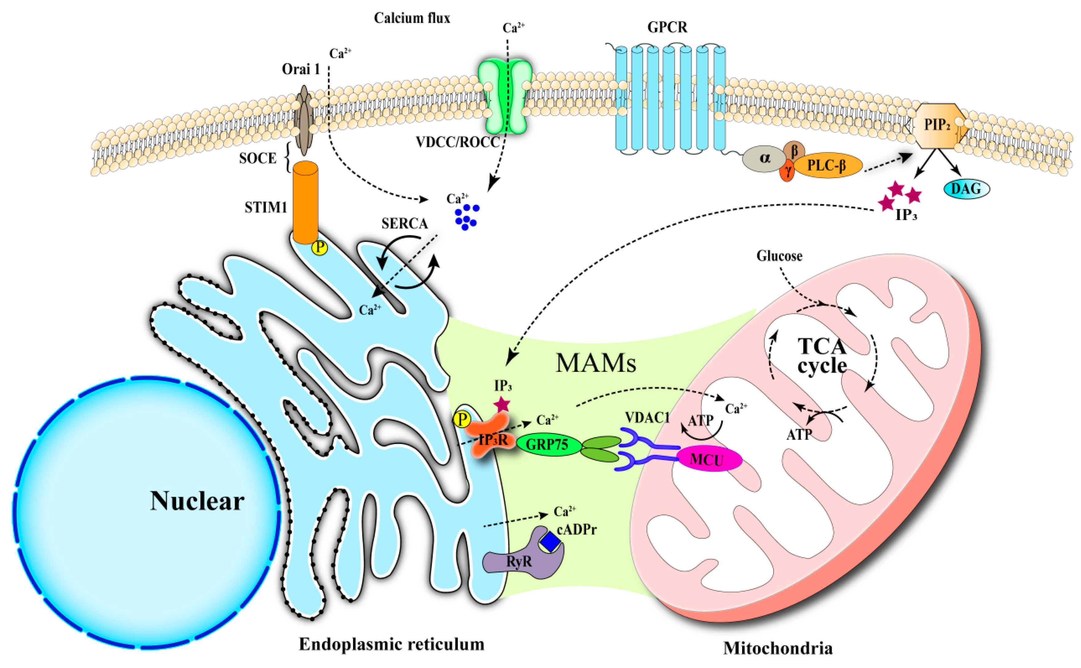
Figure 1. Core Mechanisms of Intracellular Calcium Homeostasis. (1) Calcium enters cells via plasma membrane channels (VDCCs, ROCCs, SOCE). (2) The endoplasmic reticulum (ER) acts as a major calcium store, regulated by SERCA (uptake) and IP3R/RyR (release). (3) Mitochondria uptake calcium via the MCU and MAMs to link calcium signaling with metabolism.
Biomedicines 13 02694 g001
Figure 2. Regulation of Apoptosis and Autophagy by Calcium Homeostasis and Key Signaling Pathways. ER stress-induced calcium overload activates both intrinsic and ER-specific apoptotic pathways. Mitochondrial ROS production from calcium overload promotes autophagy via CAMKKβ-AMPK signaling. Autophagy and apoptosis are interconnected, forming a feedback loop that determines cell fate.
Biomedicines 13 02694 g002
Figure 3. Calcium Dysregulation Drives Ferroptosis. ER stress and calcium release cause mitochondrial dysfunction and glutathione depletion. This leads to iron accumulation and lipid peroxide production via the Fenton reaction. The convergence of calcium imbalance and iron toxicity results in ferroptotic cell death.
Biomedicines 13 02694 g003
Figure 4. Calcium Influx Triggers Pyroptosis via the NLRP3 Inflammasome. Danger signals (PAMPs/DAMPs) prime the NF-κB pathway to express NLRP3 and pro-cytokines. Calcium overload serves as a key signal for NLRP3 inflammasome assembly. Inflammasome activation cleaves GSDMD, forming pores in the membrane and executing pyroptosis.
Biomedicines 13 02694 g004
Table 1. Proteins regulating calcium homeostasis.
Name/LocationFunctionMain RegulatorsReferences
SERCA
(ER membrane)
Ca2+ uptake from cytosol to ER lumenThapsigargin (inhibitor);
CDN1163 (activators);
Aged garlic extract (activators);
[40,41,42,43]
IP3R
(ER membrane)
Ca2+ release from ER to cytosol Xestospongin C (inhibitor);
ryanodine (inhibitor);
2-APB (inhibitor);
carbachol (activators);
IP3 (activators);
[43,44]
RyR
(ER membrane)
Ca2+ release from ER to cytosol Dantrolene (inhibitor);
High Ca2+ (inhibitor);
Low Ca2+ (activator);
Caffeine (activator);
[43,44]
STIM
(ER membrane)
Ca2+ entry from extracellular space to cytosolCa2+ depletion (activator);
Low 2APB (activator);
SKF96365 (inhibitor);
Lupenone (inhibitor);
BTP2 (inhibitor);
[20,45,46]
Orai
(Plasma Membrane)
Coupled with STIM, Ca2+ entry from
extracellular
milieu to cytosol
A chaperone complex
(Regulating agent);
BTP2 (inhibitor);
[20,46]
CaMKII
(Cytoplasm)
Regulated by the interaction between Ca2+ and calmodulinIonomycin (activator);[45,47,48,49]
VDAC
(Mitochondrial
outer membrane)
Forms a channel in the outer mitochondrial membrane and coupled to MCU to allow Ca2+ diffusionBD1047 (Regulating agent);[24,46,50]
MCU
(Mitochondrial
inner membrane)
Ca2+ uptake into the
mitochondria
Ruthenium Red (inhibitor)
BAPTA-AM (Regulating agent);
[24,46,50]
TRPML1
(Lysosome)
Mediating lysosomal Ca2+ efflux and promoting autophagyPI (3,5) P2 (activator)[51]
Table 2. Mechanisms by which different intracellular calcium channels influence cell injury.
Forms of InjuryCalcium Channels
(Activation/Inhibition)
MechanismsReferences
ApoptosisSERCA (inhibition); Improving the calcium storage in the endoplasmic reticulum and alleviating endoplasmic reticulum stress.[40,42,43,53]
IP3R, RyR (activation);Resulting in depletion of calcium ions in the endoplasmic reticulum, triggering endoplasmic reticulum stress.[43,44,62]
SOCE (activation)Increasing the intracellular calcium ion concentration, causing endoplasmic reticulum stress.[45]
AutophagyTRPML1 (activation);Mediating the release of calcium ions from lysosomes, activating Calcineurin, and inducing autophagy.[51]
CRAC (activation);Mediating the inward flow of extracellular calcium ions and promoting autophagy.[51]
PyroptosisIP3R (activation)PLCγ1 indirectly activating IP3R to release endoplasmic reticulum calcium to induce pyroptosis.[63]
CaMKII (activation)Promoting the assembly of the NLRP3 inflammasome and activating pyroptosis.[64]
L-type calcium channel (inhibition)Reducing the intracellular calcium ion concentration and inhibiting the activation of the NLRP3 inflammasome.[65]
Table 3. Mechanisms by which different calcium channels affect insulin resistance.
Calcium ChannelsAnimals/CellsIntervention ReagentsTreatment TimeDisease TypeMechanismsReferences
SERCA
(inhibitor)
ob/ob miceCDN11635 daysInsulin resistance and prediabetesActivating Ca2+-ATPase activity and improving endoplasmic reticulum stress [41]
SOCE
(activator)
Male C57BL/6 mice
HepG2 cells
Candesartan
Azilsartan, candesartan,
21 days
16 h
Obesity;
Insulin resistance
Mediated calcium influx, inhibition of AKT phosphorylation[34]
CaMKII
(inhibitor)
L6-GLUT4myc cellsLonomycin48 hMuscle Insulin resistanceActivating AMPK-PKC phosphorylation pathway to enhance GLUT4 exocytosis and endocytosis[49]
CAMK2
(activator)
Ai-CAMK2 KO mice;
OP9 cells
Tamoxifen
KN93
5 days
1 h
Obesity;
Insulin resistance
CAMK2 activation decreases the number of insulin receptors (INSR)[130]
Disclaimer/Publisher’s Note: The statements, opinions and data contained in all publications are solely those of the individual author(s) and contributor(s) and not of MDPI and/or the editor(s). MDPI and/or the editor(s) disclaim responsibility for any injury to people or property resulting from any ideas, methods, instructions or products referred to in the content.