Article

Combination of Neovestitol and Vestitol Modifies the Profile of Periodontitis-Related Subgingival Multispecies Biofilm

by
Tatiane Tiemi Macedo
1,
Larissa Matias Malavazi
2,
Gustavo Quilles Vargas
2,
Francisco Jerfeson dos Santos Gonçalves
1,
Aline Paim de Abreu Paulo Gomes
1,
Manuela Rocha Bueno
3,
Lucas Daylor Aguiar da Silva
1,
Luciene Cristina Figueiredo
1 and
Bruno Bueno-Silva
1,2,*
1
Dental Research Division, Guarulhos University, Guarulhos 07023-070, SP, Brazil
2
Departamento de Biociências, Faculdade de Odontologia de Piracicaba, Universidade de Campinas (UNICAMP), Piracicaba 13414-903, SP, Brazil
3
Faculdade São Leopoldo Mandic, Campinas 13045-775, SP, Brazil
*
Author to whom correspondence should be addressed.
Biomedicines 2024, 12(6), 1189; https://doi.org/10.3390/biomedicines12061189
Submission received: 11 April 2024 / Revised: 18 May 2024 / Accepted: 20 May 2024 / Published: 27 May 2024
(This article belongs to the Section Molecular and Translational Medicine)

Abstract

:
The aim of this study was to evaluate the effect of the combination of neovestitol–vestitol (CNV) compounds obtained from Brazilian red propolis on the microbiological profile of a mature multispecies subgingival biofilm. The biofilm with 32 bacterial species associated with periodontitis was formed for seven days using a Calgary device. Treatment with CNV (1600, 800, 400, and 200 μg/mL), amoxicillin (54 μg/mL), and vehicle control was performed for 24 h on the last day of biofilm formation. Biofilm metabolic activity and DNA–DNA hybridization (checkerboard) assays were performed. The groups treated with CNV 1600 and amoxicillin reduced 25 and 13 species, respectively, compared to the control vehicle treatment (p ≤ 0.05); both reduced P. gingivalis, while only CNV reduced T. forsythia. When the data from the two treatments (CNV and AMOXI) were compared, a statistically significant difference was observed in 13 species, particularly members of Socransky’s orange complex. Our results showed that CNV at 1600 μg/mL showed the best results regarding the metabolic activity of mature biofilms and obtained a reduction in species associated with the disease, such as T. forsythia, showing a better reduction than amoxicillin. Therefore, CNV seems to be a promising alternative to eradicate biofilms and reduce their pathogenicity.

1. Introduction

Periodontal disease represents a pathology of significant importance for oral health; its development is intrinsically linked to a complex pathogenic dental biofilm, which shelters multiple microbial communities capable of evading the defenses of the immune system [1]. As a result, an uncontrolled immuno-inflammatory response starts in the host, generating progressive damage to the protective and supporting periodontium [2]. When these microbial communities are in balance with the defense system, they have a higher proportion of bacteria associated with health and the periodontal tissues maintain health; however, oral biofilm dysbiosis associated with an exacerbated immuno-inflammatory response in the host is evidenced by the development of periodontal pockets and tooth mobility [3].
Numerous preventive strategies have been employed to reduce the number of microorganisms and, in consequence, decrease inflammation response and thus reduce the risk of periodontal disease [4]. The mechanical removal of biofilm (scaling and root planning) is considered the conventional treatment, although it is not always sufficient. Consequently, in many cases, it is essential to consider the use of adjunctive treatments, for example, systemic antibiotics like the combination of amoxicillin and metronidazole [5] or probiotics [6].
Although antibiotic therapy has been shown to improve the clinical parameters of periodontal disease, its effects on the human body can cause changes in the composition of the microbiota of other body systems due to its broad spectrum and prolonged use [7,8,9], as well as the risk of bacterial resistance, so the discovery of new drugs from natural products has aroused considerable interest in the scientific community.
Brazilian propolis is a natural substance that has a variety of pharmacological properties, including antimicrobial, antioxidant, anticancer, and anti-inflammatory activities [10,11,12]. The composition of Brazilian propolis is highly variable due to the presence of multiple components, resulting in a wide chemical diversity of bioactive compounds that vary according to the geographical location of its production [10,11].
Brazilian red propolis (BRP) is produced by Appis mellifera bees and originates mainly in the coastal region of Maceió city, Alagoas state. Its botanical origin is the resin of Dalbergia ecastophyllum [13]. This propolis has an intense red color and its main constituents are isoflavonoids and flavonoids, especially formononetin, vestitol, and neovestitol [10]. Neovestitol and vestitol appear to be a great option as local oral bioactive agents due to their antimicrobial and anti-inflammatory properties [14,15,16,17].
The natural combination of neovestitol–vestitol (60/30%) showed a minimum inhibitory concentration of 50 µg/mL against Porphyromona gingivalis and Aggregatibacter actinomycetemcomitans, inhibited the formation and development of Streptococcus mutans biofilm in vitro [18], and reduced the development of dental caries through an in vivo study at a concentration of 800 µg/mL [19], demonstrating its potential to inhibit the growth of microorganisms important in the development of biofilm associated with periodontal disease. Therefore, the aim of this study was to evaluate the effect of the combination of neovestitol–vestitol (CNV) compounds obtained from Brazilian red propolis on the microbiological profile of mature multispecies subgingival biofilm.

2. Materials and Methods

2.1. Biofilm Formation

The bacterial species below were used in the multispecies biofilm model: Actinomyces naeslundii ATCC12104, Actinomyces oris ATCC43146, Actinomyces gerencseriae ATCC23840, Actinomyces israelii ATCC12102, Veillonella parvula ATCC10790 Actinomyces odontolyticus ATCC17929, Streptococcus sanguinis ATCC10556, Streptococcus oralis ATCC35037, Streptococcus intermedius ATCC27335, Streptococcus gordonii ATCC10558, Streptococcus mitis ATCC49456, Aggregatibacter actinomycetemcomitans ATCC29523, Capnocytophaga ochracea ATCC33596, Capnocytophaga gingivalis ATCC33624, Eikenella corrodens ATCC23834, Capnocytophaga sputigena ATCC33612, Streptococcus constellatus ATCC27823, Eubacterium nodatum ATCC33099, Fusobacterium nucleatum vincentii ATCC49256, Parvimonas micra ATCC33270, Fusobacterium nucleatum polymorphum ATCC10953, Campylobacter showae ATCC51146, Fusobacterium periodonticum ATCC33693, Prevotella intermedia ATCC25611, Porphyromonas gingivalis ATCC33277, Tannerella forsythia ATCC43037, Eubacterium saburreum ATCC33271, Streptococcus anginosus ATCC33397, Selenomonas noxia ATCC43541, Propionibacterium acnes ATCC11827, Gemella morbillorum ATCC27824, and Streptococcus mutans ATCC25175.
The microorganisms were grown on tryptone soy agar with 5% sheep’s blood under anaerobic conditions (85% nitrogen, 10% carbon dioxide, and 5% hydrogen), with P. gingivalis and P. intermedia being grown on yeast extract enriched with 1% hemin, 5% menadione, and 5% sheep’s blood, while T. forsythia was grown on tryptone soy agar with yeast extract enriched with 1% hemin, 5% menadione, 5% sheep’s blood, and 1% N-acetylmuramic acid. After 48 h of growth, all bacterial species were transferred to BHI broth (Becton Dickinson, Sparks, MD, USA) supplemented with 1% hemin.
After 24 h, the optical density (OD) at 600 nm was adjusted to 0.1, corresponding to around 108 cells/mL of each species. The individual cell suspensions were diluted to obtain the final biofilm inoculum, containing 104 cells of each species. The multiple species model of subgingival biofilm was developed using the Calgary biofilm device (CBD) according to Soares et al. [20].

2.2. CNV Obtainment

CNV was obtained naturally through the fractionation of Brazilian red propolis (BRP), as described previously [19]. The red propolis studies were registered in the National System for the Management of Genetic Heritage and Associated Traditional Knowledge of the Brazilian Federal Government (SISGEN—register number A305815). Briefly, BRP samples were collected in the State of Alagoas, Brazil, and first an ethanolic extract was prepared. Liquid–liquid fractionation with two solvents (hexane and chloroform) was then performed on the ethanolic crude BRP extract. As previously described [18], the chloroform fraction has better antimicrobial activity and was subjected to two different chromatographic separations: dry column (polarity separation) and Sephadex LH-20 column (size separation). The combination of Neovestitol and Vestitol compounds used here was obtained after Sephadex LH-20, and a previous work characterized its chemical composition and demonstrated its reducing effect on S. mutans biofilm [19].

2.3. 24 h Treatment with CNV on Mature Biofilm

The biofilms were formed for 7 days, and the medium was changed on day 3 (after 72 h of incubation) and day 6 (after 144 h of formation). After 6 days of formation, the biofilms were treated for 24 h until the completion of 7 days of formation. For the treatment, the pins coated with the biofilms were washed twice with 200 µL of 1% PBS and transferred to 96-well plates containing culture medium (100 µL) and 100 µL of different concentrations of CNV-VO (1600; 800; 400 and 200 µg/mL), chosen according to previous results with crude propolis [21,22]. For this therapeutic scheme, in addition to the untreated control group, a group treated with amoxicillin at 54 µg/mL was also included (concentration chosen according to Soares et al. [20]). At the end of seven days of biofilm formation, items 2.4. (metabolic activity) and 2.5. (checkerboard) [20], described below, were carried out.

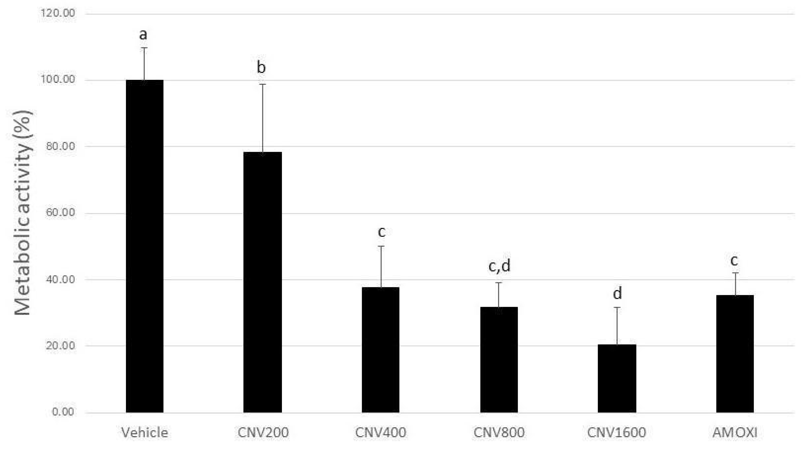
2.4. Biofilm Metabolic Activity

The percentage reduction in biofilm metabolic activity was determined using 2,3,5-triphenyltetrazolium chloride (TTC) and spectrophotometry. TTC is used to differentiate between metabolically active and inactive cells. The white substrate was enzymatically reduced to red 1,3,5-triphenyl formazan (TPHP) by living bacterial cells due to the activity of various dehydrogenases enzymes. The substrate color change was read using spectrophotometry to determine the rate of reduction, which is used as an indirect measure of bacterial metabolic activity. To measure the metabolic activity of the biofilms, the pins were washed twice with washing solution and transferred to plates with 200 µL per well of fresh BHI medium containing 1% hemin and 0.1% TTC solution. The plates were then incubated under anaerobic conditions for 6–8 h at 37 °C. TTC conversion was read at 485 nm using a spectrophotometer [20].

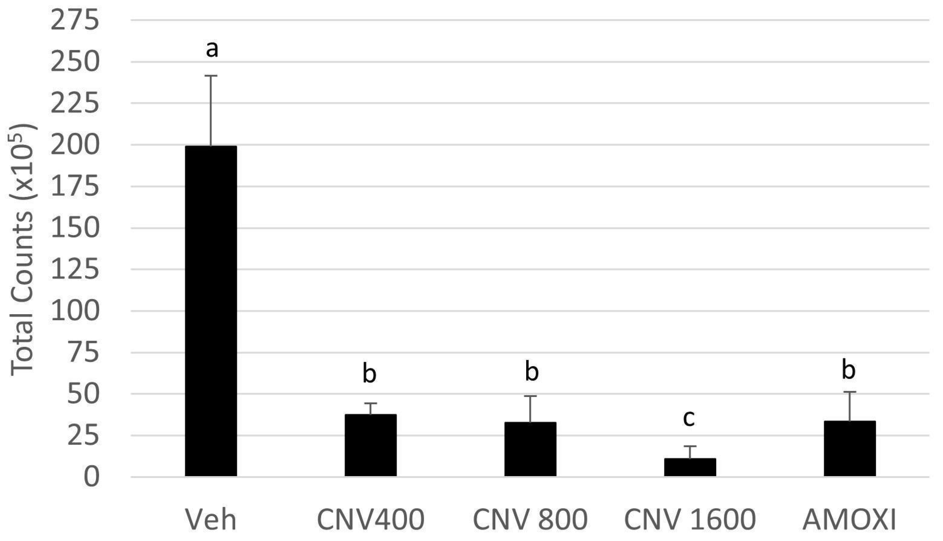
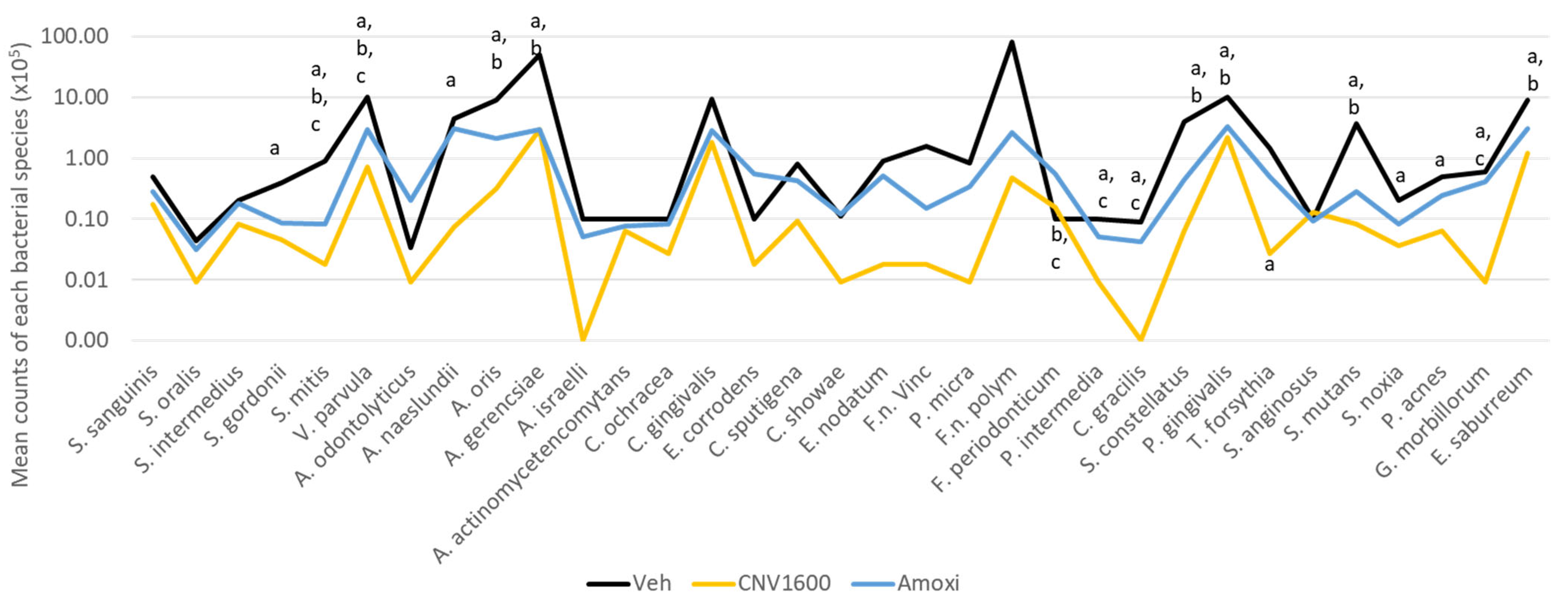
2.5. DNA–DNA Hybridization (Checkerboard)

Calgary device “pegs” coated with 7-day biofilm from each of the groups were washed twice with PBS and transferred to Eppendorf tubes containing 150 µL of TE buffer (Tris 10 mM -HCl, EDTA 1 mM [pH 7.6]), and then 100 µL of 0.5 M NaOH was added. The tubes containing the pins and the final solution were boiled for 10 min, and the solution was neutralized by adding 0.8 mL of 5 M ammonium acetate. The samples were then analyzed individually for the presence and quantity of the 30 bacterial species using the DNA–DNA hybridization technique. Briefly, after the lysis of the samples, the DNA was placed in lanes on a nylon membrane using a Minislot device (Immunetics, Cambridge, MA, USA). After fixing the DNA to the membrane, which was placed in a Miniblotter 45 (Immunetics), digoxigenin-labeled whole-genome DNA probes for the subgingival species used were hybridized to individual lanes of the Miniblotter 45. After hybridization, the membranes were washed, and the DNA probes were detected using an antibody specific for digoxigenin conjugated with alkaline phosphatase. The signals were detected using AttoPhos substrate (Amersham Life Sciences, Arlington Heights, IL, USA), and the results were read using Typhoon Trio Plus (Molecular Dynamics, Sunnyvale, CA, USA). Two lanes in each run contained the standards with 105 or 106 cells of each species. Signals obtained with the Typhoon Trio were converted into absolute counts by comparison with the standards on the same membrane. Failure to detect a signal was recorded as zero. The values obtained after treatment with the samples were compared with the pegs of the positive and negative control groups.

2.6. Statistical Analysis

Data from the biofilm’s metabolic activity test were statistically analyzed using Analysis of Variance (ANOVA) followed by Tukey’s test. The results of the checkerboard DNA–DNA hybridization were statistically analyzed using Kruskal–Wallis, followed by Dunn’s post hoc (p ≤ 0.05).

3. Results

Biofilms treated with CNV at 1600, 800, 400, and 200 µg/mL, and amoxicillin showed a statistically significant reduction in metabolic activity of 79, 68, 62, 21, and 65%, respectively when compared to the negative control group (p < 0.05) (Figure 1). The metabolic activity of mature biofilms treated with CNV at 1600 µg/mL showed the lowest absolute value (21%), showing better results even than amoxicillin (p < 0.05). The group treated with 800 µg/mL showed no statistically significant difference to the CNV 400, CNV 1600, and AMOXI groups (p > 0.05) (Figure 1).
Figure 2 shows the results of the total biofilm count formed over 7 days and treated with CNV at 1600, 800, and 400 µg/mL, amoxicillin, and control vehicle. As the group treated with CNV at 200 µg/mL showed the worst metabolic activity results, it was discarded from subsequent analyses. The group treated with CNV at 1600 µg/mL showed the lowest total count values, corroborating the metabolic activity data. The groups treated with CNV at 400 and 800 µg/mL showed no statistically significant difference to the group treated with amoxicillin (positive control).
Figure 3 shows the average count results for each species used in the biofilm model treated with Vehicle, CNV 1600, and AMOXI. Since the 1600 µg/mL concentration of CNV showed the most promising results in the previous tests, only this concentration was selected for the analysis of the count of each species. The treatments with CNV 1600 and AMOXI reduced 25 and 13 species, respectively, compared to the control vehicle treatment (p ≤ 0.05). It is noteworthy that both treatments reduced P. gingivalis counts, while only CNV reduced T. forsythia values. When comparing the data from both treatments (CNV and AMOXI), a statistically significant difference was observed in 13 species, highlighting P. intermedia, F. periodonticum, F. nucleatum polymorphum, F. nucleatum vicentii, and P. micra (all members of Socransky’s orange complex).

4. Discussion

The present article shows the effects of a combination of Neovestitol and Vestitol, a fraction isolated from Brazilian red propolis, on the periodontitis-related subgingival multispecies biofilm. Three different concentrations of CNV (1600, 800, and 400 µg/mL) were shown to have a reducing effect on the mature biofilm. The highest tested concentration (1600 ug/mL) was even better than the amoxicillin treatment, reducing the amount of 25 species compared to vehicle control and 13 species compared to amoxicillin-treated biofilms. It seems that amoxicillin is a non-selective antibiotic, acting on both health- and disease-associated bacteria, while CNV has a prominent effect on disease-associated species.
The dysbiotic subgingival biofilm is an important etiological factor of periodontitis. It is a complex, intricate community formed by several distinct bacterial species and other microorganisms. Initially, the bacteria found in periodontitis patients were divided into five groups, according to their association with healthy or diseased conditions [23]. The main species associated with diseased sites were identified as P. gingivalis, T. forsythia, and Treponema denticola. Nowadays, researchers are searching for novel putative oral pathobionts [24,25]. In recent studies, other species, such as Filifactor allocis, have been investigated as putative new pathogens, and a clinical investigation confirmed their presence in patients with periodontitis [26]. These results suggest that the composition of the biofilm associated with periodontal disease may be more complex and diverse than previously thought [27,28]. In this context, it has been shown that a consortium of bacteria, rather than a single species, is accountable for the transition of the biofilm from a state associated with oral health to one that is pathogenic. Notably, P. gingivalis, along with T. forsythia and S. gordonii (and potentially other oral bacteria), have been identified as significant contributors to the dysbiosis of the subgingival biofilm, which ultimately results in the development of periodontal disease [29].
P. gingivalis may be considered the primary periodontal pathogen responsible for biofilm dysbiosis since it has been identified as a keystone pathogen in periodontal disease due to its production of various virulence factors (for example, gingipains and FimA), with properties to subvert the human immune response, including neutrophils, macrophages, and the complement system [30]. Therefore, the effect of CNV on P. gingivalis is remarkable. The natural compound combination reduced the quantity of this keystone pathogen in the mature biofilm by almost fivefold.
Of particular note, only the biofilms treated with CNV at 1600 µg/mL exhibited a statistically significant reduction in T. forsythia (Figure 3), another periodontal pathogen related to biofilm dysbiosis. Even amoxicillin was not statistically significant in reducing the amounts of this pathogen when compared to vehicle-treated biofilms. A recent article described that, during the infection of macrophages by T. forsythia, the immune response was led to produce pro-inflammatory factors (such as BspA, sialidase, GroEL, and numerous lipoproteins) related to periodontal tissue damage through the activation of the Toll-like 2 receptor of macrophages [31].
Another significant observation is the decline in all species of the Fusobacterium genera included in the model (F. nucleatum vincentii, F. nucleatum polymorphum, and F. periodonticum) by CNV at a concentration of 1600 µg/mL. The Fusobacterium genera are notably influential in the transition from periodontal health to disease [32]. F. nucleatum is recognized as the most prevalent anaerobic, Gram-negative species during the advanced stages of periodontal disease, and has been suggested as a potential periodontal pathogen [33]. According to research by Colombo et al. (2002) [34] and Socransky and Haffajee (2002) [33], the presence of F. nucleatum is predominantly associated with individuals affected by periodontitis and periodontal abscesses. Furthermore, its abundance tends to decrease after successful periodontal therapy [32]. Acting as an intermediary colonizer within dental biofilms, Fusobacterium species are among the first Gram-negative organisms to establish a stable presence in the subgingival biofilm. This pivotal role involves facilitating interactions between Gram-positive and Gram-negative species, thereby fostering the colonization of additional anaerobic species, including those recognized as pathogens within the red complex [32].
The present in vitro biofilm model is widely used by our research group. The number of bacterial species included in the model is the main strength since, to our knowledge, this is the only model that includes almost all Socransky species (except T. denticola). The values for the reduction in metabolic activity of biofilms treated with amoxicillin are consistent with those obtained in previous studies [20,22,35], demonstrating the reproducibility of the method. It is of importance to note that among all the articles published to date with this therapeutic regimen (treatment for 12 or 24 h), CNV at 1600 µg/mL was the first test agent to show metabolic activity and total counts significantly lower than the positive control, either amoxicillin or chlorhexidine [22,35,36,37]. This result is considered very promising. Another point to note is that as this formulation is intended to be used orally, some metabolization of the bioactive compounds is to be expected. Although not carried out with the same propolis or CNV compounds, a recent study evaluated the in vitro metabolization of organic propolis containing flavonoid compounds. The results showed that the concentration of flavonoid compounds in propolis was reduced by about ¼ after gastric and intestinal metabolization [38]. It is therefore important to note that CNV at 400 µg/mL showed statistically similar results to the biofilm treated with the positive control amoxicillin, demonstrating that the oral intake of 1600 µg/mL may be effective through in vivo studies; however, this hypothesis needs to be tested in future works.
Furthermore, the efficacy of systemic antibiotics in treating periodontal disease is well documented in the literature [39]. However, it is important to acknowledge the potential side effects associated with prolonged antibiotic therapy (lasting 7 or 14 days), which include disrupting the resident microbiota, the selection of antimicrobial-resistant organisms [7,40], damage to the intestinal microbiome [41], and even microbial alterations in the skin [42]. The toxicity of the compounds Neovestitol and Vestitol is another issue that needs to be investigated. To date, in vivo studies in animals (mice and rats) have not reported any signs of toxicity after the use of the compounds [19,43,44], but future studies should focus specifically on toxicity. Another matter to be investigated in the future would be the possible effects on the gut microbiome. These findings underscore the potential value of conducting future in vivo studies on CNV to explore its potential benefits in treating periodontitis.

5. Conclusions

In conclusion, the combination of the compounds neovestitol and vestitol (CNV) proved to be effective in disrupting the mature multispecies subgingival biofilm associated with periodontal disease. The results indicate that CNV concentrations of 800 and 400 µg/mL reduced the levels of classical periodontal pathogens in a manner similar to amoxicillin, while CNV at 1600 µg/mL showed the best results with respect to the metabolic activity of mature biofilms and achieved a better reduction in disease-associated species such as T. forsythia than the positive control amoxicillin. Therefore, CNV appears to be a promising alternative to eradicate biofilm and decrease its pathogenicity. However, further studies are needed to corroborate and extend these results.

Author Contributions

Conceptualization, M.R.B. and B.B.-S.; Formal analysis, T.T.M., L.M.M., G.Q.V., F.J.d.S.G., A.P.d.A.P.G., M.R.B., L.D.A.d.S. and B.B.-S.; Funding acquisition, B.B.-S.; Methodology, T.T.M., L.M.M., G.Q.V., F.J.d.S.G., A.P.d.A.P.G., M.R.B., L.D.A.d.S., L.C.F. and B.B.-S.; Supervision, B.B.-S.; Writing—original draft, L.M.M. and G.Q.V.; Writing—review and editing, M.R.B., L.C.F. and B.B.-S. All authors have read and agreed to the published version of the manuscript.

Funding

This research was financially supported by the Sao Paulo Research Foundation (FAPESP), grant #2019/19691-0; the Brazilian National Council for Scientific and Technological Development (CNPq), grant # 428984/2018-5, the Fundo de apoio ao Ensino, Extensão e Pesquisa (FAEPEX-Unicamp), grants #3364/23 and #2878/23 and by Coordenação de Aperfeiçoamento de Pessoal de Nível Superior—Brasil (CAPES), grant code 001. T.T.M., G.Q.V., and F.J.d.S.G. received a scholarship from the Coordenação de Aperfeiçoamento de Pessoal de Nível Superior—Brasil (CAPES); A.P.d.A.P.G. had an undergraduate scholarship from the Brazilian National Council for Scientific and Technological Development (CNPq). L.M.M. had a scholarship from the Fund to support teaching, research and extension (FAEPEX–Unicamp). L.D.A.d.S. had a scholarship from the Latin American Oral Health Association (LAOHA). MRB received a scholarship from the Sao Paulo Research Foundation (FAPESP), grant # 2017/16377-7.

Institutional Review Board Statement

Not applicable.

Informed Consent Statement

Not applicable.

Data Availability Statement

Data will be available upon request.

Conflicts of Interest

The authors declare no conflicts of interest.

References

  1. Ng, E.; Tay, J.R.H.; Ong, M.M.A. Minimally invasive periodontology: A treatment philosophy and suggested approach. Int. J. Dent. 2021, 2021, 2810264. [Google Scholar] [CrossRef] [PubMed]
  2. Papapanou, P.N.; Sanz, M.; Buduneli, N.; Dietrich, T.; Feres, M.; Fine, D.H.; Flemmig, T.F.; Garcia, R.; Giannobile, W.V.; Graziani, F.; et al. Periodontitis: Consensus report of workgroup 2 of the 2017 world workshop on the classification of periodontal and peri-implant diseases and conditions. J. Periodontol. 2018, 89 (Suppl. S1), S173–S182. [Google Scholar] [CrossRef] [PubMed]
  3. Hajishengallis, G.; Lamont, R.J. Beyond the red complex and into more complexity: The polymicrobial synergy and dysbiosis (PSD) model of periodontal disease etiology. Mol. Oral Microbiol. 2012, 27, 409–419. [Google Scholar] [CrossRef] [PubMed]
  4. Van Dyke, T.E.; Bartold, P.M.; Reynolds, E.C. The Nexus between periodontal inflammation and dysbiosis. Front. Immunol. 2020, 11, 511. [Google Scholar] [CrossRef] [PubMed]
  5. Teughels, W.; Feres, M.; Oud, V.; Martin, C.; Matesanz, P.; Herrera, D. Adjunctive effect of systemic antimicrobials in periodontitis therapy: A systematic review and meta-analysis. J. Clin. Periodontol. 2020, 47 (Suppl. S20), 257–281. [Google Scholar] [CrossRef]
  6. Ng, E.; Tay, J.R.H.; Saffari, S.E.; Lim, L.P.; Chung, K.M.; Ong, M.M.A. Adjunctive probiotics after periodontal debridement versus placebo: A systematic review and meta-analysis. Acta Odontol. Scand. 2022, 80, 81–90. [Google Scholar] [CrossRef]
  7. Zhang, L.; Huang, Y.; Zhou, Y.; Buckley, T.; Wang, H.H. Antibiotic administration routes significantly influence the levels of antibiotic resistance in gut microbiota. Antimicrob. Agents Chemother. 2013, 57, 3659–3666. [Google Scholar] [CrossRef] [PubMed]
  8. Mombelli, A. Microbial colonization of the periodontal pocket and its significance for periodontal therapy. Periodontology 2000 2018, 76, 85–96. [Google Scholar] [CrossRef]
  9. Ng, E.; Tay, J.R.H.; Boey, S.K.; Laine, M.L.; Ivanovski, S.; Seneviratne, C.J. Antibiotic resistance in the microbiota of periodontitis patients: An update of current findings. Crit. Rev. Microbiol. 2024, 50, 329–340. [Google Scholar] [CrossRef]
  10. Bueno-Silva, B.; Kawamoto, D.; Ando-Suguimoto, E.S.; Alencar, S.M.; Rosalen, P.L.; Mayer, M.P.A. Brazilian red propolis attenuates inflammatory signaling cascade in lps-activated macrophages. PLoS ONE 2015, 10, e0144954. [Google Scholar] [CrossRef]
  11. Tiveron, A.P.; Rosalen, P.L.; Franchin, M.; Lacerda, R.C.C.; Bueno-Silva, B.; Benso, B.; Denny, C.; Ikegaki, M.; de Alencar, S.M. Chemical characterization and antioxidant, antimicrobial, and anti-inflammatory activities of south brazilian organic propolis. PLoS ONE 2016, 11, e0165588. [Google Scholar] [CrossRef] [PubMed]
  12. Banzato, T.P.; Gubiani, J.R.; Bernardi, D.I.; Nogueira, C.R.; Monteiro, A.F.; Juliano, F.F.; de Alencar, S.M.; Pilli, R.A.; de Lima, C.A.; Longato, G.B.; et al. Antiproliferative flavanoid dimers isolated from brazilian red propolis. J. Nat. Prod. 2020, 83, 1784–1793. [Google Scholar] [CrossRef] [PubMed]
  13. Silva, B.B.; Rosalen, P.L.; Cury, J.A.; Ikegaki, M.; Souza, V.C.; Esteves, A.; Alencar, S.M. Chemical composition and botanical origin of red propolis, a new type of brazilian propolis. Evid. Based Complement. Altern. Med. 2008, 5, 313–316. [Google Scholar] [CrossRef]
  14. Bueno-Silva, B.; Rosalen, P.L.; Alencar, S.M.; Mayer, M.P. Anti-inflammatory mechanisms of neovestitol from Brazilian red propolis in LPS-activated macrophages. J. Funct. Foods 2017, 36, 440–447. [Google Scholar] [CrossRef]
  15. Silva, N.B.S.; de Souza, J.H.; Santiago, M.B.; Aguiar, J.R.d.S.; Martins, D.O.S.; da Silva, R.A.; Santos, I.d.A.; Aldana-Mejía, J.A.; Jardim, A.C.G.; Pedroso, R.d.S.; et al. Potential in vitro anti-periodontopathogenic, anti-Chikungunya activities and in vivo toxicity of Brazilian red propolis. Sci. Rep. 2022, 12, 21165. [Google Scholar] [CrossRef] [PubMed]
  16. Freires, I.A.; de Alencar, S.M.; Rosalen, P.L. A pharmacological perspective on the use of Brazilian Red Propolis and its isolated compounds against human diseases. Eur. J. Med. Chem. 2016, 110, 267–279. [Google Scholar] [CrossRef] [PubMed]
  17. de Freitas, M.C.D.; de Miranda, M.B.; de Oliveira, D.T.; Vieira-Filho, S.A.; Caligiorne, R.B.; de Figueiredo, S.M. Biological activities of red propolis: A review. Recent Pat. Endocr. Metab. Immune Drug Discov. 2017, 11, 3–12. [Google Scholar] [CrossRef] [PubMed]
  18. Bueno-Silva, B.; Alencar, S.M.; Koo, H.; Ikegaki, M.; Silva, G.V.J.; Napimoga, M.H.; Rosalen, P.L. Anti-inflammatory and antimicrobial evaluation of neovestitol and vestitol isolated from brazilian red propolis. J. Agric. Food Chem. 2013, 61, 4546–4550. [Google Scholar] [CrossRef]
  19. Bueno-Silva, B.; Koo, H.; Falsetta, M.; Alencar, S.; Ikegaki, M.; Rosalen, P. Effect of neovestitol–vestitol containing Brazilian red propolis on accumulation of biofilm in vitro and development of dental caries in vivo. Biofouling 2013, 29, 1233–1242. [Google Scholar] [CrossRef]
  20. Soares, G.M.S.; Teles, F.; Starr, J.R.; Feres, M.; Patel, M.; Martin, L.; Teles, R. Effects of azithromycin, metronidazole, amoxicillin, and metronidazole plus amoxicillin on an in vitro polymicrobial subgingival biofilm model. Antimicrob. Agents Chemother. 2015, 59, 2791–2798. [Google Scholar] [CrossRef]
  21. Miranda, S.L.F.; Damasceno, J.T.; Faveri, M.; Figueiredo, L.; da Silva, H.D.; Alencar, S.M.d.A.; Rosalen, P.L.; Feres, M.; Bueno-Silva, B. Brazilian red propolis reduces orange-complex periodontopathogens growing in multispecies biofilms. Biofouling 2019, 35, 308–319. [Google Scholar] [CrossRef] [PubMed]
  22. de Figueiredo, K.A.; da Silva, H.D.P.; Miranda, S.L.F.; Gonçalves, F.J.d.S.; de Sousa, A.P.; de Figueiredo, L.C.; Feres, M.; Bueno-Silva, B. Brazilian red propolis is as effective as amoxicillin in controlling red-complex of multispecies subgingival mature biofilm in vitro. Antibiotics 2020, 9, 432. [Google Scholar] [CrossRef] [PubMed]
  23. Socransky, S.S.; Haffajee, A.D.; Dzink, J.L. Relationship of subgingival microbial complexes to clinical features at the sampled sites. J. Clin. Periodontol. 1988, 15, 440–444. [Google Scholar] [CrossRef] [PubMed]
  24. Hajishengallis, G.; Darveau, R.P.; Curtis, M.A. The keystone-pathogen hypothesis. Nat. Rev. Microbiol. 2012, 10, 717–725. [Google Scholar] [CrossRef]
  25. Jiao, Y.; Hasegawa, M.; Inohara, N. The role of oral pathobionts in dysbiosis during periodontitis development. J. Dent. Res. 2014, 93, 539–546. [Google Scholar] [CrossRef] [PubMed]
  26. Ng, E.; Tay, J.R.H.; Balan, P.; Ong, M.M.A.; Bostanci, N.; Belibasakis, G.N.; Seneviratne, C.J. Metagenomic sequencing provides new insights into the subgingival bacteriome and aetiopathology of periodontitis. J. Periodontal Res. 2021, 56, 205–218. [Google Scholar] [CrossRef] [PubMed]
  27. Araujo, L.L.; Lourenco, T.G.B.; Colombo, A.P.V. Periodontal disease severity is associated to pathogenic consortia comprising putative and candidate periodontal pathogens. J. Appl. Oral Sci. 2023, 31, e20220359. [Google Scholar] [CrossRef]
  28. Aja, E.; Mangar, M.; Fletcher, H.; Mishra, A. Filifactor alocis: Recent insights and advances. J. Dent. Res. 2021, 100, 790–797. [Google Scholar] [CrossRef] [PubMed]
  29. Hajishengallis, G.; Lamont, R.J.; Koo, H. Oral polymicrobial communities: Assembly, function, and impact on diseases. Cell Host Microbe 2023, 31, 528–538. [Google Scholar] [CrossRef]
  30. Hajishengallis, G. Oral bacteria and leaky endothelial junctions in remote extraoral sites. FEBS J. 2021, 288, 1475–1478. [Google Scholar] [CrossRef]
  31. Lim, Y.; Kim, H.Y.; Han, D.; Choi, B. Proteome and immune responses of extracellular vesicles derived from macrophages infected with the periodontal pathogen Tannerella forsythia. J. Extracell. Vesicles 2023, 12, e12381. [Google Scholar] [CrossRef] [PubMed]
  32. Kolenbrander, P.E. Oral Microbial Communities: Biofilms, Interactions, and Genetic Systems. Annu. Rev. Microbiol. 2000, 54, 413–437. [Google Scholar] [CrossRef] [PubMed]
  33. Socransky, S.S.; Haffajee, A.D. Dental biofilms: Difficult therapeutic targets. Periodontology 2000 2002, 28, 12–55. [Google Scholar] [CrossRef] [PubMed]
  34. Colombo, A.P.V.; Teles, R.P.; Torres, M.C.; Souto, R.; Rosalém, W., Jr.; Mendes, M.C.S.; Uzeda, M. Subgingival microbiota of brazilian subjects with untreated chronic periodontitis. J. Periodontol. 2002, 73, 360–369. [Google Scholar] [CrossRef] [PubMed]
  35. de Carvalho, R.D.P.; Moreno, J.d.A.; Roque, S.M.; Chan, D.C.H.; Torrez, W.B.; Stipp, R.N.; Bueno-Silva, B.; de Lima, P.O.; Cogo-Müller, K. Statins and oral biofilm: Simvastatin as a promising drug to control periodontal dysbiosis. Oral Dis. 2024, 30, 669–680. [Google Scholar] [CrossRef] [PubMed]
  36. Bueno-Silva, B.; Kiausinus, K.R.; Goncalves, F.J.; Moreira, M.V.C.; de Oliveira, E.G.; Junior, A.B.; Feres, M.; Figueiredo, L.C. Antimicrobial activity of Desplac® oral gel in the subgingival multispecies biofilm formation. Front. Microbiol. 2023, 14, 1122051. [Google Scholar] [CrossRef] [PubMed]
  37. Shibli, J.A.; Rocha, T.F.; Coelho, F.; de Oliveira Capote, T.S.; Saska, S.; Melo, M.A.; Pingueiro, J.M.S.; de Faveri, M.; Bueno-Silva, B. Metabolic activity of hydro-carbon-oxo-borate on a multispecies subgingival periodontal biofilm: A short communication. Clin. Oral Investig. 2021, 25, 5945–5953. [Google Scholar] [CrossRef] [PubMed]
  38. Saliba, A.S.M.C.; Quirino, D.J.G.; Favaro-Trindade, C.S.; de Oliveira Sartori, A.G.; Massarioli, A.P.; Lazarini, J.G.; de Souza Silva, A.P.; de Alencar, S.M. Effects of simulated gastrointestinal digestion/epithelial transport on phenolics and bioactivities of particles of brewer’s spent yeasts loaded with Brazilian red propolis. Food Res. Int. 2023, 173, 113345. [Google Scholar] [CrossRef]
  39. Feres, M.; Figueiredo, L.C.; Soares, G.M.S.; Faveri, M. Systemic antibiotics in the treatment of periodontitis. Periodontology 2000 2015, 67, 131–186. [Google Scholar] [CrossRef]
  40. Jepsen, K.; Jepsen, S. Antibiotics/antimicrobials: Systemic and local administration in the therapy of mild to moderately advanced periodontitis. Periodontology 2000 2016, 71, 82–112. [Google Scholar] [CrossRef]
  41. de Nies, L.; Kobras, C.M.; Stracy, M. Antibiotic-induced collateral damage to the microbiota and associated infections. Nat. Rev. Microbiol. 2023, 21, 789–804. [Google Scholar] [CrossRef] [PubMed]
  42. Jo, J.-H.; Harkins, C.P.; Schwardt, N.H.; Portillo, J.A.; Zimmerman, M.D.; Carter, C.L.; Hossen, A.; Peer, C.J.; Polley, E.C.; Dartois, V.; et al. Alterations of human skin microbiome and expansion of antimicrobial resistance after systemic antibiotics. Sci. Transl. Med. 2021, 13, eabd8077. [Google Scholar] [CrossRef] [PubMed]
  43. Franchin, M.; Colón, D.F.; da Cunha, M.G.; Castanheira, F.V.S.; Saraiva, A.L.L.; Bueno-Silva, B.; Alencar, S.M.; Cunha, T.M.; Rosalen, P.L. Neovestitol, an isoflavonoid isolated from Brazilian red propolis, reduces acute and chronic inflammation: Involvement of nitric oxide and IL-6. Sci. Rep. 2016, 6, 36401. [Google Scholar] [CrossRef] [PubMed]
  44. Franchin, M.; Cólon, D.F.; Castanheira, F.V.S.; da Cunha, M.G.; Bueno-Silva, B.; Alencar, S.M.; Cunha, T.M.; Rosalen, P.L. Vestitol isolated from brazilian red propolis inhibits neutrophils migration in the inflammatory process: Elucidation of the mechanism of action. J. Nat. Prod. 2016, 79, 954–960. [Google Scholar] [CrossRef] [PubMed]
Figure 1. Effect of CNV treatment at 200, 400, 800, and 1600 µg/mL and amoxicillin at 54 µg/mL for 24 h on the metabolic activity of biofilms formed over 7 days (n = 10–3 separate experiments). Different letters mean statistically significant differences among the groups determined using Kruskal–Wallis’s test, followed by Dunn’s post hoc test (p ≤ 0.05).
Biomedicines 12 01189 g001
Figure 2. Mean and standard deviation of the total count (×105) of biofilms formed over 6 days and treated for 24 h with control vehicle (Veh), CNV at 400, 800, and 1600 µg/mL and amoxicillin at 54 µg/mL (n = 9–3 separate experiments). Different letters mean statistically significant differences among the groups determined using Kruskal–Wallis’s test, followed by Dunn’s post hoc test (p ≤ 0.05).
Biomedicines 12 01189 g002
Figure 3. Average counts of each of the bacterial species present in the biofilm. Statistical analysis carried out using Kruskal–Wallis’s test, followed by Dunn’s post hoc test (p ≤ 0.05). The letter “a” means statistical difference between CNV1600 and Vehicle; the letter “b” means statistical difference between Vehicle and Amoxicillin (AMOXI); and the letter “c” means statistical difference between CNV1600 and AMOXI.
Biomedicines 12 01189 g003
Disclaimer/Publisher’s Note: The statements, opinions and data contained in all publications are solely those of the individual author(s) and contributor(s) and not of MDPI and/or the editor(s). MDPI and/or the editor(s) disclaim responsibility for any injury to people or property resulting from any ideas, methods, instructions or products referred to in the content.

