Article

Polydopamine Coating of Graphitic Carbon Nitride, g-C3N4, Improves Biomedical Application

1
Department of Bioengineering, Faculty of Engineering, Canakkale Onsekiz Mart University Terzioglu Campus, 17100 Canakkale, Turkey
2
Department of Chemistry, Faculty of Sciences, Canakkale Onsekiz Mart University Terzioglu Campus, 17100 Canakkale, Turkey
3
Department of Ophthalmology, Morsani College of Medicine, University of South Florida, 12901 Bruce B Downs B. Downs Blv., MDC 21, Tampa, FL 33612, USA
*
Author to whom correspondence should be addressed.
Biomedicines 2024, 12(6), 1151; https://doi.org/10.3390/biomedicines12061151
Submission received: 29 February 2024 / Revised: 7 May 2024 / Accepted: 22 May 2024 / Published: 23 May 2024
(This article belongs to the Special Issue Health-Related Applications of Natural Molecule Derived Structures)

Abstract

:
Graphitic carbon nitride (g-C3N4) is an intriguing nanomaterial that exhibits photoconductive fluorescence properties under UV–visible light. Dopamine (DA) coating of g-C3N4 prepared from melamine was accomplished via self-polymerization of DA as polydopamine (PDA). The g-C3N4 was coated with PDA 1, 3, and 5 times repeatedly as (PDA@g-C3N4) in tris buffer at pH 8.5. As the number of PDA coatings was increased on g-C3N4, the peak intensity at 1512 cm−1 for N–H bending increased. In addition, the increased weight loss values of PDA@g-C3N4 structures at 600 °C from TGA thermograms confirmed that the coating was accomplished. The band gap of g-C3N4, 2.72 eV, was reduced to 0.87 eV after five coatings with PDA. A pristine g-C3N4 was found to have an isoelectric point (IEP) of 4.0, whereas the isoelectric points of 1PDA@g-C3N4 and 3PDA@g-C3N4 are close to each other at 3.94 and 3.91, respectively. On the other hand, the IEP of 5PDA@g-C3N4 was determined at pH 5.75 assuming complete coating with g-C3N4. The biocompatibility of g-C3N4 and PDA@g-C3N4 against L929 fibroblast cell lines revealed that all PDA@g-C3N4 coatings were found to be biocompatible up to a 1000 mg/mL concentration, establishing that PDA coatings did not adversely affect the biocompatibility of the composite materials. In addition, PDA@g-C3N4 was screened for antioxidant potential via total phenol content (TPC) and total flavonoid content assays and it was found that PDA@g-C3N4 has recognizable TPC values and increased linearly with an increased number of PDA coatings. Furthermore, blood compatibility of pristine g-C3N4 is enhanced considerably upon PDA coating, affirmed by hemolysis and the blood clotting index%. Additionally, α-glucosidase inhibitory properties of PDA@g-C3N4 structures revealed that 67.6 + 9.8% of this enzyme was evenly inhibited by 3PDA@g-C3N4 structure.

Graphical Abstract

1. Introduction

Graphitic carbon nitrides (g-C3N4) are fluorescent polymeric materials made of C and N elements. They are nanomaterials with photocatalytic properties, high physicochemical stability, tunable optical properties, controllable band gap and band position, and low toxicity [1,2,3,4,5,6]. It is envisaged that g-C3N4 can be used for pH-sensitive drug delivery systems as a therapeutic agent in both modeling and applied sciences. There are studies on the use of g-C3N4 as a nanocarrier for anticancer drugs such as lonidamine [7], carboplatin [8] and/or cardiovascular drugs such as levosimendan [9] and curcumin [10,11]. Therefore, most of these studies concluded that g-C3N4-based nanomaterials can be used as pH-sensitive drug carriers in cancer imaging and therapy [12]. Despite its biocompatibility and biometabolizability properties, g-C3N4 also has some shortcomings. These include low solubility in water, a relatively large particle size, a low electrical conductivity, and a lack of absorption of visible light above 460 nm [2]. Since g-C3N4 attracts great attention due to its high chemical and physical stability as well as its promising photocatalysts [13,14,15], it can be used, for example, in photocatalysis for the degradation of organic pollutants in wastewater [10]. The tunable band structure of g-C3N4s is another plus feature for wide ranging applications.
Dopamine (DA), (3,4-dihyroxyphenyl)ethylamine and its’ polymeric forms as poly(3,4-dihyroxyphenyl) ethylamine (p(DA)) has many different biological activities [16]. Due to its catechol groups, it has antioxidant, adhesive [17], and enzyme inhibiting properties [18]. The synthesis of DA occurs via a biochemical pathway that involves the amino acid tyrosine [19]. DA is one of the most important neurotransmitters in the human body and provides transmission of signals between nerve cells, and a deficiency of DA can cause many neurological and mental diseases [20]. It was reported that through the functionalization of polydopamine nanoparticles, an electrochemical aptasensor was developed for the rapid, accurate, and economical detection of glycated albumin as a promising biomarker for glycemic management [21]. There are also reports in the literature about the use of DA as a medication [7,22]. By coating g-C3N4 with PDA, g-C3N4 can be gained with new and intriguing properties. Here, DA is used for the coating of g-C3N4 multiple times, e.g., 1, 3, and 5 times as 1PDA@g-C3N4, 3PDA@g-C3N4, and 5PDA@g-C3N4, respectively, and their characterization was carried out as well as an evaluation of their biomedical use. The enzyme α-glucosidase inhibition capacity, the cytotoxicity, as well as the antioxidant properties of PDA-coated g-C3N4 were investigated. Thus, the potential for use in the biomedical field of composite structures obtained by the PDA coating of g-C3N4 was determined.

2. Materials and Methods

2.1. Materials

To synthesize the graphitic carbon nitride (g-C3N4)-based structures, melamine (99%, Sigma-Aldrich, St. Louis, MO, USA) was used as a precursor. Dopamine hydrochloride (DA, 98%, Sigma-Aldrich) was used as received. The coating of g-C3N4 with PDA was carried out in tris buffer (99%, Sigma Aldrich). Folin Ciocalteu’s phenol reagent (Sigma-Aldrich, 99%), sodium nitrite (NaNO2, +97%, Acros, Fukuoka, Japan) and aluminum chloride (AlCl3, 99.9%, Alfa-Aesar, Haverhill, MA, USA) were used in antioxidant tests. The enzyme α-Glucosidase, from Saccharomyces cerevisiae (S. cerevisiae) (Sigma-Aldrich), and substrate p-nitrophenyl-α-D-glucopyranose (≥99%, Sigma-Aldrich) were used in the enzyme inhibition tests. Gram-negative bacteria Escherichia coli (E. coli) (ATCC 8739) and Gram-positive bacteria, Staphylococcus aureus (S. aureus) (ATCC 6538) were obtained from KWIK-STIK™ Microbiologics (St. Cloud, MN, USA) for antibacterial activity tests. Growth mediums for bacteria were nutrient agar (NA) and nutrient broth (NB), which were obtained from BD DifcoTM (Becton, Dickinson and Company, Sparks, MD, USA).

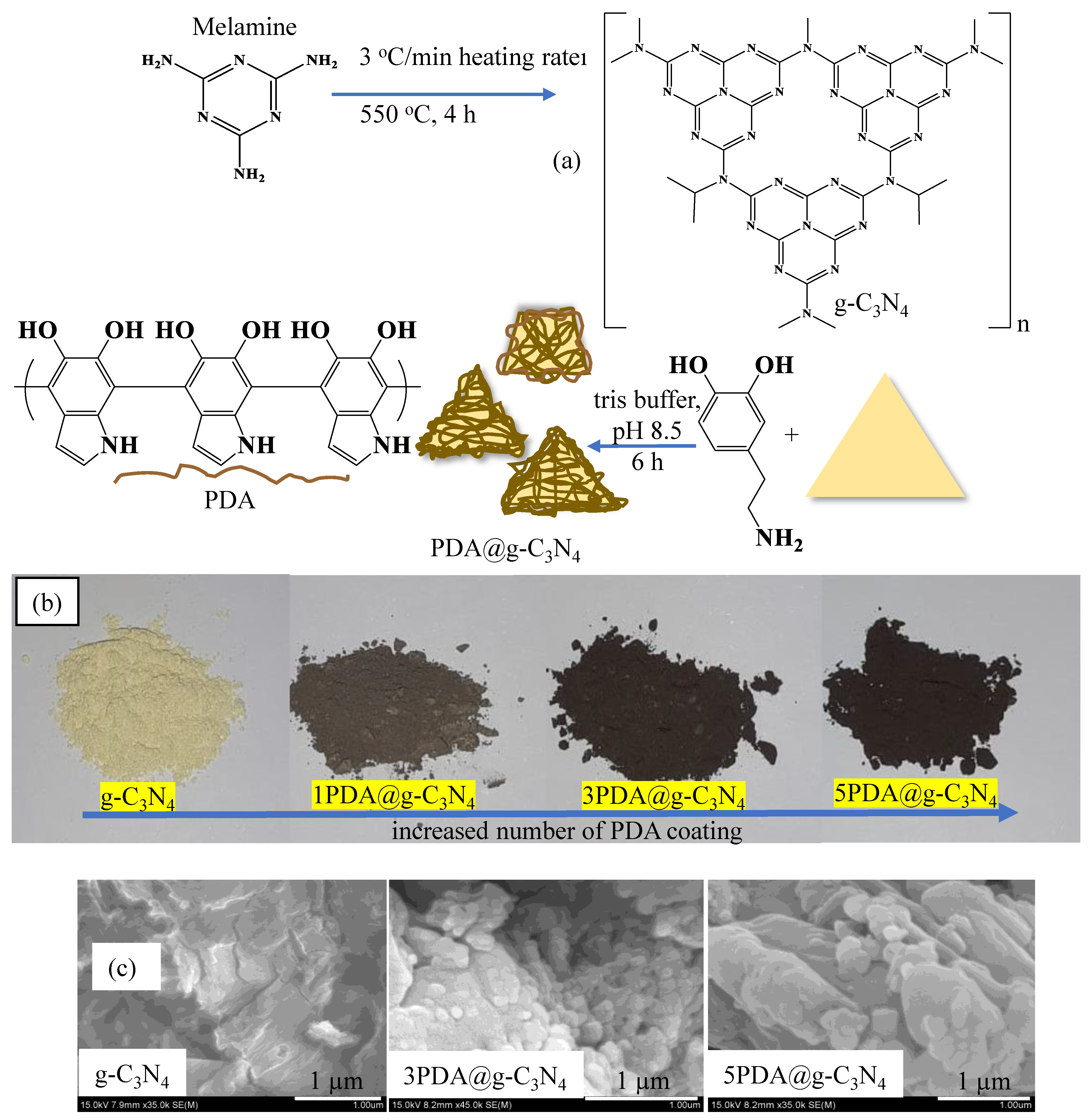
2.2. Synthesis of g-C3N4 and Its PDA Coating

The synthesis of graphitic carbon nitride (g-C3N4) was carried out according to the procedures reported in the literature with some modifications [23,24,25]. Briefly, 10 g of melamine was added to a porcelain crucible and placed in a furnace and the furnace was heated to 550 °C with 3 °C/min heating rate and kept at 550 °C for 4 h. The porcelain crucible was then cooled to room temperature and the yellowish solid obtained was ground in a porcelain mortar with a pestle and dispersed in 500 mL of water. Then, the suspension of g-C3N4 was sonicated for 5 h with stirring every 1 h. Finally, the dispersed g-C3N4 was collected by centrifugation at 10,000 rpm for 10 min and was dried in a freeze dryer (Alpha 2-4 LSC, Christ, Osterode am Harz, Germany) at −80 °C, and stored in closed tubes for further use.
The coating of g-C3N4 with polydopamine (PDA) was realized by self-polymerization of DA in tris buffer [26]. Accordingly, 4.5 g of washed and dried g-C3N4 was suspended in 450 mL of 10 mM tris buffer at pH 8.5 solution and sonicated for 3 h at 20 °C to ensure the exfoliation of g-C3N4 layers. Next, a freshly prepared 50 mL 20 mg/mL DA solution in 10 mM tris buffer at pH 8.5 was added to this solution and stirred at 1000 rpm mixing rate at room temperature. The final concentration of DA was 2 mg/mL in 500 mL 10 mM tris buffer at pH 8.5. Then, the self-polymerization of DA was carried out in the presence of g-C3N4 at 1000 rpm mixing rate for 6 h to coat g-C3N4 with PDA. Finally, PDA-coated g-C3N4s (1PDA@g-C3N4) were collected by centrifugation at 10,000 rpm for 10 min and washed twice with DI water to remove impurities from the medium. The yellowish color of g-C3N4 changed to a brownish color by coating with PDA. Then, 1PDA@g-C3N4 (1.5 g) was separated, and the remaining 3 g of 1PDA@g-C3N4 continued to be coated with PDA by applying the same coating procedure. This coating process was carried out 5 times in a row and after each two-coating process, 1.5 g of PDA@g-C3N4 was dried and separated. The brownish color of 1PDA@g-C3N4 turned black after the 5th coating with PDA. The washed and dried PDA-coated g-C3N4 structures were labelled as 1PDA@g-C3N4, 3PDA@g-C3N4, and 5PDA@g-C3N4 structures and stored in closed tubes for further use.

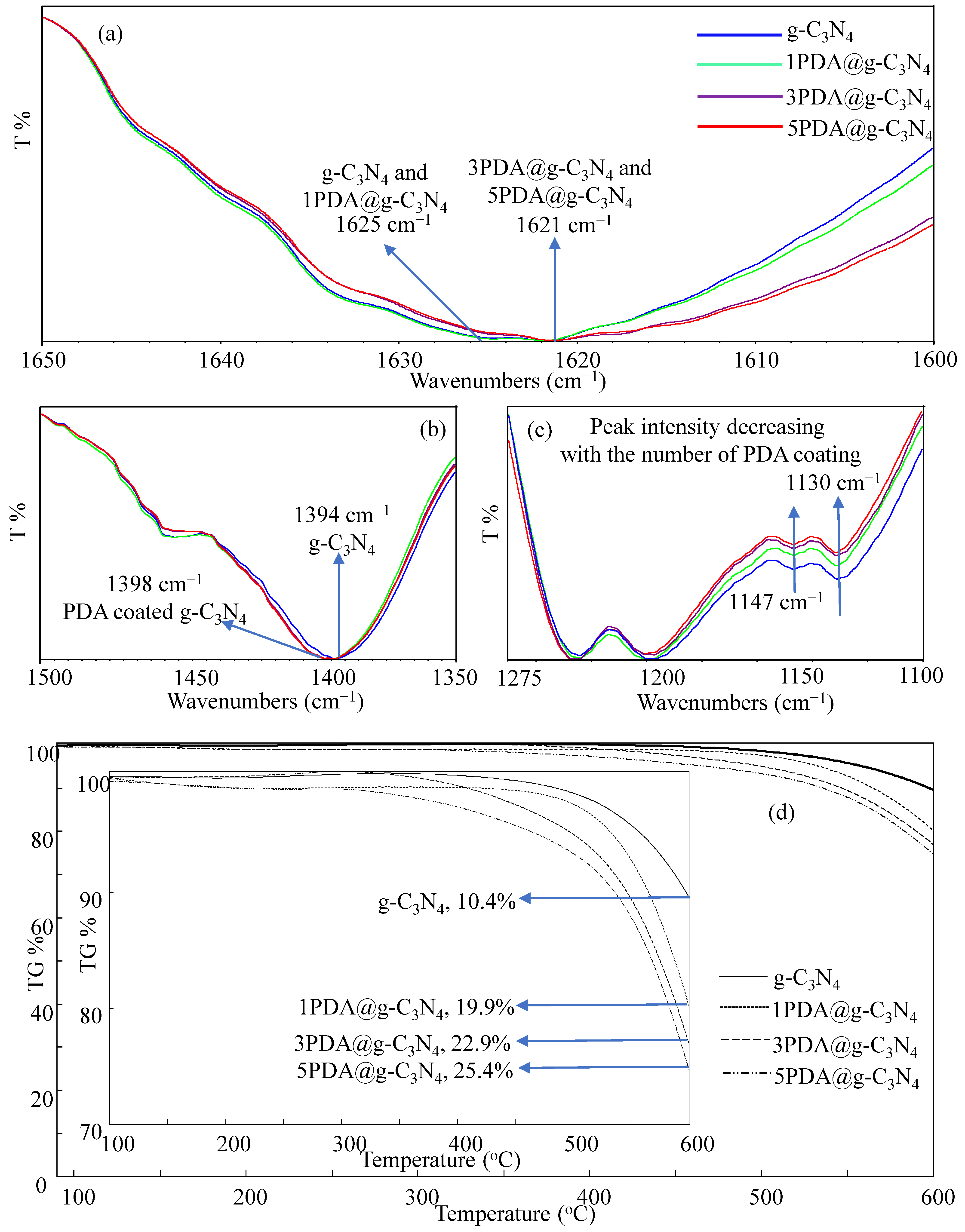
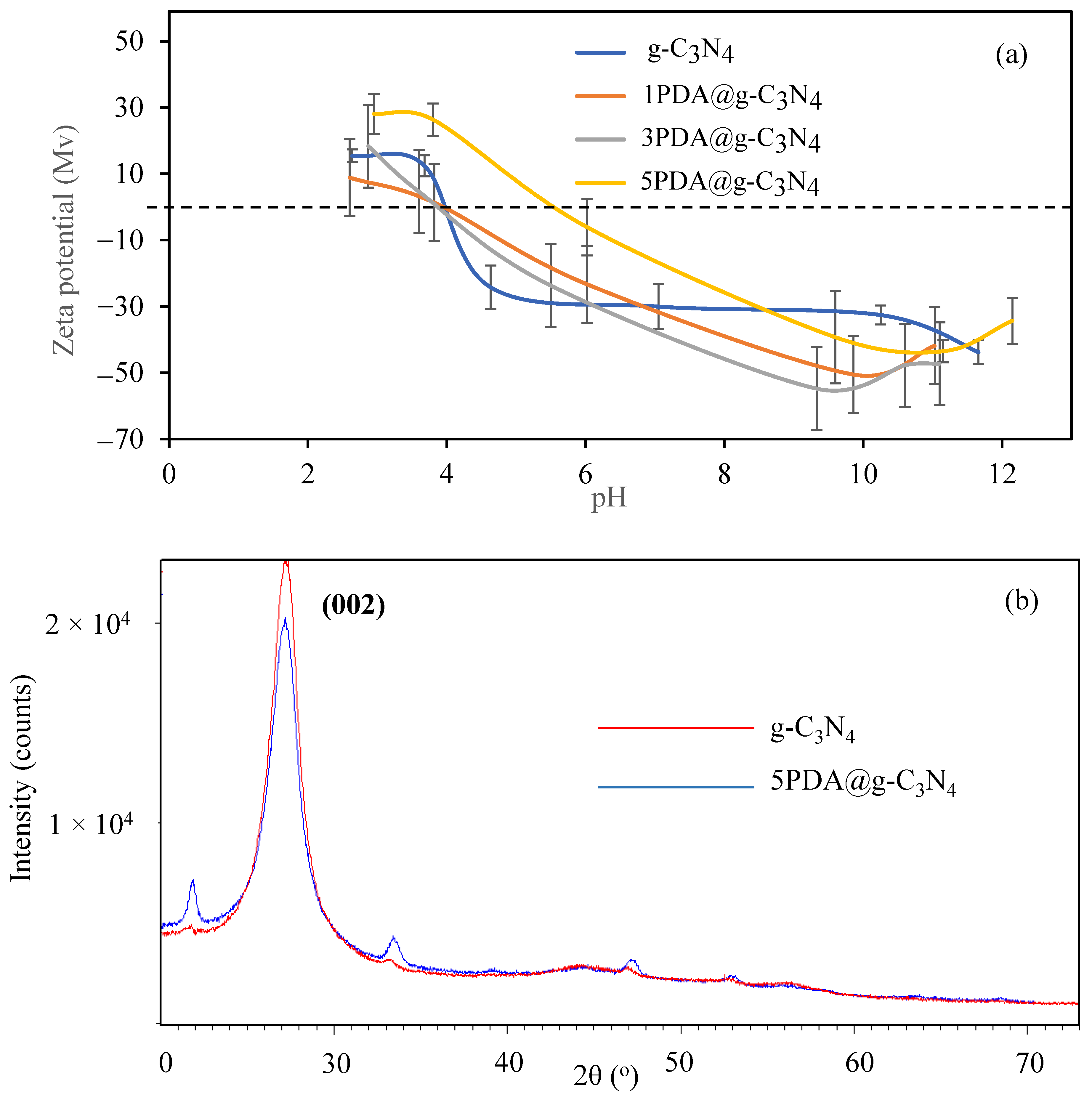
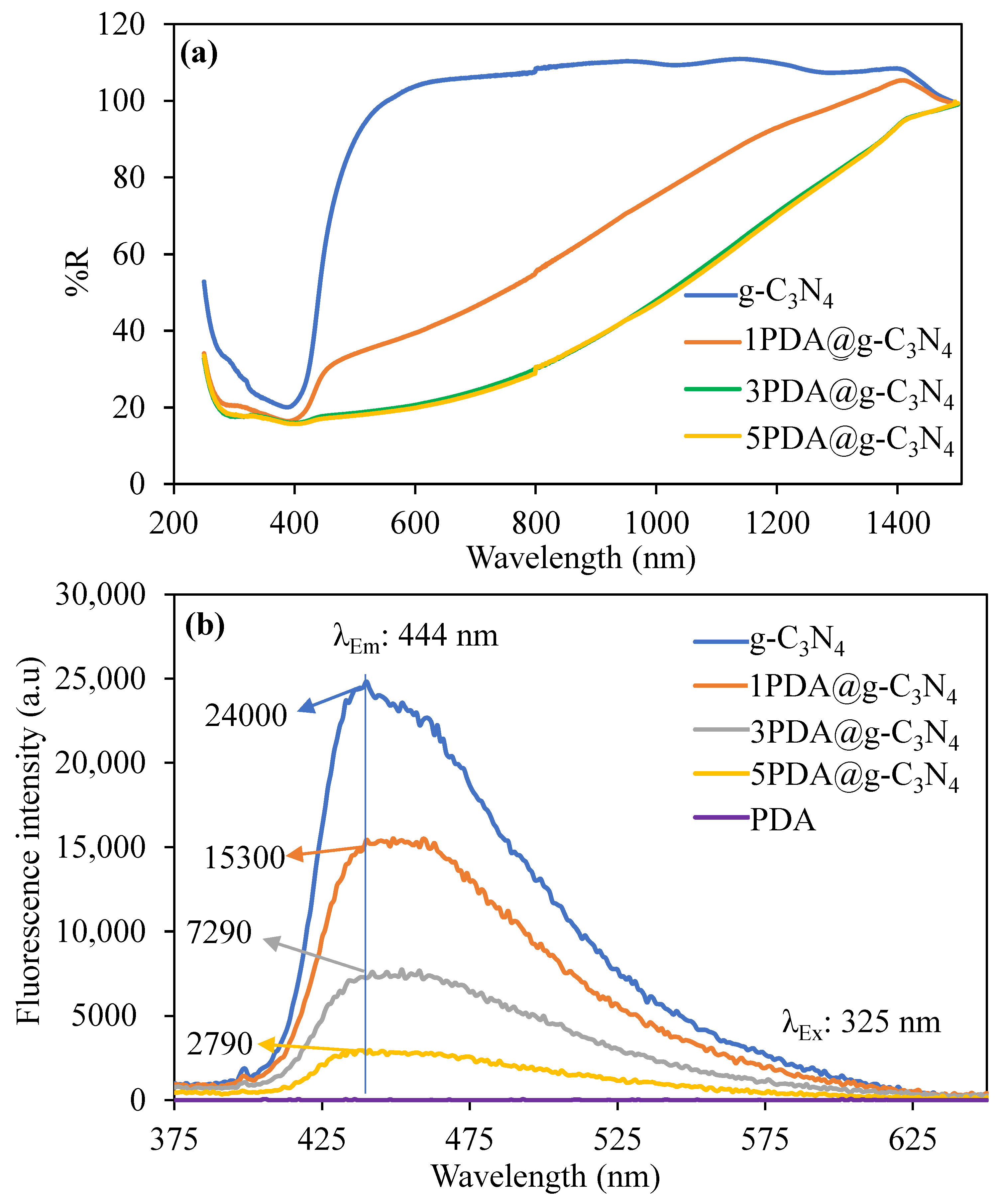
2.3. Characterization of PDA@g-C3N4

The functional group analysis of the g-C3N4 and PDA@g-C3N4 structures was performed using Fourier transform infrared spectroscopy (FT-IR) (Nicolet iS10, Thermo-Scientific, Waltham, MA, USA) in the range of 650–4000 cm−1, with an attenuated total reflection (ATR) technique, in 4 repetitions.
Thermal stabilities of the g-C3N4 and PDA@g-C3N4 structures were compared by a thermogravimetric analyzer (TGA, SII TG/DTA6300, Exstar, Seiko Instruments Corp., Tokyo, Japan). Thermogravimetric analyses were carried out by heating the samples weighing 3–5 mg (PDA@g-C3N4 samples) from 100 °C to 600 °C with a temperature increase of 10 °C/min under 200 mL/min N2 gas flow.
Zeta potential measurements of the g-C3N4 and PDA@g-C3N4 structures were carried out by suspending them in 1 mM KNO3 solutions at 1 mg/mL concentration using a zeta potential analyzer (Zeta-Pals, BIC, New York, NY, USA). Zeta potential measurements for ionic-group-containing materials (–OH, NH2, –COOH, –SO3H, etc.) are generally carried out in a KNO3 solution that has constant pH variation to prove that the solution is an inert electrolyte [27]. In addition, the isoelectric point values of the materials were calculated by measuring the zeta potential values of the g-C3N4 and PDA@g-C3N4 structures at different pH values of material suspension solutions. Furthermore, the zeta potential of the g-C3N4 and PDA@g-C3N4 structures was examined at different concentrations of KNO3 solution, 0.0, 1, 10, 100 mM, to determine the effect of KNO3 salt.
X-ray diffraction (XRD) pattern analysis of the g-C3N4 and PDA@g-C3N4 structures by a Cu Kα X-ray source (40 kV, 40 mA) at a wavelength of 1.5418 Å with a scanning 2θ range of 20°–70° was performed using an XRD analyzer (XDR, Bruker D8 Advance Diffractometer, Billerica, MA, USA) with CuKα radiation.
The optical diffuse reflectance of the g-C3N4-based materials was measured at room temperature via a UV–VIS–NIR spectrometer (UV-3600, Shimadzu, Kyoto, Japan) operating in the 200 to 2000 nm range. BaSO4 was used to obtain a reference of non-absorbing reflectance. The generated reflectance-versus-wavelength data were used to estimate the material’s bandgap by converting it to absorbance data using the Kubelka–Munk Equation (1):
α/S = (1 − R)2/2R
where R is the reflectance, and α and S are the absorption and scattering coefficients, respectively [28,29].
The UV–Vis spectrum (UV-Vis spectrometer, Genesys 180, Thermo Scientific) of the g-C3N4-based structures was recorded between 200 and 800 nm for the samples of 0.02 mg/mL concentrations of g-C3N4s. Before UV–Vis spectroscopy measurements, the prepared g-C3N4-based structure solutions in water were sonicated for 2 h to exfoliate the structures and obtain a good suspension. A fluorescence spectrometer (Spectrofluorometer FS5, Edinburgh Instruments, Livingston, UK) was used to determine the optical properties of the prepared g-C3N4 structures. The fluorescence properties of g-C3N4 and PDA@g-C3N4 mixtures prepared at a 0.2 mg/mL concentration in water were examined at a 325 nm excitation wavelength and 700 PMT V between 330 and 600 nm in wavelength range.
Moreover, the QY% values for the g-C3N4-based structures were calculated via following the literature [30]. Using quinine sulfate as a reference, the quantum yield% (QY%) values of the g-C3N4-based structures were computed. Quinine in 0.5 M H2SO4 was used as a control, with a QY value of 54% and an excitation wavelength of 345 nm. The QY% values of the g-C3N4-based structures were calculated using the Equation (2).
QY% = QY%std (I/Istd) · (ODstd/OD) · (η2std2)
where ‘QY’ is fluorescence quantum yield, ‘I’ is the integrated fluorescence intensity, ‘OD’ is the UV–vis absorbance, and ‘η’ is the refractive index of the solvents used for the g-C3N4-based structure suspension solutions, water (η = 1.333) and 0.5 M H2SO4 in water (η = 1.346).

2.4. Biomedical Properties of PDA Coated g-C3N4

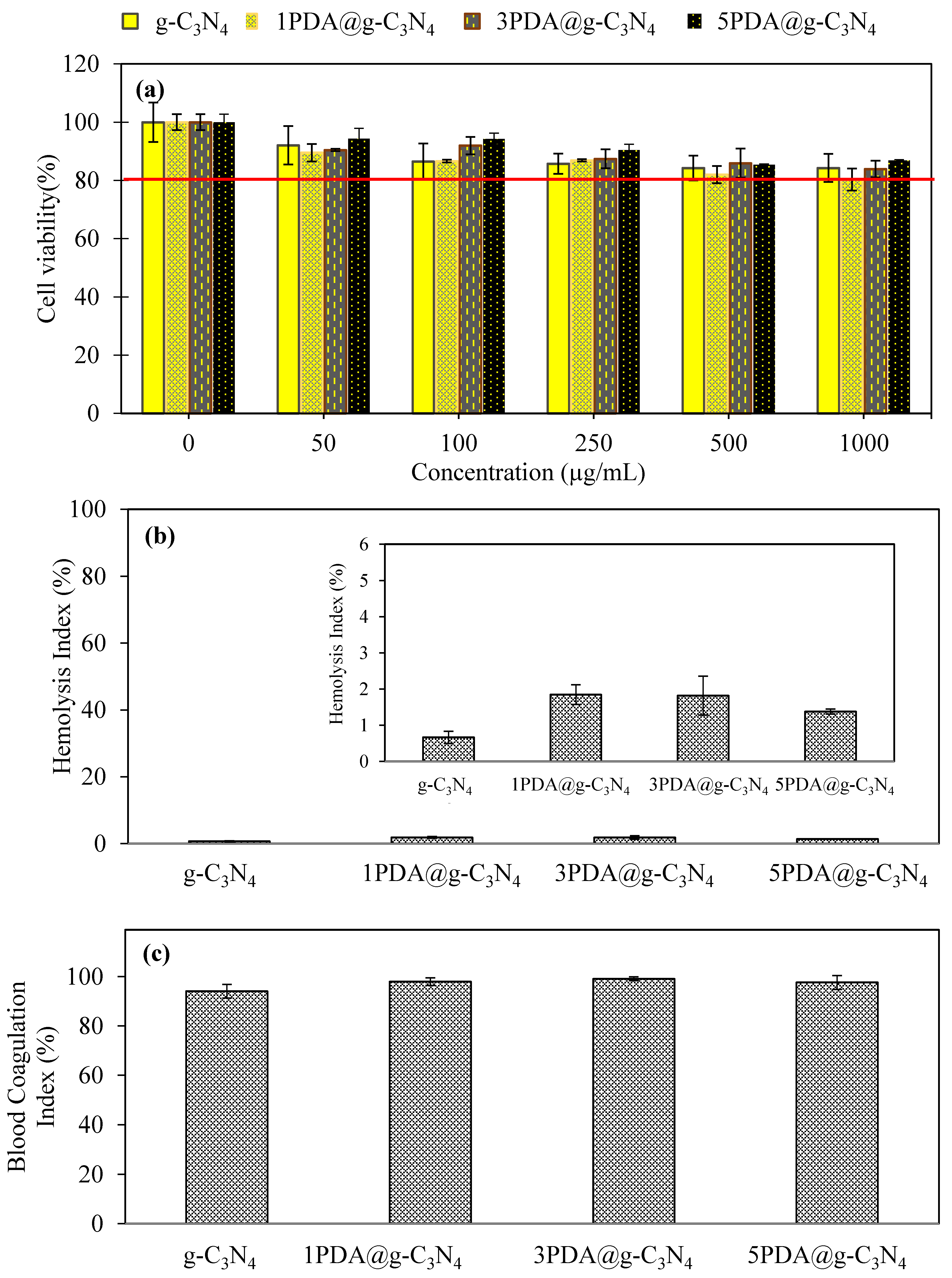
2.4.1. Cytotoxicity and Blood Compatibility of PDA-Coated g-C3N4

Biocompatibility and hemocompatibility tests were performed according to the literature [31], and details are given in the Supplementary Material.

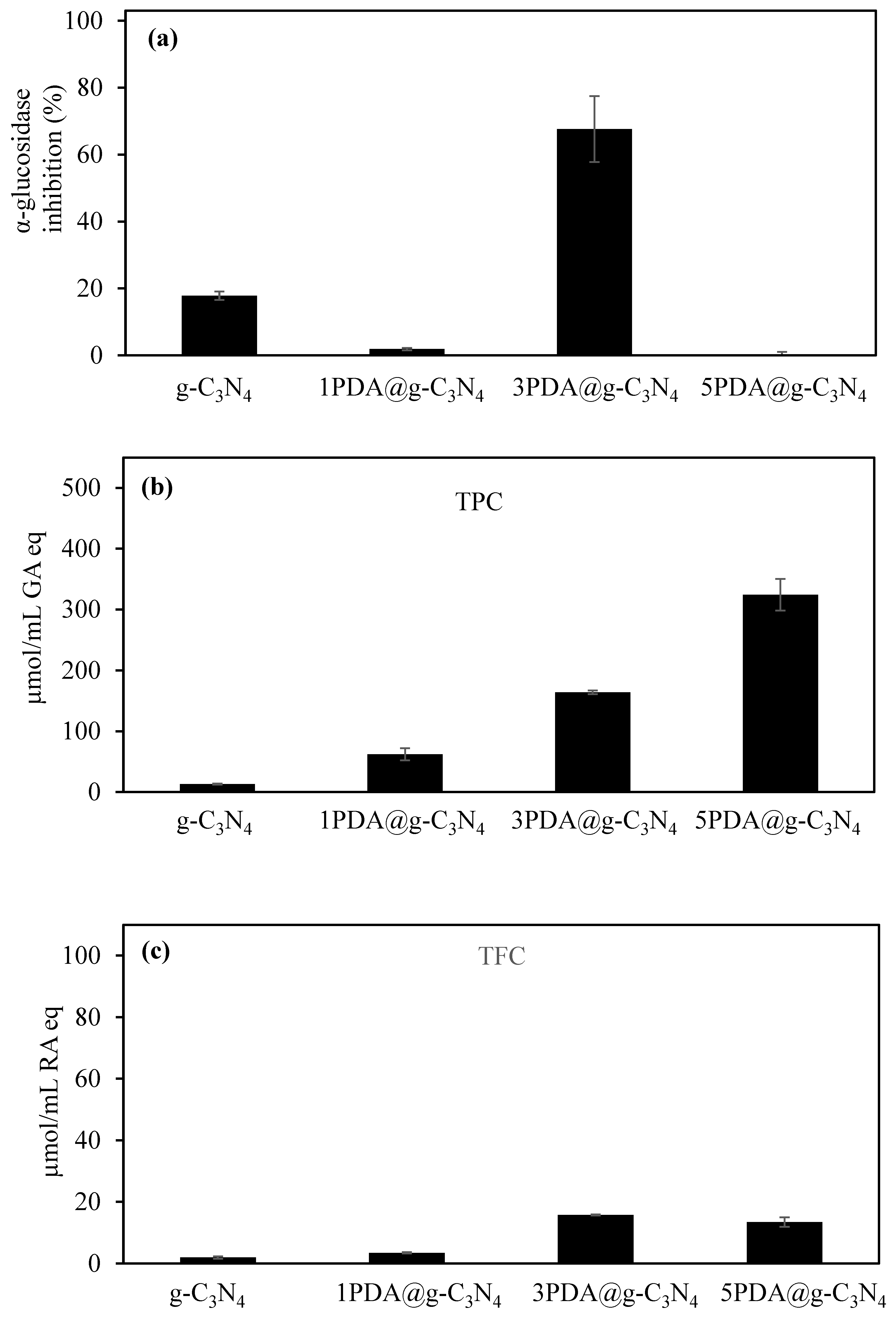
2.4.2. Antioxidant Activity Assays for PDA-Coated g-C3N4

In the determination of antioxidant capacity and enzyme inhibition tests of g-C3N4 and PDA@g-C3N4, samples weighing 2000 mg/mL were suspended in DI water and these mixtures were used for α-glucosidase enzyme inhibition, total phenol content (TFC), and total flavonoid content (TFC) tests. The α-glucosidase inhibition test was performed by modifying the method in the literature [26]. A sample solution of 700 µL and 2000 mg/mL is placed in a 10 mL tube and 700 µL of the enzyme at 0.03 units/mL was placed on it and left for 10 min. An amount of 700 µL of the substrate was added to the mixture and incubated for 20 min. At the end of the period, the solution mixture was filtered with a 0.5 µm syringe filter and the absorbance value at 405 nm was recorded on the UV–vis spectrophotometer. DI water was used as control.
Briefly, in the TFC test, 0.1 mL of sample solution was placed in 10 mL glass tubes, and a 1.25 mL of FC solution was added, followed by the addition of 1 mL of saturated bicarbonate solution. At the end of 2 h, the mixed solution was filtered, and the absorbance value was recorded at 760 nm with a UV–vis spectrophotometer. Results were calculated based on gallic acid (GA) equivalent.
For the TFC test, 0.5 mL of sample solutions were prepared and then diluted by adding 2 mL of DI water. This medium was then treated with 0.15 mL of 5% NaNO2 and then 0.15 mL of 10% AlCl3·6H2O was added to this medium. Subsequently, 1 M 1 mL NaOH was added to the mixture. The UV–Vis spectrum of this solution was recorded at 405 nm after it was kept at rest for an additional 15 min. Rosmarinic acid (RA) was used as a standard.

2.4.3. Antibacterial Activity Assay for PDA Coated g-C3N4

The antimicrobial activity of 20 mg/mL of g-C3N4-based materials was tested against Gram-negative E. coli (ATCC 8739) and Gram-positive S. aureus (ATCC 6538) for 24 h of incubation time. The g-C3N4, 1PDA@g-C3N4, and 3PDA@g-C3N4 were protonated for 1 h at room temperature interacting with 20 mL of 1 M HCl solutions. To remove excess HCl from the structure, the protonated PDA-5th@CNTs were centrifuged at 10,000 rpm and washed twice with DI water. The protonated and washed g-C3N4-based materials were dried using a freeze dryer as mentioned above. Microdilution tests were applied to the g-C3N4, 1PDA@g-C3N4, 3PDA@g-C3N4, and their protonated forms at a 20 mg/mL concentration. The suspensions were prepared in 10 mL of nutrient broth after sterilization and inoculated with 0.1 mL of bacteria suspension at 108 CFU/mL adjusted to McFarland No: 0.5. A shaker was used to cultivate the suspension at 35 °C for 24 h. The suspensions were then inoculated on nutrient agar at temperatures of 35 °C for 24 h. After incubation, the colonies were counted, and the viability of the bacteria was determined.
The blood compatibility of the prepared PDA@g-C3N4 was determined by hemolysis and blood coagulation tests in accordance with the procedures available in the literature [32]. Assay details for hemolysis and blood clotting are provided in the Supplementary Material.

3. Results

Graphitic nitride (g-C3N4) has a layered assembly of corresponding plates that is similar to graphite [33,34]. It is reported that the carbon–nitrogen (C–N) bonds within g-C3N4 structures can be constructed from various sources including urea, dicyandiamide, melamine, etc. [27,28], and therefore they are in s-triazine and heptazine [35,36,37] structures. The schematic presentation of the prepared g-C3N4 structure is illustrated in Figure 1a. Surface functionalization of graphitic materials is an important route for creating active sites to anchor as well as to tune the materials’ chemical, electrical, optical, and even biological properties [38,39]. Many techniques have been successfully employed to construct advanced carbon-based functional materials to exploit these feature for diverse applications [40]. Among the modification methods employed in g-C3N4 structures, covalent and noncovalent modification techniques are the most often utilized [40]. The covalent modification procedures are not favored due to the material’s sp2 structure being damaged and the process being difficult [40].
Noncovalent techniques, on the other hand, offer easier application processes and have no negative impact on the original structure [40]. In contrast to other approaches, surface modification via PDA coating can offer a unique ability to deposit a thin film of a biocompatible polymer onto organic and inorganic surfaces of fundamentally any shape and size of materials [41,42,43]. In this study, PDA coating was employed as the surface modification technique and applied on g-C3N4, and the related schematic representation is given in Figure 1a. Accordingly, the g-C3N4 structures at a concentration of 2 mg/mL were placed in the DA solution with 500 mL of 10 mM pH 8.5 tris buffer solution which afforded PDA coating. PDA possesses highly expressed catechol and amino functions, which enable this polymer to coat almost any substrate with a variety of forms and geometries by emulating the structure of the adhesive protein found in mussels [44,45]. The coating process with PDA was performed five times consecutively on g-C3N4, and the first, third, and fifth coatings as 1PDA@g-C3N4, 3PDA@g-C3N4, and 5PDA@g-C3N4 were tested for the application of biomedical parameters. The change in color of g-C3N4 from clear yellowish to black with an increasing number of PDA coatings is shown in digital camera images in Figure 1b. The changes in the intensity of the dark coloration of g-C3N4 with the increasing number of PDA coatings is clearly visible in the digital camera images as illustrated in Figure 1b. The increase in the number of coatings provides a higher PDA amount to the surface of the g-C3N4 structures as revealed with digital camera images. From the SEM images in Figure 1c, the morphology of g-C3N4 after coating with PDA did not significantly change regarding the size of the flakes. However, some PDA-coated g-C3N4 in nano, submicron, and micrometer sizes are visible on the surfaces.
The FT-IR spectra of the g-C3N4 and PDA@g-C3N4 structures are compared and presented in Figure S1. As can be seen, there is no noticeable change visible. To better assess the PDA coatings on g-C3N4 surfaces, the FT-IR spectra of the g-C3N4 and PDA-coated 1PDA@g-C3N4, 3PDA@g-C3N4, and 5PDA@g-C3N4 structures are compared in detail and given in in Figure 2a–c, respectively. Due to the normal stretching modes of aromatic C-N heterocyclic structures, the bands in the 1200–1600 cm−1 region were seen in all spectra. Triazine unit vibrations are present at 807 cm−1 for all g-C3N4-based materials [46,47]. At 3000–3500 cm−1, the peaks corresponding to the vibrations of terminal amino groups and surface-adsorbed –OH bands are visible also for all g-C3N4-based materials [48,49]. However, due to overlapping bands in the FT-IR spectra of both g-C3N4s and PDAs, no substantial change in the FT-IR spectra of the g-C3N4 and PDA@g-C3N4 structures was observed. Therefore, the detailed FT-IR spectra of the three regions, e.g., 1600–1650 cm−1, 1350–1500 cm−1, and 1100–1275 cm−1, are shown in Figure 2a–c, respectively. It is also apparent that there are not very significant changes in the FT-IR spectrum of these regions, as the N–H stretching and indole ring stretching peaks which are the most likely characteristic peaks to be observed for PDA at 1605 and 1514 cm−1, respectively, cannot be seen due to the overlapping of the strong peak belonging to g-C3N4 in this region [26]. However, there is a slight peak shift upon PDA coating that is observed in Figure 2a, e.g., the 1625 cm−1 peak belonging to g-C3N4 shifted to 1621 cm−1 upon PDA coating. On the other hand, the observed peak at 1394 cm−1 in the FT-IR spectrum of g-C3N4 shown in Figure 2b shifted to 1398 cm−1 after the PDA coating process. Also, there is some decrease in intensities of the peaks at 1147 and 1130 cm−1 upon PDA coating as illustrated in Figure 2c.
Because the PDA coating of the g-C3N4 structures cannot be adequately confirmed by FT-IR spectra, TGA thermograms of the uncoated g-C3N4 and PDA@g-C3N4 structures were compared to evaluate and compare the thermal stability of the structures and to validate the increasing quantity of PDA coating on g-C3N4 with the repeated coating procedures, and the corresponding thermograms are shown in Figure 2d. Because the synthesis of the g-C3N4 structures is performed at 550 °C, the uncoated g-C3N4’s thermal degradation began after around 550 °C. At 600 °C, it was observed that the g-C3N4 structures lost 10.6% of their mass. On the other hand, the PDA@g-C3N4 structures had essentially little mass loss up to 550 °C. However, at 600 °C, the thermal degradation of the 1PDA@g-C3N4, 3PDA@g-C3N4, and 5PDA@g-C3N4 structures was 19.9%, 22.9%, and 25.4%, respectively. This is due to an increase in the quantity of PDA coating on the g-C3N4 structures as the number of PDA coatings increases. The quantity of PDA in the structure after the first coating was determined to be 9.3%, and after the third and fifth coatings, the amount of PDA on the g-C3N4 structures grew to 12.3% and 14.8%, respectively.
For detailed information to confirm the increase in PDA amount on the g-C3N4 structures, the EDX analysis of the uncoated g-C3N4, 1PDA@g-C3N4, 3PDA@g-C3N4, and 5PDA@g-C3N4 structures were carried out, and corresponding results are summarized in Table 1. It was clearly seen that the elemental ingredients of uncoated g-C3N4 includes 22.1% C, 71.8% N, and 5.4% O. The amount of O in neat g-C3N4 is 5.4 wt% which comes from the air as the g-C3N4 was synthesized in air atmosphere. So, the amount of PDA coatings of g-C3N4 was determined as 4.4%, 7.7%, and 14.4% weight increases for 1PDA@g-C3N4, 3PDA@g-C3N4, and 5PDA@g-C3N4 that are normalized based on the amount of O atoms in the neat g-C3N4.
On the other hand, from the EDX analysis of the PDA-coated g-C3N4 structure, it can be clearly seen that the percentage of C and O atoms increased as the number of PDA coatings increased, while the percentage of N atoms decreased. The determined C% for the structures 1PDA@g-C3N4, 3PDA@g-C3N4, and 5PDA@g-C3N4 are 23.2%, 26.0%, and 32.6%, respectively. On the other hand, %N for the 1PDA@g-C3N4, 3PDA@g-C3N4, and 5PDA@g-C3N4 structures were calculated as 67.0, 60.9, and 48.3%, respectively. This is because for every PDA unit, there are 8 C atoms generated versus 1 N atom and 2 O atoms formed. As the number of PDA coatings increases, the amount of O which increases on the coated g-C3N4 reveals coatings of PDA is accomplished. As observed for O atoms, there are 9.8, 13.1, and 19.1 wt% for the first, third, and fifth PDA coatings on g-C3N4, versus 5.4 wt% for uncoated g-C3N4.
Furthermore, the changes in the zeta potential of the g-C3N4 and PDA@g-C3N4 structures at various pHs of the particle solution was investigated and the results are given in Figure 3a. The zeta potential values for g-C3N4, 1PDA@g-C3N4, 3PDA@g-C3N4, and 5PDA@g-C3N4 were calculated from their dispersion in 1 mM KNO3 solution at 1 mg/mL concentration, and were measured as −23.3 ± 3.0, −30.0 ± 6.8, −23.7 ± 12.6, and −6.1 ± 8.5 mV, respectively. The isoelectric points of the g-C3N4, 1PDA@g-C3N4, 3PDA@g-C3N4, and 5PDA@g-C3N4 structures were determined as pH 4.0, 4.0, 3.9, and 5.6, respectively. The zeta potential values of the structures are zero at these pH values, whereas they are positively charged at pH values lower than these values, and they become negatively charged at higher pH values. In addition, to ascertain the effect of KNO3 concentration on the zeta potential of the g-C3N4 and PDA@g-C3N4 structures, the measurements were conducted at various concentrations of KNO3 solution. As seen in Figure S2, the zeta potential values of g-C3N4 and PDA@g-C3N4 were not significantly affected by KNO3 solution concentrations ranging from 1 to 100 mM. However, when KNO3 solution is not used as the measuring medium, big jumps in pH values occur immediately upon small additions of acid or base in the medium during the adjustment of solution pHs, resulting in incorrect measurements. The ionic strength of the medium needs to be equilibrated by use of some salts, e.g., KNO3, as some materials that can develop charges upon small additions of acid or base used to arrange pH of the medium can influence the medium pH greatly. Therefore, zeta potential measurements were generally performed in salt solutions with low concentrations such as 1–100 mM [27].
Moreover, the XRD pattern comparison of the g-C3N4 and 5PDA@g-C3N4 structures are also given in Figure 3b. An XRD peak associated with in-plane structural packing is easily recognized from the exact periodic units in each layer of g-C3N4. The typical experimental XRD pattern of bulk g-C3N4 contains a unique diffraction peak positioned at 2θ = 27.40°, which is indexed as graphitic materials’ (002) diffraction plane. In the literature, two main peaks, 2θ = 13.40 and 2θ = 27.40 for g-C3N4 at (100) and (002) planes, are reported [50,51]. The first peak is the structural packing between the layers and the second peak is the characteristic interplanar staking peaks of the aromatic systems. As the 2θ range of 20°–73° is shown in Figure 3b, the peak at 27° is clearly visible. It can also be seen that the peak intensity decreases slightly due to the dopamine coating, e.g., the intensity of 5PDA@g-C3N4 is 20,315 at 2θ = 27°, whereas the intensity of g-C3N4 is 23,129 at the same 2θ value.
According to the XRD data, the g-C3N4 has a flake-like structure with an interplanar stacking distance of 0.356 nm as shown by (002) diffraction. However, there is also no significant changes in the structure of g-C3N4 after PDA coating, and the diffraction peak was observed at 27.4° 2θ, indicating that the material is still in a graphitic structure with reduced intensity as an indication of the PDA coating [49].
The change in optical properties of g-C3N4 after multiple PDA coatings was also investigated using bandgap analysis and the change in fluorescence properties of the structures. Figure 4a depicts the UV–Vis DRS spectra of the g-C3N4 and PDA@g-C3N4 structures, and Figure S3 shows the reflectivity wavelength diagram used to determine the band gaps of the g-C3N4-based structures. The band gap of g-C3N4 is determined to be 2.72 eV, which is consistent with previously reported g-C3N4 structures [31,52,53,54]. The coating of g-C3N4 with PDA, on the other hand, shifts the band gap to lower levels. The bandgap values for 1PDA@g-C3N4 after the first PDA coating were determined to be 2.57 and 0.94 eV. Because of non-homogeneous coating, the first PDA coating results in two band gap values one of which is for g-C3N4 with a value of 2.57 eV that is closer to bare g-C3N4 and another band gap with a value of 0.94 eV for incomplete PDA coating or partial coating of g-C3N4. However, following the third and fifth PDA coatings of g-C3N4, single band gap values were measured as 0.89 and 0.87 eV, respectively.
The UV–Vis spectra of neat g-C3N4 and the PDA-coated g-C3N4 structures at 0.2 mg/mL concentrations based on g-C3N4 amounts are given in Figure S4a. Based on EDX results provided in Table 1, the amount of PDA coating of the g-C3N4s structures (normalized according to the O) are calculated as 4.4%, 7.7%, and 14.4% after the frist, third, and fifth coating processes, respectively. To eliminate the concentration dependence of absorbance values, the aqueous solutions of g-C3N4, 1PDA@g-C3N4, 3PDA@g-C3N4, and 5PDA@g-C3N4 weighing 20, 20.9, 21.5, and 22.9 mg, respectively, per 1 mL water solution were prepared and diluted 1000-fold for the comparison of their corresponding UV–Vis spectra, shown in Figure S4a. Thus, each sample, g-C3N4, 1PDA@g-C3N4, 3PDA@g-C3N4, and 5PDA@g-C3N4, have 0.0200 mg g-C3N4 per 1 mL solution. As can be seen in Figure S4a, the max absorbance values for the g-C3N4-based structures were observed at 325 nm and are 0.086, 0.082, 0.069, and 0.053 for g-C3N4, 1PDA@g-C3N4, 3PDA@g-C3N4, and 5PDA@g-C3N4, respectively. So, the decrease in absorbance values at 325 nm of these g-C3N4 structures with an increasing number of PDA coatings directly related to the increased amount of PDA on the surface of g-C3N4s. The digital camera images shown in Figure S4b contain the same amounts of g-C3N4s, 0.02 mg/mL.
Accordingly, the excitation wavelengths of the g-C3N4, PDA-coated g-C3N4 structures, and PDA were determined at 325 nm, and their emission wavelengths and fluorescence intensities were compared in the 350–600 nm range. The fluorescence emission spectra of g-C3N4 the PDA-coated g-C3N4 structures, 1PDA@g-C3N4, 3PDA@g-C3N4, and 5PDA@g-C3N4, and PDA are given in Figure 4b. The emission wavelength for all structures was determined at 444 nm, but differences were observed in the fluorescence emission intensities, except for PDA. PDA did not show any fluorescence properties at this excitation wavelength. The structures were excited at 325 nm, which corresponds to the S0-to-S1 transition, and gave off fluorescence emissions around 450 nm, showing that the S1-to-S0 transition occurred [55]. Accordingly, while the observed fluorescence emission intensity for the g-C3N4 structure at 444 nm wavelength is 24,000, this value decreases to 15,300, 7290, and 2790 for the 1PDA@g-C3N4, 3PDA@g-C3N4, and 5PDA@g-C3N4 structures, respectively. As expected, there was a decrease in the fluorescence properties of the g-C3N4 structures after coating with PDA and decrease in intensity of the fluorescence properties increased as the number of coatings with PDA increases. Although the concentrations of the solutions used were the same (0.02 mg/mL based on g-C3N4) in the measurements with florescence spectroscopy, the resulting decrease in fluorescence intensity with number of PDA coatings is due to the suppression of the fluorescence feature coming from the g-C3N4 structure. Table 2 also summarizes the QY% values calculated based on Equation (2) for g-C3N4, 1PDA@g-C3N4, 3PDA@g-C3N4, and 5PDA@g-C3N4.
The QY% values of the materials with luminous characteristics are important in their optical use. The higher the fluorescence property, the higher the QY%. As given in Table 2, the QY% value of g-C3N4 is 19.8 ± 1.2, with 13.7 ± 1.1, 9.1 ± 0.7, and 4.8 ± 0.6% calculated for the 1PDA@g-C3N4, 3PDA@g-C3N4, and 5PDA@g-C3N4 structures, respectively. As expected, with the increase in number of PDA coatings of g-C3N4, the QY values decreased due to the increased extent of interaction of the excited electrons with the increased number PDA chains with a higher number of coatings.
Cytotoxicity tests of g-C3N4 and PDA@g-C3N4 were performed on L-929 fibroblast cells and the results are shown in Figure 5. Cell viability of g-C3N4 was found >80% in all concentrations in the range of 50–1000 ug/mL.
The biocompatibility results confirm that g-C3N4 is considered biocompatible up to a concentration of 1000 µg/mL. Polydopamine coating on g-C3N4 did not affect its biocompatibility, as cell viability results showed 81.9 ± 4.8% cell bioavailability for g-C3N4 for 1000 µg/mL concentration versus cell viabilities of 80.3 ± 3.8, 83.9 ± 1.0 and 86.8 ± 1.0% for the first, third, and fifth PDA-coated g-C3N4, respectively.
The hemolysis and blood clotting index values of g-C3N4 and PDA@g-C3N4 are illustrated in Figure 5b and 5c, respectively. Hemolysis index values were determined as 0.7 ± 0.2% for g-C3N4, whereas it was determined as 1.8 ± 0.5% for 3PDA@g-C3N4 which was the highest among all the PDA-coated g-C3N4s. As the hemolysis% values for all the samples are <5%, they are all blood compatible. Or in other words, g-C3N4 materials whether PDA-coated or not and independent of the number of PDA coatings, do not rupture red blood samples and can be utilized in blood contacting applications safely. Another important parameter in the blood compatibility test is the blood clotting index% which gives information on whether the material in question influences the clotting mechanism of the blood. In order not to impair the clotting mechanism of the blood, the blood clotting percentage of a material must be around 100% [56]. As shown in Figure 5c, the lowest blood clotting index value was determined for g-C3N4 with 94.1 ± 2.8%, and PDA-coated g-C3N4, 1PDA@g-C3N4, 3PDA@g-C3N4, and 5PDA@g-C3N4 demonstrated the blood clotting% values of 97.92 ± 1.2, 99.1 ± 0.8%, and 97.62 ± 1.5%, respectively, ascertaining that PDA coating improves the blood compatibility of g-C3N4 by not interfering with the mechanism of the clotting of blood.
In addition, the treatment of type II diabetes also considers other aspects that are important for biomedical application, such as the inhibition of enzymes, e.g., α-glucosidase, a hydrolysis enzyme that breaks down disaccharides and α-glucosidase inhibitors. Therefore, the α-glucosidase enzyme inhibitory ability of g-C3N4 and its PDA-coated forms was investigated, and the results are summarized in Figure 6a. As can be seen amongst all the g-C3N4-based materials, the highest α-glucosidase inhibition potency was determined for 3PDA@g-C3N4 with 67.6 ± 9.8%, suggesting the potential use of PDA-coated materials in type II diabetes.
Furthermore, the determine the antioxidant effectiveness of PDA-coated g-C3N4, the total phenol content (TPC) and total flavonoid content of (TFC) test were performed at 2000 µg/mL for g-C3N4 and PDA@g-C3N4 samples and results are shown in Figure 6b and 6c, respectively. According to the TPC test results, there is linear increase in TPC values in parallel to the PDA coating number and the highest TPC value of 314 ± 26 µmol/mL GA eq was found for 5PDA@g-C3N4. On the other hand, TFC values for the PDA coating of g-C3N4 show a slight increase with the number of PDA coatings of g-C3N4, as shown in Figure 6c. In the TFC test, the highest value obtained was 15.7 ± 0.2 mg/mL RA eq for the 3PDA@g-C3N4 sample.
While the TPC of PDA-coated samples shows a linear increase with an increased number of coatings, suggesting antioxidant properties, the TFC value showed only a marginal increase with PDA coating; this is reasonable, as DA is a phenolic compound, not a flavonoid.
To evaluate the antibacterial capabilities of g-C3N4 and its PDA-coated forms, bacteria inhibition% values against two bacterial strains were examined using 20 mg/mL concentrations of g-C3N4 and PDA@g-C3N4, and their protonated form, PDA@g-C3N4+. As shown in Figure S5, g-C3N4 showed a reducing effect on the bacterial growth of S. aureus bacterium with reduction of 91.2 ± 0.4%. The highest inhibition% was observed as 62.3 ± 1.9% against S. aureus, and 76.5 ± 1.0% inhibition against E. coli for 3PDA@g-C3N4+. Overall, upon protonation all the g-C3N4 and PDA@g-C3N4 material showed increased inhibition% values against common pathogens, e.g., S. aureus and E. coli, indicating the potential role of PDA-coated g-C3N4 composites in preventive or prophylactic usage against infections caused by these microorganisms or protection against the spread and/or contamination of infections caused by different pathogens.
PDA is a versatile antibacterial agent and can be combined or modified with other materials or compounds to achieve a further potent antibacterial potency. These materials may include metal ions, antibiotics, and other antibacterial substances on the surface, resulting in controlled release, detoxification, or other effects. Therefore, PDA coating represents a simple and universal method for functionalizing various material surfaces as excellent antibacterial materials, making it a valuable addition to any material [57].

4. Conclusions

Here, we reported the facile multiple coating of g-C3N4 by PDA through self-polymerization of DA, a phenolic neurotransmitter. It was shown that the g-C3N4 5PDA@g-C3N4, coated up to five times, retained the fluorescence properties, although the fluorescence emission at 444 nm was reduced by five times. As the coating number of the PDA increased, the intensity of fluorescence emission decreased linearly. It was found that three consecutive PDA coatings enabled complete coating with g-C3N4 and bestowed significant improvement in biomedical application potentials. For example, the cytotoxicity of the 1PDA@g-C3N4, 3PDA@g-C3N4, and 5PDA@g-C3N4 structures is retained against L929 and fibroblast cell lines and their blood compatibilities via hemolysis, and the blood clotting index% was enhanced in comparison to bare g-C3N4. Furthermore, the antioxidant capability of PDA-coated g-C3N4 via a total phenol content assay revealed a linear increased antioxidant ability in terms of GA equivalency with an increasing number of PDA coatings. Moreover, because of the presence of N atoms in g-C3N4 and the PDA structures, g-C3N4 and PDA@g-C3N4 can be easily protonated by simple acid treatments such as HCl, etc. Therefore, their protonated forms also resulted in a reduction in bacterial growth, further conforming the improved biomedical utilization of PDA-coated material. Due to the presence of a PDA coating on an optically active 2D g-C3N4, it is expected that this type of material could be used in the treatment of neurogenerative diseases such as Parkinson’s and Alzheimer’s.

Supplementary Materials

The following supporting information can be downloaded at: https://www.mdpi.com/article/10.3390/biomedicines12061151/s1, Figure S1: FT-IR spectrum of g-C3N4 and PDA-coated g-C3N4 structures. Figure S2: (a) pH–zeta potential plots of g-C3N4 and (b) 5PDA@g-C3N4 in different concentrations of KNO3 solutions (0, 1, 10, 100 mM). Figure S3: The calculated bandgap values for (a) g-C3N4, (b) 1PDA@g-C3N4, (c) 3PDA@g-C3N4, and (d) 5PDA@g-C3N4. Figure S4: (a) UV–Vis spectra and (b) digital camera images of g-C3N4 and PDA-coated g-C3N4 structure solutions at 0.2 mg/mL concentration of g-C3N4s. Figure S5: Antimicrobial activity of 20 mg/mL of g-C3N4-based materials against Gram-negative E. coli (ATCC 8739) and Gram-positive S. aureus (ATCC 6538) for 24 h of incubation time.

Author Contributions

Conceptualization, N.S.; methodology, M.S., S.D. and N.S.; validation, M.S. and S.D.; formal analysis, M.S., S.D. and N.S.; investigation, M.S. and S.D.; resources, N.S.; writing—original draft preparation, M.S. and S.D.; writing—review and editing, N.S.; visualization, N.S.; supervision, N.S.; project administration, N.S.; funding acquisition, M.S. and N.S. All authors have read and agreed to the published version of the manuscript.

Funding

The financial support from Canakkale Onsekiz Mart University Scientific Research Commission (COMU-BAP: FBA-2022-4117) is acknowledged.

Institutional Review Board Statement

Blood compatibility studies were carried out with the approval of the ethics committee (no. KAEK-2011-KAEK-27/2022-2200063689), which was approved by the Human Research Ethics Committee of Çanakkale Onsekiz Mart University.

Informed Consent Statement

Informed consent is not needed.

Data Availability Statement

Data are contained within the article and Supplementary Materials.

Acknowledgments

The authors are grateful to Ioannis Spanopoulos from USF for the optical diffuse reflectance studies.

Conflicts of Interest

The authors declare no conflicts of interest.

References

  1. Cao, S.; Low, J.; Yu, J.; Jaroniec, M. Polymeric Photocatalysts Based on Graphitic Carbon Nitride. Adv. Mater. 2015, 27, 2150–2176. [Google Scholar] [CrossRef] [PubMed]
  2. Liao, G.; He, F.; Li, Q.; Zhong, L.; Zhao, R.; Che, H.; Gao, H.; Fang, B. Emerging Graphitic Carbon Nitride-Based Materials for Biomedical Applications. Prog. Mater. Sci. 2020, 112, 100666. [Google Scholar] [CrossRef]
  3. Liu, H.; Wang, X.; Wang, H.; Nie, R. Synthesis and Biomedical Applications of Graphitic Carbon Nitride Quantum Dots. J. Mater. Chem. B 2019, 7, 5432–5448. [Google Scholar] [CrossRef] [PubMed]
  4. Luo, Y.; Yan, Y.; Zheng, S.; Xue, H.; Pang, H. Graphitic Carbon Nitride Based Materials for Electrochemical Energy Storage. J. Mater. Chem. A 2019, 7, 901–924. [Google Scholar] [CrossRef]
  5. Wang, X.; Blechert, S.; Antonietti, M. Polymeric Graphitic Carbon Nitride for Heterogeneous Photocatalysis. ACS Catal. 2012, 2, 1596–1606. [Google Scholar] [CrossRef]
  6. Zhao, Z.; Sun, Y.; Dong, F. Graphitic Carbon Nitride Based Nanocomposites: A Review. Nanoscale 2015, 7, 15–37. [Google Scholar] [CrossRef]
  7. Asif, K.; Perveen, M.; Khera, R.A.; Nazir, S.; Raza Ayub, A.; Asif, T.; Shabbir, M.; Iqbal, J. Computational and Theoretical Study of Graphitic Carbon Nitride (g-C3N4) as a Drug Delivery Carrier for Lonidamine Drug to Treat Cancer. Comput. Theor. Chem. 2021, 1206, 113459. [Google Scholar] [CrossRef]
  8. Shamim, M.; Perveen, M.; Nazir, S.; Hussnain, M.; Mehmood, R.; Khan, M.I.; Iqbal, J. DFT Study of Therapeutic Potential of Graphitic Carbon Nitride (g-C3N4) as a New Drug Delivery System for Carboplatin to Treat Cancer. J. Mol. Liq. 2021, 331, 115607. [Google Scholar] [CrossRef]
  9. Qusain Afzal, Q.; Rafique, J.; Jaffar, K.; Perveen, M.; Iqbal, J.; Al-Buriahi, M.S.; Alomairy, S.; Alrowaili, Z.A.; Somaily, H.H. DFT Study of 2D Graphitic Carbon Nitride Based Preferential Targeted Delivery of Levosimendan, a Cardiovascular Drug. Comput. Theor. Chem. 2022, 1209, 113584. [Google Scholar] [CrossRef]
  10. Rajabzadeh-Khosroshahi, M.; Pourmadadi, M.; Yazdian, F.; Rashedi, H.; Navaei-Nigjeh, M.; Rasekh, B. Chitosan/Agarose/Graphitic Carbon Nitride Nanocomposite as an Efficient PH-Sensitive Drug Delivery System for Anticancer Curcumin Releasing. J. Drug Deliv. Sci. Technol. 2022, 74, 103443. [Google Scholar] [CrossRef]
  11. Ilyas, M.; Ayu, A.R.; Shehzad, R.A.; Khan, M.A.; Perveen, M.; Amin, S.; Muhammad, S.; Iqbal, J. A DFT Approach for Finding Therapeutic Potential of Two Dimensional (2D) Graphitic Carbon Nitride (GCN) as a Drug Delivery Carrier for Curcumin to Treat Cardiovascular Diseases. J. Mol. Struct. 2022, 1257, 132547. [Google Scholar] [CrossRef]
  12. Lin, L.-S.; Cong, Z.-X.; Li, J.; Ke, K.-M.; Guo, S.-S.; Yang, H.-H.; Chen, G.-N. Graphitic-Phase C3N4 Nanosheets as Efficient Photosensitizers and PH-Responsive Drug Nanocarriers for Cancer Imaging and Therapy. J. Mater. Chem. B 2014, 2, 1031. [Google Scholar] [CrossRef] [PubMed]
  13. Fei, Z.-Y.; Liu, Y.-X. Effective Route to Graphitic Carbon Nitride from Ball-Milled Amorphous Carbon in NH3 Atmosphere Under Annealing. Chin. Phys. Lett. 2003, 20, 1554–1557. [Google Scholar] [CrossRef]
  14. Li, L.; Zhang, J.; Zhang, Q.; Wang, X.; Dai, W.-L. Superior Sponge-like Carbon Self-Doping Graphitic Carbon Nitride Nanosheets Derived from Supramolecular Pre-Assembly of a Melamine–Cyanuric Acid Complex for Photocatalytic H2 Evolution. Nanotechnology 2021, 32, 155604. [Google Scholar] [CrossRef] [PubMed]
  15. Zhang, Y.; Yuan, J.; Ding, Y.; Liu, B.; Zhao, L.; Zhang, S. Research Progress on g–C3N4–Based Photocatalysts for Organic Pollutants Degradation in Wastewater: From Exciton and Carrier Perspectives. Ceram. Int. 2021, 47, 31005–31030. [Google Scholar] [CrossRef]
  16. Demirci, S.; Sahiner, M.; Suner, S.S.; Sahiner, N. Improved Biomedical Properties of Polydopamine-Coated Carbon Nanotubes. Micromachines 2021, 12, 1280. [Google Scholar] [CrossRef] [PubMed]
  17. Du, Z.; Guo, R.; Lan, J.; Jiang, S.; Lin, S.; Cheng, C.; Zhao, L. Bismuth Tungstate Coating on Polyester Fabric Modified with Dopamine for Photocatalytic Property under Visible Light Irradiation. Surf. Coat. Technol. 2017, 319, 219–229. [Google Scholar] [CrossRef]
  18. Sahiner, N.; Sagbas, S.; Sahiner, M.; Blake, D.A.; Reed, W.F. Polydopamine Particles as Nontoxic, Blood Compatible, Antioxidant and Drug Delivery Materials. Colloids Surf. B Biointerfaces 2018, 172, 618–626. [Google Scholar] [CrossRef]
  19. Moura, C.; Correia, A.S.; Vale, N. Exploring the Interaction of Indole-3-Acetonitrile with Neuroblastoma Cells: Understanding the Connection with the Serotonin and Dopamine Pathways. Biomedicines 2023, 11, 3325. [Google Scholar] [CrossRef]
  20. Abramova, O.; Zorkina, Y.; Ushakova, V.; Zubkov, E.; Morozova, A.; Chekhonin, V. The Role of Oxytocin and Vasopressin Dysfunction in Cognitive Impairment and Mental Disorders. Neuropeptides 2020, 83, 102079. [Google Scholar] [CrossRef]
  21. Maraming, P.; Aye, N.N.S.; Boonsiri, P.; Daduang, S.; Buhome, O.; Daduang, J. Polydopamine Nanoparticles Functionalized Electrochemical DNA Aptasensor for Serum Glycated Albumin Detection. Int. J. Mol. Sci. 2022, 23, 13699. [Google Scholar] [CrossRef] [PubMed]
  22. Liu, K.; Dong, X.; Wang, Y.; Wu, X.; Dai, H. Dopamine-Modified Chitosan Hydrogel for Spinal Cord Injury. Carbohydr. Polym. 2022, 298, 120047. [Google Scholar] [CrossRef] [PubMed]
  23. Yan, S.C.; Li, Z.S.; Zou, Z.G. Photodegradation Performance of g-C3N4 Fabricated by Directly Heating Melamine. Langmuir 2009, 25, 10397–10401. [Google Scholar] [CrossRef]
  24. Wu, M.; Yan, J.-M.; Zhang, X.; Zhao, M. Synthesis of g-C3N4 with Heating Acetic Acid Treated Melamine and Its Photocatalytic Activity for Hydrogen Evolution. Appl. Surf. Sci. 2015, 354, 196–200. [Google Scholar] [CrossRef]
  25. Jia, L.; Cheng, X.; Wang, X.; Cai, H.; He, P.; Ma, J.; Li, L.; Ding, Y.; Fan, X. Large-Scale Preparation of g-C3N4 Porous Nanotubes with Enhanced Photocatalytic Activity by Using Salicylic Acid and Melamine. Ind. Eng. Chem. Res. 2020, 59, 1065–1072. [Google Scholar] [CrossRef]
  26. Sahiner, M.; Demirci, S.; Sahiner, N. Enhanced Bioactive Properties of Halloysite Nanotubes via Polydopamine Coating. Polymers 2022, 14, 4346. [Google Scholar] [CrossRef] [PubMed]
  27. Rāutiu, R.; White, D.A.; Adeleye, S.A.; Adkins, L. The Use of Zeta Potential Measurements in Inorganic Ion Exchange Studies. Hydrometallurgy 1994, 35, 361–374. [Google Scholar] [CrossRef]
  28. Stoumpos, C.C.; Malliakas, C.D.; Kanatzidis, M.G. Semiconducting Tin and Lead Iodide Perovskites with Organic Cations: Phase Transitions, High Mobilities, and Near-Infrared Photoluminescent Properties. Inorg. Chem. 2013, 52, 9019–9038. [Google Scholar] [CrossRef]
  29. Ke, W.; Stoumpos, C.C.; Zhu, M.; Mao, L.; Spanopoulos, I.; Liu, J.; Kontsevoi, O.Y.; Chen, M.; Sarma, D.; Zhang, Y.; et al. Enhanced Photovoltaic Performance and Stability with a New Type of Hollow 3D Perovskite {en}FASnI3. Sci. Adv. 2017, 3, e1701293. [Google Scholar] [CrossRef]
  30. Gao, F.; Ma, S.; Li, J.; Dai, K.; Xiao, X.; Zhao, D.; Gong, W. Rational Design of High Quality Citric Acid-Derived Carbon Dots by Selecting Efficient Chemical Structure Motifs. Carbon 2017, 112, 131–141. [Google Scholar] [CrossRef]
  31. Demirci, S.; Suner, S.S.; Neli, O.U.; Koca, A.; Sahiner, N. B, P, and S Heteroatom Doped, Bio- and Hemo-Compatible 2D Graphitic-Carbon Nitride (g-C3N4) with Antioxidant, Light-Induced Antibacterial, and Bioimaging Endeavors. Nanotechnology 2023, 35, 025101. [Google Scholar] [CrossRef] [PubMed]
  32. Sahiner, M.; Blake, D.A.; Fullerton, M.L.; Suner, S.S.; Sunol, A.K.; Sahiner, N. Enhancement of Biocompatibility and Carbohydrate Absorption Control Potential of Rosmarinic Acid through Crosslinking into Microparticles. Int. J. Biol. Macromol. 2019, 137, 836–843. [Google Scholar] [CrossRef] [PubMed]
  33. Bahuguna, A.; Sasson, Y. Functionalized Graphitic Carbon Nitride Decorated with Palladium: An Efficient Heterogeneous Catalyst for Hydrogenation Reactions Using KHCO2 as a Mild and Noncorrosive Source of Hydrogen. ACS Omega 2020, 5, 12302–12312. [Google Scholar] [CrossRef] [PubMed]
  34. Sakakibara, N.; Shizuno, M.; Kanazawa, T.; Kato, K.; Yamakata, A.; Nozawa, S.; Ito, T.; Terashima, K.; Maeda, K.; Tamaki, Y.; et al. Surface-Specific Modification of Graphitic Carbon Nitride by Plasma for Enhanced Durability and Selectivity of Photocatalytic CO2 Reduction with a Supramolecular Photocatalyst. ACS Appl. Mater. Interfaces 2023, 15, 13205–13218. [Google Scholar] [CrossRef] [PubMed]
  35. Inagaki, M.; Tsumura, T.; Kinumoto, T.; Toyoda, M. Graphitic Carbon Nitrides (g-C3N4) with Comparative Discussion to Carbon Materials. Carbon 2019, 141, 580–607. [Google Scholar] [CrossRef]
  36. Rono, N.; Kibet, J.K.; Martincigh, B.S.; Nyamori, V.O. A Review of the Current Status of Graphitic Carbon Nitride. Crit. Rev. Solid State Mater. Sci. 2021, 46, 189–217. [Google Scholar] [CrossRef]
  37. Veerakumar, P.; Thanasekaran, P.; Subburaj, T.; Lin, K.-C. A Metal-Free Carbon-Based Catalyst: An Overview and Directions for Future Research. C 2018, 4, 54. [Google Scholar] [CrossRef]
  38. Clancy, A.J.; Bayazit, M.K.; Hodge, S.A.; Skipper, N.T.; Howard, C.A.; Shaffer, M.S.P. Charged Carbon Nanomaterials: Redox Chemistries of Fullerenes, Carbon Nanotubes, and Graphenes. Chem. Rev. 2018, 118, 7363–7408. [Google Scholar] [CrossRef] [PubMed]
  39. Hodge, S.A.; Bayazit, M.K.; Coleman, K.S.; Shaffer, M.S.P. Unweaving the Rainbow: A Review of the Relationship between Single-Walled Carbon Nanotube Molecular Structures and Their Chemical Reactivity. Chem. Soc. Rev. 2012, 41, 4409. [Google Scholar] [CrossRef] [PubMed]
  40. Fidan, T.; Torabfam, M.; Saleem, Q.; Wang, C.; Kurt, H.; Yüce, M.; Tang, J.; Bayazit, M.K. Functionalized Graphitic Carbon Nitrides for Environmental and Sensing Applications. Adv. Energy Sustain. Res. 2021, 2, 2000073. [Google Scholar] [CrossRef]
  41. Ryu, J.H.; Messersmith, P.B.; Lee, H. Polydopamine Surface Chemistry: A Decade of Discovery. ACS Appl. Mater. Interfaces 2018, 10, 7523–7540. [Google Scholar] [CrossRef] [PubMed]
  42. Davidsen, M.B.; Teixeira, J.F.L.; Dehli, J.; Karlsson, C.; Kraft, D.; Souza, P.P.C.; Foss, M. Post-Treatments of Polydopamine Coatings Influence Cellular Response. Colloids Surf. B Biointerfaces 2021, 207, 111972. [Google Scholar] [CrossRef] [PubMed]
  43. Xia, P.; Liu, M.; Cheng, B.; Yu, J.; Zhang, L. Dopamine Modified g-C3N4 and Its Enhanced Visible-Light Photocatalytic H2-Production Activity. ACS Sustain. Chem. Eng. 2018, 6, 8945–8953. [Google Scholar] [CrossRef]
  44. Fan, K.W.; Roberts, J.J.; Martens, P.J.; Stenzel, M.H.; Granville, A.M. Copolymerization of an Indazole Ligand into the Self-Polymerization of Dopamine for Enhanced Binding with Metal Ions. J. Mater. Chem. B 2015, 3, 7457–7465. [Google Scholar] [CrossRef] [PubMed]
  45. Lee, H.; Dellatore, S.M.; Miller, W.M.; Messersmith, P.B. Mussel-Inspired Surface Chemistry for Multifunctional Coatings. Science 2007, 318, 426–430. [Google Scholar] [CrossRef] [PubMed]
  46. Saka, C. Metal-Free Catalysts with Phosphorus and Oxygen Doped on Carbon-Based on Chlorella Vulgaris Microalgae for Hydrogen Generation via Sodium Borohydride Methanolysis Reaction. Int. J. Hydrogen Energy 2021, 46, 5150–5157. [Google Scholar] [CrossRef]
  47. Saka, C. Facile Fabrication of P-Doped g-C3N4 Particles with Nitrogen Vacancies for Efficient Dehydrogenation of Sodium Borohydride Methanolysis. Fuel 2022, 313, 122688. [Google Scholar] [CrossRef]
  48. Li, G.; Yang, N.; Wang, W.; Zhang, W.F. Synthesis, Photophysical and Photocatalytic Properties of N-Doped Sodium Niobate Sensitized by Carbon Nitride. J. Phys. Chem. C 2009, 113, 14829–14833. [Google Scholar] [CrossRef]
  49. Wickramaarachchi, K.; Minakshi, M.; Aravindh, S.A.; Dabare, R.; Gao, X.; Jiang, Z.-T.; Wong, K.W. Repurposing N-Doped Grape Marc for the Fabrication of Supercapacitors with Theoretical and Machine Learning Models. Nanomaterials 2022, 12, 1847. [Google Scholar] [CrossRef]
  50. Tan, L.; Xu, J.; Zhang, X.; Hang, Z.; Jia, Y.; Wang, S. Synthesis of g-C3N4/CeO2 Nanocomposites with Improved Catalytic Activity on the Thermal Decomposition of Ammonium Perchlorate. Appl. Surf. Sci. 2015, 356, 447–453. [Google Scholar] [CrossRef]
  51. Yu, Z.; Li, F.; Yang, Q.; Shi, H.; Chen, Q.; Xu, M. Nature-Mimic Method to Fabricate Polydopamine/Graphitic Carbon Nitride for Enhancing Photocatalytic Degradation Performance. ACS Sustain. Chem. Eng. 2017, 5, 7840–7850. [Google Scholar] [CrossRef]
  52. Zhang, D.; Tan, G.; Wang, M.; Li, B.; Dang, M.; Ren, H.; Xia, A. The Modulation of g-C3N4 Energy Band Structure by Excitons Capture and Dissociation. Mater. Res. Bull. 2020, 122, 110685. [Google Scholar] [CrossRef]
  53. Zhu, D.; Zhou, Q. Nitrogen Doped g-C3N4 with the Extremely Narrow Band Gap for Excellent Photocatalytic Activities under Visible Light. Appl. Catal. B Environ. 2021, 281, 119474. [Google Scholar] [CrossRef]
  54. Xu, Y.; Gao, S.-P. Band Gap of C3N4 in the GW Approximation. Int. J. Hydrogen Energy 2012, 37, 11072–11080. [Google Scholar] [CrossRef]
  55. del Valle, J.C.; Catalán, J. Kasha’s Rule: A Reappraisal. Phys. Chem. Chem. Phys. 2019, 21, 10061–10069. [Google Scholar] [CrossRef] [PubMed]
  56. Chou, C.-C.; Hsin, S.-W.; Lin, H.-C.; Yeh, C.-H.; Wu, R.; Cherng, W.-J. Oxidized Dopamine as the Interlayer between Heparin/Collagen Polyelectrolyte Multilayers and Titanium Substrate: An Investigation of the Coating’s Adhesion and Hemocompatibility. Surf. Coat. Technol. 2016, 303, 277–282. [Google Scholar] [CrossRef]
  57. Xu, Z.; Wang, T.; Liu, J. Recent Development of Polydopamine Anti-Bacterial Nanomaterials. Int. J. Mol. Sci. 2022, 23, 7278. [Google Scholar] [CrossRef]
Figure 1. (a) Schematic representation of synthesis and PDA coating of g-C3N4, and (b) digital camera images of g-C3N4 and PDA@g-C3N4s, and (c) SEM images of g-C3N4 and PDA-coated g-C3N4, 3PDA@g-C3N4 and 3PDA@g-C3N4.
Biomedicines 12 01151 g001
Figure 2. Comparison of FT-IR spectrum in details between (a) 1600–1650 cm−1, (b) 1350–1500 cm−1, (c) 1100–1275 cm−1, and (d) TGA thermograms of g-C3N4 and PDA@g-C3N4 structures.
Biomedicines 12 01151 g002
Figure 3. Comparison of (a) pH–zeta potential graphs, and (b) XRD patterns of g-C3N4 and PDA@g-C3N4 structures.
Biomedicines 12 01151 g003
Figure 4. Comparison of (a) reflectance spectrums, and (b) fluorescence spectra of g-C3N4 and PDA@g-C3N4 structures.
Biomedicines 12 01151 g004
Figure 5. (a) Biocompatibility against fibroblast cells, (b) hemolysis and (c) blood clotting index of g-C3N4 and PDA@g-C3N4 structures.
Biomedicines 12 01151 g005
Figure 6. (a) α-Glucosidase inhibition, (b) total phenol content (TPC), and (c) total flavonoid content (TFC) of g-C3N4 and PDA@g-C3N4 structures.
Biomedicines 12 01151 g006
Table 1. EDX analysis results of g-C3N4-based structures.
MaterialsElements (wt%)
CNO
g-C3N422.171.85.4
1PDA@g-C3N423.267.09.8
3PDA@g-C3N426.060.913.1
5PDA@g-C3N432.648.319.1
Table 2. The quantum yield% (QY%) values of g-C3N4 and PDA@g-C3N4 structures.
Samplesg-C3N41PDA@g-C3N43PDA@g-C3N45PDA@g-C3N4
QY%19.8 ± 1.213.7 ± 1.19.1 ± 0.74.8 ± 0.6
Disclaimer/Publisher’s Note: The statements, opinions and data contained in all publications are solely those of the individual author(s) and contributor(s) and not of MDPI and/or the editor(s). MDPI and/or the editor(s) disclaim responsibility for any injury to people or property resulting from any ideas, methods, instructions or products referred to in the content.

