
Microvascular and Structural Characterization of Birdshot Chorioretinitis in Active and Inactive Phases

 , and
, and
Abstract
1. Introduction
2. Materials and Methods
Statistical Analyses
3. Results
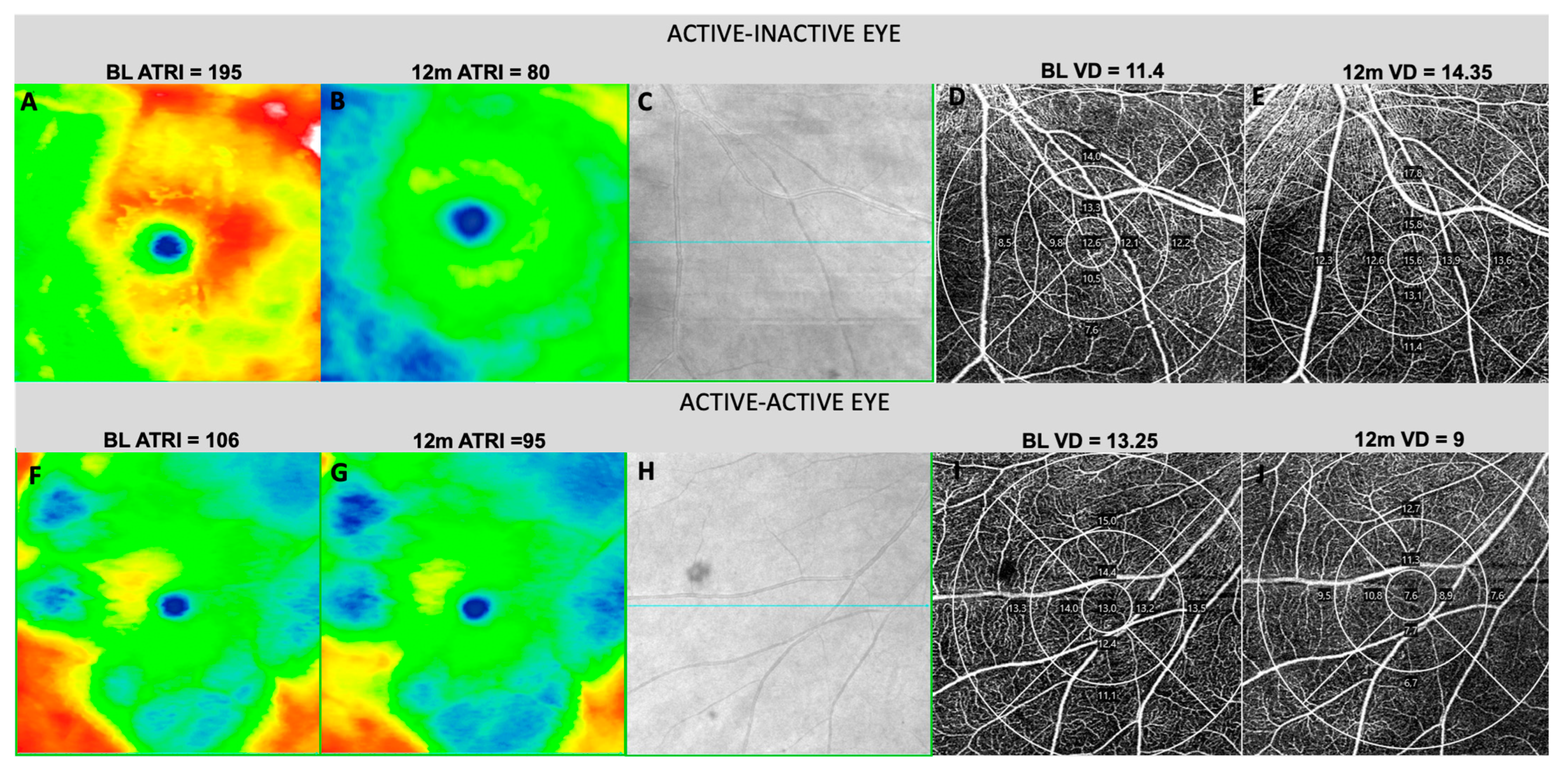
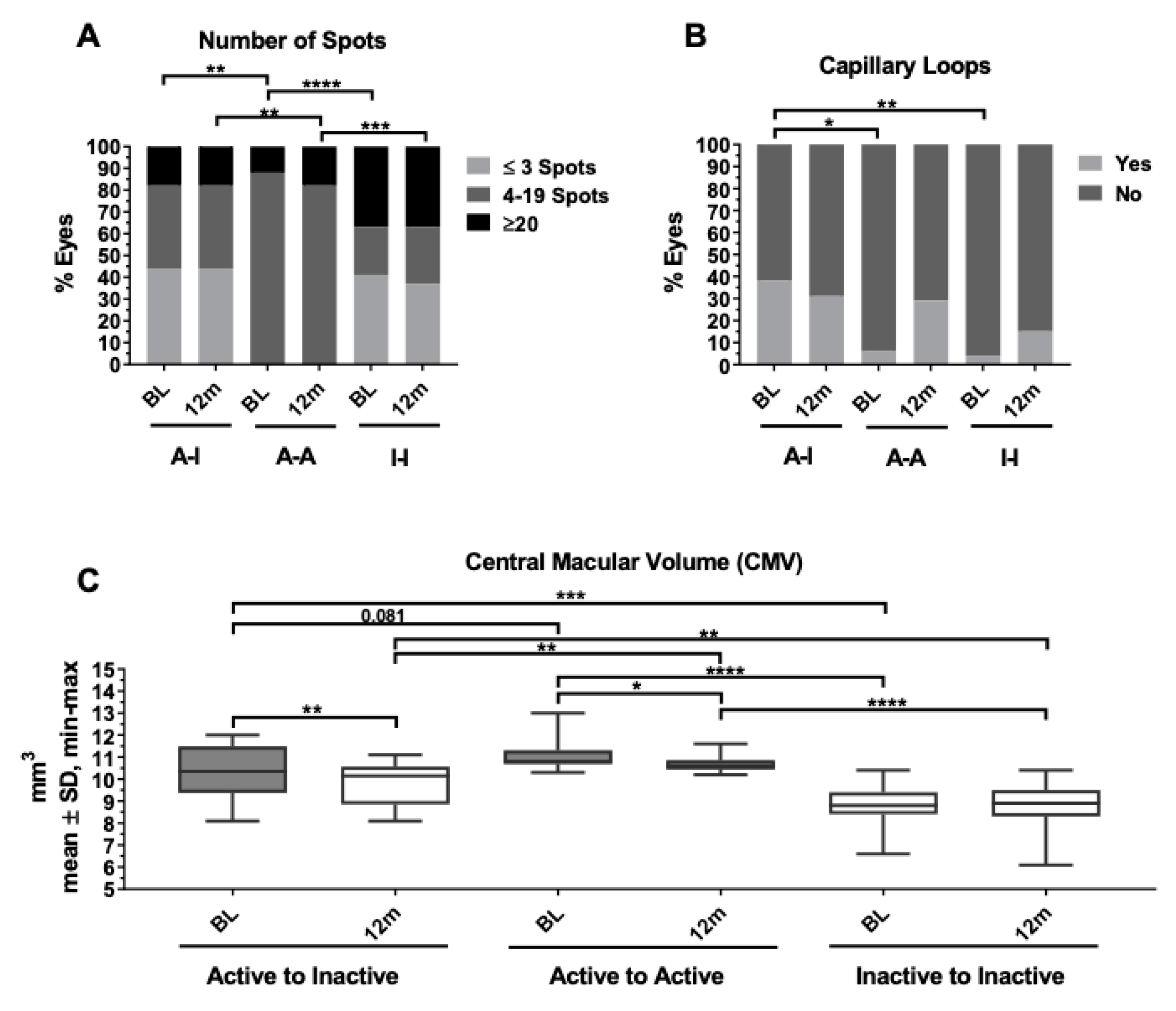
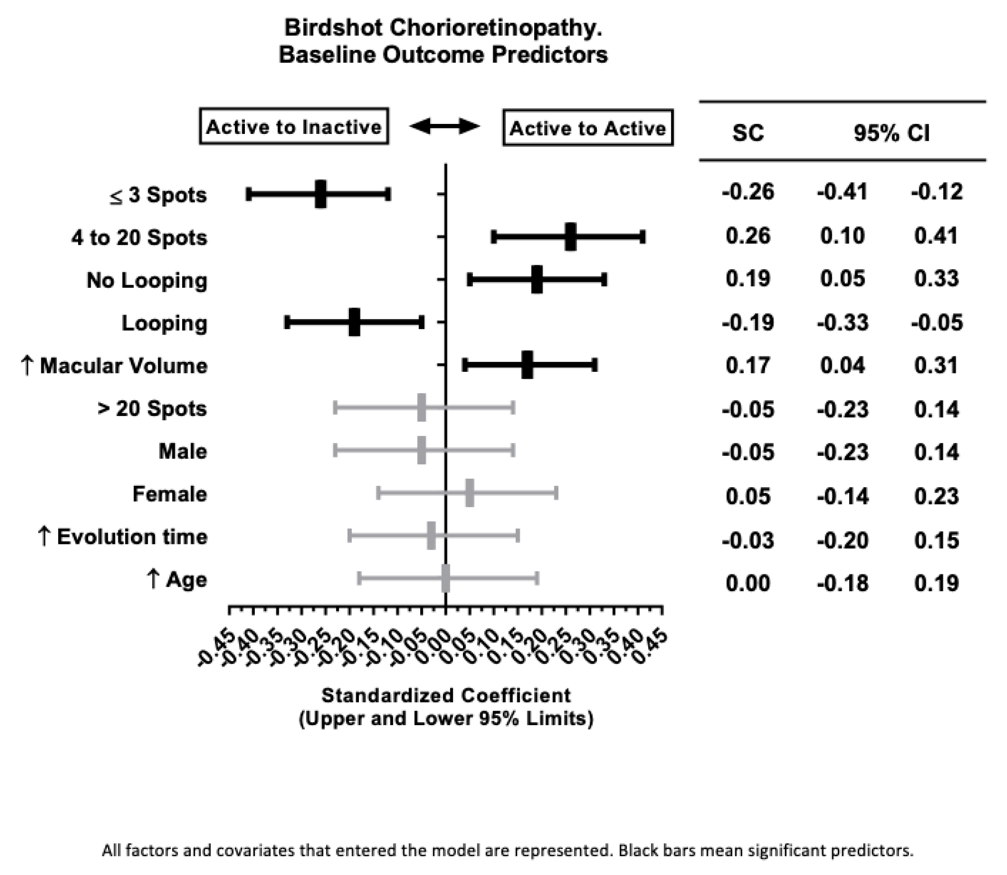
3.1. Active-Inactive Subgroup
3.2. Active-Active Subgroup
3.3. Inactive-Inactive Subgroup
4. Discussion
5. Conclusions
Supplementary Materials
Author Contributions
Funding
Institutional Review Board Statement
Informed Consent Statement
Data Availability Statement
Conflicts of Interest
References
- Pagnoux, C.; Mahr, A.; Aouba, A.; Bérezné, A.; Monnet, D.; Cohen, P.; Levinson, R.D.; Brézin, A.P.; Guillevin, L. Extraocular Manifestations of Birdshot Chorioretinopathy in 118 French Patients. Presse Med. 2010, 39, e97–e102. [Google Scholar] [CrossRef] [PubMed]
- Bousquet, E.; Duraffour, P.; Debillon, L.; Somisetty, S.; Monnet, D.; Brézin, A.P. Birdshot Chorioretinopathy: A Review. J. Clin. Med. 2022, 11, 4772. [Google Scholar] [CrossRef] [PubMed]
- Levinson, R.D.; Rajalingam, R.; Park, M.S.; Reed, E.F.; Gjertson, D.W.; Kappel, P.J.; See, R.F.; Rao, N.A.; Holland, G.N. Human Leukocyte Antigen A29 Subtypes Associated with Birdshot Retinochoroidopathy. Am. J. Ophthalmol. 2004, 138, 631–634. [Google Scholar] [CrossRef] [PubMed]
- Brézin, A.P.; Monnet, D.; Cohen, J.H.M.; Levinson, R.D. HLA-A29 and Birdshot Chorioretinopathy. Ocul. Immunol. Inflamm. 2011, 19, 397–400. [Google Scholar] [CrossRef] [PubMed]
- Herbort, C.P.; Pavésio, C.; LeHoang, P.; Bodaghi, B.; Fardeau, C.; Kestelyn, P.; Neri, P.; Papadia, M. Why Birdshot Retinochoroiditis Should Rather Be Called “HLA-A29 Uveitis”? Br. J. Ophthalmol. 2017, 101, 851–855. [Google Scholar] [CrossRef] [PubMed]
- Monnet, D.; Brézin, A.P. Birdshot Chorioretinopathy. Curr. Opin. Ophthalmol. 2006, 17, 545–550. [Google Scholar] [CrossRef]
- The Standardization of Uveitis Nomenclature (SUN) Working Group. Classification Criteria for Birdshot Chorioretinitis. Am. J. Ophthalmol. 2021, 228, 65–71. [Google Scholar] [CrossRef]
- Lages, V.; Skvortsova, N.; Jeannin, B.; Gasc, A.; Herbort, C.P. Low-Grade “Benign” Birdshot Retinochoroiditis: Prevalence and Characteristics. Int. Ophthalmol. 2019, 39, 2111–2120. [Google Scholar] [CrossRef]
- Gordon, L.K.; Goldhardt, R.; Holland, G.N.; Yu, F.; Levinson, R.D. Standardized Visual Field Assessment for Patients with Birdshot Chorioretinopathy. Ocul. Immunol. Inflamm. 2006, 14, 325–332. [Google Scholar] [CrossRef]
- Thorne, J.E.; Jabs, D.A.; Kedhar, S.R.; Peters, G.B.; Dunn, J.P. Loss of Visual Field Among Patients with Birdshot Chorioretinopathy. Am. J. Ophthalmol. 2008, 145, 23–28.e2. [Google Scholar] [CrossRef]
- Holder, G.E.; Robson, A.G.; Pavesio, C.; Graham, E.M. Electrophysiological Characterisation and Monitoring in the Management of Birdshot Chorioretinopathy. Br. J. Ophthalmol. 2005, 89, 709–718. [Google Scholar] [CrossRef] [PubMed]
- De Carlo, T.E.; Bonini Filho, M.A.; Adhi, M.; Duker, J.S. Retinal and Choroidal Vasculature in Birdshot Chorioretinopathy Analyzed Using Spectral Domain Optical Coherence Tomography Angiography. Retina 2015, 35, 2392–2399. [Google Scholar] [CrossRef]
- Böni, C.; Thorne, J.E.; Spaide, R.F.; Ostheimer, T.A.; Sarraf, D.; Levinson, R.D.; Goldstein, D.A.; Rifkin, L.M.; Vitale, A.T.; Jaffe, G.J.; et al. Multimodal Imaging of the Disease Progression of Birdshot Chorioretinopathy. Ocul. Immunol. Inflamm. 2017, 25, 185–187. [Google Scholar]
- Levinson, R.D.; Brezin, A.; Rothova, A.; Accorinti, M.; Holland, G.N. Research Criteria for the Diagnosis of Birdshot Chorioretinopathy: Results of an International Consensus Conference. Am. J. Ophthalmol. 2006, 141, 185–187. [Google Scholar] [CrossRef]
- Tugal-Tutkun, I.; Herbort, C.P.; Khairallah, M.; Allegri, P.; Biziorek, B.; Bodaghi, B.; Bouchenaki, N.; Cimino, L.; Fardeau, C.; Gupta, A.; et al. Scoring of Dual Fluorescein and ICG Inflammatory Angiographic Signs for the Grading of Posterior Segment Inflammation (Dual Fluorescein and ICG Angiographic Scoring System for Uveitis). Int. Ophthalmol. 2010, 30, 539–552. [Google Scholar] [CrossRef]
- Llorenç, V.; Serrano, A.R.; Mesquida, M.; Lin, P.; Esquinas, C.; Sainz-de-la-Maza, M.; Metea, C.; Bosch, A.; Calvo, M.; Balter, A.; et al. Swept-Source Optical Coherence Tomography Objective Composite Activity Score for Uveitis. Acta Ophthalmol. 2021, 99, 756–764. [Google Scholar] [CrossRef]
- Pichi, F.; Lembo, A.; Nucci, P.; Neri, P. Optical Coherence Tomography Angiography in Birdshot Chorioretinopathy. Eur. J. Ophthalmol. 2023, 34, 781–788. [Google Scholar] [CrossRef]
- Roberts, P.K.; Nesper, P.L.; Goldstein, D.A.; Fawzi, A.A. Retinal Capillary Density in Patients with Birdshot Chorioretinopathy. Retina 2018, 38, 387–394. [Google Scholar] [CrossRef] [PubMed]
- Pohlmann, D.; Macedo, S.; Stübiger, N.; Pleyer, U.; Joussen, A.M.; Winterhalter, S. Multimodal Imaging in Birdshot Retinochoroiditis. Ocul. Immunol. Inflamm. 2017, 25, 621–632. [Google Scholar] [CrossRef]
- Forte, R.; Saleh, M.; Aptel, F.; Chiquet, C. Evaluation of Photoreceptors, Retinal Capillary Plexuses, and Choriocapillaris in Patients With Birdshot Chorioretinopathy. Retina 2020, 40, 977–988. [Google Scholar] [CrossRef]
- Monnet, D.; Brézin, A.P.; Holland, G.N.; Yu, F.; Mahr, A.; Gordon, L.K.; Levinson, R.D. Longitudinal Cohort Study of Patients with Birdshot Chorioretinopathy. I. Baseline Clinical Characteristics. Am. J. Ophthalmol. 2006, 141, 135–142. [Google Scholar] [CrossRef] [PubMed]
- Minos, E.; Barry, R.J.; Southworth, S.; Folkard, A.; Murray, P.I.; Duker, J.S.; Keane, P.A.; Denniston, A.K. Birdshot Chorioretinopathy: Current Knowledge and New Concepts in Pathophysiology, Diagnosis, Monitoring and Treatment. Orphanet J. Rare Dis. 2016, 11, 61. [Google Scholar] [CrossRef] [PubMed]
- Kuiper, J.J.W.; Van Setten, J.; Ripke, S.; Van ’T Slot, R.; Mulder, F.; Missotten, T.; Baarsma, G.S.; Francioli, L.C.; Pulit, S.L.; De Kovel, C.G.F.; et al. A Genome-Wide Association Study Identifies a Functional ERAP2 Haplotype Associated with Birdshot Chorioretinopathy. Hum. Mol. Genet. 2014, 23, 6081–6087. [Google Scholar] [CrossRef] [PubMed]
- Knickelbein, J.E.; Tucker, W.; Kodati, S.; Akanda, M.; Sen, H.N. Non-Invasive Method of Monitoring Retinal Vasculitis in Patients with Birdshot Chorioretinopathy Using Optical Coherence Tomography. Br. J. Ophthalmol. 2018, 102, 815–820. [Google Scholar] [CrossRef] [PubMed]
- Thomas, A.S.; Hatef, A.L.; Stinnett, S.S.; Keenan, R.T.; Jaffe, G.J. Perivascular Thickening on Optical Coherence Tomography as a Marker of Inflammation in Birdshot Retinochoroiditis. Retina 2019, 39, 956–963. [Google Scholar] [CrossRef]
- Teussink, M.M.; Huis in het Veld, P.I.; de Vries, L.A.M.; Hoyng, C.B.; Klevering, B.J.; Theelen, T. Multimodal Imaging of the Disease Progression of Birdshot Chorioretinopathy. Acta Ophthalmol. 2016, 94, 815–823. [Google Scholar] [CrossRef]
- Rothova, A.; Berendschot, T.T.J.M.; Probst, K.; Van Kooij, B.; Baarsma, G.S. Birdshot Chorioretinopathy: Long-Term Manifestations and Visual Prognosis. Ophthalmology 2004, 111, 954–959. [Google Scholar] [CrossRef]
- Keane, P.A.; Allie, M.; Turner, S.J.; Southworth, H.S.; Sadda, S.R.; Murray, P.I.; Denniston, A.K. Characterization of Birdshot Chorioretinopathy Using Extramacular Enhanced Depth Optical Coherence Tomography. JAMA Ophthalmol. 2013, 131, 341–350. [Google Scholar] [CrossRef]


| Active-Inactive | p-Value | Active-Active | p-Value | Inactive-Inactive | p-Value | ||||
|---|---|---|---|---|---|---|---|---|---|
| n | % | n | % | n | % | ||||
| Patients | 13 | 43.3 | 12 | 40.0 | 18 | 60.0 | |||
| Eyes | 16 | 26.7 | 17 | 28.3 | 27 | 45.0 | |||
| mean | ±SD | mean | ±SD | mean | ±SD | ||||
| Age (years) | 58.70 | 11.10 | 0.692 | 58.90 | 14.80 | 0.754 | 60.90 | 10.90 | 0.525 |
| Evolution Time (months) | 97.50 | 49.90 | 0.331 | 92.00 | 51.40 | 0.158 | 132.20 | 77.80 | 0.032 |
| Inflammation VH (NEI) | n | % | n | % | n | % | |||
| BL 0+ | 13 | 81.25 | 0.345 | 14 | 82.35 | 0.401 | 26 | 96.30 | 0.100 |
| 12 m 0+ | 15 | 93.75 | 0.533 | 17 | 100.00 | 0.510 | 26 | 96.30 | 0.900 |
| ∆ 12 m − BL | 2.00 | 12.50 | 0.533 | 3.00 | 17.65 | 0.510 | 0.00 | 0.00 | 0.157 |
| BL 0.5+/1+ | 3 | 18.75 | 0.345 | 3 | 17.65 | 0.401 | 1 | 3.70 | 0.100 |
| 12 m 0.5+/1+ | 1 | 6.25 | 0.533 | 0 | 0.00 | 0.510 | 1 | 3.70 | 0.900 |
| ∆ 12 m − BL | −2.00 | −12.50 | 0.345 | −3.00 | −17.65 | 0.401 | 0.00 | 0.00 | 0.157 |
| UWF Imaging: n Spots | n | % | n | % | n | % | |||
| BL ≤ 3 | 7 | 43.75 | 0.185 | 0 | 0.00 | 0.001 | 11 | 40.74 | 0.114 |
| 12 m ≤ 3 | 7 | 43.75 | 0.133 | 0 | 0.00 | 0.001 | 10 | 37.04 | 0.194 |
| ∆ 12 m − BL | 0 | 0 | 0.534 | 0 | 0 | 0.567 | −1 | −4 | 0.900 |
| BL 4–20 | 6 | 37.50 | 0.502 | 15 | 88.24 | <0.0001 | 6 | 22.22 | 0.002 |
| 12 m 4–20 | 6 | 37.50 | 0.502 | 14 | 82.35 | <0.0001 | 7 | 25.93 | 0.009 |
| ∆ 12 m − BL | 0 | 0 | 0.534 | −1 | −6 | 0.567 | 1 | 4 | 0.900 |
| BL > 20 | 3 | 18.75 | 0.534 | 2 | 11.77 | 0.149 | 10 | 37.04 | 0.052 |
| 12 m > 20 | 3 | 18.75 | 0.433 | 3 | 17.65 | 0.347 | 10 | 37.04 | 0.115 |
| ∆ 12 m − BL | 0 | 0 | 0.534 | 1 | 6 | 0.567 | 0 | 0 | 0.900 |
| UWF FA Score | n | % | n | % | n | % | |||
| BL > 5 | 11 | 68.75 | 0.164 | 11 | 64.71 | 0.286 | 10 | 37.04 | 0.026 |
| 12 m > 5 | 10 | 62.50 | 0.332 | 12 | 70.59 | 0.074 | 9 | 33.33 | 0.012 |
| ∆ 12 m − BL | −1 | −6 | 0.991 | 1 | 6 | 0.830 | −1 | −4 | 0.509 |
| mean | ±SD | mean | ±SD | mean | ±SD | ||||
| BL FA Score | 10.06 | 5.02 | 0.068 | 9.94 | 5.92 | 0.072 | 5.26 | 4.43 | 0.001 |
| 12 m FA Score | 6.25 | 4.78 | 0.778 | 7.53 | 4.43 | 0.105 | 4.82 | 4.39 | 0.085 |
| ∆ 12 m − BL | −4 | 5.30 | 0.026 | −2 | 3.37 | 0.534 | 0 | 2.60 | 0.011 |
| OCT Macular Cube | mean | ±SD | mean | ±SD | mean | ±SD | |||
| BL CMT (μm) | 271.38 | 59.96 | 0.158 | 285.12 | 82.96 | 0.014 | 217.30 | 37.44 | 0.000 |
| 12 m CMT (μm) | 243.63 | 37.54 | 0.595 | 268.71 | 54.77 | 0.002 | 215.41 | 37.95 | 0.001 |
| ∆ 12 m − BL | −27.75 | 42.51 | 0.106 | −16.41 | 65.01 | 0.691 | −1.89 | 6.52 | 0.073 |
| BL MV (mm3) | 10.39 | 1.17 | 0.088 | 11.10 | 0.66 | <0.0001 | 8.86 | 0.82 | <0.0001 |
| 12 m MV (mm3) | 9.78 | 0.92 | 0.462 | 10.71 | 0.40 | <0.0001 | 8.80 | 0.90 | <0.0001 |
| ∆ 12 m − BL | −0.61 | 0.62 | 0.020 | −0.39 | 0.70 | 0.479 | −0.06 | 0.42 | 0.007 |
| n | % | n | % | n | % | ||||
| BL IRF | 4 | 25.00 | 0.571 | 6 | 35.29 | 0.085 | 2 | 7.41 | 0.031 |
| 12 m IRF | 1 | 6.25 | 0.795 | 4 | 23.53 | 0.021 | 0 | 0.00 | 0.043 |
| ∆ 12 m − BL | −3 | −19 | 0.345 | −2 | −12 | 0.957 | −2 | −7 | 0.393 |
| Active-Inactive | p-Value | Active-Active | p-Value | Inactive-Inactive | p-Value | ||||
|---|---|---|---|---|---|---|---|---|---|
| n | % | n | % | n | % | ||||
| Eyes | 16 | 26.7 | 17 | 28.3 | 27 | 45.0 | |||
| Macular Cube OCT | mean | ±SD | mean | ±SD | mean | ±SD | |||
| BL ATRI | 65.93 | 45.89 | 0.070 | 82.36 | 37.99 | <0.0001 | 17.96 | 13.53 | <0.0001 |
| 12 m ATRI | 30.27 | 24.25 | 0.556 | 64.75 | 25.81 | <0.0001 | 16.66 | 12.26 | <0.0001 |
| ∆ 12 m − BL | −35.66 | 39.83 | 0.005 | −17.61 | 42.91 | 0.721 | −1.30 | 7.37 | 0.005 |
| BL AARI | 75.19 | 47.50 | 0.022 | 57.02 | 26.10 | <0.0001 | 142.70 | 26.27 | <0.0001 |
| 12 m AARI | 102.09 | 42.41 | 0.323 | 69.43 | 25.54 | <0.0001 | 143.88 | 27.65 | <0.0001 |
| ∆ 12 m − BL | 26.90 | 25.54 | 0.001 | 12.41 | 24.94 | 0.798 | 1.18 | 10.40 | 0.002 |
| Macular Ellipsoid: | n | % | n | % | n | % | |||
| BL Normal | 6 | 37.50 | 0.570 | 8 | 47.06 | 0.127 | 5 | 18.51 | 0.054 |
| 12 m Normal | 8 | 50.00 | 0.118 | 8 | 47.06 | 0.177 | 4 | 14.81 | 0.007 |
| ∆ 12 m − BL | 2 | 13 | 0.387 | 0 | 0 | 0.957 | −1 | −4 | 0.470 |
| BL Damaged | 9 | 56.25 | 0.727 | 9 | 52.94 | 0.499 | 8 | 66.67 | 0.357 |
| 12 m Damaged | 7 | 43.75 | 0.186 | 9 | 52.94 | 0.607 | 19 | 70.37 | 0.097 |
| ∆ 12 m − BL | −2 | −13 | 0.341 | 0 | 0 | 0.957 | 11 | 4 | 0.509 |
| BL Absent | 1 | 6.25 | 0.795 | 0 | 0.00 | 0.176 | 4 | 14.82 | 0.136 |
| 12 m Absent | 1 | 6.25 | 0.795 | 0 | 0.00 | 0.176 | 4 | 14.82 | 0.136 |
| ∆ 12 m-BL | 0 | 0 | 0.879 | 0 | 0 | 0.957 | 0 | 0 | 0.918 |
| Nasal Field Cube OCT | mean | ±SD | mean | ±SD | mean | ±SD | |||
| BL NRT (μm) | 231.56 | 31.67 | 0.191 | 240.29 | 29.75 | 0.008 | 205.74 | 25.47 | <0.001 |
| 12 m NRT (μm) | 204.04 | 54.08 | 0.251 | 231.12 | 22.45 | 0.021 | 207.48 | 27.30 | 0.281 |
| ∆ 12 m − BL | −27.52 | 66.84 | 0.044 | −9.17 | 30.04 | 0.998 | 1.74 | 20.48 | 0.073 |
| BL Nasal Volume (mm3) | 8.11 | 1.35 | 0.008 | 7.28 | 1.51 | 0.798 | 6.94 | 0.94 | 0.032 |
| 12 m Nasal Volume (mm3) | 7.63 | 0.78 | 0.145 | 7.59 | 1.26 | 0.183 | 6.89 | 1.03 | 0.012 |
| ∆12 m − BL | −0.48 | 0.85 | 0.050 | 0.31 | 1.32 | 0.066 | −0.05 | 0.66 | 0.936 |
| Nasal Ellipsoid: | n | % | n | % | n | % | |||
| BL Normal | 5 | 31.25 | 0.635 | 7 | 41.18 | 0.133 | 4 | 14.82 | 0.068 |
| 12 m Normal | 7 | 43.75 | 0.060 | 4 | 23.53 | 0.892 | 4 | 14.82 | 0.111 |
| ∆12 m − BL | 2 | 13 | 0.278 | −3 | −18 | 0.510 | 0 | 0 | 0.172 |
| BL Damaged | 10 | 62.50 | 0.930 | 8 | 47.06 | 0.117 | 20 | 74.07 | 0.130 |
| 12 m Damaged | 8 | 50.00 | 0.218 | 11 | 64.71 | 0.903 | 19 | 70.37 | 0.324 |
| ∆12 m − BL | −2 | −13 | 0.068 | 3 | 18 | 0.070 | −1 | −4 | 0.298 |
| BL Absent | 1 | 6.25 | 0.629 | 2 | 11.77 | 0.767 | 3 | 11.11 | 0.807 |
| 12 m Absent | 1 | 6.25 | 0.491 | 2 | 11.77 | 0.957 | 4 | 14.82 | 0.524 |
| ∆12 m − BL | 0 | 0 | 0.698 | 0 | 0 | 0.265 | 1 | 4 | 0.470 |
| Active-Inactive | p-Value | Active-Active | p-Value | Inactive-Inactive | p-Value | ||||
|---|---|---|---|---|---|---|---|---|---|
| n | % | n | % | n | % | ||||
| Eyes | 16 | 26.70 | 17 | 28.30 | 27 | 45.00 | |||
| Fundus field OCTA Qualitative Assessment | n | % | n | % | n | % | |||
| BL Capillary Loops | 6 | 37.50 | 0.003 | 1 | 5.88 | 0.327 | 1 | 3.70 | 0.056 |
| 12 m Capillary Loops | 5 | 31.25 | 0.0405 | 5 | 29.41 | 0.500 | 4 | 14.82 | 0.175 |
| ∆ 12 m − BL | −1.00 | −6.25 | 0.048 | 1.00 | 23.53 | 0.398 | 3.00 | 11.12 | 0.749 |
| BL Telangiectasia | 9 | 56.25 | 0.091 | 14 | 82.35 | 0.347 | 21 | 77.78 | 0.502 |
| 12 m Telangiectasia | 12 | 75.00 | 0.884 | 10 | 58.82 | 0.133 | 22 | 81.48 | 0.214 |
| ∆ 12 m − BL | 3.00 | 18.75 | 0.148 | −4.00 | −23.53 | 0.039 | 1.00 | 3.70 | 0.676 |
| BL Increased IC spaces | 14 | 87.50 | 0.879 | 15 | 88.24 | 0.957 | 24 | 88.89 | 0.918 |
| 12 m Increased IC spaces | 14 | 87.50 | 0.879 | 14 | 82.35 | 0.401 | 25 | 92.59 | 0.393 |
| ∆ 12 m − BL | 0.00 | 0.00 | 0.949 | −1.00 | −5.89 | 0.382 | 1.00 | 3.70 | 0.848 |
| BL Capillary Irregularity | 13 | 81.25 | 0.534 | 12 | 70.58 | 0.627 | 20 | 74.07 | 0.883 |
| 12 m Capillary Irregularity | 9 | 56.25 | 0.060 | 13 | 76.47 | 0.892 | 23 | 85.19 | 0.111 |
| ∆ 12 m − BL | −4.00 | −25.00 | 0.016 | 1.00 | 5.89 | 0.737 | 3.00 | 11.12 | 0.136 |
| Inferonasal Field OCTA Vascular Density (VD) mm−2 | mean | ±SD | mean | ±SD | mean | ±SD | |||
| BL Total VD | 6.81 | 3.20 | 0.851 | 8.33 | 4.97 | 0.043 | 5.49 | 3.37 | 0.046 |
| 12 m Total VD | 7.63 | 4.13 | 0.106 | 6.65 | 4.28 | 0.563 | 4.93 | 3.98 | 0.050 |
| ∆ 12 m − BL | 0.82 | 4.36 | 0.134 | −1.68 | 3.79 | 0.165 | −0.55 | 3.86 | 0.939 |
| BL Central VD | 5.76 | 4.43 | 0.911 | 7.21 | 5.48 | 0.096 | 4.60 | 3.54 | 0.108 |
| 12 m Central VD | 6.56 | 5.37 | 0.161 | 5.32 | 4.06 | 0.829 | 4.11 | 4.64 | 0.149 |
| ∆ 12 m − BL | 0.81 | 6.46 | 0.255 | −1.89 | 5.15 | 0.234 | −0.49 | 4.79 | 0.946 |
| BL Internal VD | 6.10 | 3.73 | 0.954 | 8.05 | 5.14 | 0.032 | 4.99 | 3.52 | 0.059 |
| 12 m Internal VD | 7.05 | 4.85 | 0.136 | 6.11 | 4.19 | 0.586 | 4.41 | 4.17 | 0.069 |
| ∆ 12 m − BL | 0.95 | 5.37 | 0.115 | −1.95 | 3.83 | 0.130 | −0.57 | 3.83 | 0.978 |
| BL External VD | 7.09 | 3.09 | 0.770 | 8.46 | 4.95 | 0.051 | 5.67 | 3.42 | 0.042 |
| 12 m External VD | 7.84 | 4.00 | 0.106 | 6.88 | 4.41 | 0.555 | 5.15 | 3.95 | 0.049 |
| ∆ 12 m − BL | 0.75 | 4.19 | 0.162 | −1.59 | 3.80 | 0.192 | −0.52 | 3.98 | 0.952 |
| Inferonasal Field OCTA Perfusion Index (PI)% | mean | ±SD | mean | ±SD | mean | ±SD | |||
| BL Total PI | 16.19 | 7.97 | 0.844 | 20.36 | 12.48 | 0.203 | 14.85 | 16.32 | 0.328 |
| 12 m Total PI | 18.33 | 10.11 | 0.106 | 16.12 | 10.82 | 0.499 | 11.51 | 9.79 | 0.040 |
| ∆ 12 m − BL | 2.14 | 10.73 | 0.135 | −4.25 | 9.46 | 0.441 | −3.34 | 15.83 | 0.528 |
| BL Central PI | 13.64 | 10.93 | 0.978 | 17.70 | 13.33 | 0.074 | 10.94 | 8.72 | 0.101 |
| 12 m Central PI | 15.46 | 13.12 | 0.175 | 12.29 | 9.64 | 0.913 | 9.81 | 11.59 | 0.192 |
| ∆ 12 m − BL | 1.82 | 16.38 | 0.251 | −5.41 | 12.49 | 0.172 | −1.13 | 11.80 | 0.828 |
| BL Internal PI | 14.46 | 9.30 | 0.894 | 19.91 | 13.00 | 0.021 | 11.72 | 8.53 | 0.048 |
| 12 m Internal PI | 17.00 | 12.00 | 0.150 | 15.16 | 10.73 | 0.477 | 10.38 | 10.25 | 0.054 |
| ∆ 12 m − BL | 2.54 | 13.44 | 0.109 | −4.75 | 9.69 | 0.126 | −1.34 | 9.29 | 0.967 |
| BL External PI | 16.84 | 7.70 | 0.785 | 20.64 | 12.41 | 0.034 | 13.14 | 8.27 | 0.031 |
| 12 m External PI | 18.81 | 9.81 | 0.103 | 16.56 | 11.15 | 0.504 | 11.97 | 9.68 | 0.040 |
| ∆ 12 m − BL | 1.97 | 10.29 | 0.150 | −4.08 | 9.57 | 0.161 | −1.17 | 9.64 | 0.992 |
Disclaimer/Publisher’s Note: The statements, opinions and data contained in all publications are solely those of the individual author(s) and contributor(s) and not of MDPI and/or the editor(s). MDPI and/or the editor(s) disclaim responsibility for any injury to people or property resulting from any ideas, methods, instructions or products referred to in the content. |
© 2024 by the authors. Licensee MDPI, Basel, Switzerland. This article is an open access article distributed under the terms and conditions of the Creative Commons Attribution (CC BY) license (https://creativecommons.org/licenses/by/4.0/).
Share and Cite
Moll-Udina, A.; Dotti-Boada, M.; Rodríguez, A.; Sainz-de-la-Maza, M.; Adán, A.; Llorenç, V. Microvascular and Structural Characterization of Birdshot Chorioretinitis in Active and Inactive Phases. Biomedicines 2024, 12, 2414. https://doi.org/10.3390/biomedicines12102414
Moll-Udina A, Dotti-Boada M, Rodríguez A, Sainz-de-la-Maza M, Adán A, Llorenç V. Microvascular and Structural Characterization of Birdshot Chorioretinitis in Active and Inactive Phases. Biomedicines. 2024; 12(10):2414. https://doi.org/10.3390/biomedicines12102414
Chicago/Turabian StyleMoll-Udina, Aina, Marina Dotti-Boada, Anabel Rodríguez, Maite Sainz-de-la-Maza, Alfredo Adán, and Victor Llorenç. 2024. "Microvascular and Structural Characterization of Birdshot Chorioretinitis in Active and Inactive Phases" Biomedicines 12, no. 10: 2414. https://doi.org/10.3390/biomedicines12102414
APA StyleMoll-Udina, A., Dotti-Boada, M., Rodríguez, A., Sainz-de-la-Maza, M., Adán, A., & Llorenç, V. (2024). Microvascular and Structural Characterization of Birdshot Chorioretinitis in Active and Inactive Phases. Biomedicines, 12(10), 2414. https://doi.org/10.3390/biomedicines12102414