Article

Luteinizing Hormone Surge-Induced Krüppel-like Factor 4 Inhibits Cyp17A1 Expression in Preovulatory Granulosa Cells

1
Laboratory of Reproductive Endocrinology, Department of Anatomy & Cell Biology, College of Medicine, Hanyang University, Seoul 04763, Republic of Korea
2
Department of Obstetrics & Gynecology, College of Medicine, Hanyang University, Guri-si 11923, Republic of Korea
*
Author to whom correspondence should be addressed.
These authors contributed equally to this work.
Biomedicines 2024, 12(1), 71; https://doi.org/10.3390/biomedicines12010071
Submission received: 30 November 2023 / Revised: 21 December 2023 / Accepted: 26 December 2023 / Published: 27 December 2023
(This article belongs to the Special Issue Reproductive Medicine: Focus on Cell and Molecule 2.0)

Abstract

:
Previous in vivo and in vitro studies have demonstrated a dramatic up-regulation of Krüppel-like factor 4 (Klf4) in rat preovulatory granulosa cells (GCs) after LH/hCG treatment and its role in regulating Cyp19A1 expression during the luteal shift in steroidogenesis. In this study, we examined whether Klf4 also mediates the LH-induced repression of Cyp17A1 expression in primary rat preovulatory GCs. In response to LH treatment of GCs in vitro, Cyp17A1 expression declined to less than half of its initial value by 1 h, remaining low for 24 h of culture. Overexpression of Klf4 decreased basal and Sf1-induced Cyp17A1 expressions and increased progesterone secretion. Reduction of endogenous Klf4 by siRNA elevated basal Cyp17A1 expression but did not affect LH-stimulated progesterone production. Overexpression of Klf4 also significantly attenuated Sf1-induced Cyp17A1 promoter activity. On the other hand, mutation of the conserved Sp1/Klf binding motif in the promoter revealed that this motif is not required for Klf4-mediated repression. Taken together, these data indicate that the Cyp17A1 gene may be one of the downstream targets of Klf4, which is induced by LH in preovulatory GCs. This information may help in identifying potential targets for preventing the molecular changes occurring in hyperandrogenic disorders.

1. Introduction

Preovulatory granulosa cells (GCs) are characterized by a high content of steroidogenic enzymes and the acquisition of luteinizing hormone (LH) receptors [1]. During the LH surge, GCs undergo molecular and morphological changes, leading to terminal differentiation into luteal cells [2]. These changes are mediated by the coordinated regulation of genes encoding various cell cycle modulators and steroidogenic factors in preovulatory follicles and drive the greater transformation seen in GCs than in theca cells (TCs) as they undergo luteinization [3,4]. As a consequence of luteinization, GCs develop the enzymatic machinery to synthesize progesterone [4,5,6]. A major factor in this steroidogenic shift is a dramatic reduction in the expression of Cyp19A1 [7,8] along with the acquisition of progesterone-producing steroidogenic enzymes such as Cyp11A1 and 3βHSD [9]. GCs start synthesizing progesterone at a greater rate than TCs [10], indicating that they make a major contribution to the marked increase in progesterone production after the LH surge in the corpus luteum (CL).
On the other hand, Cyp17A1 plays a role in converting C21 precursors (progesterone) to androgens. Cyp17A1 transcripts also decline to very low or even undetectable levels during the LH surge in the CL [11,12], which may contribute to the accumulation of its precursor, progesterone. Although the expression of Cyp17A1 is reported to be restricted to TCs [11], it has also been noted in preovulatory GCs [13,14] and even increases slightly in rat GCs after exposure to pregnant mare serum gonadotropin [13]. As it does in TCs, the LH surge reduces Cyp17A1 to low levels in bovine and human preovulatory GCs [3,14].
Transcriptional regulation of Cyp17A1 in TCs is well studied [11,15]. In bovine TCs, Cyp17A1 activity is repressed in response to LH, via the protein kinase A signaling pathway [16]. However, the mechanism of further reduction in Cyp17A1 after the LH surge, during the reprogramming of GCs to luteal cells, has not been elucidated.
Krüppel-like factor 4 (Klf4) is a zinc finger transcription factor involved in the terminal differentiation of various types of epithelial cells [17]. It is highly expressed in preovulatory GCs after LH/hCG treatment and plays a role in the luteal transition of steroidogenesis by mediating LH-induced repression of Cyp19A1 [18,19]. Also, clinical investigations point to dysregulation of Klf4 and Cyp17A1 expression in the ovaries of patients with polycystic ovary syndrome (PCOS) [20,21]. Based on these findings, it seemed likely that Klf4 played a role in regulating the transcription of the Cyp17A1 gene in luteinizing GCs.
Therefore, the present study was undertaken to examine the expression of Cyp17A1 in cultured GCs in response to LH and to investigate whether the induction of Klf4 plays a role in regulating Cyp17A1 expression and progesterone levels. In this work, we used preovulatory GCs isolated from pregnant mare serum gonadotropin-primed immature rat ovaries, which are well-established models of preovulatory GCs.

2. Materials and Methods

2.1. Animals and Reagents

Immature female Sprague Dawley rats (21 days of age) were purchased from Samtako Biokorea (Osan, Republic of Korea) and housed under controlled temperature, humidity, and light conditions (22–24 °C, humidity 40–50%, 12 h light–dark cycle), with free access to standard rat chow and water. The rats were allowed to acclimate until 26 days of age before being used in experiments. Animal care was consistent with institutional guidelines, and the Hanyang University ACUC committee approved all procedures involving animals (HY-IACUC-2021-0067). Ovine LH (NIH-LH-23) was obtained from the National Hormone and Pituitary Distribution Program (Baltimore, MD, USA). Pregnant mare serum gonadotropin (G4527) was from Sigma–Aldrich (St. Louis, MO, USA). Culture media (McCoy’s 5a medium, Leibovitz L-15 medium), fetal bovine serum (FBS), L-glutamine, and penicillin–streptomycin were purchased from Invitrogen Life Technologies (Carlsbad, CA, USA).

2.2. Preparation of Granulosa Cells

In order to induce growth of multiple preovulatory follicles, pregnant mare serum gonadotropin (PMSG) (10 IU) was injected intraperitoneally into immature rats (26 days of age, body weight 55–60 g). Ovaries were dissected 48 h after injection of the PMSG, and preovulatory follicles were punctured in L-15 Leibovitz medium to obtain GCs. Ovarian debris and small follicles were removed, and the remaining medium containing GCs was collected after low-speed centrifugation (500× g) for 10 min. The GCs were washed twice with PBS, and suspended in the culture medium (McCoy’s 5a supplemented with 100 U/mL penicillin, 100 μg/mL streptomycin, and 2 mM L-glutamine).

2.3. Plasmid Constructs

pCMV3 × FLAG-Klf4 (FLAG-Klf4) was made by subcloning full-length rat Klf4 cDNA (kindly provided by Prof. H. Kook; Chonnam National University Medical School, Kwangju, Republic of Korea). The plasmid encoding a Cyp17A1 promoter construct (containing the −2860/+40 region of the human promoter) and steroidogenic factor-1 (Sf1) were generous gifts from K. Parker (University of Texas Southwestern Medical Center, Dallas, TX, USA).
The QuickChange site-directed mutagenesis kit (Stratagene, La Jolla, CA, USA) was used to generate the Cyp17A1 promoter constructs with mutated CACCC motifs (−1252/−1241).
Using Stratagene’s web-based QuickChange primer design program, the primers were designed as follows; 5′-GTGTTACAACGAATTCTCAAAAGATCAAGAG ATT-3′ (sense) and 5′-ACAGCAGAGAACCGGTCTGACCACAAATTTACCAGGGCGGAGTTTTTatCttCCCTAGTAAGCCTGAGG-3′ (antisense). The mutant constructs are designated as follows; (ΔKLF/luc) 5′-TAGGGTGGGGAA-3′→5′-TAGGGaaGatAA-3′. Methylated parental DNA templates were digested with DpnI at 37 °C for 1 h and purified. Mutations were confirmed by direct sequencing. The promoterless luciferase reporter plasmid, pGL3-Basic vector, was purchased from Promega (Madison, WI, USA).

2.4. Transient Transfection of Granulosa Cells

GCs were prepared as described above and resuspended in electroporation buffer (MPK1025; Thermo Fisher Scientific, Waltham, MA, USA) and then mixed with siRNA or plasmids as indicated in figure legends. Electroporation was performed using a Neon™ Transfection System (MPK5000, Thermo Fisher Scientific, Waltham, MA, USA) and the choice of conditions was a single pulse of 1000 V, 40 ms based on previous studies [18,19]. Transfected cells (1 × 10⁵ cells/well) were resuspended in culture medium and plated in 24-well culture plates. After 6 h of transfection, the medium was replaced and then cells were cultured for 24–36 h. At the end of the culture, the cells were harvested and frozen for total RNA/protein extraction or immediately used for luciferase assays. Conditioned culture media were collected and stored at −20 °C for measurement of progesterone levels. To knockdown Klf4 with small interfering RNA (siRNA), silence-selected pre-designed and validated siRNA targeting Klf4 (Klf4-siRNA) was purchased from Genepharma (GenePharma Co., Ltd., Shanghai, China); the sequences of the 25-nucleotide sense and antisense RNAs were 5′-CCAUUAUCAAGAGCUCAUGCCACCG-3′ (sense) and 5′-CGGUGGCAUGAGCUCUUGAUAAUGG-3′ (antisense) (accession no: NM_053713). Non-targeting control siRNA (Universal Scrambled siRNA) was also obtained from Genepharma. All experiments were performed at least three times with measurements of duplicate cultures.

2.5. Real-Time Quantitative PCR

An RNeasy extraction kit (Qiagen Inc., Valencia, CA, USA) was used to extract total RNA. Total RNA (1 µg) was annealed (5 min at 70 °C) to oligo(dT)18 primers and then reverse transcribed using cDNA synthesis platinum master mix (GenDEPOT, Katy, TX, USA). The Primer-BLAST program (NCBI, Bethesda, MD, USA) was used to design the primers as follows: Klf4 forward, 5′-GAGAGGAACTCTCTCACATGAAGC-3′ and reverse, 5′-AAGGATAAAGTCTAGGTCCAGGAGA-3′ (NM_053713.1); Cyp17A1 forward, 5′-CAAGGCTAACGTTGACTCCAG-3′ and reverse, 5′-TGGGTGTAATGAGATGGCTTC-3′ (NM_012753.2). Amplified 18S ribosomal RNA (18S rRNA) (forward, 5′-GCAATTATTCCCCATGAACG-3′ and reverse, 5′-GGCCTCACTAAACCATCCAA-3′) (NM_001025002.1) was used to normalize each reaction (amplification product sizes 185, 167, and 123 bp for Klf4, Cyp17A1, and 18S rRNA, respectively). Real-time PCR reactions were carried out in total volumes of 20 μL with Prime Q-Master Mix (with SYBR Green I) (GeNet Bio Inc., Daejeon, Republic of Korea) using a LightCycler 480 II System (Roche Molecular Diagnostics, Indianapolis, IN, USA). PCR conditions were 10 min at 95 °C, 45 cycles of 95 °C for 10 s, 58~60 °C for 10 s, and 72 °C for 10 s. All reactions were run in triplicate (Roche) and relative amounts of the transcript were calculated by comparing mean values with the control values. Data are expressed as means ± standard deviations (SDs) of triplicate measurements in three independent experiments.

2.6. Western Blot Analysis

GCs grown as described above were collected and washed with cold PBS before lysis in Laemmli buffer containing β-mercaptoethanol, and the resulting cell lysate was boiled for 3 min to denature proteins. Samples of 30 μg were loaded per lane and resolved by 8% SDS-PAGE gel electrophoresis, and proteins were transferred onto nitrocellulose membranes (Amersham Pharmacia Biotech, Arlington Heights, IL, USA). Membranes were blocked for 2 h at room temperature (RT) in TBS-0.1% Tween containing 5% fat-free dry milk, and incubated at 4 °C overnight with anti-KLF4 antibody (abx006830, Abbexa Ltd., Cambridge, UK) or anti-CYP17A1 antibody (MBS820246, MyBioScience, Inc., San Diego, CA, USA) diluted 1:1000 and 1:500, respectively, in TBS-0.1% Tween solution. The membranes were washed with TBS-0.1% Tween and blotted with peroxidase-conjugated donkey anti-rabbit secondary antibody (1:8000) (Boehringer Mannheim, Indianapolis, IN, USA) for 2 h. Immunolabeled proteins were detected with an enhanced chemiluminescence kit (Amersham Pharmacia Biotech., Little Chalfont, UK). The 55 kDa KLF4 and 50 kDa CYP17A1 proteins are indicated in the figures. To ensure that lysates were loaded equally, the blots were stripped and incubated with β-actin (1:3000) (ab8227, Abcam, Waltham, MA, USA).

2.7. Assessment of Progesterone Production

GCs (1 × 10⁵ cells/well) were transfected with Klf4 expression plasmid (0.1 and 0.3 μg/well) or Klf4 siRNA (200 nM/well). After 6 h, they were transferred to LH (200 ng/mL) or control medium for 24–36 h, and conditioned media were harvested for the hormone assay. Progesterone levels were measured using an enzyme-linked immunosorbent assay (ELISA) kit (CSB-E07282r, Cusabio Biotech Co., Ltd., Wuhan, China) following the manufacturer’s protocol. Intra- and inter-assay coefficients of variation were less than 15%, and the limit of detection was 0.8 ng/mL under our conditions. To achieve concentrations within the assay range, the conditioned media were diluted, and serial dilutions were found to behave in a linear fashion using progesterone standards. Absorbance was read against a blanking well at 450 nm within 15 min in an ELISA Reader (BioRad, Hercules, CA, USA). All samples were run in duplicate. Data were collected from three independent experiments.

2.8. Luciferase Assays

GCs were prepared as described above. Cells (1 × 10⁵ cells/well) were cotransfected with 1.5 μg of Cyp17A1 promoter luciferase plasmid (wild-type or mutant constructs) or pGL3-Luc, empty vector, and increasing concentrations of plasmids encoding Klf4 (10, 30, 100 ng/well) and/or Sf1 (10, 30 ng/well) using a Neon™ Transfection System. In order to correct for differences in transfection efficiency, a Renilla luciferase reporter vector (50 ng/well) (Promega) was co-transfected as an internal control. Transfected GCs plated in 24-well plates were cultured for 24–36 h. To harvest cells, cells were washed with PBS, Reporter Lysis Buffer (100 μL) (Promega Corp., Madison, WI, USA) was added into each well, and 20 μL of the supernatant was used for the luciferase assay using a luminometer (FB12, Berthold Technologies, Bad Wildbad, Germany). Firefly luciferase activities were normalized by Renilla luciferase activities and data are expressed as means ± SD of triplicate measurements in three independent experiments.

2.9. Data Analysis

Data are expressed as means with standard deviations (SDs) of at least three independent experiments. IBM SPSS Statistics 26 for Windows (IBM Corp., Armonk, NY, USA) was used for all data analysis. Statistical significant differences were determined using the Kruskal–Wallis test followed by Dunnett’s post hoc test for multiple-group comparisons and the Mann–Whitney U-test for two-group comparisons. p < 0.05 was considered significant.

3. Results

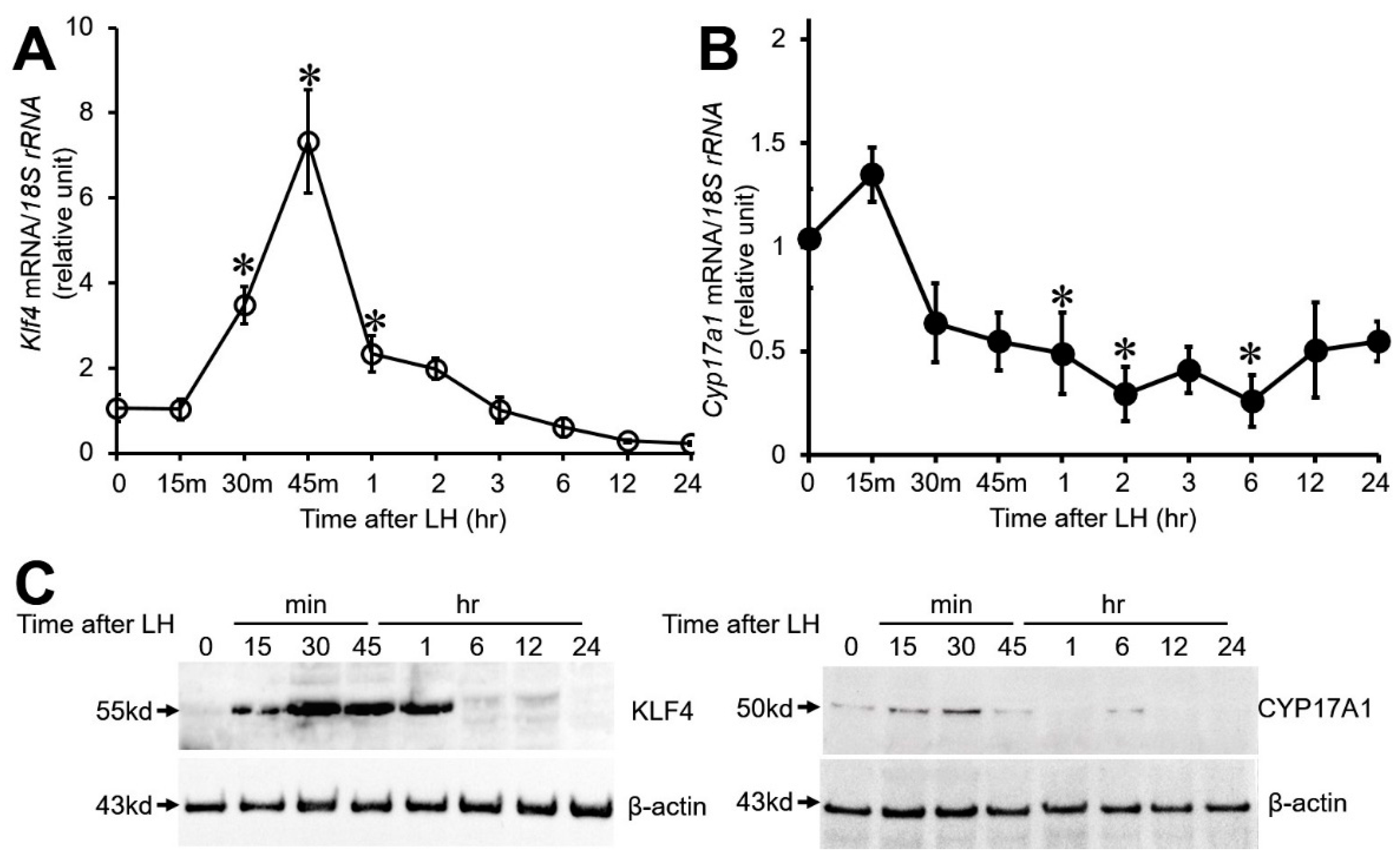
3.1. Effect of LH on Klf4 and Cyp17A1 Expression in Cultured Preovulatory GCs

To characterize the LH-induced regulation of Klf4 and Cyp17A1 expression in GCs, preovulatory GCs were treated with a luteinizing dose of LH (200 ng/mL) for the indicated times and collected for real-time PCR and Western blot analysis. As previously demonstrated [18], LH treatment rapidly and transiently increased Klf4 mRNA (Figure 1A) and protein levels (Figure 1C, upper panel) within 1 h. On the other hand, Cyp17A1 mRNA levels began to decrease rapidly from 30 min and a marked decrease was noted by 1 h (p < 0.001 vs. 0 h). After that, Cyp17A1 mRNA levels remained low throughout the 24 h culture period (Figure 1B). Cyp17A1 protein levels also declined very quickly after peaking at 30 min (Figure 1C, right panel).

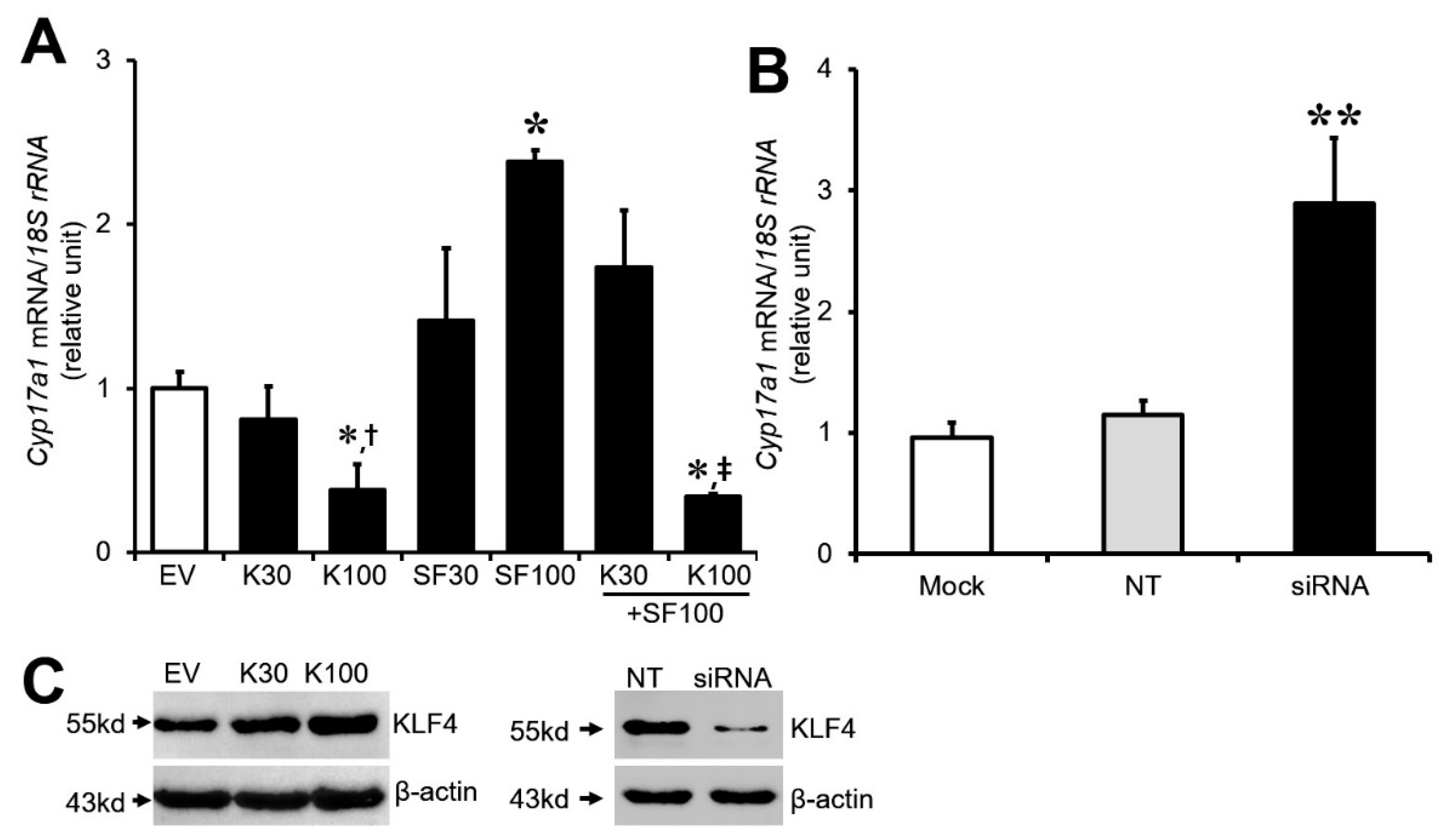
3.2. Effect of Klf4 on Basal and Sf1-Mediated Cyp17A1 Gene Expression in Cultured GCs

Klf4 has been linked to Cyp19A1 repression after the LH surge in luteinizing GCs [19]. Furthermore, Klf4 was most strongly expressed in the GCs of preovulatory follicles as well as granulosa lutein cells of newly formed or mature corpus luteum, although its expression was heterogeneous (Supplemental Figure S1). We therefore hypothesized that the ovarian increase in Klf4 after the LH surge also participates in the regulation of Cyp17A1 in preovulatory GCs. To determine whether Klf4 modulates the expression of Cyp17A1, GCs isolated from preovulatory follicles were transiently transfected with a Klf4 expression plasmid or Klf4-specific siRNA. After 24–36 h, Cyp17A1 mRNA levels were quantified by real-time RT-PCR. Overexpression of and reduction in Klf4, respectively, were confirmed by immunoblot analysis (Figure 2C). Overexpression of Klf4 led to a significant reduction in the basal level of Cyp17A1 mRNA, which was about 0.4-fold of the control at 100 ng of Klf4 plasmid (p < 0.01), and Sf1-stimulated expression was also completely abolished in the cells receiving 100 ng Klf4 expression plasmid (Figure 2A). Conversely, the knockdown of Klf4 expression with siRNA significantly increased basal Cyp17A1 mRNA compared with scrambled siRNA-transfected cells (2.89-fold) (p < 0.05) (Figure 2B). These data demonstrate an inhibitory effect of Klf4 on Cyp17A1 expression.

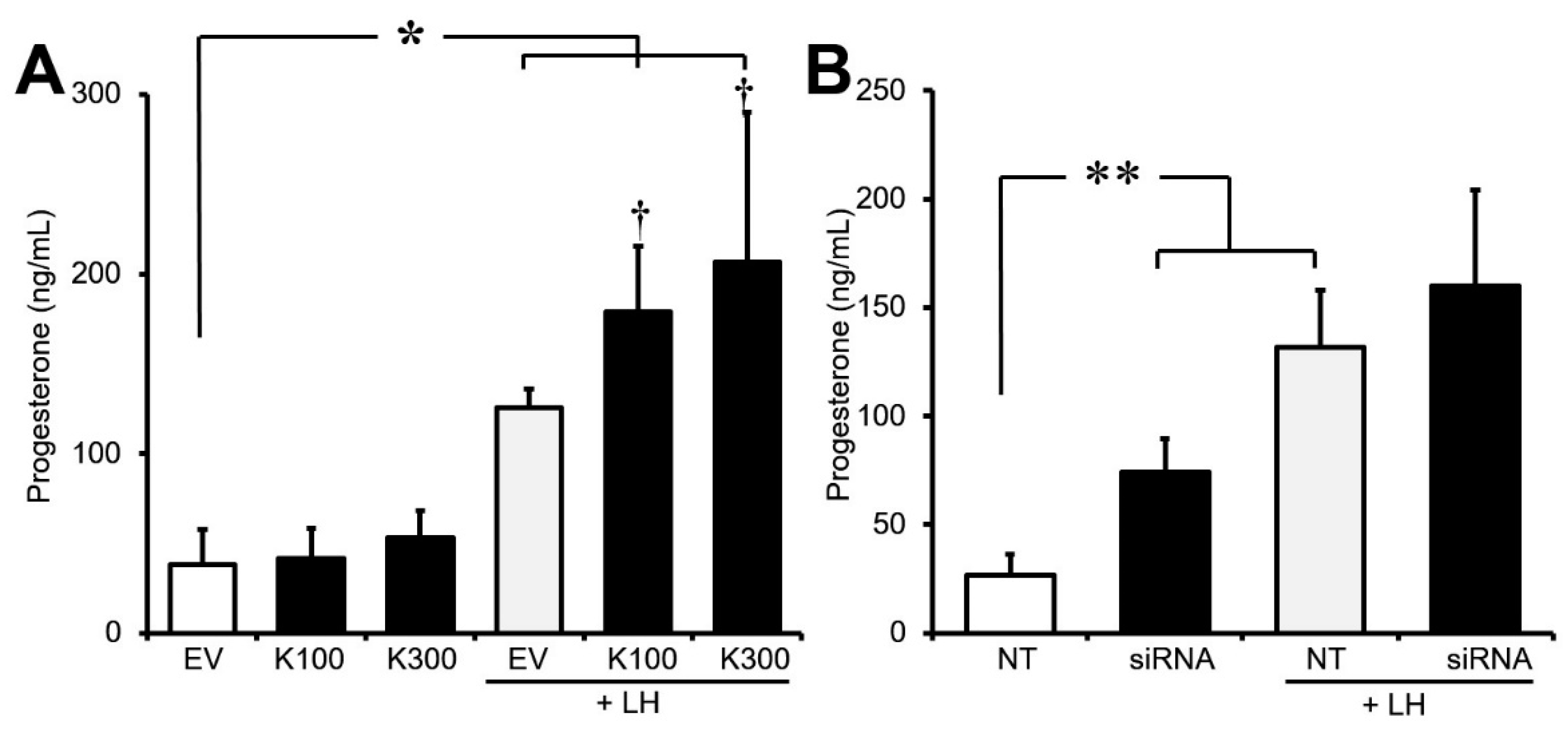
3.3. Effect of Klf4 on Basal and LH-Stimulated Progesterone Production in Cultured GCs

Since Cyp17A1 is known to have the capacity to convert C21 precursors (progesterone) to androgens, Klf4 may increase progesterone levels by directly downregulating the transcription of Cyp17A1 in luteinizing GCs. To evaluate whether Klf4 affected progesterone levels, GCs transfected with the Klf4 overexpression construct or Klf4-specific siRNA were cultured in the absence or presence of LH for 24–36 h, and supernatant progesterone concentrations were measured. Klf4 overexpression significantly increased LH-stimulated progesterone production (p < 0.05 vs. CT) (Figure 3A). Unexpectedly, a reduction in Klf4 expression with siRNA also significantly enhanced basal progesterone production (2.8-fold), whereas inhibition of Klf4 expression had no effect on LH-stimulated progesterone levels (Figure 3B).

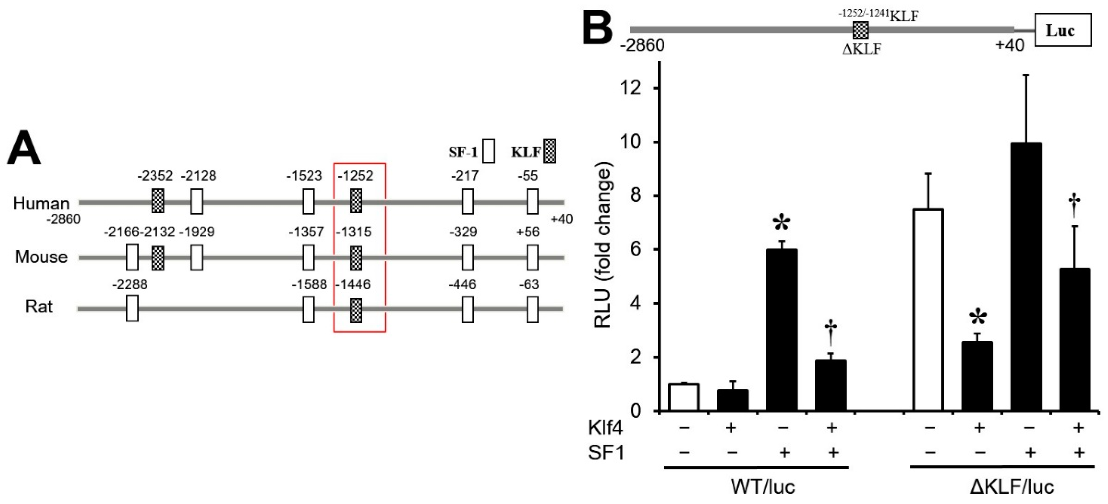
3.4. Regulation of Cyp17A1 Promoter Activity by Klf4

To determine whether Klf4 acts on the Cyp17A1 promoter, the Klf4 expression vector and a Cyp17A1 promoter construct were cotransfected into cultured GCs. Klf4 overexpression caused a dose-dependent decrease in basal promoter activity, although the effect did not attain statistical significance. In addition, the Sf1-stimulated transcriptional activity of the Cyp17A1 promoter was significantly attenuated by cotransfection of the Klf4 expression plasmid (Figure 4). These findings imply that Cyp17A1 is directly or indirectly affected by LH-induced Klf4.

3.5. Sp1/Klf4-Binding Sequences Are Not Involved in Klf4 Repression of Cyp17A1

Next, we examined the putative promoter region (~2900 bp upstream of the transcription start site) of human Cyp17A1 using a web-based transcription factor prediction program. This revealed the presence of three Sf1- and one Sp1/Klf4-binding motif (CACCC) (−1252/−1241 bp), all of which are conserved in humans, mice, and rats (Figure 5A). To assess the functional importance of the conserved Klf4-binding motif in the inhibition of Cyp17A1 expression by Klf4, we mutated the CACCC motif (generating plasmid ∆KLF/luc) (Figure 5B, upper panel) and co-transfected it with the Klf4 and/or the Sf1 expression vector into preovulatory GCs. Mutation of the Klf4 binding site did not alter the inhibitory effect of Klf4 on basal and Sf1-stimulated transcriptional activity (Figure 5B). Unexpectedly, the basal luciferase activity of ΔKLF/luc was markedly enhanced (more than seven-fold) compared with WT/luc. These results indicate that the Sp1/Klf4-binding motif (−1252/−1241 bp region) in the human Cyp17A1 promoter is not responsible for the Klf4-mediated repression of Cyp17A1 promoter activity.

4. Discussion

A major focus of this study was to understand the mechanism by which Cyp17A1 expression is repressed in preovulatory GCs after the LH surge, and we have presented evidence that Klf4 mediates the LH-induced repression of Cyp17A1 expression in these cells.
It is well established that the LH surge initiates a shift in GC steroidogenesis. After the surge, luteinized GCs constitute the major component of the corpora lutea and are the main source of ovarian progesterone [3,4,5,6]. The decline in estradiol and rise in progesterone synthesis after the LH surge appear to be related to a decline in androgen biosynthesis and loss of Cyp17A1 activity [11,12]. Cyp17A1 has a key enzyme role in androgen synthesis, converting progesterone to androstenedione. Previous studies have shown that Cyp17A1 expression drops to low levels in the GCs and TCs of preovulatory follicles at the time of luteinization [12,22]. Although Cyp17A1 expression occurs mainly in TCs [11], weak expression has also been found in GCs [13,14]. Several groups have reported that the LH surge reduces Cyp17A1 expression to low levels in preovulatory GCs as it does in TCs, but they examined only one time point after LH exposure [3,13,14]. In agreement with these observations, we found that Cyp17A1 mRNA expression fell to approximately 0.3-fold of its basal level by 2 h of LH treatment and remained low during a further 24 h of culture (Figure 1B). Similarly, Cyp17A1 protein levels declined very quickly after peaking at 30 min. (Figure 1C).
Klf4 is one of several transcription factors that are transiently induced via the protein kinase A signaling pathway within 30 min of the LH surge in preovulatory GCs (Figure 1) [18] and strongly expressed in the GCs of preovulatory follicles and luteinized GCs of newly formed CLs in the rat ovary (Supplementary Figure S1). Previous studies have indicated that Klf4 participates in regulating steroidogenic genes such as StAR and Cyp11A1 in porcine granulosa luteal cells [23] and Cyp19A1 in rat preovulatory GCs [19]. In the present study, Klf4 overexpression was shown to down-regulate basal and Sf1-stimulated Cyp17A1 expression in preovulatory GCs, leading to increased C21 precursor and progesterone levels (Figure 3A), whereas knockdown of endogenous Klf4 expression led to increased Cyp17A1 expression (Figure 3B), as it did in the case of Cyp19A1 [19]. On the other hand, inhibition of Klf4 expression had no effect on LH-stimulated progesterone levels (Figure 3B). These results suggest that a specific level of Klf4 may be needed for the optimal regulation of progesterone production during the luteal transition of GCs. According to published studies, Cyp17A1 expression in luteinizing TCs is inhibited as a result of repression of Sf1 by c-fos [24]. Therefore, it will be of interest to determine whether c-fos also contributes to Cyp17A1 expression in luteinizing GCs.
Klf4 has been shown to interact with CACCC and GC-rich motifs [17,25], and a conserved CACCC motif was noted in the Cyp17A1 promoter (Figure 5A). However, alteration of the CACCC sequence did not impair Klf4-mediated repression; instead, and unexpectedly, it enhanced the transcriptional activity of a Cyp17A1 promoter reporter construct in preovulatory GCs (Figure 5B), indicating that this motif is not required for Klf4-mediated repression and that other elements must be involved on the CACCC motif.
Sf1 (Steroidogenic factor-1), an orphan nuclear receptor, is essential for gonadal development [26] and positively regulates the transcription of enzymes involved in ovarian steroidogenesis including Cyp11A1, Cyp19A1, and Cyp17A1 by binding to specific sites in their promoter regions [22,27]. In rodents, Sf1 is widely expressed in the ovary and its expression in preovulatory GCs is modulated by gonadotropins [28]. Klf4 is able to suppress the well-established Sf1 induction of pro-proliferative genes and Cyp19A1 by interfering with the recruitment of Sf1 or competing with Sf1 for binding to recognition motifs within the promoter region [19,29]. Although a number of possible mechanisms may be responsible for the inhibition of Cyp17A1 transcription by Klf4, Klf4 may interact with Sf1 to competitively inhibit its binding to the Cyp17A1 promoter as it does in the case of Cyp19A1 [19]. Furthermore, Sf1 expression in GCs is rapidly reduced to very low levels in response to the LH surge [30]. Along with this, LH-induced Klf4 may also inhibit Sf1 activity, ultimately leading to the sharp reduction in Cyp17A1 expression that we observed. Although additional studies are required to clarify the exact mechanism controlling the expression of this gene in GCs, these data provide experimental evidence that Cyp17A1 is one of the downstream targets of Klf4 in preovulatory GCs. However, this needs to be confirmed in human GCs from preovulatory follicles, as findings in humans may differ from those in experimental animals.
Hyperandrogenism is one of the most prominent clinical features of patients with PCOS [20,21]. Elevated expression of Cyp17A1 has been reported in the GCs, TCs, and even preovulatory oocytes of PCOS patients [20,21,31], suggesting that gene alterations in GCs and oocytes are responsible for the poor oocyte quality and decreased fertilization rates in IVF patients with PCOS [32]. Furthermore, Klf4 expression is down-regulated in the ovaries of PCOS patients [20]. In view of the involvement of Klf4 in Cyp17A1 expression, down-regulation of Klf4 may contribute to the increased Cyp17A1 expression found in PCOS patients.
Increased understanding of the ability of Klf4 to inhibit Cyp17A1 production may suggest a target for overcoming the molecular aberrations underlying hyperandrogenic disorders such as PCOS.

Supplementary Materials

The following supporting information can be downloaded at: https://www.mdpi.com/article/10.3390/biomedicines12010071/s1, Figure S1: Immunohistochemical localization of KLF4 in rat ovaries. Ovaries were sectioned at 0, 2, 24, and 48 h after injection of pregnant mare serum gonadotropin-primed rats with hCG and incubated with KLF4 antibody (NBP2-24749). Positive staining appears as a dark brown precipitate. (A) Representative images of ovaries from rats treated with pregnant mare serum gonadotropin for 48 h. Positive signals were detected in the GCs (arrows) of preovulatory follicles, but not in TCs. (B) Representative images of an ovarian section obtained 2 h after hCG injection. Positive signals were detected in the GCs of follicles, with stronger expression in the cumulus and antral GCs (arrows) of ovulatory follicles. Positive signals were also seen in TCs (arrowheads). Representative images of a section of an ovary obtained (C) 24 h and (D) 48 h after hCG injection. Luteinized GCs of the newly formed corpus luteum (nCL) and mature CL (arrow) stained more intensely than those of TCs. GCs, granulosa cells; TCs, theca cells; SF, secondary follicle; POF, preovulatory follicle; nCL, newly formed corpus luteum; CL, mature corpus luteum. Bar = 100 μm.

Author Contributions

Y.C. and O.L. contributed equally to this work as the first author. Y.C., O.L., K.R. and J.R. conceived and designed the research; Y.C., O.L. and K.R. participated in the experiments, data collection, and data analysis; J.R. analyzed the data and interpreted the results of experiments; Y.C. and O.L. drafted the manuscript; J.R. edited and revised the manuscript; Y.C., O.L., K.R. and J.R. all read and approved the final version; J.R. takes responsibility for the integrity of the data analysis. All authors have read and agreed to the published version of the manuscript.

Funding

This study was supported by a National Research Foundation of Korea Grant funded by the Korean Government (KRF-2022R1F1A1069408).

Institutional Review Board Statement

The animal study protocol was approved by the Institutional Review Board of the Hanyang University ACUC committee (HY-IACUC-2021-0067).

Informed Consent Statement

Not applicable.

Data Availability Statement

The authors confirm that the data supporting the findings of this study are available within the article and its Supplementary Materials.

Conflicts of Interest

The authors declare no conflicts of interest.

References

  1. Hsueh, A.; Jones, P.; Adashi, E.; Wang, C.; Zhuang, L.-Z.; Welsh, T. Intraovarian mechanisms in the hormonal control of granulosa cell differentiation in rats. Reproduction 1983, 69, 325–342. [Google Scholar] [CrossRef]
  2. Murphy, B.D. Models of luteinization. Biol. Reprod. 2000, 63, 2–11. [Google Scholar] [CrossRef] [PubMed]
  3. Christenson, L.K.; Gunewardena, S.; Hong, X.; Spitschak, M.; Baufeld, A.; Vanselow, J. Research resource: Preovulatory LH surge effects on follicular theca and granulosa transcriptomes. Mol. Endocrinol. 2013, 27, 1153–1171. [Google Scholar] [CrossRef] [PubMed]
  4. Espey, L.L.; Richards, J.S. Temporal and spatial patterns of ovarian gene transcription following an ovulatory dose of gonadotropin in the rat. Biol. Reprod. 2002, 67, 1662–1670. [Google Scholar] [CrossRef] [PubMed]
  5. Rajapaksha, W.; McBride, M.; Robertson, L.; O’shaughnessy, P. Sequence of the bovine HDL-receptor (SR-BI) cDNA and changes in receptor mRNA expression during granulosa cell luteinization in vivo and in vitro. Mol. Cell. Endocrinol. 1997, 134, 59–67. [Google Scholar] [CrossRef]
  6. Rodgers, R.; Waterman, M.; Simpson, E. Cytochromes P-450scc, P-45017 α, Adrenodoxin, and Reduced Nicotinamide Adenine Dinucleotide Phosphate-Cytochrome P-450 Reductase in Bovine Follicles and Corpora Lutea. Changes in Specific Contents during the Ovarian Cycle. Endocrinology 1986, 118, 1366–1374. [Google Scholar] [CrossRef]
  7. Fitzpatrick, S.L.; Carlone, D.L.; Robker, R.L.; Richards, J.S. Expression of aromatase in the ovary: Down-regulation of mRNA by the ovulatory luteinizing hormone surge. Steroids 1997, 62, 197–206. [Google Scholar] [CrossRef]
  8. Hickey, G.J.; Krasnow, J.S.; Beattie, W.G.; Richards, J.S. Aromatase cytochrome P450 in rat ovarian granulosa cells before and after luteinization: Adenosine 3′, 5′-monophosphate-dependent and independent regulation. Cloning and sequencing of rat aromatase cDNA and 5′ genomic DNA. Mol. Endocrinol. 1990, 4, 3–12. [Google Scholar] [CrossRef]
  9. Carletti, M.Z.; Christenson, L.K. Rapid effects of luteinizing hormone on gene expression in the mural granulosa cells of mouse preovulatory follicles. Reproduction 2009, 137, 843. [Google Scholar] [CrossRef]
  10. Aflalo, L.; Meidan, R. The hormonal regulation of cholesterol side-chain cleavage cytochrome P450, adrenodoxin, and their messenger ribonucleic acid expression in bovine small-like and large-like luteal cells: Relationship with progesterone production. Endocrinology 1993, 132, 410–416. [Google Scholar] [CrossRef]
  11. Conley, A.J.; Kaminski, M.A.; Dubowsky, S.A.; Ablonka-Shariff, A.; Redmer, D.A.; Reynolds, L.P. Immunohistochemical localization of 3β-hydroxysteroid dehydrogenase and P450 17α-hydroxylase during follicular and luteal development in pigs, sheep, and cows. Biol. Reprod. 1995, 52, 1081–1094. [Google Scholar] [CrossRef] [PubMed]
  12. Nimz, M.; Spitschak, M.; Fürbass, R.; Vanselow, J. The pre-ovulatory luteinizing hormone surge is followed by down-regulation of CYP19A1, HSD3B1, and CYP17A1 and chromatin condensation of the corresponding promoters in bovine follicles. Mol. Reprod. Dev. 2010, 77, 1040–1048. [Google Scholar] [CrossRef] [PubMed]
  13. Jo, M.; Gieske, M.C.; Payne, C.E.; Wheeler-Price, S.E.; Gieske, J.B.; Ignatius, I.V.; Curry, T.E.; Ko, C. Development and application of a rat ovarian gene expression database. Endocrinology 2004, 145, 5384–5396. [Google Scholar] [CrossRef] [PubMed]
  14. Oktem, O.; Akin, N.; Bildik, G.; Yakin, K.; Alper, E.; Balaban, B.; Urman, B. FSH Stimulation promotes progesterone synthesis and output from human granulosa cells without luteinization. Hum. Reprod. 2017, 32, 643–652. [Google Scholar] [CrossRef] [PubMed]
  15. Komar, C.; Berndtson, A.; Evans, A.; Fortune, J. Decline in circulating estradiol during the preovulatory period is correlated with decreases in estradiol and androgen, and in messenger RNA for P450 aromatase and P450 17α-hydroxylase, in bovine preovulatory follicles. Biol. Reprod. 2001, 64, 1797–1805. [Google Scholar] [CrossRef] [PubMed]
  16. Magoffin, D.A.; Weitsman, S.R. Differentiation of ovarian theca-interstitial cells in vitro: Regulation of 17 alpha-hydroxylase messenger ribonucleic acid expression by luteinizing hormone and insulin-like growth factor-I. Endocrinology 1993, 132, 1945–1951. [Google Scholar] [CrossRef]
  17. Garrett-Sinha, L.A.; Eberspaecher, H.; Seldin, M.F.; de Crombrugghe, B. A gene for a novel zinc-finger protein expressed in differentiated epithelial cells and transiently in certain mesenchymal cells. J. Biol. Chem. 1996, 271, 31384–31390. [Google Scholar] [CrossRef]
  18. Choi, H.; Roh, J. LH-induced transcriptional regulation of KLF4 expression in granulosa cells occurs via the camp/pka pathway and requires a putative sp1 binding site. Int. J. Mol. Sci. 2020, 21, 7385. [Google Scholar] [CrossRef]
  19. Choi, H.; Ryu, K.-Y.; Roh, J. Krüppel-like factor 4 plays a role in the luteal transition in steroidogenesis by downregulating Cyp19A1 expression. Am. J. Physiol. Endocrinol. Metab. 2019, 316, E1071–E1080. [Google Scholar] [CrossRef]
  20. Jansen, E.; Laven, J.S.; Dommerholt, H.B.; Polman, J.; van Rijt, C.; van den Hurk, C.; Westland, J.; Mosselman, S.; Fauser, B.C. Abnormal gene expression profiles in human ovaries from polycystic ovary syndrome patients. Mol. Endocrinol. 2004, 18, 3050–3063. [Google Scholar] [CrossRef]
  21. Wickenheisser, J.K.; Quinn, P.G.; Nelson, V.L.; Legro, R.S.; Strauss, J.F., III; McAllister, J.M. Differential activity of the cytochrome P450 17α-hydroxylase and steroidogenic acute regulatory protein gene promoters in normal and polycystic ovary syndrome theca cells. J. Clin. Endocrinol. Metab. 2000, 85, 2304–2311. [Google Scholar] [CrossRef] [PubMed]
  22. Borroni, R.; Liu, Z.; Simpson, E.R.; Hinshelwood, M.M. A putative binding site for Sp1 is involved in transcriptional regulation of CYP17 gene expression in bovine ovary. Endocrinology 1997, 138, 2011–2020. [Google Scholar] [CrossRef] [PubMed]
  23. Natesampillai, S.; Kerkvliet, J.; Leung, P.C.; Veldhuis, J.D. Regulation of Kruppel-like factor 4, 9, and 13 genes and the steroidogenic genes LDLR, StAR, and CYP11A in ovarian granulosa cells. Am. J. Physiol.-Endocrinol. Metab. 2008, 294, E385–E391. [Google Scholar] [CrossRef] [PubMed]
  24. Patel, S.S.; Beshay, V.E.; Escobar, J.C.; Carr, B.R. 17α-Hydroxylase (CYP17) expression and subsequent androstenedione production in the human ovary. Reprod. Sci. 2010, 17, 978–986. [Google Scholar] [CrossRef] [PubMed]
  25. Shields, J.M.; Yang, V.W. Identification of the DNA sequence that interacts with the gut-enriched Krüppel-like factor. Nucleic Acids Res. 1998, 26, 796–802. [Google Scholar] [CrossRef] [PubMed]
  26. Luo, X.; Ikeda, Y.; Parker, K.L. A cell-specific nuclear receptor is essential for adrenal and gonadal development and sexual differentiation. Cell 1994, 77, 481–490. [Google Scholar] [CrossRef] [PubMed]
  27. Parker, K.L.; Rice, D.A.; Lala, D.S.; Ikeda, Y.; Luo, X.; Wong, M.; Bakke, M.; Zhao, L.; Frigeri, C.; Hanley, N.A.; et al. Steroidogenic factor 1: An essential mediator of endocrine development. Recent Prog. Horm. Res. 2002, 57, 19–36. [Google Scholar] [CrossRef] [PubMed]
  28. Falender, A.E.; Lanz, R.; Malenfant, D.; Belanger, L.; Richards, J.S. Differential expression of steroidogenic factor-1 and FTF/LRH-1 in the rodent ovary. Endocrinology 2003, 144, 3598–3610. [Google Scholar] [CrossRef]
  29. Lomberk, G.; Urrutia, R. The family feud: Turning off Sp1 by Sp1-like KLF proteins. Biochem. J. 2005, 392, 1–11. [Google Scholar] [CrossRef]
  30. Agarwal, P.; Peluso, J.J.; White, B.A. Steroidogenic factor-1 expression is transiently repressed and c-myc expression and deoxyribonucleic acid synthesis are induced in rat granulosa cells during the preovulatory period. Biol. Reprod. 1996, 55, 1271–1275. [Google Scholar] [CrossRef]
  31. Al-Omar, Z.; Ozbakir, B.; Tulay, P. Differential expression of genes involved in steroidogenesis pathway in human oocytes obtained from patients with polycystic ovaries. J. Reprod. Immunol. 2020, 142, 103191. [Google Scholar] [CrossRef] [PubMed]
  32. Qiao, J.; Feng, H.L. Extra-and intra-ovarian factors in polycystic ovary syndrome: Impact on oocyte maturation and embryo developmental competence. Hum. Reprod. Update 2011, 17, 17–33. [Google Scholar] [CrossRef] [PubMed]
Figure 1. Effect of LH on Klf4 and Cyp17A1 mRNA and protein levels in cultured preovulatory GCs. Real-time RT-PCR analysis of (A) Klf4 and (B) Cyp17A1 mRNA levels. GCs obtained from rat preovulatory follicles (as described above) were cultured with LH (200 ng/mL) for the indicated times. 18S rRNA was used to normalize reactions. Values were calculated as fold changes relative to values at 0 h and are expressed as means ± SDs of three independent preparations of GCs. * p < 0.001 vs. 0 h. (C) Immunoblot analysis of KLF4 (left panel) and CYP17A1 (right panel). Lysates were immunoblotted with anti-KLF4 antibody (1:1000) (abx006830) or anti-CYP17A1 antibody (1:500) (MBS820246). Arrows indicate bands corresponding to KLF4 (55 kDa), CYP17A1 (50 kDa), and β-actin (43 kDa).
Biomedicines 12 00071 g001
Figure 2. Regulation of Cyp17A1 expression by Klf4 in preovulatory granulosa cells. GCs were obtained from preovulatory follicles of rat ovaries 48 h after injection of pregnant mare serum gonadotropin. (A) Real-time RT-PCR analysis of Cyp17A1 mRNA levels in GCs cotransfected with increasing amounts of Klf4 plasmid (30, 100 ng/well), Sf1 plasmid (30, 100 ng/well), or empty vector (EV). EV, empty vector; SF30, Sf1 30 ng/well; SF100, Sf1 100 ng/well; K30, Klf4 30 ng/well; K100, Klf4 100 ng/well. * p < 0.05 vs. EV;  p < 0.05 vs. K30;  p < 0.001 vs. SF100. (B) Real-time RT-PCR analysis of Cyp17A1 mRNA levels in GCs transfected with Klf4 siRNA (200 nM) or control siRNA (200 nM) (NT). Mock, mock-transfected control; NT, non-target control siRNA; siRNA, Klf4-specific siRNA. ** p < 0.05 vs. NT. 18S rRNA was used to normalize reactions. Values were calculated as fold changes relative to controls (EV or NT) and are expressed as means ± SDs of at least three independent experiments. (C) Immunoblot analyses of KLF4 protein in lysates of transfected GCs to confirm Klf4 overexpression (left panel) and knockdown (right panel). Arrows indicate bands corresponding to KLF4 (55 kDa) and β-actin (42 kDa).
Biomedicines 12 00071 g002
Figure 3. Effects of Klf4 overexpression and knockdown on basal and LH-stimulated progesterone production by cultured granulosa cells. GCs were obtained from preovulatory follicles of rat ovaries 48 h after injection of pregnant mare serum gonadotropin. (A) For overexpression, GCs were transfected with Klf4 plasmid (100, 300 ng/well) or empty vector (EV), and (B) for knockdown, they were transfected with Klf4 siRNA (200 nM) or control non-target siRNA (NT) and cultured for 24–36 h in the presence or absence of LH (200 ng/mL). At the end of the incubation, supernatant progesterone levels were assayed by ELISA. Values were calculated as fold changes relative to the value for EV-transfected cells or NT-transfected cells and are expressed as means ± SDs of at least three independent experiments. EV, empty vector; Klf4, FLAG-Klf4 plasmid; NT, non-target control siRNA; siRNA, Klf4-specific siRNA. * p < 0.05 vs. EV;  p < 0.05 vs. EV + LH; ** p < 0.05 vs. NT.
Biomedicines 12 00071 g003
Figure 4. Klf4 inhibition of basal and Sf1-stimulated Cyp17A1 promoter activity in cultured granulosa cells. GCs were obtained from preovulatory follicles of rat ovaries 48 h after injection of pregnant mare serum gonadotropin. Cells were co-transfected with a Cyp17A1 promoter luciferase reporter construct (1.5 μg/well) consisting of the proximal 2900 bp of the promoter with increasing amounts of Sf1 plasmid (10 and 30 ng/well) with/without Klf4 plasmid (10, 30, and 100 ng/well), and cultured for 24–36 h. Cell lysates were assayed for luciferase, expressed as relative light units (RLU), and normalized to Renilla luciferase activity in co-transfected cells. Values are fold changes relative to control (CT) and are means ± SDs of three independent experiments, each performed in triplicate. CT, cells transfected with Cyp17A1 promoter only. * p < 0.05 vs. CT;  p < 0.05 vs. SF10 only;  p < 0.05 vs. SF30 only.
Biomedicines 12 00071 g004
Figure 5. The CACCC motif is not required for Klf4-mediated inhibition of Cyp17A1 promoter activity. (A) Schematic representation of the human, rat, and mouse Cyp17A1 promoter indicating the relative position of the conserved Klf4 binding site (indicated in red square). Sf1, steroidogenic factor 1; KLF, CACCC motif. (B) Schematic showing the mutated regions of the Cyp17A1 promoter (upper panel). Cyp17A1 promoter constructs (1.5 μg/well) containing the wild-type or mutated CACCC motif were co-transfected into GCs together with Klf4 plasmid (30 ng/well) or Sf1 plasmid (30 ng/well). Values were calculated as fold changes relative to control and are expressed as the means ± SDs of three independent experiments, each performed in triplicate. Control (CT, cells transfected with either wild-type or mutated Cyp17A1 promoter only); WT/luc, wild-type Cyp17A1 promoter; ΔKLF/luc, promoter with mutation at −1252/−1241KLF site (GTGGGGA→GaaGatA). * p < 0.05 vs. CT;  p < 0.05 vs. CT + SF1.
Biomedicines 12 00071 g005
Disclaimer/Publisher’s Note: The statements, opinions and data contained in all publications are solely those of the individual author(s) and contributor(s) and not of MDPI and/or the editor(s). MDPI and/or the editor(s) disclaim responsibility for any injury to people or property resulting from any ideas, methods, instructions or products referred to in the content.

