Abstract
Objectives: This study aimed to assess the efficacy of shockwave-enhanced emission photoacoustic streaming (SWEEPS) plus antimicrobial photodynamic therapy (aPDT) using indocyanine green (ICG) for the elimination of Enterococcus faecalis biofilm from infected root canals. Materials and Methods: thirty sound human single-canal teeth were chosen and standardized to have 12 mm of root length. The root canals were shaped and prepared by means of ProTaper rotary files. After sterilization of the teeth, the canals were inoculated with E. faecalis for 2 weeks. The teeth were then randomly divided into six groups (n = five) of control, ICG, ICG + 808 nm diode laser, ICG + SWEEPS, ICG + 808 nm diode laser + SWEEPS, and 5.25% sodium hypochlorite (NaOCl). Following treatment, the number of colony-forming units (CFUs)/mL were calculated for each group. Statistical analysis was carried out using one-way ANOVA. For multiple comparisons, Tukey’s test was used as the post hoc test. Results: NaOCl alone showed the highest efficacy (p < 0.001). The ICG + 808 nm diode laser + SWEEPS group displayed significantly lower amounts of bacteria than either the ICG + 808 nm diode laser or SWEEPS (p < 0.001). There was a statistically significant difference detected between the ICG + 808 nm diode laser and ICG + SWEEPS (p = 0.035). Conclusions: SWEEPS can effectively increase the photosensitizer distribution in the root canal space, and its application along with irrigants can bring about promising results.
1. Introduction
Endodontic treatments aim to effectively reduce the microorganisms responsible for endodontic infections [1]. However, the complete elimination of endodontic pathogens is extremely difficult, if not impossible, with the commonly used instrument methods, due to the complex anatomy of the root canal system and the presence of lateral canals, isthmi, ramifications and fins [2]. Enterococcus faecalis is associated with secondary endodontic infections, refractory infected lesions and periapical biofilms, resulting in endodontic treatment failure [3]. Teeth with failed endodontic treatment are more likely than non-endodontically treated teeth to contain this microorganism in their root canal system [3]. The resistance of this bacterium to the challenges of survival within the root canal space is related to the ability to invade the dentinal tubules and bond to collagen fibers, biofilm formation, and its capacity to endure harsh environments [3].
Root canal irrigation is performed along with mechanical cleaning and instrumentation of canals to chemically decrease the intracanal microbial load. Syringe irrigation is the standard method of root canal irrigation. The elimination of bacterial biofilm is not possible merely by the chemical action of irrigants or mechanical instrumentation alone, and chemical irrigants should be used in combination with physical manipulation of the canal in order to be able to access all parts of the root canal system [4]. Instrumentation with rotary and hand files cannot efficiently clean the isthmi and canal irregularities, and approximately 35% of the canal surface always remains intact [5]. In addition, rotary instruments create significant amounts of dentinal debris that may accumulate in the canal irregularities and isthmi. The presence of debris prevents the optimal sealing of the canal with root filling materials, and can impair efficient root canal disinfection [6]. Sodium hypochlorite (NaOCl) is a root canal irrigating solution that is currently the most popular, since it can remove bacteria and their biofilm and dissolve the residual vital and necrotic tissues [7]. Nevertheless, NaOCl has neurotoxic and cytotoxic effects, and exhibits a destructive effect on mineralized dentin [8]. Different techniques are used to enhance the efficacy and penetration depth of irrigants into the canal irregularities, such as sonic and ultrasonic instruments and different types of lasers [9].
Laser application for the activation of root canal irrigants and elimination of debris accumulated in the canal has gained increasing attention in recent years. In antimicrobial photodynamic therapy (aPDT), the root canals are filled with a light-sensitive material known as photosensitizer, which is then activated with the appropriate wavelength of light, and produces singlet oxygen and other free radicals in the presence of oxygen molecules. Free oxygen radicals damage the microbial molecules such as proteins, membrane lipids and nucleic acid, and cause microbial death [10].
Indocyanine green (ICG) (4,5-benzoindotricarbocyanine—C43H47N2NaO6S2), also known as cardio green, is a polymethine dye with 775 kDa molecular weight, and is a water-soluble anionic photosensitizer. Its negative charge decreases its interaction with negatively charged cell membranes. This photosensitizer has a higher absorbance peak (~800 nm) than the conventional photosensitizers [11]. Unlike other photosensitizers, the primary effect of ICG is due to its photothermal, rather than photochemical effects [12]. Thus, it can more effectively excite the electrons and transfer energy for the generation of free radicals. In fact, due to combined photothermal and photochemical effects, ICG is a suitable agent for effective elimination of endodontic pathogens from hard-to-reach and inaccessible areas. This photosensitizer has a simple application, low cytotoxicity, and is quickly excreted from the body [11].
Laser-activated irrigation (LAI) refers to the activation of irrigants with a specific laser wavelength. Lasers used for this purpose include erbium lasers such as the erbium chromium: yttrium-scandium-gallium-garnet (Er,Cr:YSGG) laser with 2780 nm wavelength, and the erbium: yttrium-aluminum-garnet (Er:YAG) laser with 2940 nm wavelength, which are well absorbed in water, and with their mechanism of action based on causing cavitation in irrigating solutions [13,14].
Shockwave-enhanced emission photoacoustic streaming (SWEEPS) is a novel LAI technique suggested for more efficient cleaning of the root canals by using irrigants [15]. In this method, the Er:YAG laser fiber tip is placed in the access cavity filled with irrigant to irradiate the irrigant with paired pulses [16,17]. In this technique, during the collapse of the bubble primarily created by laser irradiation, the second pulse is emitted, creating another bubble, which causes a faster and more violent collapse of the first bubble. The accelerated collapse of the primary bubble, as well as the collapse of the secondary bubble, result in the generation of a shockwave in the irrigant which increases the efficacy of canal disinfection [18]. In other words, the secondary bubble exerts pressure on the primary one and causes its movement into deeper areas and the turbulent movement of the irrigant. For this reason, this method is more efficient than ultrasonic techniques and photon-induced photoacoustic streaming (PIPS) in the elimination of canal debris. In this technique, the determination of the optimal pulse interval is not possible for the clinicians [18]. In auto-SWEEPS mode, which is a more recent technology, this time interval is automatically adjusted between 300–650 µs in 10 µs steps [19]. This study aimed to assess the efficacy of the SWEEPS technique plus aPDT with ICG in eliminating E. faecalis biofilm from infected root canals.
2. Materials and Methods
2.1. Sample Preparation
The study protocol was approved by the Ethics Committee of the Tehran University of Medical Sciences (IR. TUMS. DENTISTRY.REC. 1401. 143). Thirty single-rooted teeth with completely formed roots and mature apices that had been extracted for purposes not related to this study were collected. Immediately after extraction, the teeth were cleaned of tissue residues using a brush, and were stored in saline. Next, the teeth were decoronated at the cementoenamel junction using a high-speed handpiece and diamond fissure bur under air and water spray, such that the root length was standardized to be 12 mm. A #15 K-file (Mani Inc., Tochigi, Japan) was introduced into the canal until its tip was visible at the apex. The working length was determined to be 0.5 mm shorter than this length. The canals were then instrumented with the ProTaper rotary system (Dentsply Maillefer, Ballaigues, Switzerland) up to F4 to the working length with the single length technique, as instructed by the manufacturer. In the process of cleaning and shaping, the root canals were irrigated with NaOCl. In addition, 1 mL of 17% ethylenediaminetetraacetic acid (EDTA) (Masterdent, New York, USA) was used for 3 min for smear layer removal, followed by irrigation with 1 mL of saline, NaOCl, for 3 min, and, as the final irrigation step, the canals were rinsed with 5 mL of sterile saline [20]. The root canals were then dried with #40 paper points. To prevent apical leakage through the apex, the apex of the teeth was sealed with auto-polymerizing glass ionomer (GC Gold Label, Kyoto, Japan). To prevent external microbial contamination, the external root surfaces, except for the canal orifice, were coated with one layer of nail varnish. The teeth were then autoclave-sterilized at 121 °C and 15 Psi pressure for 20 min.
2.2. Bacterial Culture
The microorganism used in this study was E. faecalis (IBRC-M 11,130), which was obtained from the Iranian Biological Resource Center (Tehran, Iran). E. faecalis was cultured in brain heart infusion broth (Ibresco, Iran) under aerobic conditions at 37 °C, overnight. Bacterial suspension with 0.5 McFarland standard concentration (1.5 × 10⁸ colony-forming unit (CFU)/mL) was prepared using a spectrophotometer (optical density (OD) 600 nm: 0.08–0.13). After sterilization of the teeth, the root canals were inoculated with 10 µL of E. faecalis bacterial suspension (1.5 × 10⁷ CFU/mL) using a micropipette, and the teeth were incubated at 37 °C for 2 weeks. Ten microliters of fresh microbial suspension were inoculated into the canals every 48 h. After termination of the incubation period, the teeth were rinsed with sterile saline, and randomly assigned to 6 groups.
2.3. Scanning Electron Microscope (SEM) Measurements
After performing the above steps, in order to confirm the formation of biofilm, the teeth were sectioned vertically into two parts and fixed in 1% aqueous osmium tetroxide followed by an ethanol gradient wash, and then sputter coated with gold. The samples were imaged with a SEM-EDAX apparatus (FEI SEM QUANTA 200 EDAX EDS SILICON DRIFT 2017, Hillsborough, OR, USA) at a magnification of 3000×.
2.4. Study Groups
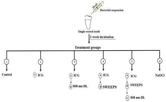
The treatment steps were as follows (Figure 1):

Figure 1.
Schematic representation of experimental setup. ICG: Indocyanine green, nm: nanometer, DL: diode laser, SWEEPS: shockwave-enhanced emission photoacoustic streaming, NaOCl: 5.25% sodium hypochlorite.
Group 1. Control group: the teeth did not undergo any intervention.
Group 2. ICG: the root canals were filled with 10 µL of ICG (Green + I, NovaTeb Pars, Tehran, Iran) at a concentration of 1000 µg/mL, and placed at room temperature in the dark for 5 min.
Group 3. ICG + 808 nm diode laser: the root canals were filled with 10 µL of ICG (1000 µg/mL) and placed at room temperature in the dark for 5 min. They were then subjected to 808 nm diode laser (DX82, Konftec, New Taipei City, Taiwan) with output power of 250 mW and total energy of 15 J, for 60 s. The 3D diffuser tip was used in an up-and-down motion from the apex to the coronal part.
Group 4. ICG + SWEEPS: the root canals were filled with 10 µL of ICG (1000 µg/mL) and placed at room temperature in the dark for 5 min. They were then subjected to Er:YAG laser irradiation with 2940 nm wavelength (LightWalker AT, Fotona, LjuBlijana, Slovenia) with an H14 handpiece and SWEEPS tip with the Fotona protocol for SWEEPS (25 µs, SWEEPS mode, 15 Hz, 20 mJ, 0.3 W). The tip of SWEEPS was placed in the pulp chamber and activated for 90 s.
Group 5. ICG + 808 nm diode laser + SWEEPS: the root canals were treated by ICG-mediated SWEEPS similar to group 4, and then, after 5 min, 808-nm diode laser irradiation was performed, similar to group 3.
Group 6. NaOCl: the root canals were filled with 5.25% NaOCl for 1 min.
2.5. Microbiological Process
After treatment, the teeth were placed in a microtube containing 1 mL BHI broth, and vortexed for 1 min. Next, 10 µL of the suspension was serially diluted 5 times, and 10 µL of each dilution was cultured on BHI agar (Ibresco), and incubated at 37 °C for 24 h. The colonies were then counted [11].
2.6. Statistical Analysis
Statistical analysis was carried out using one-way ANOVA (SPSS, version 23.0, Chicago, IL, USA). For multiple comparisons, Tukey’s test was used as the post hoc test. p value < 0.05 was considered statistically significant.
3. Results
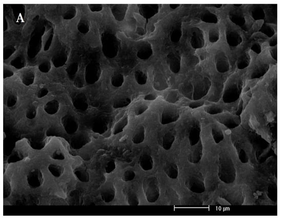
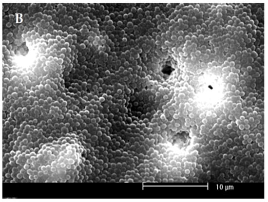
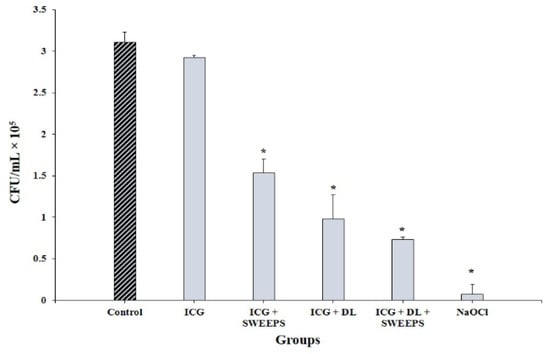
The SEM of the root canal without bacterial biofilm and E. faecalis biofilm on the root canal walls and in the dentinal tubules 2 weeks after inoculation are shown in Figure 2A,B, respectively. The results in Figure 3 and Table 1 demonstrate that, except for ICG alone, all experimental groups could decrease the viability of E. faecalis, compared with the control (p < 0.001). The results revealed that NaOCl decreased the microbial count to almost zero (p < 0.001). A significant difference was found between the ICG and ICG + SWEEPS or 808 nm diode laser, or both (p < 0.001) regarding the reduction in the E. faecalis count. Accordingly, ICG + 808 nm diode laser + SWEEPS had a 2.1- and 1.3-fold anti-biofilm effect compared to ICG + SWEEPS and ICG + 808 nm diode laser, respectively. In addition, there was a significant difference between the ICG + SWEEPS and ICG + 808 nm diode laser + SWEEPS (p = 0.002), whereas there was no significant difference found between the ICG + 808 nm diode laser and ICG + 808 nm diode laser + SWEEPS (p = 0.64). Furthermore, ICG + SWEEPS and ICG + 808 nm diode laser groups showed significant differences (p = 0.035).


Figure 2.
(A) Scanning electron microscope of the root canal without bacterial biofilm. (B) Scanning electron microscope of Enterococcus faecalis biofilm on the root canal walls at a magnification of 3000×.

Figure 3.
Effect of different treatment groups on cell viability of Enterococcus faecalis biofilm. * Significantly different from the control group, p < 0.001. ICG: indocyanine green, DL: 808 nm diode laser, SWEEPS: shockwave-enhanced emission photoacoustic streaming, NaOCl: 5.25% sodium hypochlorite.

Table 1.
Multiple comparison post hoc test results.
4. Discussion
This study evaluated the efficacy of SWEEPS plus aPDT with ICG for the reduction of E. faecalis biofilm from the root canal space. Since studies on the efficacy of the SWEEPS technique in actual root canals are highly limited, this study assessed the efficacy of the above-mentioned techniques in actual root canals with the usual anatomical complexities.
The present results revealed that the application of NaOCl alone had a significantly superior efficacy than the other groups in relation to the reduction of the E. faecalis count in the root canal system. For higher disinfecting efficacy, antimicrobial agents need to be in direct contact with the bacteria; however, the lodging of bacteria in anatomical complexities of the canal such as anastomoses, fins, and ramifications make it impossible for the irrigants to directly contact the microorganisms [20].
In the present study, aPDT with 808 nm diode laser and ICG caused a significant reduction in the E faecalis count, which is in consistent with the results of most studies conducted on the efficacy of aPDT with ICG on E. faecalis elimination [21,22]. A study has also shown that there is no evidence of cytotoxicity of ICG to MG-63 human osteoblast-like cells [23]. Furthermore, the results of this study show that ICG + SWEEPS has significant efficacy in removing E. faecalis biofilms compared to the control (p < 0.001). Wang et al. [24] confirmed the efficiency of the auto-SWEEPS in eliminating E. faecalis biofilm in root canals compared to 3% NaOCl alone and PIPS, using SEM images. They explained that strong shockwaves are eventually generated throughout the root canal, which significantly improves clearance efficacy.
In the SWEEPS technique, a photothermal effect does not occur, due to subablative laser irradiation. This technique generates powerful waves in the irrigating liquid, and produces a high fluid flow rate [25]. The maximum speed of the irrigating solution in accessory canals in the application of the SWEEPS technique is 10 m/s, which is much higher than the reported speed for other methods of root canal irrigation [26]. In addition, the penetration depth of the irrigant into the accessory canals in the SWEEPS technique is more than 1 mm [26]. According to the literature, the optimal efficacy of the SWEEPS technique is attributed to the emission of two pulses with an optimal time interval. Accordingly, the effect of the primary bubbles is reinforced by the generation of secondary bubbles, without increasing the risk of extrusion of the irrigant. Thus, this technique increases the efficacy of irrigation and debris removal from the root canal system [27]. Su et al. [26] described the breath mode for the streaming of irrigants, in which the irrigant repeatedly enters and exits the main and accessory canals. According to their study, another advantage of the SWEEPS technique is that the in-and-out movement of the liquid, which resembles inhalation and exhalation, generates alternating shear stresses in the root canal, which plays a pivotal role in improving the quality of the debridement process. No risk of intracanal instrument fracture is another advantage of the SWEEPS technique, since in this technique the tip of the handpiece is positioned in the access cavity and above the root canals, whereas in sonic and ultrasonic techniques, the instrument is introduced into the canal and proceeds to the apex for the activation of the intracanal irrgant [28]. In addition, a previous study has shown that photosensitizer extrusion is not harmful [29].
According to the present results, combination therapy with the application of SWEEPS plus aPDT yielded superior results. The SWEEPS technique mechanically detaches the biofilm from the root canal, therefore increasing the penetration depth and efficacy of the photosensitizer, due to the frequent generation of cavitation [1]. Consistent with the present results, previous studies also showed that application of SWEEPS with a 660 and 980 nm diode laser or light-emitting diode (LED) can cause a more significant decrease in the root canal infected with E. faecalis [25,30]. However, in contrast with the results of this study, the diode laser and SWEEPS did not show significant differences with methylene blue in their use as a photosensitizer [30]. Since the type of photosensitizer, light source, and irradiation time affect the bactericidal properties, this difference might be related to the fact that the mechanism of action of ICG is different from that of other photosensitizers. The main effect of ICG is due to the photothermal effect, which causes cell damage by increasing the intracellular temperature [12]. In photothermal therapy, the energy of laser radiation is absorbed by ICG, effectively raising the local temperature [31]. In addition to photothermic effects, ICG was demonstrated to have a photodynamic effect via the production of reactive oxygen species [32]. On the other hand, the 810 nm diode laser compared to other wavelengths used for toluidine blue O and methylene blue allows more penetration depth [12]. The application of only one type of photosensitizer was among the limitations of this study. The in vitro design was another limitation of this study that limits the clinical generalizability of the results. Therefore, future studies are required on other types of photosensitizers and irrigants, and also in the clinical setting.
5. Conclusions
The application of ICG along with the SWEEPS plus aPDT significantly improve its efficacy compared with its application alone. The results of this study will probably make an important contribution in the future to improving the efficiency of root canal treatments.
Author Contributions
Conceptualization, S.A., S.B., A.S. and N.C.; methodology, S.A. and N.C.; software, S.A.; writing—original draft preparation, G.R. and S.A.; writing—review and editing, S.A. All authors have read and agreed to the published version of the manuscript.
Funding
This research received no external funding.
Institutional Review Board Statement
Not applicable.
Informed Consent Statement
Not applicable.
Data Availability Statement
Not applicable.
Conflicts of Interest
The authors declare no conflict of interest.
References
- Ivanusic, T.; Lukac, M.; Lukac, N.; Jezersek, M. SSP/SWEEPS endodontics with the SkyPulse Er: YAG dental laser. J. LAHA 2019, 2019, 1–10. [Google Scholar]
- Miranda, T.C.; Andrade, J.F.M.; Gelfuso, G.M.; Cunha-Filho, M.; Oliveira, L.A.; Gratieri, T. Novel technologies to improve the treatment of endodontic microbial infections: Inputs from a drug delivery perspective. Int. J. Pharm. 2023, 635, 122794. [Google Scholar] [CrossRef]
- Stuart, C.H.; Schwartz, S.A.; Beeson, T.J.; Owatz, C.B. Enterococcus faecalis: Its role in root canal treatment failure and current concepts in retreatment. J. Endod. 2006, 32, 93–98. [Google Scholar] [CrossRef]
- Nagahashi, T.; Yahata, Y.; Handa, K.; Nakano, M.; Suzuki, S.; Kakiuchi, Y.; Tanaka, T.; Kanehira, M.; Suresh Venkataiah, V.; Saito, M. Er: YAG laser-induced cavitation can activate irrigation for the removal of intraradicular biofilm. Sci. Rep. 2022, 12, 4897. [Google Scholar] [CrossRef]
- Peters, O.A.; Schönenberger, K.; Laib, A. Effects of four Ni-Ti preparation techniques on root canal geometry assessed by micro computed tomography. Int. Endod. J. 2001, 34, 221–230. [Google Scholar] [CrossRef]
- Yang, Q.; Liu, M.; Zhu, L.; Peng, B. Micro-CT study on the removal of accumulated hard-tissue debris from the root canal system of mandibular molars when using a novel laser-activated irrigation approach. Int. Endod. J. 2020, 53, 529–538. [Google Scholar] [CrossRef] [PubMed]
- Oliveira, L.; Carvalho, C.A.T.; Nunes, W.; Valera, M.C.; Camargo, C.H.R.; Jorge, A.O.C. Effects of chlorhexidine and sodium hypochlorite on the microhardness of root canal dentin. Oral. Surg. Oral. Med. Oral. Pathol. Oral. Radiol. Endodontol. 2007, 104, 125–128. [Google Scholar] [CrossRef]
- Paiva, S.S.M.; Siqueira Jr, J.F.; Rôças, I.N.; Carmo, F.L.; Leite, D.C.A.; Ferreira, D.C.; Rachid, C.T.C.; Rosado, A.S. Clinical antimicrobial efficacy of NiTi rotary instrumentation with NaOCl irrigation, final rinse with chlorhexidine and interappointment medication: A molecular study. Int. Endod. J. 2012, 46, 225–233. [Google Scholar] [CrossRef]
- Dioguardi, M.; Di Gioia, G.; Illuzzi, G.; Laneve, E.; Cocco, A.; Troiano, G. Endodontic irrigants: Different methods to improve efficacy and related problems. Eur. J. Dent. 2018, 12, 459. [Google Scholar] [CrossRef]
- Cieplik, F.; Deng, D.; Crielaard, W.; Buchalla, W.; Hellwig, E.; Al-Ahmad, A.; Maisch, T. Antimicrobial photodynamic therapy—What we know and what we don’t. Crit. Rev. Microbiol. 2018, 44, 571–589. [Google Scholar] [CrossRef]
- Beltes, C.; Economides, N.; Sakkas, H.; Papadopoulou, C.; Lambrianidis, T. Evaluation of antimicrobial photodynamic therapy using indocyanine green and near-infrared diode laser against Enterococcus faecalis in infected human root canals. Photomed. Laser Surg. 2017, 35, 264–269. [Google Scholar] [CrossRef]
- Monzavi, A.; Chinipardaz, Z.; Mousavi, M.; Fekrazad, R.; Moslemi, N.; Azaripour, A.; Bagherpasand, O.; Chiniforush, N. Antimicrobial photodynamic therapy using diode laser activated indocyanine green as an adjunct in the treatment of chronic periodontitis: A randomized clinical trial. Photodiagn. Photodyn. Ther. 2016, 14, 93–97. [Google Scholar] [CrossRef]
- Kuştarcı, A.; Er, K. Efficacy of laser activated irrigation on apically extruded debris with different preparation systems. Photomed. Laser Surg. 2015, 33, 384–389. [Google Scholar] [CrossRef]
- Lukač, M.; Olivi, G.; Constantin, M.; Lukač, N.; Jezeršek, M. Determination of Optimal Separation Times for Dual-Pulse SWEEPS Laser-Assisted Irrigation in Different Endodontic Access Cavities. Lasers Surg. Med. 2021, 53, 998–1004. [Google Scholar] [CrossRef]
- Lei, L.; Wang, F.; Wang, Y.; Li, Y.; Huang, X. Laser Activated Irrigation with SWEEPS Modality Reduces Concentration of Sodium Hypochlorite in Root Canal Irrigation. Photodiagnosis. Photodyn. Ther. 2022, 39, 102873. [Google Scholar] [CrossRef] [PubMed]
- Panthangi, S.; Vishwaja, U.; Reddy, C.L.C.; Babu, M.B.; Podili, S. Novel sweeps technology in endodontics—A review. IP Indian J. Conserv. Endod. 2021, 6, 134–142. [Google Scholar]
- Wang, M.; Ma, L.; Li, Q.; Yang, W. Efficacy of Er: YAG laser-assisted direct pulp capping in permanent teeth with cariously exposed pulp: A pilot study. Aust. Endod. J. 2020, 46, 351–357. [Google Scholar] [CrossRef]
- Lukač, M.; Lukač, N.; Jezeršek, M. Characteristics of bubble oscillations during laser-activated irrigation of root canals and method of improvement. Lasers Surg. Med. 2020, 52, 907–915. [Google Scholar] [CrossRef]
- Lukac, N.; Muc, B.T.; Jezersek, M.; Lukac, M. Photoacoustic endodontics using the novel SWEEPS Er: YAG laser modality. J. Laser Health Acad. 2017, 1, 1–7. [Google Scholar]
- Balić, M.; Lucić, R.; Mehadžić, K.; Bago, I.; Anić, I.; Jakovljević, S.; Plečko, V. The efficacy of photon-initiated photoacoustic streaming and sonic-activated irrigation combined with QMiX solution or sodium hypochlorite against intracanal E. faecalis biofilm. Lasers Med. Sci. 2016, 31, 335–342. [Google Scholar] [CrossRef]
- Beltes, C.; Sakkas, H.; Economides, N.; Papadopoulou, C. Antimicrobial photodynamic therapy using Indocyanine green and near-infrared diode laser in reducing Entrerococcus faecalis. Photodiagn. Photodyn. Ther. 2017, 17, 5–8. [Google Scholar] [CrossRef]
- Chiniforush, N.; Pourhajibagher, M.; Parker, S.; Shahabi, S.; Bahador, A. The in vitro effect of antimicrobial photodynamic therapy with indocyanine green on Enterococcus faecalis: Influence of a washing vs. non-washing procedure. Photodiagn. Photodyn. Ther. 2016, 16, 119–123. [Google Scholar] [CrossRef]
- Aghayan, S.; Yazdanfar, A.; Seyedjafari, E.; Noroozian, M.; Ioana Bordea, R.; Chiniforush, N. Evaluation of indocyanine-mediated photodynamic therapy cytotoxicity in human osteoblast-like cells: An in vitro study. Folia Med. 2022, 64, 932–937. [Google Scholar] [CrossRef] [PubMed]
- Wang, X.-N.; Shi, J. Shock wave-enhanced emission photoacoustic streaming versus photon-induced photoacoustic streaming modes for clearing root canal bacteria using erbium-doped yttrium aluminum garnet lasers: An in vitro study. Res. Sq. 2020. [Google Scholar] [CrossRef]
- Ensafi, F.; Fazlyab, M.; Chiniforush, N.; Akhavan, H. Comparative effects of SWEEPS technique and antimicrobial photodynamic therapy by using curcumin and nano-curcumin on Enterococcus faecalis biofilm in root canal treatment. Photodiagn. Photodyn. Ther. 2022, 40, 103130. [Google Scholar] [CrossRef]
- Su, Z.; Li, Z.; Shen, Y.; Bai, Y.; Zheng, Y.; Pan, C.; Hou, B. Characteristics of the Irrigant Flow in a Simulated Lateral Canal Under Two Typical Laser-Activated Irrigation Regimens. Lasers Surg. Med. 2021, 53, 587–594. [Google Scholar] [CrossRef]
- Jezeršek, M.; Lukač, N.; Lukač, M.; Tenyi, A.; Olivi, G.; Fidler, A. Measurement of pressures generated in root canal during Er: YAG laser-activated irrigation. Photobiomodul. Photomed. Laser Surg. 2020, 38, 625–631. [Google Scholar] [CrossRef]
- Widbiller, M.; Keim, L.; Schlichting, R.; Striegl, B.; Hiller, K.-A.; Jungbauer, R.; Buchalla, W.; Galler, K.M. Debris removal by activation of endodontic irrigants in complex root canal systems: A standardized in-vitro-study. Appl. Sci. 2021, 11, 7331. [Google Scholar] [CrossRef]
- Bolhari, B.; Meraji, N.; Seddighi, R.; Ebrahimi, N.; Chiniforush, N. Effect of SWEEPS and PIPS techniques on dye extrusion in photodynamic therapy procedure after root canal preparation. Photodiagn. Photodyn. Ther. 2023, 42, 103345. [Google Scholar] [CrossRef]
- Afrasiabi, S.; Parker, S.; Chiniforush, N. Synergistic antimicrobial effect of photodynamic inactivation and SWEEPS in combined treatment against Enterococcus faecalis in a root canal biofilm model: An in vitro study. Appl. Sci. 2023, 13, 5668. [Google Scholar] [CrossRef]
- Sukumar, K.; Tadepalli, A.; Parthasarathy, H.; Ponnaiyan, D. Evaluation of combined efficacy of photodynamic therapy using indocyanine green photosensitizer and non-surgical periodontal therapy on clinical and microbial parameters in the management of chronic periodontitis subjects: A randomized split-mouth design. Photodiagn. Photodyn. Ther. 2020, 31, 101949. [Google Scholar] [CrossRef] [PubMed]
- You, Q.; Sun, Q.; Wang, J.; Tan, X.; Pang, X.; Liu, L.; Yu, M.; Tan, F.; Li, N. A single-light triggered and dual-imaging guided multifunctional platform for combined photothermal and photodynamic therapy based on TD-controlled and ICG-loaded CuS@mSiO2. Nanoscale. 2017, 9, 3784–3796. [Google Scholar] [CrossRef] [PubMed]
Disclaimer/Publisher’s Note: The statements, opinions and data contained in all publications are solely those of the individual author(s) and contributor(s) and not of MDPI and/or the editor(s). MDPI and/or the editor(s) disclaim responsibility for any injury to people or property resulting from any ideas, methods, instructions or products referred to in the content. |
© 2023 by the authors. Licensee MDPI, Basel, Switzerland. This article is an open access article distributed under the terms and conditions of the Creative Commons Attribution (CC BY) license (https://creativecommons.org/licenses/by/4.0/).