Review

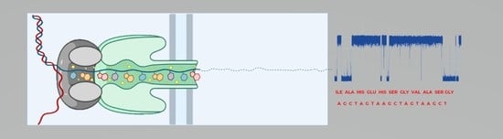
Detection of Biological Molecules Using Nanopore Sensing Techniques

1
Integrated Center for Research, Development and Innovation for Advanced Materials, Nanotechnologies, Manufacturing and Control Distributed Systems (MANSiD), Stefan cel Mare University of Suceava, 720229 Suceava, Romania
2
Department of Biomedical Sciences, Stefan cel Mare University of Suceava, 720229 Suceava, Romania
3
Department of Computer, Electronics and Automation, Stefan cel Mare University of Suceava, 720229 Suceava, Romania
*
Author to whom correspondence should be addressed.
Biomedicines 2023, 11(6), 1625; https://doi.org/10.3390/biomedicines11061625
Submission received: 16 May 2023 / Revised: 28 May 2023 / Accepted: 29 May 2023 / Published: 2 June 2023
(This article belongs to the Special Issue Advances in Nanomedicine for Disease Treatment and Diagnosis)

Abstract

:
Modern biomedical sensing techniques have significantly increased in precision and accuracy due to new technologies that enable speed and that can be tailored to be highly specific for markers of a particular disease. Diagnosing early-stage conditions is paramount to treating serious diseases. Usually, in the early stages of the disease, the number of specific biomarkers is very low and sometimes difficult to detect using classical diagnostic methods. Among detection methods, biosensors are currently attracting significant interest in medicine, for advantages such as easy operation, speed, and portability, with additional benefits of low costs and repeated reliable results. Single-molecule sensors such as nanopores that can detect biomolecules at low concentrations have the potential to become clinically relevant. As such, several applications have been introduced in this field for the detection of blood markers, nucleic acids, or proteins. The use of nanopores has yet to reach maturity for standardization as diagnostic techniques, however, they promise enormous potential, as progress is made into stabilizing nanopore structures, enhancing chemistries, and improving data collection and bioinformatic analysis. This review offers a new perspective on current biomolecule sensing techniques, based on various types of nanopores, challenges, and approaches toward implementation in clinical settings.

Graphical Abstract

1. Introduction

Each year, new health conditions and their underlying mechanisms are identified as a result of continuous changes in lifestyle and due to advancements in understanding and exploring the human body. Since causes and manifestations of diseases intrinsically have molecular foundations, the detection of specific molecules involved is paramount for human health. Examples of diseases that would benefit from such detection are Alzheimer’s, Parkinson’s, or cancer, which, if not diagnosed early, may significantly reduce life expectancy and quality. An interesting and important approach in this direction is the detection of biomarkers at the single molecule level by means of nanopores [1,2].
Nanopores are structures that naturally occur as proteic polymers or can be manufactured from synthetic materials such as nanoscale silicon or graphene. When a nanopore is embedded in a dielectric membrane, it can be used to detect biomolecules, particularly DNA, RNA, and proteins [3], due to changes in the local microenvironment. Over time, various organic and synthetic nanopores, from the discovery of α-HL (α-hemolysin) organization in lipidic solutions, to solid nanopores based on silicon, graphene, etc., have been used to generate consistent information in molecular biology (Figure 1). Solid nanopores are widely used because of their flexible geometry and shapes and can be manufactured as a function of the analyte to be detected [4]. Thus, the solid-state nanopore does not undergo selective translocations and, as a result, it only performs several detections with a low degree of selectivity. Recent studies have investigated this issue and functionalized stable surfaces with recognition molecules that allow them to identify a specific molecular entity [5]. A current is established during the application of an electrical potential across a nanopore. Passing a molecule into the nanopores causes a complete or partial blockage. This blockage is characterized by the change in current and dwell time, which corresponds to the size and respective electric charge of the molecule [6,7].
Physically, the current flowing into a nanopore is a measurement of the net transport of charged loads per unit of time (Figure 2). As a result, the species entering the nanopore may influence the amplitude of the current, which is closely related to the properties of the analyte, nanopore, and solution [11]. The hydrodynamic effects (electro-osmosis), the molecular interactions, the surface of the local charge distribution (polarization, concentration of condensation ions), as well as the properties of the translocating analyte are important to determine the type of molecules that cross the nanopore [12]. Changing the properties of solid nanopores may change translocation time, which is the time it takes for the molecule to pass through the nanopore from one side to the other, independent of other experimental parameters. For example, any slight change in the superficial load may result in a significant change in the dwell time, which is the amount of time a molecule spends inside the nanopore of the analyte [13]. Thus, the passage of a molecule through a nanopore is influenced by the potential applied from the outside, the electrophysiological solution (electrolyte), and the physicochemical properties of the molecule of interest (analyte) [14]. This review aims to highlight the versatility of the nanopore analysis method and, at the same time, provide insight into the challenges the method raises, based on the molecule to be studied. The paper includes several specific experimental aspects for various molecules with important relevance for human health

2. Applications in Sensing and Diagnostic

As a result of their biosensing capabilities, nanopores are the basis for the development of new technologies for the rapid detection of various specific molecules involved in several pathologies. The importance of using nanopores has been highlighted in an increasing number of studies. For example, genetic analyses have been reported to be carried out using nanopores in approximately 7 h, with patients receiving a clear diagnosis in a short time [15,16], while they are usually performed in a few days or even weeks using classical methods.
Moreover, the sequence, structure, and components of proteins in living organisms contain information of great importance to normal or pathological physiological processes, however, numerous challenges and deficits in this field still remain [17,18]. For example, passing molecules through the nanopore too quickly presents a problem in identifying signal fluctuations that should be specific to the translocated molecule. This is why researchers explored means to slow down molecular transport. To date, there are numerous strategies that improve nanopore analysis methods by adjusting various experimental parameters to achieve high resolution [19].
However, information about the identity of an analyte is, most times, contained in the average duration of current blockages and the amplitude of blockages [20]. Each protein has a sequence of amino acids, a three-dimensional structure, and different charge profiles. This information is reflected in the detected current signal when passing through a nanopore. According to this principle, different properties of proteins or other molecules of interest in the nanopore domain have been investigated [12,21]. Nanopores are also used according to the properties of the molecule of interest. For instance, solid-state nanopores are used for large molecules. Several nanopore techniques for certain molecules are listed in Table 1.

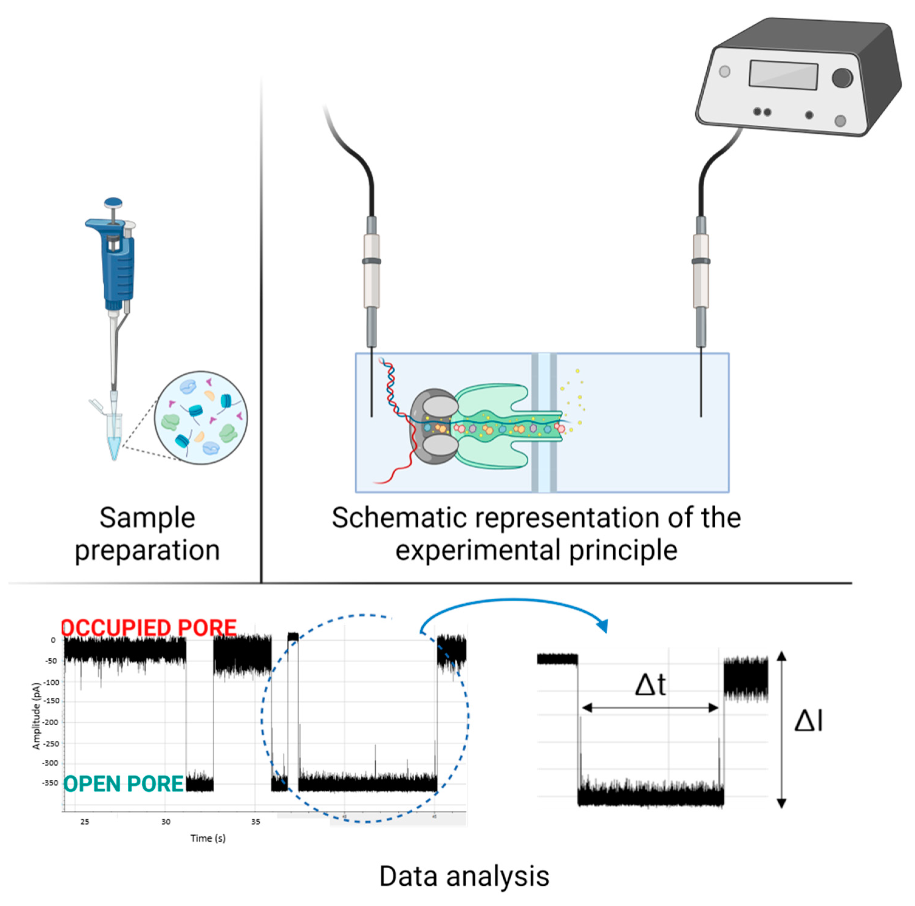
3. Methodological Approaches and Challenges

The use of nanopores as a method of analysis for the diagnosis of various diseases may be an innovative method that promises to go far beyond the analyses currently performed in hospitals. The nanopore assay method promises to revolutionize both classical molecule analysis of different biomarkers and to offer the detection of molecules at low concentrations and with high resolution. The technology has become so compact that its use at the point of care represents an enormous potential [44]. One of the most important advantages of the nanopore analysis technique is real-time analysis, which offers the possibility of dynamic monitoring of biological processes [45]. However, there are a number of challenges that make this method not yet widely available. Apart from the physical and chemical optimizations required for each type of nanopore, including the structure and materials from which nanopores are made, whether synthetic or biological, there are numerous challenges in terms of molecule capture, translocation rate control, and blockages due to the analysis of molecules that are too large or that interact with the pore surface. For better resolution in nanopore experiments, it is necessary to pay attention both to the sample and to experimental parameters such as applied voltage, buffer pH, external noise, and purity of solutions. For example, the pH of the electrophysiological solution can have effects on nanopores such as in the case of OmpG nanopores which, at an acidic pH, can close [46] or influence transport processes through the nanopore [47]. The method is being fine-tuned both in terms of working time and in terms of results. With MinION™ for example, results can be obtained in about 6 h compared to 1–2 days using culture media [48]. Another factor influencing the results is the bioinformatics analysis of the signal. Bioinformatics refers to the analysis and interpretation of the resulting electrical signal to arrive at biologically or medically conclusive results, as in the case of DNA sequencing when the base-calling method is used to determine the DNA structure [49]. There are numerous algorithms that attempt to analyze the signal and use advanced methods such as machine learning, Markov models, or neural networks [50,51]. All these factors contribute to the development of nanopore analysis methods, significantly improving time-to-value ratios. At the same time, the methods need to be optimized specifically for the application. In the following sections, we discuss specific challenges and existing approaches for specific molecules of interest, to provide an overview of the field of nanopore sensing development.

3.1. Metabolic Markers

3.1.1. Glucose Detection

Apart from diabetes, glucose is important in other physiological conditions, for example, it is the main energy source in cancer cells and, in general, glucose plays a fundamental role in providing energy for cellular metabolism. Malignant cells need more glucose than normal cells to survive, thus the amount of glucose contained in cancer cells is different from that in normal cells. It follows that accurate detection of glucose levels in individual cells is important for the diagnosis and treatment of cancers. Therefore, it would be of enormous clinical significance to distinguish the difference in glucose content in normal cells and neoplastic cells in biopsies of heterogeneous tissues, as well as to detect changes in glucose content in starved cancer cells [52]. Although methods for glucose detection in people with diabetes are widespread, as this is important for the prevention and treatment of this disease, determining glucose at the level of the individual cell is a challenge. One of the limitations in the fabrication of glucose biosensors is the charge transfer between the enzyme and the electrodes. For this problem, conductive polymers are used to facilitate the interaction between enzymes and electrodes by making charge transport more efficient [53]. Membrane glucose sensors have also been developed that have the main advantage of preventing direct electro-oxidation of chemicals in the physiological environment. The first detection of glucose without enzymes using a microchip was performed in 2006. This microchip system consisted of a network of microfluidic transport channels and a miniaturized electrochemical cell for the non-enzymatic detection of glucose. The sample and buffer were transferred to the cell by electro-osmotic flow. A Pt electrode was used to determine the glucose concentrations in the saline solution by direct oxidation of glucose without any separation process [54].
Ref. [52] Single-molecule intracellular investigations are difficult to perform due to a lack of non-destructive analytical sensors and controlled cell-targeting technology. Due to the small pore diameter, a technological solution such as a nanopipette can be introduced into individual cells repeatedly, as the tip is less than 100 nm [55]. Bio-recognition materials such as antibodies that selectively bind to specific cells can be inserted at the tip of the nanopipette [56,57]. Single-molecule glucose detection is a diagnostic tool in research for distinguishing between malignant and benign cells in biopsies and a tool for monitoring cancer progression in situ. The research seeks to automate the system to increase single-molecule analysis efficiency to 120 cells/hour [58].

3.1.2. Vitamin B1

B-complex vitamins are essential for both the physical and mental state, having important functions in the cognitive, digestive, muscular, and cardiac systems, and playing an important role in energy metabolism [59]. Vitamin B deficiency can lead to mild symptoms such as fatigue but can also result in permanent neuronal degeneration and significant disorders in the human body [60]. Vitamin B1 (Thiamine) molecules are distinct from the other molecules in the B complex in that they contain a thiazole and a pyrimidine ring [59,61]. The determination of vitamin B1 status is usually performed in medical laboratories by various techniques, such as liquid chromatography [62] and UV-VIS spectrometry [63], within a few hours. The nanopore technique also tends to make the methods more efficient in terms of time. To this end, a biological nanopore of ClyA (Cytolysin A) has been used to determine the concentration of thiamine in urine using automatic data analysis that takes less than 1 min [26]. The particularity of this technique consists in attaching to the nanopore a protein that has the property of binding to thiamine when translocated through the nanopore. By replacing a residue (Y27A), the affinity between TbpA (periplasmic thiamine-binding protein) and thiamine is increased [64,65], which improves the signal resulting from the translocation of the molecule through the nanopore, allowing the identification of thiamine.

3.1.3. Uric Acid Detection

Uric acid (UA) is the main product of the breakdown of the metabolism of purine nucleotides (e.g., adenine, guanine). High serum uric acid levels are associated with many diseases, such as gout, arthritis, kidney disease, and others. Current clinical tests such as the colorimetric method or enzymatic method used to measure uric acid in urine and serum involve the use of uricase enzymes and other specific reagents [66,67]. Enzymatic methods suffer from interferences caused by chemical reactions taking place, and the analysis requires careful pH control. Therefore, it is worth developing reliable and inexpensive non-enzymatic methods for determining uric acid levels. The glass capillary nano-pipette method is a non-enzymatic strategy for the determination of uric acid. This involves a glass capillary nanopore over which an ultrathin gold substrate is inserted by chemical reduction of HAuCl4 on a poly (L-histidine) nanoshell attached to the substrate in the presence of NH2OH-HCl [28]. This nanopore is functionalized with 2-thiouracil by self-assembly and tested for non-enzymatic detection of uric acid. This method of uric acid detection is selective and reproducible. In addition, such a glass nanopore could be used for the practical analysis of serum samples. A detection limit of 1.9 μM was observed in the proposed experimental setting and the concentration in serum samples detected in hospital laboratories was 360 μM [27].

3.2. Detection of Nucleic Acids

3.2.1. Bacterial Pathogen Identification

The detection of pathogenic bacteria is important to ensure adequate responses in clinical settings, such as ICUs (Intensive care units), for timely detection of nosocomial infections and catheter infections but also in quality assurance in the food industry. To avoid pathologies caused by bacteria, the process of detection and identification is essential [68]. Hospital infections are the cause of many deaths, for example, bacteria that cause sepsis in plasma can now be detected by microbiological culture which takes 1–2 days. For every hour that the administration of the specific antibiotic is delayed, the risk of the patient dying increases. More specifically, if appropriate therapy is not started within 1 h of shock, the risk of death increases from 1.67 in 2 h to 92.54 for delays > 36 h [69,70]. During this time, patients receive broad-spectrum empiric treatment to cover as many action spectra as possible, which can lead to multiple side effects and can also lead to antibiotic-resistant bacteria. Rapid detection of pathogens is required for early administration of antibiotics, thus avoiding the aforementioned problems resulting from diagnostic delays. Recent research that focused on Oxford Nanopore Technology sequencing (ONT) showed that detection of bacteria is possible within 5–6 h of sample collection, thus enabling much faster diagnoses [48], possibly even in clinical settings. The Nanopore technique was compared with classical techniques (Illumina) to identify the influenza A virus directly from respiratory samples. The data showed that the read ratio generated by Nanopore and Illumina metagenomic sequencing is very similar, therefore the detection limit is probably very close for these two platforms [71]. For example, when comparing 15 samples of IAV (Influenza A viruses) with 16 HMPV (Human Metapneumovirus) samples at a sequencing depth of 10×, the agreement between the two methods (Nanopore and Illumina) was 99.95%. In addition, the high error rate of nanopore reads is compensated by the increased read length, which could provide an advantage in the taxonomic assignment of individual reads [72].
With the spread of antibiotic-resistant pathogens and the associated increase in human morbidity and mortality, antibiotic-resistant genes have been identified as emerging environmental pollutants by the World Health Organization [73]. Oxford Nanopore Technology provides rapid real-time sequencing and has been used for the rapid detection of pathogens, including the diagnosis of bacterial meningitis, bacterial respiratory tract diseases, infective endocarditis, pneumonia, and other bacterial infections. The method is also very useful for detecting antibiotic-resistant bacteria or other mutations resulting from antibacterial treatments. As bacteria develop new antibiotic resistance mechanisms, this causes significant problems in the treatment of infections. Because the use of broad-spectrum antibiotics may generate antibiotic resistance, accurate and rapid detection of both bacteria and antibiotic resistance is essential in treating infections [74]. For example, the MinION™ has been quite successful in identifying pathogens. In the case of bacterial meningitis, 16S amplicon sequencing took only 10 min, and in the case of some respiratory tract-influencing bacteria, MinION™ analysis was performed in 6 h, compared to 2 days using the culture method [75,76]. Nanopore Targeted Sequencing (NTS) is an accurate and rapid analytical method for the detection of infection-causing pathogens found mainly in body fluids. Another comparison between classical methods and nanopores: the culture, NTS, and Sanger sequencing methods demonstrate the accuracy and precision of nanopores. When samples from patients with and without infections were tested by classical and NTS methods, 290 out of 307 (94.5%) patients with infectious diseases were diagnosed with NTS, while routine culture determined 116 out of 307 (37.8%) infectious diagnoses [77]. Another study carried out in 2020 confirms the results presented above, as 58.7% of specimens were detected by the NTS method, compared to 22.1% detected by the culture method [78].
Nanopore-based sequencing technology offers the ability to produce long reads from individual molecules in real time, with the potential to introduce new sequencing methods with clinical applications. Since its commercial launch, ONT has suffered from a high error rate that had to be mitigated by experimental methods, computational corrections, or signal filtering. Owing to many updates in the chemistry tools and software, the raw read accuracy has steadily increased from 60% to 85% and beyond. At the moment, the data produced by MinION™ is accurate enough to sequence with an accuracy of > 99% [79], as stated in the manufacturer’s manual: “In addition to high single-pass accuracy, the nanopore platform is now able to deliver single molecule consensus accuracy of 99.995% with UMIs, and Q50 (99.999%) consensus sequencing data” [80]. The quality (Q) of the assembled genome depends not only on the chemical corrections and filters applied but also on the properties of the sequenced molecule, for example in the case of rare species (e.g., Proteus vulgaris Q27 to Q32), the degree of polishing is lower than in common species (e.g., K. pneumonia Q26 to Q38) [81].
Pathogen detection might be more prone to interference from antibiotics in conventional culture testing, while NTS is much less affected. Some bacteria with specific nutritional requirements are difficult to cultivate even on supplemented medium or are slow-growing. As such, several bacterial species in normal and pathogenic microflora are unculturable, which poses a challenge to their detection. For these species, sequencing has proved a viable method, which may allow precise diagnostics, whereas Nanopore sequencing, due to fast protocols and sequencing times, may further provide rapid tools, even in clinical settings. Thus, NTS plays an important role in the diagnosis of infectious diseases, especially after antibiotic treatment or infections caused by slow-growing or precocious microorganisms. A rapid and accurate method such as NTS would contribute to the early administration of targeted treatment and reduce the use of broad-spectrum antibiotics. Classical culture media have a response time of about 24–48 h while in the case of NTS, the time to obtain a result varies from 6–18 h, thus the method is much more reliable with an acceptable cost since a rapid determination of the pathogen would reduce treatment costs and duration of hospitalization [82].

3.2.2. Genetic Markers

A biomarker is a characteristic indicator of a specific biological condition, which can be associated with a disease. For example, the identification of biomarkers provides information about the process of change in cancer cells [83].
MicroRNAs have been investigated as potential biomarkers, and although they are relatively short sequences of about 18–22 nucleotides which should make sequencing more accessible, detection of microRNAs in the bloodstream remains difficult. The results also show that the nanopore HL sensor can detect microRNA structures from plasma samples taken from lung cancer patients without the need for label addition or sequence amplification. The method is based on the difference in concentration of the electrolyte solution between the cis and trans parts which favors the flow of ions under the influence of the concentration gradient. This is also observable at low concentrations. The technique facilitates translocation at concentrations down to 1 pM [33]. In the case of solid (glass) nanopores, faster translocation is based on the dielectrophoretic force that is applied when opening a nanopore, a technique that appears to be more efficient, with translocation occurring up to concentrations of 5 fM [84]. It has subsequently been shown that, in the early stages of cancer, the concentration of microRNA is in the femtomolar range. In fact, in early-stage lung cancer, the microRNA concentration is about 1 fM, which makes the newly developed methods more sensitive and faster. Molecule analysis at concentrations of 1 fM was achieved by combining oligonucleotide amplification and the asymmetric method [85]. In recent studies, microRNAs have also been detected at concentrations of 0.5 fM in plasma samples, demonstrating the versatility and accuracy of the nanopore. Improved mechanisms increase the detection sensitivity of nanopores which will contribute in this case to the characterization of liquid biopsies [86].
Nanopore sequencing allows for variant detection, as shown for six SNPs at different loci, with the potential for assessing perioperative outcomes [87] or even insertions and deletions several base pairs long [88]. A hallmark of genomic instability that has a major influence on cancers is so-called structural variants (SV), which include structural changes such as translocations, amplifications, or inversions. The ability of methods using nanopores to read through repetitive regions could make them an ideal tool for detecting structural variants associated with tumors [89]. In pancreatic cancer, the tumor genes CDKN2A/p16 and SMAD4/DPC4 are difficult to detect, given the transformational processes they go through such as inversions or translocations, which virtually inactivate the tumor genes. Using PCR amplicon mixtures, it has been shown that the nanopore sequencing technique can detect translocations, inversions, and dilutions up to 1:100, with only 500 reads per sample [89,90].
In the case of breast cancer, for example, genome-wide sequencing analysis of DNA methylation (which involves long reads) would provide important clinical information. An improved variant of nanopore sequencing for this context is based on enzymatic base conversion, where an unmethylated cytosine should be converted to thymine. This type of analysis is reliable for a minimum of 1 ng genomic DNA, obtaining a read length of 3.4–7.6 kb [91].
mDNA (DNA methylation) detection is an essential process in the analysis and control of gene evolution. Methylation detection techniques such as bisulfite sequencing [92] and MeDIP-seq (Methylated DNA immunoprecipitation sequencing) [93] are widely used. However, they can be time-consuming and expensive. Nanopore sequencing technology offers a real-time alternative. The advantage of the technique is that it can facilitate the detection of methylation in long repetitive regions and can also detect methylation in low-volume samples [94]. Unlike classical methods, methylation detection with nanopores can be bioinformatically challenging, requiring specialized expertise. Although the nanopore methylation analysis method is not widely used, it is a promising route to real-time, high-throughput detection. Since the nanopore technique can correlate the values of the electric current intensity with the properties of the molecule, the identification of methylated genes is performed on the same principle, observing differences between unmodified and modified bases [95]. It is not enough just to identify differences in current amplitude, for a satisfactory yield, it is necessary to directly call the modified bases, and for these different algorithms are used [50], such as the Markov model [96] and neural networks [97,98]. Currently, there are multiple software tools developed by researchers for this purpose, but most of them do not fully satisfy the requirements as the methylation levels are not linearly distributed [99].
An example of a practical application is the change produced by the active substances in cigarette smoke on DNA profiles, effects which have also been observed in passive smokers or even in newborns who were exposed during pregnancy [100]. The study was carried out on 9389 subjects, of which 2433 were current smokers. The results showed that 2623 of those monitored had changes in DNA methylation. A CRISPR-Cas9 targeted sequencing with Nanopores was used as a method to determine the mDNA sequences. The method showed satisfactory results, identifying 1779 CpG regions. These were associated with three genomic regions that are known to be related to smoking [101]. CRISPR-Cas is used for rapid and sensitive detection of DNA or RNA targets. A combination of CRISPR-Cas-based bioanalytical systems and nanopore sequencing can be performed for better accuracy, with CRISPR-Cas specifically enriching DNA sequences for better and rapid nanopore sequencing. In the meantime, CRISPR-based sensing tools can accurately recognize specific sequences or molecules and perform electrochemical tag modifications, thus changing currents through the sensing device [102,103]. Another direct approach to identifying 5-methylcytosine (5mC) highlights the selectivity of nanopores. The method is based on the interaction of the modified cytosine with anionic acids in biological nanopores without any further chemical modification of the DNA [104].

3.3. Protein Detection

Proteins are present in all living organisms and play an important role in the structure and function of cells, with proteins made of up to thousands of amino acids. Identifying the properties of proteins involved in different pathologies would lead to faster and more efficient diagnosis [105,106]. However, proteins are more difficult to characterize than DNA, firstly because of the three-dimensional structure of proteins, the dimensions of which are comparable to, or larger than, the diameter of nanopores. Secondly, proteins are non-uniform charge-carrying structures due to having a large number of amino acids which makes translocation through a nanopore even more difficult compared to DNA which has a uniform charge distribution along the length of the molecule [107]. To overcome these difficulties, other approaches such as dielectrophoretic capture and electro-osmotic flow have been studied [108]. It has been demonstrated that, in certain conditions, such as neurological disorders (Alzheimer’s, Parkinson’s, and Prion diseases), the proteins involved undergo structural changes. Early diagnosis of these diseases can improve quality of life. For example, an increasing number of studies validate the accuracy of nanopore sequencing for rapid diagnosis of prion disease and highlight the need for single-molecule sequencing methods for somatic mutation detection. The study established a new long-read sequencing strategy, succeeding in reading the full length of the PRNP (Prion gene) of 16.7 kb [109]. However, the technique presented can be improved, since it currently requires amplification of PRNP in two fragments. If the experimental procedure was simplified, PRNP detection could be done in the field. Researchers have considered using a targeted sequence, for example, CRISPR-Cas9 [110] or the “read to...” mode of Oxford Nanopore devices [111]. Among the diseases that irreversibly affect neurological functions, Alzheimer’s disease affects 6.2 million Americans, a number estimated to double in the next 30 years [112]. Symptoms of Alzheimer’s disease progress over time, which is why early detection of disease triggers is essential. It is known that the aggregation of Aᏸ peptides is the cause of this disease, therefore the determination of the factors that favor aggregation is desirable [113]. The use of nanopores to detect changes in Aᏸ 40 sequences is an important step since nanopores have the sensitivity to identify conformational changes [113]. The level of quantification attainable using molecular sensing is not achieved by classical spectroscopic, gel electrophoretic, or ELISA-based detection techniques [114]. Techniques using nanopores propose a simple way to simultaneously obtain information about the size, shape, and distribution of heterogeneous protein aggregates using a small volume of solution [115]. However, this is not without challenges due to the complex structure and specific electrical charge of the protein. One way to improve protein detection is to slow down their movement to increase the dwell time in the nanopore [116]. Since the charge of the protein influences the translocation process through the nanopore, one solution is to work at a pH close to the isoelectric point of the protein [12]. This makes the net charge of the protein molecules almost zero, consequently, electrophoretic mobility is decreased, which slows down the protein translocation [117]. Another way to slow down the passage is to modify the surface properties of the nanopore. For example, the SiNx nanopore can acquire a slightly negative charge at a given pH or under the influence of light intensity. This factor can alter the translocation process [116,118]. Another method is to bind a ligand to the protein. For example, DNA chains containing aptamer sequences have been linked to specific target proteins. Protein detection was determined by analyzing the amplitude of the ionic current when DNA chains translocated through the pore [119]. The use of the Monte Carlo method to introduce a probabilistic model using aptamer-functionalized nanopores is intended to identify proteins in serum at pM concentrations. In this regard, studies examined the identification of thrombin at a concentration of 50 pM in serum. The technique has been verified over 25 repetitions, and the results have shown lower detection limits, allowing the platform to advance toward clinical testing [120].
Another marker is αS (α-synuclein), which is a biomarker of synucleinopathies (Parkinson’s disease, Lewy body dementia, multiple system atrophy), and rapid detection from a small amount of the substance is ideal for early diagnosis. A nanopipette combining in a single system a reaction vessel for accelerated amplification and a nanopore sensor allows the detection of αS assemblies in a given sample in only 90 min by adding a small amount (35 μL at 100 nM) of recombinant αS for amplification [121]. A rapid and correct diagnosis is critical to increase the chance of life and maintain quality of life. In this respect, nanopores are a promising technology that can help. For example, cancer is one of the diseases with a more or less chaotic evolution that requires early and precise identification of biomarkers that are involved in the decline of the disease. Moreover, new tumor entities have been identified that cannot be characterized histologically, leading to the need to characterize cancers molecularly. For example, the World Health Organization explicitly requires molecular testing for central nervous system tumors. However, the implementation of integrated histomolecular testing requires a multitude of technologies, and the time to implement it can be several weeks, making standardized implementation difficult [122]. Things can also be improved in cancer cases that already have an established diagnosis and treatment protocol. For example, in the case of prostate cancer, patients with high levels of PSA (Prostate-Specific Serum Antigen) undergo painful and expensive procedures such as biopsy. The test can be improved by finding a protein that is selectively secreted by malignant cells. To this end, researchers have discovered a protein called NEM (neuroendocrine marker) that is secreted only by malignant prostate cells and released into the bloodstream [123]. Improved systems that make the nanopore technique reliable in clinical testing are increasingly being tested. For example, one system that can measure NEM and PSA consists of an optofluidic chip made up of nanopore-based sensors made of anodic aluminum oxide. The results show that the chip had improved sensitivity, requiring only 1 μL of serum to measure PSA and NEM. In addition, compared to ELISA tests for PSA, the nanopore-based technique is 50–100 times more sensitive [38].
Another important protein, hemoglobin, is an essential, iron-containing protein found in red blood cells. Its function is to carry oxygen from the lungs to every cell in the body and to take the carbon dioxide resulting from metabolic burning to the lungs for disposal [124]. Folded hemoglobin can also be detected in blood samples by techniques using nanopores, for example, PlyAB (pleurotolysin) biological nanopores show promise for the real-time analysis of folded proteins [40]. It has been possible to identify hemoglobin even in variants in which a single amino acid differs, representing an accuracy of 97%. The concentrations identified as the normal range is between 15–17 g/dL (2.3–2.6 mM), and the nanopore is able to identify proteins even in concentrations of 1 mg/dL (150 nM). Therefore, the use of nanopores for automated analysis of blood samples would be useful in the identification of hemoglobin variants and thus in the diagnosis of specific diseases caused by changes in hemoglobin levels [125]. Likewise, the presence of insulin antibodies is a predictor of type 1 diabetes for people who do not have diabetes but are genetically predisposed. Early identification of this factor helps to prevent and manage diabetic disease accurately [126]. To facilitate the passage of insulin through a solid nanopore, the system must be adjusted to slow translocation. Thus, the nanopore with a metallic film was fabricated by electron beam sputter deposition of Au on a SiNx layer. Finally, the Au surface was modified with Hcy (homocysteine), which led to a significant change in the current-voltage characteristics, practically after the modification the translocation time of insulin through the pore increases, which is explained by hydrophobic interactions between the protein (insulin) and the Hcy layer [127]. The use of homocysteine-modified metal nanopores has been shown to be effective, as protein uptake on chemically modified nanopores slows the translocation process to tens of milliseconds [128]. Another approach to the analysis and subsequent sequencing of proteins is tried with the ONT method, which is now used for DNA sequencing. There are a few articles using the ONT method for protein analysis, one of the most relevant uses an R9.4.1 cell and a specific modified script and protein tags (NTERS). A negatively charged protein (NTERY00) is used to develop this method. The negative loading of the C-terminal end facilitates the capture of the protein by the nanopore. Protein analysis with MinION™ using NTER barcode engineering is promising as the method reveals both protein expression and specific post-translational modifications [129].

4. Protein Sequencing

Single-molecule protein sequencing would greatly impact detecting and treating rare diseases or diseases involving a specific protein such as Alzheimer’s. For example, the difficulty of sequencing using biological nanopores is the small inner diameter, making it almost impossible to pass the secondary or tertiary structures of proteins, requiring protein blunting processes. In fact, the protein must spend a certain detectable time inside the nanopore so that the events taking place can be analyzed. The latest research in the field focuses on single-molecule protein sequencing, and studies can now be carried out that target interaction recognition, structure characterization, etc. [130] Although for DNA sequencing, the recognition of the four deoxyribonucleotides has been achieved, the sensitivity of biological nanopores is not sufficient to identify 20 amino acids. The use of the aerolysin (AeL) pore in protein sequencing seems to be the most optimal option. The aerolysin detection zone allows the protein to stay inside the pore for up to a few milliseconds, which facilitates the analysis of the blocking time and thus the electrical current amplitude [131]. However, progress is being made in single-molecule protein sequencing. For example, using an AeL nanopore, six peptides have been identified that differ in a single amino acid. Current fluctuations were regulated by changing the pH [132,133]. Moreover, different variants of the aerolysin nanopore can be associated with synthetic nanopores, resulting in so-called hybrid nanopores. This type of nanopore is expected to combine robustness and sensitivity to facilitate protein translocation [134]. Solid-state nanopores have been used for short protein sequencing. In order to read whole proteins, unfolding of the proteins is required, which involves denaturation. Solid-state nanopores have the great advantage of being stable and not undergoing changes due to chemical processes of protein denaturation. Moreover, temperature also enhances the denaturation process but increases the translocation rate, which prevents the detection of specific events. Identifying the optimal single-molecule protein sequencing technique would revolutionize proteomics research. In addition, identifying proteins at low levels in biological fluids would allow the identification of pathology-specific biomarkers [135].

5. Emerging Nanopore Technologies

Single-molecule DNA sequencing has come to be integrated into an efficient device such as one from Oxford Nanopore Technologies. However, single-molecule protein sequencing technology processes are more difficult, mainly due to a large number of amino acids, the unique nature of each polypeptide, which cannot be replicated as with DNA, and also the movement of non-uniformly charged proteins [136,137]. Plasmonic nanopores have recently been developed and are distinguished by the electromagnetic field projected around a nanoporous sensor which allows for improved optical spectroscopy, temperature control, etc. [138]. Future research directions are multiple, for example in the case of plasmonic nanopores it is envisaged to use other device media such as 2D materials to facilitate high-resolution detection, or to use plasmonic nanopores as filtering devices considering correct surface functionalization. In terms of denaturation and linearization of a protein molecule to facilitate translocation, plasmonic heating can be used [136]. Biological aerolysin nanopores are extremely versatile, being suitable for peptide and genomic detection due to their high sensitivity and specificity [139]. Moreover, aerolysin nanopores have the ability to accurately read digitally encoded information, making the detection limit no longer a barrier [140]. In order to improve the reading accuracy, the techniques have to be adapted according to the pore and environmental chemistry, the type of molecules to be studied, the base calling algorithm, or different external factors [141]. The most optimal choices will lead to an accurate and reliable nanopore sequencing method.

6. Conclusions and Perspectives

Nanopore sequencing is of major interest to the future of medicine. In order to make the transition from experimental variants to clinical routine, simplified and reliable working methods are needed that can be performed quickly and at a reasonable cost. Nanopore-based technology appears to be one of the newest and most promising methods for single-molecule analysis. Protein pores of biological origin are used mainly because of their selective properties for sequencing DNA, RNA, proteins, and metal ion molecules.
On the other hand, solid nanopores are easier to handle due to their flexible geometry, being manufactured according to their use. Such pores detect only the ionic current that occurs at a given time in the system due to the translocation of the molecule. The optimization of the type of nanopore used, correlated with the molecule of interest and with experimental parameters, can lead to a new, reliable technique that can later be implemented for accurate and rapid clinical diagnostics. The advantages of these techniques include long read lengths, low substrate volume, direct identification of base changes, direct RNA sequencing, and portability. Nanopore methods are increasingly complex and can be tailored to the specific molecule, which is why expanding the knowledge and research will provide the basis for developing new techniques. Here an attempt has been made to bring to the forefront some of the methods using nanopores, aimed at rapid and efficient diagnosis of the most common and current diseases, amplifying the importance of developing and improving current methods. The versatility of using nanopores in the context of various molecules of significant importance in human health is also noted, highlighting the challenges that this method raises, as well as the ability of nanopores to analyze in real-time at the single-molecule level. Techniques using nanopores have comparable accuracy to the classical methods that are currently used, however, the time in which the sequencing result is provided is significantly shorter, thus finding the best methods for a fast and accurate diagnosis is of significant interest. The future of nanopore technology holds great promise in a variety of fields, starting with genomic sequencing, where significant progress has already been made, disease diagnosis, single molecule analysis, and even in synthetic biology and nanomedicine. The improvement of the technique should focus first on reliability and accuracy, but also on aspects such as miniaturization and portability, improved speed, integration with other technologies, and detection and analysis of new molecules.

Author Contributions

Conceptualization, I.Ș. and A.L.; methodology, I.Ș. and A.L.; validation, M.D. and M.C.; formal analysis, M.D.; writing—original draft preparation, I.Ș. and A.L.; writing—review and editing, I.Ș. and M.C.; supervision, M.D. and A.L. All authors have read and agreed to the published version of the manuscript.

Funding

This work was funded by Ministry of Research, Innovation and Digitalization within Program 1—Development of national research and development system, Subprogram 1.2—Institutional Performance—RDI excellence funding projects, under contract no. 10PFE/2021.

Institutional Review Board Statement

Not applicable.

Informed Consent Statement

Not applicable.

Data Availability Statement

Not applicable.

Conflicts of Interest

The authors declare no conflict of interest.

References

  1. Kolmogorov, M.; Billingsley, K.J.; Meredith, M.; Monlong, J.; Lorig-Roach, R.; Asri, M.; Jerez, P.A.; Malik, L.; Dewan, R.; Reed, X.; et al. Scalable Nanopore sequencing of human genomes provides a comprehensive view of haplotype-resolved variation and methylation. bioRxiv 2023. [Google Scholar] [CrossRef]
  2. Kono, N.; Arakawa, K. Nanopore sequencing: Review of potential applications in functional genomics. Dev. Growth Differ. 2019, 61, 316–326. [Google Scholar] [CrossRef] [PubMed] [Green Version]
  3. Shi, W.; Friedman, A.K.; Baker, L.A. Nanopore Sensing. Anal. Chem. 2017, 89, 157–188. [Google Scholar] [CrossRef] [Green Version]
  4. Smeets, R.M.M.; Keyser, U.F.; Krapf, D.; Wu, M.-Y.; Dekker, N.H.; Dekker, C. Salt Dependence of Ion Transport and DNA Translocation through Solid-State Nanopores. Nano Lett. 2005, 6, 89–95. [Google Scholar] [CrossRef]
  5. Branton, D.; Deamer, D.W.; Marziali, A.; Bayley, H.; A Benner, S.; Butler, T.; Di Ventra, M.; Garaj, S.; Hibbs, A.; Huang, X.; et al. The potential and challenges of nanopore sequencing. Nat. Biotechnol. 2008, 26, 1146–1153. [Google Scholar] [CrossRef] [PubMed]
  6. Keyser, U.; Koeleman, B.N.; Van Dorp, S.; Krapf, D.; Smeets, R.M.M.; Lemay, S.G.; Dekker, N.; Dekker, C. Direct force measurements on DNA in a solid-state nanopore. Nat. Phys. 2006, 2, 473–477. [Google Scholar] [CrossRef] [Green Version]
  7. Crnković, A.; Srnko, M.; Anderluh, G. Biological Nanopores: Engineering on Demand. Life 2021, 11, 27. [Google Scholar] [CrossRef]
  8. Yuan, W.; Chen, J.; Shi, G. Nanoporous graphene materials. Mater. Today 2014, 17, 77–85. [Google Scholar] [CrossRef]
  9. Venta, K.; Shemer, G.; Puster, M.; Rodríguez-Manzo, J.A.; Balan, A.; Rosenstein, J.K.; Shepard, K.; Drndić, M. Differentiation of Short, Single-Stranded DNA Homopolymers in Solid-State Nanopores. ACS Nano 2013, 7, 4629–4636. [Google Scholar] [CrossRef]
  10. Bienert, S.; Waterhouse, A.; de Beer, T.A.P.; Tauriello, G.; Studer, G.; Bordoli, L.; Schwede, T. The SWISS-MODEL Repository-new features and functionality. Nucleic Acids Res. 2017, 45, D313–D319. [Google Scholar] [CrossRef] [Green Version]
  11. Albrecht, T. Progress in single-biomolecule analysis with solid-state nanopores. Curr. Opin. Electrochem. 2017, 4, 159–165. [Google Scholar] [CrossRef]
  12. Firnkes, M.; Pedone, D.; Knezevic, J.; Döblinger, M.; Rant, U. Electrically Facilitated Translocations of Proteins through Silicon Nitride Nanopores: Conjoint and Competitive Action of Diffusion, Electrophoresis, and Electroosmosis. Nano Lett. 2010, 10, 2162–2167. [Google Scholar] [CrossRef]
  13. Nehra, A.; Ahlawat, S.; Singh, K.P. A biosensing expedition of nanopore: A review. Sens. Actuators B Chem. 2018, 284, 595–622. [Google Scholar] [CrossRef]
  14. Li, J.; Golovchenko, J.A. Solid-State Nanopore for Detecting Individual Biopolymers. Micro Nano Technol. Bioanal. Methods Protoc. 2009, 544, 81–93. [Google Scholar] [CrossRef] [Green Version]
  15. Gorzynski, J.E.; Goenka, S.D.; Shafin, K.; Jensen, T.D.; Fisk, D.G.; Grove, M.E.; Spiteri, E.; Pesout, T.; Monlong, J.; Baid, G.; et al. Ultrarapid Nanopore Genome Sequencing in a Critical Care Setting. N. Engl. J. Med. 2022, 386, 700–702. [Google Scholar] [CrossRef]
  16. Goenka, S.D.; Gorzynski, J.E.; Shafin, K.; Fisk, D.G.; Pesout, T.; Jensen, T.D.; Monlong, J.; Chang, P.-C.; Baid, G.; Bernstein, J.A.; et al. Accelerated identification of disease-causing variants with ultra-rapid nanopore genome sequencing. Nat. Biotechnol. 2022, 40, 1035–1041. [Google Scholar] [CrossRef] [PubMed]
  17. Luo, Y.; Wu, L.; Tu, J.; Lu, Z. Application of Solid-State Nanopore in Protein Detection. Int. J. Mol. Sci. 2020, 21, 2808. [Google Scholar] [CrossRef] [Green Version]
  18. Ahmad, M.; Ha, J.-H.; Mayse, L.A.; Presti, M.F.; Wolfe, A.J.; Moody, K.J.; Loh, S.N.; Movileanu, L. A generalizable nanopore sensor for highly specific protein detection at single-molecule precision. Nat. Commun. 2023, 14, 1374. [Google Scholar] [CrossRef]
  19. Wang, G.; Wang, L.; Han, Y.; Zhou, S.; Guan, X. Nanopore Stochastic Detection: Diversity, Sensitivity, and Beyond. Acc. Chem. Res. 2013, 46, 2867–2877. [Google Scholar] [CrossRef]
  20. Bayley, H. Nanopore Sequencing: From Imagination to Reality. Clin. Chem. 2015, 61, 25–31. [Google Scholar] [CrossRef] [Green Version]
  21. Yusko, E.C.; Bruhn, B.R.; Eggenberger, O.; Houghtaling, J.; Rollings, R.C.; Walsh, N.C.; Nandivada, S.; Pindrus, M.; Hall, A.R.; Sept, D.; et al. Real-time shape approximation and fingerprinting of single proteins using a nanopore. Nat. Nanotechnol. 2016, 12, 360–367. [Google Scholar] [CrossRef] [Green Version]
  22. Mercado, R.; Moussy, F. In vitro and in vivo mineralization of Nafion membrane used for implantable glucose sensors. Biosens. Bioelectron. 1998, 13, 133–145. [Google Scholar] [CrossRef]
  23. Li, Q.; Luo, G.; Feng, J.; Zhou, Q.; Zhang, L.; Zhu, Y. Amperometric Detection of Glucose with Glucose Oxidase Absorbed on Porous Nanocrystalline TiO2 Film. Electroanalysis 2001, 13, 413–416. [Google Scholar] [CrossRef]
  24. Wei, A.; Sun, X.W.; Wang, J.X.; Lei, Y.; Cai, X.P.; Li, C.M.; Dong, Z.L.; Huang, W. Enzymatic glucose biosensor based on ZnO nanorod array grown by hydrothermal decomposition. Appl. Phys. Lett. 2006, 89, 123902. [Google Scholar] [CrossRef]
  25. Saha, S.; Arya, S.K.; Singh, S.; Sreenivas, K.; Malhotra, B.; Gupta, V. Zinc oxide–potassium ferricyanide composite thin film matrix for biosensing applications. Anal. Chim. Acta 2009, 653, 212–216. [Google Scholar] [CrossRef]
  26. Lucas, F.L.R.; Piso, T.R.C.; Heide, N.J.; Galenkamp, N.S.; Hermans, J.; Wloka, C.; Maglia, G. Automated Electrical Quantification of Vitamin B1 in a Bodily Fluid using an Engineered Nanopore Sensor. Angew. Chem. Int. Ed. 2021, 60, 22849–22855. [Google Scholar] [CrossRef]
  27. He, H.; Xu, X.; Wang, P.; Chen, L.; Jin, Y. The facile surface chemical modification of a single glass nanopore and its use in the nonenzymatic detection of uric acid. Chem. Commun. 2014, 51, 1914–1917. [Google Scholar] [CrossRef]
  28. Cao, S.; Ding, S.; Liu, Y.; Zhu, A.; Shi, G. Biomimetic Mineralization of Gold Nanoclusters as Multifunctional Thin Films for Glass Nanopore Modification, Characterization, and Sensing. Anal. Chem. 2017, 89, 7886–7892. [Google Scholar] [CrossRef]
  29. Tang, Y.; Li, Z.; Luo, Q.; Liu, J.; Wu, J. Bacteria detection based on its blockage effect on silicon nanopore array. Biosens. Bioelectron. 2016, 79, 715–720. [Google Scholar] [CrossRef]
  30. Cheng, M.S.; Lau, S.H.; Chow, V.T.; Toh, C.-S. Membrane-Based Electrochemical Nanobiosensor for Escherichia coli Detection and Analysis of Cells Viability. Environ. Sci. Technol. 2011, 45, 6453–6459. [Google Scholar] [CrossRef]
  31. Tourancheau, A.; Mead, E.A.; Zhang, X.-S.; Fang, G. Discovering multiple types of DNA methylation from bacteria and microbiome using nanopore sequencing. Nat. Methods 2021, 18, 491–498. [Google Scholar] [CrossRef]
  32. Schmidt, K.; Mwaigwisya, S.; Crossman, L.C.; Doumith, M.; Munroe, D.; Pires, C.; Khan, A.M.; Woodford, N.; Saunders, N.J.; Wain, J.; et al. Identification of bacterial pathogens and antimicrobial resistance directly from clinical urines by nanopore-based metagenomic sequencing. J. Antimicrob. Chemother. 2016, 72, 104–114. [Google Scholar] [CrossRef] [Green Version]
  33. Wang, Y.; Zheng, D.; Tan, Q.; Wang, M.X.; Gu, L.-Q. Nanopore-based detection of circulating microRNAs in lung cancer patients. Nat. Nanotechnol. 2011, 6, 668–674. [Google Scholar] [CrossRef] [PubMed]
  34. Xi, D.; Li, Z.; Liu, L.; Ai, S.; Zhang, S. Ultrasensitive Detection of Cancer Cells Combining Enzymatic Signal Amplification with an Aerolysin Nanopore. Anal. Chem. 2017, 90, 1029–1034. [Google Scholar] [CrossRef]
  35. Liu, L.; Li, T.; Zhang, S.; Song, P.; Guo, B.; Zhao, Y.; Wu, H.-C. Simultaneous Quantification of Multiple Cancer Biomarkers in Blood Samples through DNA-Assisted Nanopore Sensing. Angew. Chem. Int. Ed. 2018, 57, 11882–11887. [Google Scholar] [CrossRef]
  36. Bhatti, H.; Lu, Z.; Liu, Q. Nanopore Detection of Cancer Biomarkers: A Challenge to Science. Technol. Cancer Res. Treat. 2022, 21, 153303382210766. [Google Scholar] [CrossRef] [PubMed]
  37. Henry, N.L.; Hayes, D.F. Cancer biomarkers. Mol. Oncol. 2012, 6, 140–146. [Google Scholar] [CrossRef] [Green Version]
  38. Alzghoul, S.; Hailat, M.; Zivanovic, S.; Que, L.; Shah, G.V. Measurement of serum prostate cancer markers using a nanopore thin film based optofluidic chip. Biosens. Bioelectron. 2016, 77, 491–498. [Google Scholar] [CrossRef] [PubMed] [Green Version]
  39. Stefureac, R.; Long, Y.T.; Kraatz, H.B.; Howard, P.; Lee, J.S. Transport of α-Helical Peptides through α-Hemolysin and Aerolysin Pores. Biochemistry 2006, 45, 9172–9179. [Google Scholar] [CrossRef] [PubMed]
  40. Soskine, M.; Biesemans, A.; Moeyaert, B.; Cheley, S.; Bayley, H.; Maglia, G. An Engineered ClyA Nanopore Detects Folded Target Proteins by Selective External Association and Pore Entry. Nano Lett. 2012, 12, 4895–4900. [Google Scholar] [CrossRef] [Green Version]
  41. Mereuta, L.; Roy, M.; Asandei, A.; Lee, J.K.; Park, Y.; Andricioaei, I.; Luchian, T. Slowing down single-molecule trafficking through a protein nanopore reveals intermediates for peptide translocation. Sci. Rep. 2014, 4, 3885. [Google Scholar] [CrossRef] [Green Version]
  42. Freedman, K.J.; Bastian, A.R.; Chaiken, I.; Kim, M.J. Solid-State Nanopore Detection of Protein Complexes: Applications in Healthcare and Protein Kinetics. Small 2012, 9, 750–759. [Google Scholar] [CrossRef]
  43. Turko, I.V.; Murad, F. Protein Nitration in Cardiovascular Diseases. 2002. Available online: http://pharmrev.aspetjournals.org (accessed on 3 June 2022).
  44. Chen, P.; Sun, Z.; Wang, J.; Liu, X.; Bai, Y.; Chen, J.; Liu, A.; Qiao, F.; Chen, Y.; Yuan, C.; et al. Portable nanopore-sequencing technology: Trends in development and applications. Front. Microbiol. 2023, 14, 1043967. [Google Scholar] [CrossRef] [PubMed]
  45. Munro, R.; Holmes, N.; Moore, C.; Carlile, M.; Payne, A.; Tyson, J.R.; Williams, T.; Alder, C.; Snell, L.B.; Nebbia, G.; et al. A framework for real-time monitoring, analysis and adaptive sampling of viral amplicon nanopore sequencing. Front. Genet. 2023, 14, 1138582. [Google Scholar] [CrossRef] [PubMed]
  46. Fahie, M.A.; Li, F.; Palmer, C.; Yoon, C.; Chen, M. Modifying the pH sensitivity of OmpG nanopore for improved detection at acidic pH. Biophys. J. 2022, 121, 731–741. [Google Scholar] [CrossRef]
  47. Keyser, U.F. Controlling molecular transport through nanopores. J. R. Soc. Interface 2011, 8, 1369–1378. [Google Scholar] [CrossRef] [PubMed] [Green Version]
  48. Moragues-Solanas, L.; Scotti, R.; O’grady, J. Rapid metagenomics for diagnosis of bloodstream and respiratory tract nosocomial infections: Current status and future prospects. Expert Rev. Mol. Diagn. 2021, 21, 371–380. [Google Scholar] [CrossRef]
  49. Makałowski, W.; Shabardina, V. Bioinformatics of nanopore sequencing. J. Hum. Genet. 2019, 65, 61–67. [Google Scholar] [CrossRef]
  50. White, L.K.; Hesselberth, J.R. Modification mapping by nanopore sequencing. Front. Genet. 2022, 13, 1037134. [Google Scholar] [CrossRef]
  51. Wan, Y.K.; Hendra, C.; Pratanwanich, P.N.; Göke, J. Beyond sequencing: Machine learning algorithms extract biology hidden in Nanopore signal data. Trends Genet. 2021, 38, 246–257. [Google Scholar] [CrossRef]
  52. Wang, D.; Qi, G.; Zhou, Y.; Li, H.; Zhang, Y.; Xu, C.; Hu, P.; Jin, Y. Glucose level determination in single cells in their satiety and starvation states using an enzymatic functional glass nanopore. Chem. Commun. 2020, 56, 5393–5396. [Google Scholar] [CrossRef]
  53. Ramanavicius, S.; Ramanavicius, A. Charge Transfer and Biocompatibility Aspects in Conducting Polymer-Based Enzymatic Biosensors and Biofuel Cells. Nanomaterials 2021, 11, 371. [Google Scholar] [CrossRef]
  54. Joo, S.; Park, S.; Chung, T.D.; Kim, H.C. Integration of a Nanoporous Platinum Thin Film into a Microfluidic System for Non-enzymatic Electrochemical Glucose Sensing. Anal. Sci. 2007, 23, 277–281. [Google Scholar] [CrossRef] [Green Version]
  55. Özel, R.E.; Lohith, A.; Mak, W.H.; Pourmand, N. Single-cell intracellular nano-pH probes. RSC Adv. 2015, 5, 52436–52443. [Google Scholar] [CrossRef] [Green Version]
  56. Umehara, S.; Pourmand, N.; Webb, C.D.; Davis, R.W.; Yasuda, K.; Karhanek, M. Current Rectification with Poly-l-Lysine-Coated Quartz Nanopipettes. Nano Lett. 2006, 6, 2486–2492. [Google Scholar] [CrossRef] [Green Version]
  57. Vilozny, B.; Actis, P.; Seger, R.A.; Vallmajo-Martin, Q.; Pourmand, N. Reversible Cation Response with a Protein-Modified Nanopipette. Anal. Chem. 2011, 83, 6121–6126. [Google Scholar] [CrossRef] [Green Version]
  58. Nascimento, R.A.S.; Özel, R.E.; Mak, W.H.; Mulato, M.; Singaram, B.; Pourmand, N. Single Cell “Glucose Nanosensor” Verifies Elevated Glucose Levels in Individual Cancer Cells. Nano Lett. 2016, 16, 1194–1200. [Google Scholar] [CrossRef] [Green Version]
  59. Collie, J.T.; Greaves, R.F.; Jones, O.A.; Lam, Q.; Eastwood, G.M.; Bellomo, R. Vitamin B1 in critically ill patients: Needs and challenges. Clin. Chem. Lab. Med. 2017, 55, 1652–1668. [Google Scholar] [CrossRef]
  60. Hrubša, M.; Siatka, T.; Nejmanová, I.; Vopršalová, M.; Krčmová, L.K.; Matoušová, K.; Javorská, L.; Macáková, K.; Mercolini, L.; Remião, F.; et al. Biological Properties of Vitamins of the B-Complex, Part 1: Vitamins B1, B2, B3, and B5. Nutrients 2022, 14, 484. [Google Scholar] [CrossRef]
  61. Fattal-Valevski, A. Thiamine (Vitamin B1). J. Evid. Based Complement. Altern. Med. 2011, 16, 12–20. [Google Scholar] [CrossRef]
  62. Basiri, B.; Sutton, J.M.; Hanberry, B.S.; Zastre, J.A.; Bartlett, M.G. Ion pair liquid chromatography method for the determination of thiamine (vitamin B1) homeostasis. Biomed. Chromatogr. 2015, 30, 35–41. [Google Scholar] [CrossRef]
  63. Liu, S.; Zhang, Z.; Liu, Q.; Luo, H.; Zheng, W. Spectrophotometric determination of vitamin B1 in a pharmaceutical formulation using triphenylmethane acid dyes. J. Pharm. Biomed. Anal. 2002, 30, 685–694. [Google Scholar] [CrossRef] [PubMed]
  64. Frank, R.A.W.; Leeper, F.J.; Luisi, B.F. Structure, mechanism and catalytic duality of thiamine-dependent enzymes. Cell. Mol. Life Sci. 2007, 64, 892–905. [Google Scholar] [CrossRef]
  65. Soriano, E.V.; Rajashankar, K.R.; Hanes, J.W.; Bale, S.; Begley, T.P.; Ealick, S.E. Structural Similarities between Thiamin-Binding Protein and Thiaminase-I Suggest a Common Ancestor. Biochemistry 2008, 47, 1346–1357. [Google Scholar] [CrossRef] [PubMed]
  66. Li, L.; Wang, J.; Chen, Z. Colorimetric determination of uric acid based on the suppression of oxidative etching of silver nanoparticles by chloroauric acid. Microchim. Acta 2019, 187, 18. [Google Scholar] [CrossRef] [PubMed]
  67. Kayamori, Y.; Katayama, Y.; Matsuyama, T.; Urata, T. Enzymatic Method for Assaying Uric Acid in Serum with a New Tetrazolium Salt Produces Water-Soluble Formazan Dye. Clin. Biochem. 1997, 30, 595–599. [Google Scholar] [CrossRef]
  68. Mo, Y.; Fei, T. Nanoporous membrane for biosensing applications. Nano Life 2012, 2, 1230003. [Google Scholar] [CrossRef]
  69. Burillo, A.; Bouza, E. Use of rapid diagnostic techniques in ICU patients with infections. BMC Infect. Dis. 2014, 14, 593. [Google Scholar] [CrossRef] [Green Version]
  70. Kollef, M.H.; Shorr, A.F.; Bassetti, M.; Timsit, J.-F.; Micek, S.T.; Michelson, A.P.; Garnacho-Montero, J. Timing of antibiotic therapy in the ICU. Crit. Care 2021, 25, 360. [Google Scholar] [CrossRef]
  71. Xu, Y.; Lewandowski, K.; O Downs, L.; Kavanagh, J.; Hender, T.; Lumley, S.; Jeffery, K.; Foster, D.; Sanderson, N.D.; Vaughan, A.; et al. Nanopore metagenomic sequencing of influenza virus directly from respiratory samples: Diagnosis, drug resistance and nosocomial transmission, United Kingdom, 2018/19 influenza season. Eurosurveillance 2021, 26, 2000004. [Google Scholar] [CrossRef]
  72. Xu, Y.; Lewandowski, K.; Jeffery, K.; Downs, L.O.; Foster, D.; Sanderson, N.D.; Kavanagh, J.; Vaughan, A.; Salvagno, C.; Vipond, R.; et al. Nanopore metagenomic sequencing to investigate nosocomial transmission of human metapneumovirus from a unique genetic group among haematology patients in the United Kingdom. J. Infect. 2020, 80, 571–577. [Google Scholar] [CrossRef] [Green Version]
  73. Xia, Y.; Li, A.-D.; Deng, Y.; Jiang, X.-T.; Li, L.-G.; Zhang, T. MinION Nanopore Sequencing Enables Correlation between Resistome Phenotype and Genotype of Coliform Bacteria in Municipal Sewage. Front. Microbiol. 2017, 8, 2105. [Google Scholar] [CrossRef] [PubMed]
  74. Dever, L.A. Mechanisms of Bacterial Resistance to Antibiotics. Arch. Intern. Med. 1991, 151, 886–895. [Google Scholar] [CrossRef] [PubMed]
  75. Wang, Y.; Zhao, Y.; Bollas, A.; Wang, Y.; Au, K.F. Nanopore sequencing technology, bioinformatics and applications. Nat. Biotechnol. 2021, 39, 1348–1365. [Google Scholar] [CrossRef] [PubMed]
  76. Charalampous, T.; Kay, G.L.; Richardson, H.; Aydin, A.; Baldan, R.; Jeanes, C.; Rae, D.; Grundy, S.; Turner, D.J.; Wain, J.; et al. Nanopore metagenomics enables rapid clinical diagnosis of bacterial lower respiratory infection. Nat. Biotechnol. 2019, 37, 783–792. [Google Scholar] [CrossRef] [PubMed]
  77. Fu, Y.; Chen, Q.; Xiong, M.; Zhao, J.; Shen, S.; Chen, L.; Pan, Y.; Li, Z.; Li, Y. Clinical Performance of Nanopore Targeted Sequencing for Diagnosing Infectious Diseases. Microbiol. Spectr. 2022, 10, e00270-22. [Google Scholar] [CrossRef] [PubMed]
  78. Wang, M.; Wang, M.; Fu, A.; Hu, B.; Shen, G.; Liu, R.; Zhao, W.; Jiang, S.; Cai, X.; Li, C.; et al. Same-day simultaneous diagnosis of bacterial and fungal infections in clinical practice by nanopore targeted sequencing. MedRxiv 2020. [Google Scholar] [CrossRef] [Green Version]
  79. Rang, F.J.; Kloosterman, W.P.; de Ridder, J. From squiggle to basepair: Computational approaches for improving nanopore sequencing read accuracy. Genome Biol. 2018, 19, 90. [Google Scholar] [CrossRef] [Green Version]
  80. Available online: https://nanoporetech.com/about-us/news/new-nanopore-sequencing-chemistry-developers-hands-set-deliver-q20-99-raw-read (accessed on 21 June 2022).
  81. Huang, Y.-T.; Liu, P.-Y.; Shih, P.-W. Homopolish: A method for the removal of systematic errors in nanopore sequencing by homologous polishing. Genome Biol. 2021, 22, 95. [Google Scholar] [CrossRef]
  82. Li, J.-Y.; Shen, G.-G.; Liu, T.-G.; Tang, L.V.; Xia, L.-H.; Hu, Y. Nanopore-targeted sequencing for simultaneous diagnosis of suspected sepsis and early targeted therapy. Ann. Transl. Med. 2021, 9, 1749. [Google Scholar] [CrossRef] [PubMed]
  83. Choi, Y.-E.; Kwak, J.-W.; Park, J.W. Nanotechnology for Early Cancer Detection. Sensors 2010, 10, 428–455. [Google Scholar] [CrossRef]
  84. Freedman, K.J.; Otto, L.M.; Ivanov, A.P.; Barik, A.; Oh, S.-H.; Edel, J.B. Nanopore sensing at ultra-low concentrations using single-molecule dielectrophoretic trapping. Nat. Commun. 2016, 7, 10217. [Google Scholar] [CrossRef] [PubMed] [Green Version]
  85. Zhang, H.; Hiratani, M.; Nagaoka, K.; Kawano, R. MicroRNA detection at femtomolar concentrations with isothermal amplification and a biological nanopore. Nanoscale 2017, 9, 16124–16127. [Google Scholar] [CrossRef] [PubMed]
  86. Takeuchi, N.; Kawano, R. MicroRNA detection at femtomolar concentration using a probe-based nanopore technique. Biophys. J. 2022, 121, 539a. [Google Scholar] [CrossRef]
  87. Tabata, Y.; Matsuo, Y.; Fujii, Y.; Ohta, A.; Hirota, K. Rapid detection of single nucleotide polymorphisms using the MinION nanopore sequencer: A feasibility study for perioperative precision medicine. JA Clin. Rep. 2022, 8, 17. [Google Scholar] [CrossRef]
  88. Watson, C.M.; Holliday, D.L.; Crinnion, L.A.; Bonthron, D.T. Long-read nanopore DNA sequencing can resolve complex intragenic duplication/deletion variants, providing information to enable preimplantation genetic diagnosis. Prenat. Diagn. 2022, 42, 226–232. [Google Scholar] [CrossRef]
  89. Norris, A.L.; Workman, R.E.; Fan, Y.; Eshleman, J.R.; Timp, W. Nanopore sequencing detects structural variants in cancer. Cancer Biol. Ther. 2016, 17, 246–253. [Google Scholar] [CrossRef]
  90. Norris, A.L.; Kamiyama, H.; Makohon-Moore, A.; Pallavajjala, A.; Morsberger, L.A.; Lee, K.; Batista, D.; Iacobuzio-Donahue, C.A.; Lin, M.-T.; Klein, A.P.; et al. Transflip mutations produce deletions in pancreatic cancer. Genes Chromosom. Cancer 2015, 54, 472–481. [Google Scholar] [CrossRef]
  91. Sakamoto, Y.; Zaha, S.; Nagasawa, S.; Miyake, S.; Kojima, Y.; Suzuki, A.; Suzuki, Y.; Seki, M. Long-read whole-genome methylation patterning using enzymatic base conversion and nanopore sequencing. Nucleic Acids Res. 2021, 49, e81. [Google Scholar] [CrossRef]
  92. Li, Y.; Tollefsbol, T.O. DNA Methylation Detection: Bisulfite Genomic Sequencing Analysis. Epigenet. Protoc. 2011, 791, 11–21. [Google Scholar] [CrossRef] [Green Version]
  93. Staunstrup, N.H.; Starnawska, A.; Nyegaard, M.; Christiansen, L.; Nielsen, A.L.; Børglum, A.; Mors, O. Genome-wide DNA methylation profiling with MeDIP-seq using archived dried blood spots. Clin. Epigenet. 2016, 8, 81. [Google Scholar] [CrossRef] [Green Version]
  94. Available online: https://nanoporetech.com/applications/investigation/epigenetics (accessed on 21 April 2023).
  95. Liu, Y.; Rosikiewicz, W.; Pan, Z.; Jillette, N.; Wang, P.; Taghbalout, A.; Foox, J.; Mason, C.; Carroll, M.; Cheng, A.; et al. DNA methylation-calling tools for Oxford Nanopore sequencing: A survey and human epigenome-wide evaluation. Genome Biol. 2021, 22, 295. [Google Scholar] [CrossRef]
  96. Simpson, J.T.; E Workman, R.; Zuzarte, P.C.; David, M.; Dursi, L.J.; Timp, W. Detecting DNA cytosine methylation using nanopore sequencing. Nat. Methods 2017, 14, 407–410. [Google Scholar] [CrossRef]
  97. McIntyre, A.B.R.; Alexander, N.; Grigorev, K.; Bezdan, D.; Sichtig, H.; Chiu, C.Y.; Mason, C.E. Single-molecule sequencing detection of N6-methyladenine in microbial reference materials. Nat. Commun. 2019, 10, 579. [Google Scholar] [CrossRef] [PubMed]
  98. Ni, P.; Huang, N.; Zhang, Z.; Wang, D.-P.; Liang, F.; Miao, Y.; Xiao, C.-L.; Luo, F.; Wang, J. DeepSignal: Detecting DNA methylation state from Nanopore sequencing reads using deep-learning. Bioinformatics 2019, 35, 4586–4595. [Google Scholar] [CrossRef] [PubMed]
  99. Greenberg, M.V.C.; Bourc’His, D. The diverse roles of DNA methylation in mammalian development and disease. Nat. Rev. Mol. Cell Biol. 2019, 20, 590–607. [Google Scholar] [CrossRef]
  100. Hannon, E.; Schendel, D.; Ladd-Acosta, C.; Grove, J.; Hansen, C.S.; Hougaard, D.M.; Bresnahan, M.; Mors, O.; Hollegaard, M.V.; Bækvad-Hansen, M.; et al. Variable DNA methylation in neonates mediates the association between prenatal smoking and birth weight. Philos. Trans. R. Soc. B Biol. Sci. 2019, 374, 20180120. [Google Scholar] [CrossRef] [Green Version]
  101. Flynn, R.; Washer, S.; Jeffries, A.R.; Andrayas, A.; Shireby, G.; Kumari, M.; Schalkwyk, L.C.; Mill, J.; Hannon, E. Evaluation of nanopore sequencing for epigenetic epidemiology: A comparison with DNA methylation microarrays. Hum. Mol. Genet. 2022, 31, 3181–3190. [Google Scholar] [CrossRef]
  102. Dronina, J.; Bubniene, U.S.; Ramanavicius, A. The application of DNA polymerases and Cas9 as representative of DNA-modifying enzymes group in DNA sensor design (review). Biosens. Bioelectron. 2021, 175, 112867. [Google Scholar] [CrossRef]
  103. Dronina, J.; Samukaite-Bubniene, U.; Ramanavicius, A. Towards application of CRISPR-Cas12a in the design of modern viral DNA detection tools (Review). J. Nanobiotechnol. 2022, 20, 41. [Google Scholar] [CrossRef]
  104. Liu, P. Direct and high-throughput detection of DNA methylation and demethylation intermediates using biological nanopore. Biophys. J. 2023, 122, 288a. [Google Scholar] [CrossRef]
  105. Powers, A.D.; Palecek, S.P. Protein Analytical Assays for Diagnosing, Monitoring, and Choosing Treatment for Cancer Patients. J. Healthc. Eng. 2012, 3, 503–534. [Google Scholar] [CrossRef] [Green Version]
  106. Wang, S.; Zhao, Z.; Haque, F.; Guo, P. Engineering of protein nanopores for sequencing, chemical or protein sensing and disease diagnosis. Curr. Opin. Biotechnol. 2018, 51, 80–89. [Google Scholar] [CrossRef]
  107. Plesa, C.; Kowalczyk, S.W.; Zinsmeester, R.; Grosberg, A.Y.; Rabin, Y.; Dekker, C. Fast Translocation of Proteins through Solid State Nanopores. Nano Lett. 2013, 13, 658–663. [Google Scholar] [CrossRef] [Green Version]
  108. Di Muccio, G.; Rossini, A.E.; Di Marino, D.; Zollo, G.; Chinappi, M. Insights into protein sequencing with an α-Hemolysin nanopore by atomistic simulations. Sci. Rep. 2019, 9, 6440. [Google Scholar] [CrossRef] [Green Version]
  109. Kroll, F.; Dimitriadis, A.; Campbell, T.; Darwent, L.; Collinge, J.; Mead, S.; Vire, E. Prion protein gene mutation detection using long-read Nanopore sequencing. Sci. Rep. 2022, 12, 8284. [Google Scholar] [CrossRef]
  110. Gilpatrick, T.; Lee, I.; Graham, J.E.; Raimondeau, E.; Bowen, R.; Heron, A.; Downs, B.; Sukumar, S.; Sedlazeck, F.J.; Timp, W. Targeted nanopore sequencing with Cas9-guided adapter ligation. Nat. Biotechnol. 2020, 38, 433–438. [Google Scholar] [CrossRef]
  111. Stevanovski, I.; Chintalaphani, S.R.; Gamaarachchi, H.; Ferguson, J.M.; Pineda, S.S.; Scriba, C.K.; Tchan, M.; Fung, V.; Ng, K.; Cortese, A.; et al. Comprehensive genetic diagnosis of tandem repeat expansion disorders with programmable targeted nanopore sequencing. Sci. Adv. 2022, 8, eabm5386. [Google Scholar] [CrossRef]
  112. Alzheimer’s Association. 2021 Alzheimer’s disease facts and figures. Alzheimer’s Dement. 2021, 17, 327–406. [Google Scholar] [CrossRef]
  113. Lenhart, B.; Wei, X.; Watson, B.; Wang, X.; Zhang, Z.; Li, C.-Z.; Moss, M.; Liu, C. In vitro biosensing of β-Amyloid peptide aggregation dynamics using a biological nanopore. Sens. Actuators B Chem. 2021, 338, 129863. [Google Scholar] [CrossRef]
  114. Sharma, N. Exploring Biomarkers for Alzheimer’s Disease. J. Clin. Diagn. Res. 2016, 10, KE01. [Google Scholar] [CrossRef]
  115. Giamblanco, N.; Coglitore, D.; Janot, J.-M.; Coulon, P.E.; Charlot, B.; Balme, S. Detection of protein aggregate morphology through single antifouling nanopore. Sens. Actuators B Chem. 2018, 260, 736–745. [Google Scholar] [CrossRef]
  116. Meyer, N.; Abrao-Nemeir, I.; Janot, J.-M.; Torrent, J.; Lepoitevin, M.; Balme, S. Solid-state and polymer nanopores for protein sensing: A review. Adv. Colloid Interface Sci. 2021, 298, 102561. [Google Scholar] [CrossRef]
  117. INir, I.; Huttner, D.; Meller, A. Direct Sensing and Discrimination among Ubiquitin and Ubiquitin Chains Using Solid-State Nanopores. Biophys. J. 2015, 108, 2340–2349. [Google Scholar] [CrossRef] [Green Version]
  118. Zhang, Y.; Zhao, J.; Si, W.; Kan, Y.; Xu, Z.; Sha, J.; Chen, Y. Electroosmotic Facilitated Protein Capture and Transport through Solid-State Nanopores with Diameter Larger than Length. Small Methods 2020, 4, 1900893. [Google Scholar] [CrossRef]
  119. Wei, R.; Gatterdam, V.; Wieneke, R.; Tampé, R.; Rant, U. Stochastic sensing of proteins with receptor-modified solid-state nanopores. Nat. Nanotechnol. 2012, 7, 257–263. [Google Scholar] [CrossRef]
  120. Das, N.; Ray, R.; Ray, S.; Roychaudhuri, C. Intelligent Quantification of Picomolar Protein Concentration in Serum by Functionalized Nanopores. IEEE Sens. J. 2018, 18, 10183–10191. [Google Scholar] [CrossRef]
  121. Meyer, N.; Janot, J.-M.; Torrent, J.; Balme, S. Real-Time Fast Amyloid Seeding and Translocation of α-Synuclein with a Nanopipette. ACS Cent. Sci. 2022, 8, 441–448. [Google Scholar] [CrossRef]
  122. Euskirchen, P.; Bielle, F.; Labreche, K.; Kloosterman, W.P.; Rosenberg, S.; Daniau, M.; Schmitt, C.; Masliah-Planchon, J.; Bourdeaut, F.; Dehais, C.; et al. Same-day genomic and epigenomic diagnosis of brain tumors using real-time nanopore sequencing. Acta Neuropathol. 2017, 134, 691–703. [Google Scholar] [CrossRef] [Green Version]
  123. Shah, G. Human Cancer Cell Specific Gene Transcript. Google Patents 7,713,693, 11 May 2010. [Google Scholar]
  124. Ouellette, R.J.; Rawn, J.D. Amino Acids, Peptides, and Proteins. In Organic Chemistry; Elsevier: Amsterdam, The Netherlands, 2018; pp. 929–971. [Google Scholar] [CrossRef]
  125. Huang, G.; Corstiaan, R.; Versloot, A.; Maglia, G. Detection of single amino acid differences in haemoglobin from blood samples using a nanopore. ChemRxiv 2021. [Google Scholar] [CrossRef]
  126. Farrokhnia, M.; Amoabediny, G.; Ebrahimi, M.; Ganjali, M.; Arjmand, M. Ultrasensitive early detection of insulin antibody employing novel electrochemical nano-biosensor based on controllable electro-fabrication process. Talanta 2021, 238, 122947. [Google Scholar] [CrossRef]
  127. Rutkowska, A.; Freedman, K.; Skalkowska, J.; Kim, M.J.; Edel, J.B.; Albrecht, T. Electrodeposition and Bipolar Effects in Metallized Nanopores and Their Use in the Detection of Insulin. Anal. Chem. 2015, 87, 2337–2344. [Google Scholar] [CrossRef] [PubMed]
  128. Lastra, L.S.; Sharma, V.; Farajpour, N.; Nguyen, M.; Freedman, K.J. Nanodiagnostics: A review of the medical capabilities of nanopores. Nanomed. Nanotechnol. Biol. Med. 2021, 37, 102425. [Google Scholar] [CrossRef] [PubMed]
  129. Cardozo, N.; Zhang, K.; Doroschak, K.; Nguyen, A.; Siddiqui, Z.; Bogard, N.; Strauss, K.; Ceze, L.; Nivala, J. Multiplexed direct detection of barcoded protein reporters on a nanopore array. Nat. Biotechnol. 2021, 40, 42–46. [Google Scholar] [CrossRef] [PubMed]
  130. Hu, Z.L.; Huo, M.Z.; Ying, Y.L.; Long, Y.T. Biological Nanopore Approach for Single-Molecule Protein Sequencing. Angew. Chem. Int. Ed. Engl. 2020, 60, 14738–14749. [Google Scholar] [CrossRef] [PubMed]
  131. Ouldali, H.; Sarthak, K.; Ensslen, T.; Piguet, F.; Manivet, P.; Pelta, J.; Behrends, J.C.; Aksimentiev, A.; Oukhaled, A. Electrical recognition of the twenty proteinogenic amino acids using an aerolysin nanopore. Nat. Biotechnol. 2019, 38, 176–181. [Google Scholar] [CrossRef]
  132. Piguet, F.; Ouldali, H.; Pastoriza-Gallego, M.; Manivet, P.; Pelta, J.; Oukhaled, A. Identification of single amino acid differences in uniformly charged homopolymeric peptides with aerolysin nanopore. Nat. Commun. 2018, 9, 996. [Google Scholar] [CrossRef]
  133. Chavis, A.E.; Brady, K.T.; Hatmaker, G.A.; Angevine, C.E.; Kothalawala, N.; Dass, A.; Robertson, J.W.F.; Reiner, J.E. Single Molecule Nanopore Spectrometry for Peptide Detection. ACS Sens. 2017, 2, 1319–1328. [Google Scholar] [CrossRef] [Green Version]
  134. Lu, S.; Wu, X.; Li, M.; Ying, Y.; Long, Y. Diversified exploitation of aerolysin nanopore in single-molecule sensing and protein sequencing. View 2020, 1, 20200006. [Google Scholar] [CrossRef]
  135. Restrepo-Pérez, L.; Joo, C.; Dekker, C. Paving the way to single-molecule protein sequencing. Nat. Nanotechnol. 2018, 13, 786–796. [Google Scholar] [CrossRef] [Green Version]
  136. Li, W.; Zhou, J.; Maccaferri, N.; Krahne, R.; Wang, K.; Garoli, D. Enhanced Optical Spectroscopy for Multiplexed DNA and Protein-Sequencing with Plasmonic Nanopores: Challenges and Prospects. Anal. Chem. 2022, 94, 503–514. [Google Scholar] [CrossRef]
  137. Zhao, Y.; Iarossi, M.; De Fazio, A.F.; Huang, J.-A.; De Angelis, F. Label-Free Optical Analysis of Biomolecules in Solid-State Nanopores: Toward Single-Molecule Protein Sequencing. ACS Photonics 2022, 9, 730–742. [Google Scholar] [CrossRef] [PubMed]
  138. Garoli, D.; Yamazaki, H.; Maccaferri, N.; Wanunu, M. Plasmonic Nanopores for Single-Molecule Detection and Manipulation: Toward Sequencing Applications. Nano Lett. 2019, 19, 7553–7562. [Google Scholar] [CrossRef] [PubMed] [Green Version]
  139. Wang, Y.; Gu, L.-Q.; Tian, K. The aerolysin nanopore: From peptidomic to genomic applications. Nanoscale 2018, 10, 13857–13866. [Google Scholar] [CrossRef] [PubMed]
  140. Cao, C.; Krapp, L.F.; Al Ouahabi, A.; König, N.F.; Cirauqui, N.; Radenovic, A.; Lutz, J.-F.; Peraro, M.D. Aerolysin nanopores decode digital information stored in tailored macromolecular analytes. Sci. Adv. 2020, 6, eabc2661. [Google Scholar] [CrossRef]
  141. Patil, S.; Augustine, D.; Sowmya, S.; Haragannavar, V.C.; Gujjar, N.; Yousef, A.; Kashyap, S. Nanopore Sequencing Technology in Oral Oncology: A Comprehensive Insight. J. Contemp. Dent. Pr. 2022, 23, 268–275. [Google Scholar] [CrossRef]
Figure 1. Types of nanopores with examples of biological nanopore structures (left, based on SWISS-MODEL repository) and microscopic representations of synthetic nanopores (right), adapted from refs. [8,9,10]. ONT’s commercial technique—the MinION™ (bottom). (Created with BioRender.com accessed on 15 May 2023).
Biomedicines 11 01625 g001
Figure 2. Schematic representation of the nanopore experimental protocol. First, the sample is prepared, then introduced into the nanopore system which is connected to an amplifier and the acquisition system. After this, the data analysis follows, the two states of the nanopore (occupied pore—I = 0 pA)/open pore—I) and the duration of such an event are highlighted. (Created with BioRender.com accessed on 15 May 2023).
Biomedicines 11 01625 g002
Table 1. Examples of biomarkers with implications in different pathologies or physiological conditions that can be detected with nanopores.
Molecule of
Interest
Disease/Physiological StatusExperimental SetupReference
Metabolic markersGlucoseDiabetes, cancerGlass nanopore based on nano pipettes[22,23,24,25]
Vitamin B1Normal functioning of the human bodyClyA[26]
Uric acidGout, arthritis, kidney diseaseGlass capillary nano-pipette[27,28]
Detection of nucleic acidsBacteriaInfectious diseasesα-HL, silicon nanopore, MinION™[29,30,31,32]
Genetic markers: microRNA, pancreatic cancer marker, breast cancer markerCancerBiological and solid-state nanopores[33,34,35,36,37,38]
Protein detectionPrion gene, ᏸ-amyloid, α-synuclein, PSA, insulin, hemoglobinNeurologic diseases, cancer, autoimmune diseases, etc.α-HL, ClyA, aerolysin, SiN, graphene Al2O3, SiO2, PlyAB[17,39,40,41,42,43]
Disclaimer/Publisher’s Note: The statements, opinions and data contained in all publications are solely those of the individual author(s) and contributor(s) and not of MDPI and/or the editor(s). MDPI and/or the editor(s) disclaim responsibility for any injury to people or property resulting from any ideas, methods, instructions or products referred to in the content.

