Systematic Review

The Utility of Faecal Calprotectin, Lactoferrin and Other Faecal Biomarkers in Discriminating Endoscopic Activity in Crohn’s Disease: A Systematic Review and Meta-Analysis

1
Department of Gastroenterology, Eastern Health, Box Hill, Melbourne, VIC 3128, Australia
2
Department of Gastroenterology, Northern Health, Epping, Melbourne, VIC 3076, Australia
3
Department of Gastroenterology, Duke University Health System, Durham, NC 27710, USA
4
Northern Health Clinical School, University of Melbourne, Epping, Melbourne, VIC 3076, Australia
5
Department of Gastroenterology, Royal Melbourne Hospital, Parkville, Melbourne, VIC 3050, Australia
*
Author to whom correspondence should be addressed.
Biomedicines 2023, 11(5), 1408; https://doi.org/10.3390/biomedicines11051408
Submission received: 17 April 2023 / Revised: 1 May 2023 / Accepted: 3 May 2023 / Published: 9 May 2023

Abstract

:
Introduction: Currently, faecal calprotectin (FC) is the predominate faecal biomarker utilised in clinical practice to monitor Crohn’s disease (CD) activity. However, there are several potential faecal biomarkers described in the literature. We performed a meta-analysis to determine the accuracy of faecal biomarkers in discriminating endoscopic activity and mucosal healing in CD. Methods: We searched the medical literature using MEDLINE, EMBASE, and PubMed from 1978 to 8 August 2022. Descriptive statistics, including sensitivity, specificity of the primary studies, their positive and negative likelihood ratios, and their diagnostic odds ratio (DOR), were calculated. The methodological quality of the included studies was evaluated using the Quality Assessment of Diagnostic Accuracy Studies-2 (QUADAS) criteria. Results: The search found 2382 studies, of which 33 were included for analysis after screening. FC was found to have a pooled sensitivity and specificity, DOR, and negative predictive value (NPV) in discriminating active endoscopic disease (versus inactive) of 81%, 74%, 13.93, and 0.27, respectively. Faecal lactoferrin (FL) had a pooled sensitivity and specificity, DOR, and NPV in discriminating active endoscopic disease of 75%, 80%, 13.41, and 0.34, respectively. FC demonstrated a pooled sensitivity and specificity, DOR, and NPV of 88%, 72%, 18.17, and 0.19 in predicting mucosal healing. Conclusion: FC remains an accurate faecal biomarker. Further evaluation of the utility of novel faecal biomarkers is needed.

1. Introduction

Crohn’s disease (CD) is a chronic immune-inflammatory condition with a growing incidence in developed countries [1,2,3]. Whilst inflammatory changes including ulceration can manifest throughout the GIT, the most commonly affected luminal regions include the small bowel in 70% of patients and CD limited to the colon in 20% [4]. Left untreated, the inflammatory burden of CD can result in complications including stenosis formation and subsequent intestinal obstruction, fistula formation, as well as increased risk of infective complications such as intra-abdominal abscess and development of colorectal cancer [5,6]. Thus, current therapy for CD is geared toward an overall reduction in inflammatory burden and prevention of complications associated with longstanding disease [7,8,9,10]. Current treatment targets in CD include endoscopic remission assessed via the simplified endoscopic score for CD (SES-CD) and/or the CD endoscopic index of severity (CDEIS) and/or radiological targets such as transmural healing which is best evaluated via magnetic resonance enterography (MRE) and/or intestinal ultrasound (IUS) [11,12]).
Current STRIDE-2 guidelines recommend serial, objective assessment of disease activity at 6–12 monthly intervals [13,14]. Whilst ileocolonoscopic assessment may be desirable, multiple limitations preclude regular and repeated endoscopic assessment, including cost and resource constraints, the need for bowel preparation and sedation anaesthesia, plus procedural risks including bowel perforation and bleeding. Thus, there is a need for cheap, rapid, objective, and patient-friendly alternatives for disease assessment. Faecal biomarkers are attractive in this setting, and their capacity for discriminating endoscopic activity in CD has been studied extensively, thus promoting their utility in serial monitoring of CD activity. Currently, faecal calprotectin (FC) is the most commonly used faecal biomarker in CD. Calprotectin is a calcium- and zinc-binding protein of the s-100 protein family released predominantly by neutrophils that migrate to the small and large intestines during periods of active bowel inflammation [15,16]. The concentration of FC has been shown to correlate with active neutrophilic inflammation within the bowel, although it is not specific to CD or inflammatory bowel disease (IBD) [15,16]. Furthermore, a number of other faecal biomarkers of promising utility for discriminating endoscopic CD activity have been described in the literature with the potential to be incorporated more widely into routine clinical practice, including faecal lactoferrin (FL), faecal immunochemical test (FIT), neopterin, metalloprotease-9 (MMP-9), myeloperoxidase, faecal lipocalin-2 (FCLN-2), chitinase 3-like 1 (CHI3L1), polymorphonuclear neutrophil elastase (PMN-e), microRNA, S100A12, and alpha-1 antitrypsin. Therefore, this meta-analysis was conducted to assess and reaffirm the accuracy of FC and examine other studied faecal biomarkers in discriminating endoscopic activity in CD.

2. Materials and Methods

2.1. Literature Search

We searched the medical literature using MEDLINE (via Ovid), EMBASE, and PubMed from 1 January 1978 to 8 August 2022 inclusive. To identify published abstracts, we hand searched conference proceedings from the United European Gastroenterology Week, European Crohn’s and Colitis Organisation, British Society of Gastroenterology, and Digestive Diseases Week from September 2016 to August 2022 inclusive.
We searched the medical literature using the terms in the appendix using both medical subject headings [MeSH] and free-text terms. Only English language manuscripts were reviewed. We hand searched references from eligible studies and reviews for any further studies to be included. This process adhered to a standard, prespecified study protocol which was registered on the International Prospective Register of Systematic Reviews(PROSPERO) and given the study identification number: CRD42022354526 [17].

2.2. Study Selection

The search was uploaded to Covidence, a webtool for systematic reviews, where eligibility was assessed by two independent reviewers (A.B and G.M.). Articles were first screened independently (A.B. and G.M.) on the basis of title and abstract. A subsequent full-text review was performed by the same reviewers to determine inclusion. All disagreements occurring at the title, abstract, and/or full-text review went to a third reviewer (J.P.S.) for a consensus.
A study was included if it met one or more of the following inclusion criteria: (1) the study evaluated an FC, FL, FIT, neopterin, MMP-9, myeloperoxidase, FCLN-2, CHI3L1, PMN-e, microRNA, S100A12, or alpha-1 antitrypsin for discriminating endoscopically active versus inactive Crohn’s disease in adults (age > 18 years); (2) an endoscopic scoring system or endoscopic description of activity was documented as a reference standard to assess inflammatory activity; (3) the study provided sufficient details to calculate true-positive (TN), false-positive (FP), false-negative (FN), and true-negative (TN) results to (re)construct a two-by-two table.

2.3. Data Extraction and Quality Assessment

Two reviewers (A.B. and G.M.) independently retrieved specific data from each full-text article using a standard data extraction form, which included the author, year of publication, nation, method of endoscopic examination (ileocolonoscopy versus balloon assisted enteroscopy), type of faecal biomarker, and endoscopic cut-off values used for each biomarker to discriminate active versus inactive endoscopic CD. The published values for TP, FP, TN, and FN were identified and used to construct a 2 × 2 contingency table. Disagreements between the two reviewers were resolved by consensus. If consensus between the two reviewers could not be reached, a third investigator (J.P.S.) was referred for consensus.
The methodological quality of the included studies was evaluated using the Quality Assessment of Diagnostic Accuracy Studies-2 (QUADAS) criteria [18].

2.4. Statistical Analysis

Descriptive statistics were calculated based on the sensitivity, specificity, and false-positive rate of the primary studies, their positive, and negative likelihood ratios (LR+, LR−) and their diagnostic odds ratios (DOR). χ2 tests were performed to assess the heterogeneity of sensitivities and specificities. Crosshair plots were performed, and summary receiver operating characteristic (sROC) curves were produced to graphically demonstrate sensitivity and specificity.
Univariate diagnostic odds ratios and negative likelihood ratios were calculated using a fixed effects model and the Mantel–Haensz method with the i2 statistic applied to assess heterogeneity. A bivariate model was used to generate pooled sensitivity and specificities. However, as illustrated by Glas et al., these paired indicators are not valid for comparing multiple diagnostic tests, especially if one test was not superior to the other across both indicators [19]. In addition, these parameters cannot be analysed in the traditional framework of network meta-analysis or indirect comparison models. We therefore performed a bivariate analysis of the diagnostic odds ratio (DOR) which is a single global indicator for diagnostic test performance and is defined as the ratio of the odds of positive test results in subjects with the disease, compared to the odds in those without the disease. This bivariate model applied logit transformation of pairs of sensitivity and specificity to provide pooled sensitivity and specificity estimates and to derive the area under the curve analyses.
If 2 × 2 data were reported at multiple thresholds, data related to the optimal cut-off value were extracted. Global statistical heterogeneity was assessed across all comparisons using the i2 measure with the meta-statistical package [20]. Heterogeneity was defined as the following: 0% to 40%, might not be important; 30% to 60%, may represent moderate heterogeneity; 50% to 90%, may represent substantial heterogeneity; and 75% to 100%, considerable heterogeneity [21].
The meta-analysis was performed using the statistical package meta in R (version 4.1.3, R Core Team, The R Foundation, Vienna, Austria). The results were reported according to the PRISMA extension statement for meta-analyses [22]. Finally, comparison-adjusted funnel plots were generated to evaluate publication bias and small-study bias, where sufficient studies (≥10) existed [23].

2.5. Deviations from Protocol

In the prespecified protocol for this review [17], the target population was described as patients with IBD. Given the differences in disease patterns and distribution, we focused solely on patients with CD, with a future scope for another review with ulcerative colitis as the target population. In addition, it was originally proposed that the reference standard for diagnosis should include radiological and histological endpoints. However, given that cross-sectional imaging treatment targets and histological treatment targets remained poorly defined at the time of writing, this review was focused on ileocolonoscopy or balloon-assisted endoscopy assessment, which is the current gold standard of CD assessment, as the reference standard. A subanalysis of faecal biomarkers in determining mucosal healing in CD utilising endoscopic descriptions and/or definitions was also performed (where possible) or was otherwise reported with original sensitivities and specificities. In addition, to avoid the risk of duplicate patient inclusion, where studies from the same author in the same year were found, this analysis included the study with the larger number of patients.
Finally, the review also planned to determine the accuracy of faecal biomarkers based on IBD location. However, given the limited number of location-specific studies that were performed across multiple different modalities (e.g., ileocolonoscopy, radiology, and capsule endoscopy), this was not undertaken because the accuracy of faecal biomarker assessment was degraded using the variable application of multiple modalities as the reference standard.

3. Results

The initial literature search identified a total of 2382 reports; of these, 2117 were excluded based on their titles and abstracts. A review of the full text of the remaining 266 articles led to an additional 240 being excluded due to failure to meet the inclusion criteria, lacking the data required to create a contingency table, or due to the same author publishing twice within the same year. A further seven studies were identified through reference checking of the included studies. Hence, 33 eligible studies were included (see Figure 1: PRISMA flow chart).

3.1. Study Characteristics
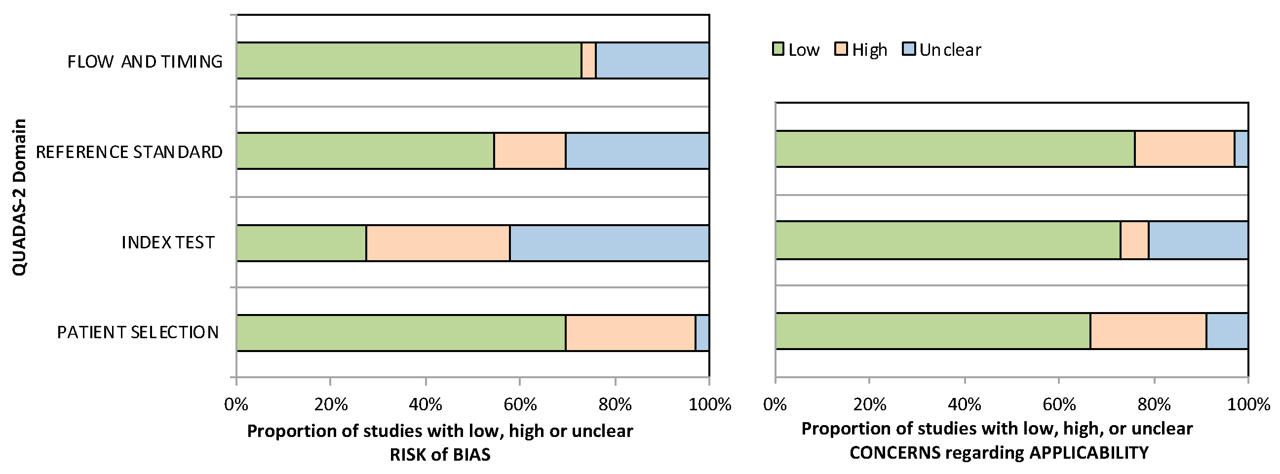
The 33 eligible studies included a population of CD patients who undertook faecal biomarker assessment with FC, FL, CHI3L1, MMP-9, FCLN-2, FIT, PMN-e, neopterrin, and/or myeloperoxidase against endoscopic assessment as the reference standard. Thirty-two studies assessed FC, four studies assessed FL, two studies assessed FCLN-2 and FIT, and one study assessed each of PMN-e, MMP-9, neopterrin, CHI3L1, and myeloperoxidase, respectively. A total of 2511 and 230 patients with CD were included in the FC and FL analyses, discriminating between active and inactive endoscopic disease. A total of 1086 patients were included in a separate subanalysis evaluating the capacity of FC to discriminate endoscopic mucosal healing. Endoscopic definitions of CD activity were via the simplified endoscopy score for CD (SES-CD), Crohn’s disease endoscopic index of severity (CDEIS), presence of endoscopic ulcers, and one study used an internally developed author’s score. SES-CD scores to discriminate between endoscopic CD activity and mucosal healing ranged between 0 and 3, and CDEIS between 0 and 6. Han et al. utilised a partial SES-CD whereby the presence of stenosis was excluded from the original SES-CD in determining endoscopic activity [24]. Iwamoto et al. utilised an extended SES-CD whereby CD activity from the proximal ileum and jejunum were included in determining endoscopic activity [25].

3.2. Study Quality

The 33 included studies underwent quality assessment with the QUADAS-2 criteria for diagnostic studies, with a summary presented in Figure 2. Twenty-three studies randomly and/or consecutively enrolled patients with CD, whereas the remaining studies were either unclear about or engendered bias via their method selection of patients. Moreover, in 15 studies it was unclear whether blinding of the reference standard was performed, and in 24 studies it was unclear whether blinding of the index test was performed. Overall, concerns regarding the applicability of the included studies across all domains were minimal.

3.3. Faecal Calprotectin

There were 32 studies that were eligible for assessment of faecal calprotectin [15,16,24,25,26,27,28,29,30,31,32,33,34,35,36,37,38,39,40,41,42,43,44,45,46,47,48,49,50,51,52,53]. Faecal calprotectin assays were performed via a lateral flow method in six studies, enzyme-linked immunosorbent assay (ELISA) in 25 studies, and the method was not described in one study. Multiple different proprietary faecal calprotectin kits were used, including Calprest (Eurospital Spa, Trieste, Italy), Bühlmann Calprotectin ELISA kit (Bühlmann, Schönenbuch, Switzerland), CALPRO ELISA (Calpro AS, Lysaker, Norway), EliA Calprotectin 2 (Thermo Fisher Scientific, Tokyo, Japan), Quantum Blue test (Buhlmann Laboratories, Schönenbuch, Switzerland), PhiCal (Immundiagnostik AG, Bensheim, Germany), RIDASCREEN® CALPROTECTIN (R-Biopharm AG, Darmstadt, Germany), and PhiCal (Genova Diagnostics Laboratories, Asheville, NC, USA). Definitions of endoscopic activity varied within the included studies, with nine studies applying an SES-CD > 2, five using SES-CD > 3, three using the presence of endoscopic ulcers, two using a CDEIS ≥ 3, and one using a CDEIS > 3. The remaining definitions (extended SES-CD ≥ 1, partial SES-CD ≥ 1, and an internally developed authors’ score) were each used in one study, respectively. Additionally, definitions of mucosal healing varied within the included studies, with three studies applying an SES-CD ≤ 2, three using an SES-CD = 0, and one each using a CDEIS ≤ 3, pSES-CD = 0, and the absence of endoscopic ulcers and/or inflammation.

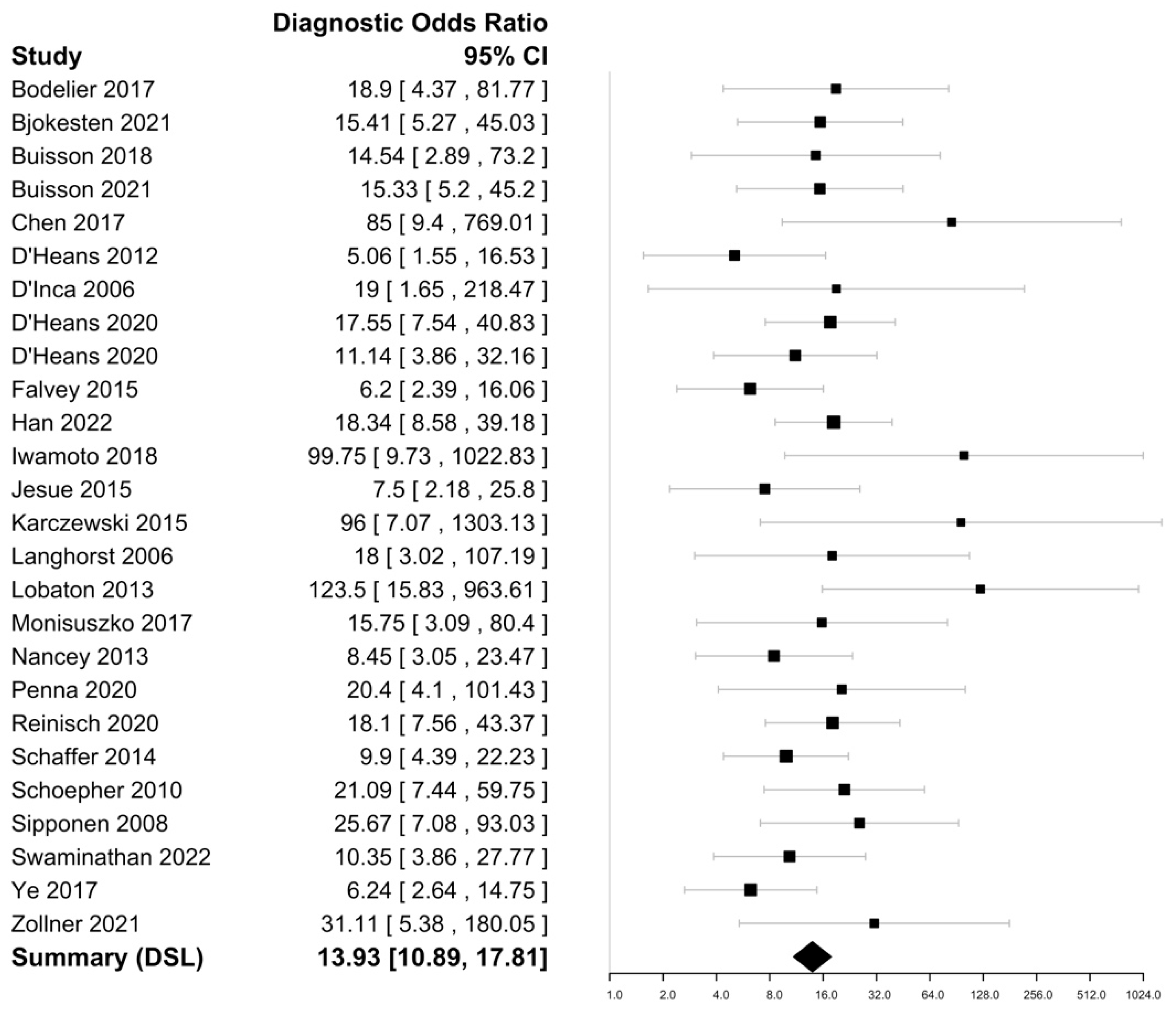
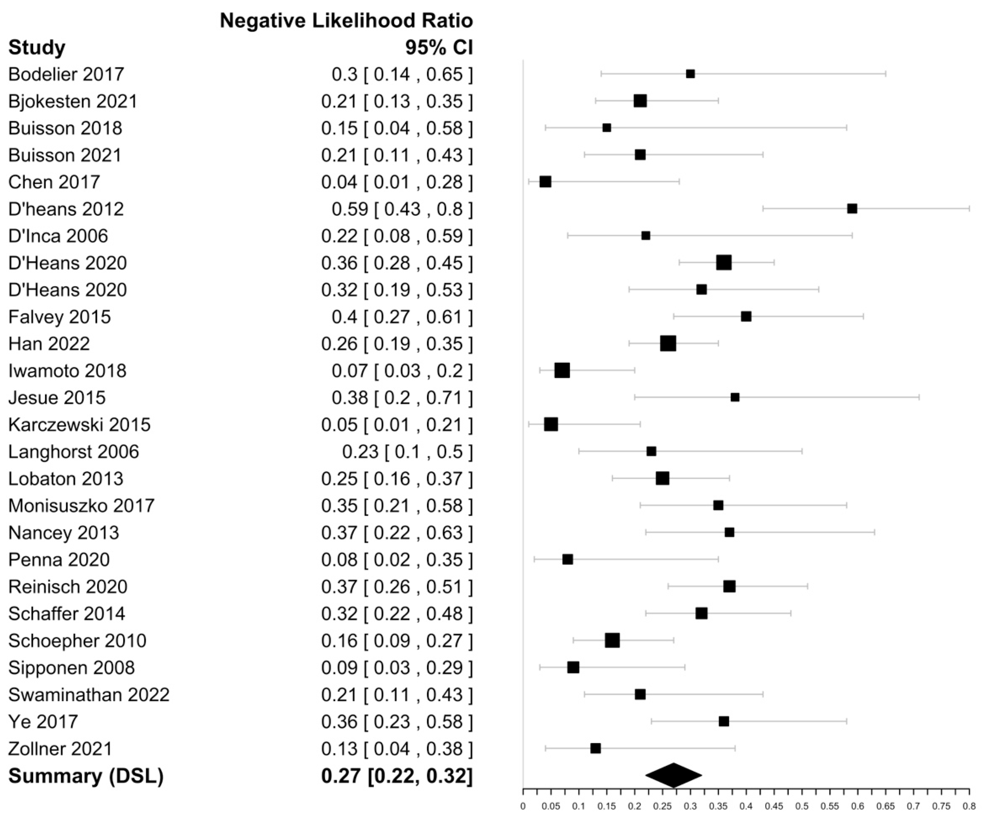
3.3.1. Detection of Endoscopic Activity

Within the included 25 studies (Table 1), FC sensitivities in discriminating endoscopically active CD ranged from 52 to 97% with a specificity ranging from 45 to 98%. The highest sensitivity was reported by Chen et al. (97%), with the highest specificity reported by Lobaton et al. (98%) [30,39]. The DOR was 13.93 (95% CI 10.89–17.81) with an i2 value of 1.34%, suggesting an insignificant heterogeneity and a negative likelihood ratio of 0.27 (95% CI 0.22–0.33) (Figure 3 and Figure 4). The Spearman’s correlation between sensitivity and false positive rate was 0.52. Using bivariate analysis, the pooled sensitivity was 81% (95% CI, 77–84%) with a specificity of 74% (95% CI, 70–80%) and an AUC of 0.85. On assessment using Deek’s funnel plot, there was no evidence of publication bias (p = 0.41).

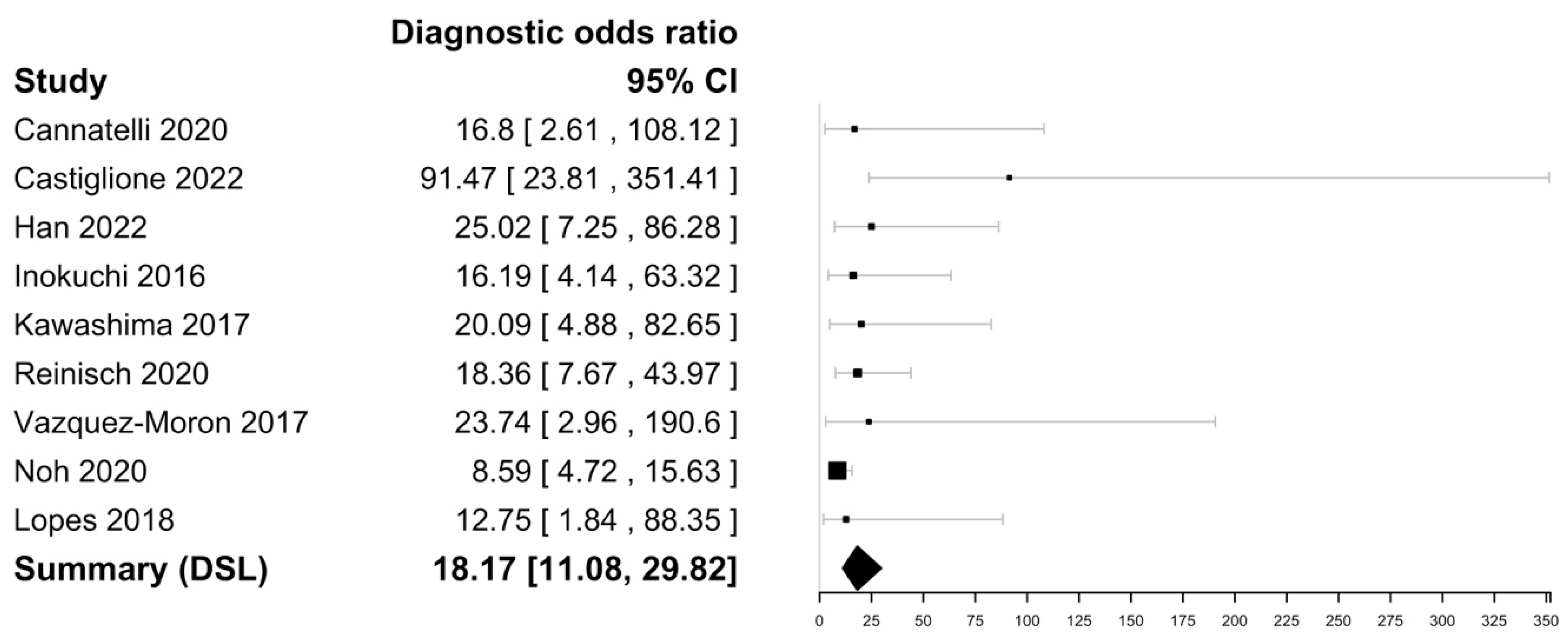
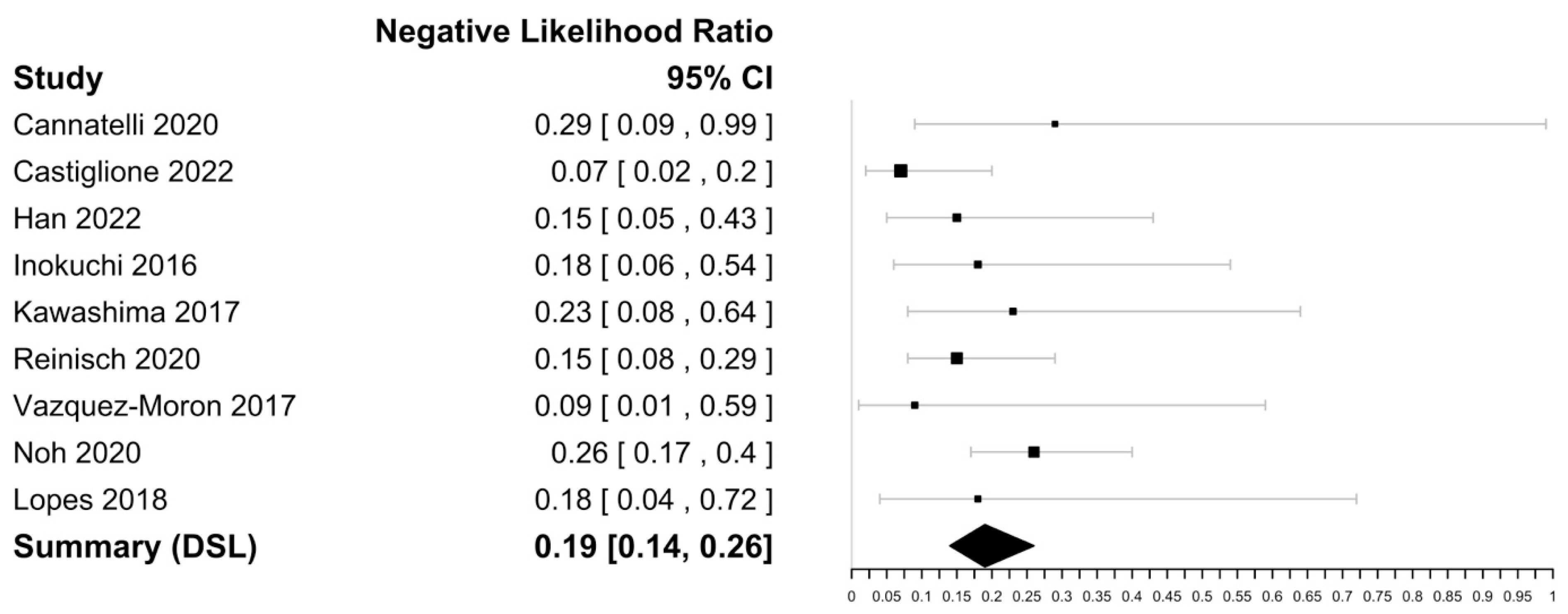
3.3.2. Prediction of Mucosal Healing

Within the nine included studies (Table 2), FC sensitivities in mucosal healing in CD ranged from 75 to 95% with a specificity ranging from 53 to 85%. The highest sensitivity was reported by Vazquez-Moron et al. (95%), and the highest specificity was reported by Castiglione and Cannatelli et al. (85%) [47,53,54]. The DOR was 18.17 (95% CI [11.08–29.82] with an i2 value of 0% implying no significant heterogeneity and a negative likelihood ratio of 0.19 [0.14–0.26]) (Figure 5 and Figure 6). The Spearman’s correlation between sensitivity and false positive rate was 0.48. Using bivariate analysis, the pooled sensitivity was 88% (84–90), specificity was 72% (64–79), and AUC was 0.88. There were not enough studies to assess for publication bias.

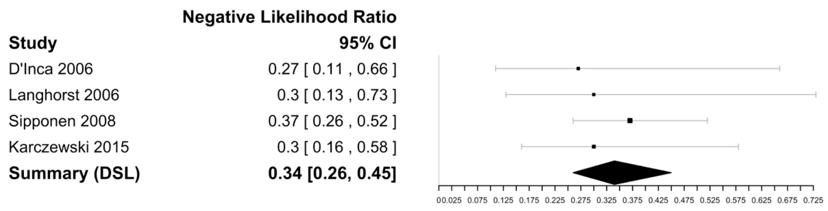
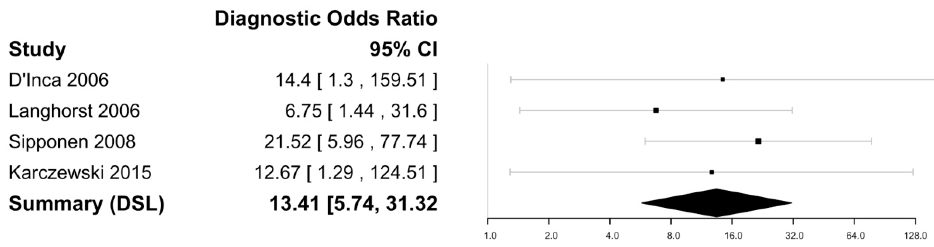
3.4. Faecal Lactoferrin

Lactoferrin is a component of polymorphonuclear neutrophils, which are implicated in the acute inflammatory response as seen in CD [35]. Four studies were eligible for the assessment of sensitivity and specificity for faecal lactoferrin in discriminating endoscopic activity (see Table 3) [31,34,35,43]. All lactoferrin assays were performed via ELISA, with four studies using the IBD-SCAN® (Techlab, Blacksburg, VA, USA) kit and one study using the IBD-CHEK® (Techlab, Blacksburg, VA, USA) kit [31,34,35,43]. Definitions of endoscopic activity varied within the included studies, with one study each using a CDEIS > 3, CDEIS > 2, SES-CD > 3, and an internally developed authors’ score to discriminate between active and inactive endoscopic CD [31,34,35,43].
Figure 7 and Figure 8 show the forest plots for the negative likelihood ratio and DOR of FL in discriminating endoscopically active and inactive CD. Sensitivities ranged from 66 to 81%, with the highest sensitivity reported as 81% by Langhorst et al. [35]. The specificity ranged from 59 to 91%, with the highest specificity of 91% reported by Sipponen et al. [43]. The negative likelihood ratio was 0.34 (95% CI [0.26–0.45] and the DOR was 13.42 (95% CI: 5.74–31.32) with an I2 value of 0%, suggesting no heterogeneity. The Spearman’s correlation between sensitivity and false positivity was 0.89. Using bivariate analysis, the pooled sensitivity was 75% (65–83) and the pooled specificity was 80% (57–92), with an AUC of 0.81. There were not enough studies to assess for publication bias.

3.5. Other Biomarkers

  • Faecal Lipocalin-2
FCLN-2 is a glycoprotein produced by intestinal epithelial cells and released into the gut lumen in response to proinflammatory stimuli, as occurs in CD [27,46]. FCLN-2 assays are performed via ELISA and have been described in two studies as potential discriminant faecal biomarkers of endoscopically active CD [27,46]. In a cohort of 54 patients with CD, Buisson et al. demonstrated an optimal cut-off level of 6700 ng/g for discriminating between the absence and presence of endoscopic ulcers, with a sensitivity and specificity of 86% and 46%, respectively [27]. More recently, in a cohort of 72 patients with CD, Zollner et al. demonstrated an optimal cut-off of 0.56 µg/g for discriminating endoscopically active and inactive CD (defined as an SES-CD > 3 for active CD), with a sensitivity and specificity of 91% and 77%, respectively [46]. The discrepancy in optimal cut-offs between the two studies may be explained by the different endoscopic standards applied (i.e., ulceration versus defined SES-CD score).
  • Faecal metalloprotease 9
Matrix metalloproteases are known to activate or degrade a variety of substrates within an immune response [27]. MMP-9 is expressed in inflamed intestinal mucosa by macrophages and neutrophils and released into faeces during active CD [27]. MMP-9 assays are performed via ELISA and have been described in one study as a potential discriminant faecal biomarker of endoscopically active CD. In a cohort of 54 patients with CD, Buisson et al. demonstrated an optimal cut-off level of 350 ng/g for discriminating between the absence and presence of endoscopic ulcers, with a sensitivity and specificity of 90% and 64%, respectively [27].
  • Neopterin
Neopterin is produced and released primarily from macrophages in response to stimulation from activated T-cells, as occurs in CD [38]. Neopterin assays are performed via ELISA and have been described in one study as a potential faecal biomarker for discriminating endoscopically active CD [38]. In a cohort of 78 patients with CD, Nancey et al. demonstrated sensitivity and specificity of 74% and 73%, utilising a neopterin cut-off value of 200 pmol/g for discriminating endoscopically active CD (defined as an SES-CD > 3) [38].
  • Faecal myeloperoxidase
Myeloperoxidase is an enzyme present within neutrophils with a putative role in inflammatory tissue damage [44]. Its presence within faeces is detectable via ELISA, with one study demonstrating its potential as a biomarker for discriminating endoscopically active CD [44]. In a cohort of 100 patients with CD, Swaminathan et al. demonstrated an optimal faecal myeloperoxidase cut-off level of 10.25 µg/g for discriminating endoscopically active CD (defined as an SES-CD > 3) [44]. At this cut-off, faecal myeloperoxidase demonstrated a sensitivity and specificity of 63 and 69%, respectively [44].
  • Faecal chitinase 3-like 1
Chitinase 3-like 1(CH3L1) is expressed in a variety of cells such as macrophages and neutrophils and is upregulated in the colonic epithelial cells and lamina propria macrophages of inflamed mucosa in CD [52]. Its presence within faeces is detectable via ELISA, with one study demonstrating its potential as a biomarker for discriminating endoscopically active CD [52]. In a study of 54 patients with CD, Buisson et al. determined an optimal faecal CH3L1 cut-off level of 15 ng/g for discriminating endoscopic ulceration in CD [52]. At this cut-off level, faecal CH3L1 demonstrated a sensitivity and specificity of 100% and 64%, respectively [52].
  • PMN-e
Polymorphonuclear neutrophil elastase (PMN-e) is an enzyme stored in polymorphonuclear neutrophils and released during the activation of these cells as a mediator of inflammation [35]. Its presence within faeces is detectable via ELISA, with one study demonstrating its potential as a biomarker capable of discriminating endoscopically active CD [35]. In a study of 43 patients with CD, Langhorst et al. determined an optimal faecal PMN-e cut-off level of 0.062 µg/mL for discriminating endoscopically active CD (using an internally developed endoscopic score by the authors as the reference standard) [35]. At this cut-off level, faecal PMN-E demonstrated a sensitivity and specificity of 82% and 70%, respectively [35].
  • FIT
FIT quantitatively measures faecal haemoglobin concentrations and forms the backbone of colon cancer surveillance programs worldwide [48]. Quantitative measurements are widely performed using the proprietary test from the Eiken Chemical Co. (Tokyo, Japan). In a cohort of 69 patients, Iwamoto et al. determined an FIT cut-off of 13 ng/mL to have a sensitivity and specificity of 73% and 71% in discriminating endoscopically active CD (defined as an eSES-CD ≥ 1) [25]. Regarding mucosal healing, in a cohort of 71 patients, Inokuchi et al. determined that a FIT cut-off level of 52 ng/mL was predictive of mucosal healing in CD (defined as an SES-CD = 0), with a sensitivity and specificity of 96% and 48%, respectively [48]. Other biomarkers used to detect endoscopic activity in CD are summarized in Table 4.

4. Discussion

With regular objective assessment now recommended by international guidelines, there is a growing imperative for cheaper, rapidly accessible yet reliable tests for assessing disease activity in CD. Clinical activity scores, such as the Harvey-Bradshaw index or CD activity index (CDAI), are predominantly symptom-based, and therefore subjective and prone to providing falsely reassuring results [32]. Moreover, biochemical tests, such as C-reactive protein, remain nonspecific to IBD and are subject to confounding in multiple clinical settings. Whilst ileocolonoscopy and cross-sectional imaging remain important options due to their informational capacity, neither are cheap nor necessarily rapidly available. Furthermore, both require significant patient participation, preparation, and risks that might be deemed unacceptable for repetitive testing over short six-monthly assessment intervals, as recommended by STRIDE-2 guidelines. Thus, faecal biomarkers play a significant role in the routine, serial monitoring of IBD patients.
This meta-analysis strengthens the existing knowledge regarding FC as a capable biomarker in discriminating between active/inactive endoscopic disease and mucosal healing and provides context for FC amongst other more novel faecal biomarkers and their respective performance in discriminating endoscopically active versus inactive CD. This meta-analysis reaffirmed that both FC and FL are highly sensitive and specific for discriminating active versus inactive endoscopic disease in CD, with the inclusion of a number of more recent studies since the previous meta-analysis was performed many years ago [55]. In this meta-analysis, FC demonstrated a pooled sensitivity of 81% and specificity of 74% (AuROC and DOR of 0.85 and 13.93, respectively), implying that FC retains good diagnostic capacity across all disease subtypes and locations of the heterogeneous entity that is CD. These pooled data are similar to those previously reported for FC in the literature [55]. Moreover, FC exhibits an excellent capacity for prediction of mucosal healing with a summary DOR of 18.17 (95% CI 11.08–29.82) in this study.
Many FC manufacturers recommend 50 µg/g as a cut-off value for representing active intestinal inflammation. However, in this meta-analysis, 15 of 25 (60%) studies reported an FC > 200 µg/g as their optimal cut-off when discriminating between endoscopically active and inactive disease, albeit with heterogeneous definitions of endoscopically active CD. Comparatively, with respect to mucosal healing, only two studies derived a FC > 200 µg/g as optimal and most (5/9) applied a normal endoscopy (SES-CD = 0 and/or no ulcers/inflammation) as their definition of mucosal healing. Hence, the capacity of a faecal biomarker to surrogately represent mucosal healing may be a more appropriate ‘harder’ endpoint by which to determine its accuracy and utility when correlating to endoscopy as a reference standard. While determining the optimal cut-off for FC in detecting endoscopically active or mucosal healing in CD was beyond the scope of this analysis, it appears that a lower calprotectin cut-off at a higher sensitivity is demonstrated in studies addressing mucosal healing compared to endoscopic disease activity in CD. Ultimately, further studies examining newer calprotectin assays and similar definitions of endoscopic activity (and/or histological activity as some recent studies have explored) are needed.
Within our meta-analysis, FL demonstrated a pooled sensitivity, specificity, AuROC, and DOR of 75%, 80%, 0.81, and 13.41, respectively. Compared to FC, the performance of FL was similar, yet the role of FL, for unknown reasons beyond the scope of this study, has not been incorporated into widespread clinical practice. The diagnostic performance of alternative faecal biomarkers was included in the study, yet most were limited to single studies. Thus, it remains difficult to draw meaningful conclusions from these biomarkers regarding their capacity to discriminate between active and inactive endoscopic CD or mucosal healing. Further studies of biomarkers beyond FC and FL are clearly needed to determine and establish their potential role in CD assessment, especially concerning whether they offer advantages over FC in terms of cost, accuracy, and/or convenience. In addition, combinations of low-cost faecal biomarkers should be explored in an effort to enhance diagnostic performance.
This meta-analysis was subject to several weaknesses and limitations. The data were drawn from predominately retrospective studies with their inherent biases. Moreover, our endoscopic reference standard was variably applied via multiple, different endoscopic scores and cut-off levels therein used to discriminate CD activity which may have impacted the observed results. In addition, FC and FL assays were performed via different methods (i.e., lateral flow and ELISA) which may not be of equivalent accuracy.

5. Conclusions

Based on this rigorous meta-analysis, FC and FL were both shown to perform robustly in discriminating between active and inactive CD. Both are relatively cheap, rapid, and reliable tests that are therefore amenable to serial testing at frequent intervals to confidently monitor CD activity. Newer and novel faecal biomarkers have been proposed but require further evaluation before any could be considered as a replacement, let alone superior, to FC in routine clinical practice. There is clearly an unmet need to incisively examine the capacity of faecal biomarkers to assess CD activity according to disease location, extent, and specific subtypes. Moreover, in this context, the application of multiple biomarkers (faecal, serum, and/or other) in a combined diagnostics matrix, perhaps harnessing artificial intelligence-based algorithms, may provide even greater diagnostic and predictive power. Faecal biomarkers are here to stay and, with further research, are likely to continue to increasingly dominate the diagnostics landscape in routine management of CD.

Supplementary Materials

The following supporting information can be downloaded at: https://www.mdpi.com/article/10.3390/biomedicines11051408/s1, Table S1: FC to CD endoscopic activity dataset; Table S2: FC to CD mucosal healing activity dataset; Table S3: FL to CD endoscopic dataset; Table S4: Literature search strategy; Table S5: Tabular presentation of QUADAS-2 results; PRISMA Checklist. Ref. [56] is cited in the supplementary materials.

Author Contributions

A.B.: literature search, data collation and extraction, writing, editing, statistical analysis; D.R.V.L.: critical revision and supervision; A.V.: critical revision and supervision; G.M.: literature search, data collation and extraction; D.L.: statistical analysis, J.P.S.: literature search, data collation and extraction, writing, editing, statistical analysis and critical revision. All authors have read and agreed to the published version of the manuscript.

Funding

This research received no external funding.

Institutional Review Board Statement

Not applicable.

Informed Consent Statement

Not applicable.

Data Availability Statement

Available in body of text and Supplementary Materials.

Acknowledgments

Wendy Wong, Eastern Health Library Services for assistance with the literature search.

Conflicts of Interest

D.R.V.L. has received educational grants or research support from Janssen, Medtronic, and Pfizer, as well as speaker fees from Janssen, Takeda, and Pfizer, and is on the advisory boards for Janssen, Takeda, Pfizer, and AbbVie. J.P.S. has received speaker fees for Janssen, Abbvie, and Takeda.

References

  1. Ng, S.C.; Shi, H.Y.; Hamidi, N.; Underwood, F.E.; Tang, W.; Benchimol, E.I.; Panaccione, R.; Ghosh, S.; Wu, J.C.Y.; Chan, F.K.L.; et al. Worldwide incidence and prevalence of inflammatory bowel disease in the 21st century: A systematic review of population-based studies. Lancet 2017, 390, 2769–2778. [Google Scholar] [CrossRef] [PubMed]
  2. Agrawal, M.; Christensen, H.S.; Bøgsted, M.; Colombel, J.F.; Jess, T.; Allin, K.H. The Rising Burden of Inflammatory Bowel Disease in Denmark Over Two Decades: A Nationwide Cohort Study. Gastroenterology 2022, 163, 1547–1554.e5. [Google Scholar] [CrossRef] [PubMed]
  3. Lee, J.W.; Eun, C.S. Inflammatory bowel disease in Korea: Epidemiology and pathophysiology. Korean J. Intern. Med. 2022, 37, 885–894. [Google Scholar] [CrossRef] [PubMed]
  4. Argüelles-Arias, F.; Rodríguez-Oballe, J.; Duarte-Chang, C.; Castro-Laria, L.; García-Montes, J.M.; Caunedo-Álvarez, A.; Herrerías-Gutiérrez, J.M. Capsule endoscopy in the small bowel Crohn’s disease. Gastroenterol. Res. Pract. 2014, 2014, 529136. [Google Scholar] [CrossRef]
  5. Peyrin-Biroulet, L.; Loftus, E.V.; Colombel, J.F.; Sandborn, W.J. Long-term complications, extraintestinal manifestations, and mortality in adult Crohn’s disease in population-based cohorts. Inflamm. Bowel Dis. 2011, 17, 471–478. [Google Scholar] [CrossRef]
  6. Cho, C.W.; You, M.W.; Oh, C.H.; Lee, C.K.; Moon, S.K. Long-term Disease Course of Crohn’s Disease: Changes in Disease Location, Phenotype, Activities, and Predictive Factors. Gut Liver 2022, 16, 157–170. [Google Scholar] [CrossRef]
  7. Adamina, M.; Bonovas, S.; Raine, T.; Spinelli, A.; Warusavitarne, J.; Armuzzi, A.; Bachmann, O.; Bager, P.; Biancone, L.; Bokemeyer, B.; et al. ECCO Guidelines on Therapeutics in Crohn’s Disease: Surgical Treatment. J. Crohn's Colitis 2020, 14, 155–168. [Google Scholar] [CrossRef]
  8. Rieder, F.; Latella, G.; Magro, F.; Yuksel, E.S.; Higgins, P.D.; Di Sabatino, A.; de Bruyn, J.R.; Rimola, J.; Brito, J.; Bettenworth, D.; et al. European Crohn’s and Colitis Organisation Topical Review on Prediction, Diagnosis and Management of Fibrostenosing Crohn’s Disease. J. Crohn's Colitis 2016, 10, 873–885. [Google Scholar] [CrossRef]
  9. Torres, J.; Bonovas, S.; Doherty, G.; Kucharzik, T.; Gisbert, J.P.; Raine, T.; Adamina, M.; Armuzzi, A.; Bachmann, O.; Bager, P.; et al. ECCO Guidelines on Therapeutics in Crohn’s Disease: Medical Treatment. J. Crohn's Colitis 2020, 14, 4–22. [Google Scholar] [CrossRef]
  10. Kumar, A.; Cole, A.; Segal, J.; Smith, P.; Limdi, J.K. A review of the therapeutic management of Crohn’s disease. Therap. Adv. Gastroenterol. 2022, 15, 17562848221078456. [Google Scholar] [CrossRef]
  11. Maaser, C.; Sturm, A.; Vavricka, S.R.; Kucharzik, T.; Fiorino, G.; Annese, V.; Calabrese, E.; Baumgart, D.C.; Bettenworth, D.; Borralho Nunes, P.; et al. ECCO-ESGAR Guideline for Diagnostic Assessment in IBD Part 1: Initial diagnosis, monitoring of known IBD, detection of complications. J. Crohn's Colitis 2019, 13, 144–164. [Google Scholar] [CrossRef]
  12. Geyl, S.; Guillo, L.; Laurent, V.; D’Amico, F.; Danese, S.; Peyrin-Biroulet, L. Transmural healing as a therapeutic goal in Crohn’s disease: A systematic review. Lancet Gastroenterol. Hepatol. 2021, 6, 659–667. [Google Scholar] [CrossRef] [PubMed]
  13. Peyrin-Biroulet, L.; Sandborn, W.; Sands, B.E.; Reinisch, W.; Bemelman, W.; Bryant, R.V.; D’Haens, G.; Dotan, I.; Dubinsky, M.; Feagan, B.; et al. Selecting Therapeutic Targets in Inflammatory Bowel Disease (STRIDE): Determining Therapeutic Goals for Treat-to-Target. Am. J. Gastroenterol. 2015, 110, 1324–1338. [Google Scholar] [CrossRef] [PubMed]
  14. Turner, D.; Ricciuto, A.; Lewis, A.; D’Amico, F.; Dhaliwal, J.; Griffiths, A.M.; Bettenworth, D.; Sandborn, W.J.; Sands, B.E.; Reinisch, W.; et al. STRIDE-II: An Update on the Selecting Therapeutic Targets in Inflammatory Bowel Disease (STRIDE) Initiative of the International Organization for the Study of IBD (IOIBD): Determining Therapeutic Goals for Treat-to-Target strategies in IBD. Gastroenterology 2021, 160, 1570–1583. [Google Scholar] [CrossRef]
  15. af Björkesten, C.G.; Nieminen, U.; Turunen, U.; Arkkila, P.; Sipponen, T.; Färkkilä, M. Surrogate markers and clinical indices, alone or combined, as indicators for endoscopic remission in anti-TNF-treated luminal Crohn’s disease. Scand. J. Gastroenterol. 2012, 47, 528–537. [Google Scholar] [CrossRef] [PubMed]
  16. Buisson, A.; Mak, W.Y.; Andersen, M.J.; Lei, D.; Pekow, J.; Cohen, R.D.; Kahn, S.A.; Pereira, B.; Rubin, D.T. Fecal Calprotectin Is Highly Effective to Detect Endoscopic Ulcerations in Crohn’s Disease Regardless of Disease Location. Inflamm. Bowel Dis. 2021, 27, 1008–1016. [Google Scholar] [CrossRef]
  17. Mohamed, G.; Bohra, A.; Segal, J. Sensitivity and Specificity of Stool Markers in Inflammatory Bowel Disease: A Systematic Review and Meta-Analysis. PROSPERO 2022 CRD42022354526. Available online: https://www.crd.york.ac.uk/prospero/display_record.php?ID=CRD42022354526 (accessed on 13 April 2023).
  18. Whiting, P.F.; Rutjes, A.W.; Westwood, M.E.; Mallett, S.; Deeks, J.J.; Reitsma, J.B.; Leeflang, M.M.; Sterne, J.A.; Bossuyt, P.M.; QUADAS-2 Group. QUADAS-2: A revised tool for the quality assessment of diagnostic accuracy studies. Ann. Intern. Med. 2011, 155, 529–536. [Google Scholar] [CrossRef]
  19. Glas, A.S.; Lijmer, J.G.; Prins, M.H.; Bonsel, G.J.; Bossuyt, P.M. The diagnostic odds ratio: A single indicator of test performance. J. Clin. Epidemiol. 2003, 56, 1129–1135. [Google Scholar] [CrossRef]
  20. Doebler, P. Comprehensive R Archive Network Mada: Meta-Analysis of Diagnostic Accuracy. Available online: https://cran.r-project.org/web/packages/mada/index.html (accessed on 13 April 2023).
  21. Higgins, J.P.; Thompson, S.G. Quantifying heterogeneity in a meta-analysis. Stat. Med. 2002, 21, 1539–1558. [Google Scholar] [CrossRef]
  22. Moher, D.; Liberati, A.; Tetzlaff, J.; Altman, D.G.; The PRISMA Group. Preferred reporting items for systematic reviews and meta-analyses: The PRISMA statement. PLoS Med. 2009, 6, e1000097. [Google Scholar] [CrossRef]
  23. Deeks, J.J.; Macaskill, P.; Irwig, L. The performance of tests of publication bias and other sample size effects in systematic reviews of diagnostic test accuracy was assessed. J. Clin. Epidemiol. 2005, 58, 882–893. [Google Scholar] [CrossRef] [PubMed]
  24. Han, W.; Wu, J.; Zhang, P.; Hu, N.; Mei, Q.; Hu, J. Fecal calprotectin predicts endoscopic activity and mucosal healing of small bowel Crohn’s disease evaluated by double-balloon endoscopy. Int. J. Color. Dis. 2022, 37, 1953–1961. [Google Scholar] [CrossRef] [PubMed]
  25. Iwamoto, F.; Matsuoka, K.; Motobayashi, M.; Takenaka, K.; Kuno, T.; Tanaka, K.; Tsukui, Y.; Kobayashi, S.; Yoshida, T.; Fujii, T.; et al. Prediction of disease activity of Crohn’s disease through fecal calprotectin evaluated by balloon-assisted endoscopy. J. Gastroenterol. Hepatol. 2018, 33, 1984–1989. [Google Scholar] [CrossRef] [PubMed]
  26. Bodelier, A.G.; Jonkers, D.; van den Heuvel, T.; de Boer, E.; Hameeteman, W.; Masclee, A.A.; Pierik, M.J. High Percentage of IBD Patients with Indefinite Fecal Calprotectin Levels: Additional Value of a Combination Score. Dig. Dis. Sci. 2017, 62, 465–472. [Google Scholar] [CrossRef]
  27. Buisson, A.; Vazeille, E.; Minet-Quinard, R.; Goutte, M.; Bouvier, D.; Goutorbe, F.; Pereira, B.; Barnich, N.; Bommelaer, G. Fecal Matrix Metalloprotease-9 and Lipocalin-2 as Biomarkers in Detecting Endoscopic Activity in Patients With Inflammatory Bowel Diseases. J. Clin. Gastroenterol. 2018, 52, e53–e62. [Google Scholar] [CrossRef]
  28. Chen, J.M.; Liu, T.; Gao, S.; Tong, X.D.; Deng, F.H.; Nie, B. Efficacy of noninvasive evaluations in monitoring inflammatory bowel disease activity: A prospective study in China. World J. Gastroenterol. 2017, 23, 8235–8247. [Google Scholar] [CrossRef]
  29. D’Haens, G.; Ferrante, M.; Vermeire, S.; Baert, F.; Noman, M.; Moortgat, L.; Geens, P.; Iwens, D.; Aerden, I.; Van Assche, G.; et al. Fecal calprotectin is a surrogate marker for endoscopic lesions in inflammatory bowel disease. Inflamm. Bowel Dis. 2012, 18, 2218–2224. [Google Scholar] [CrossRef]
  30. D’Haens, G.; Kelly, O.; Battat, R.; Silverberg, M.S.; Laharie, D.; Louis, E.; Savarino, E.; Bodini, G.; Yarur, A.; Boland, B.S.; et al. Development and Validation of a Test to Monitor Endoscopic Activity in Patients With Crohn’s Disease Based on Serum Levels of Proteins. Gastroenterology 2020, 158, 515–526.e10. [Google Scholar] [CrossRef]
  31. D’Incà, R.; Dal Pont, E.; Di Leo, V.; Ferronato, A.; Fries, W.; Vettorato, M.G.; Martines, D.; Sturniolo, G.C. Calprotectin and lactoferrin in the assessment of intestinal inflammation and organic disease. Int. J. Color. Dis. 2007, 22, 429–437. [Google Scholar] [CrossRef]
  32. Falvey, J.D.; Hoskin, T.; Meijer, B.; Ashcroft, A.; Walmsley, R.; Day, A.S.; Gearry, R.B. Disease activity assessment in IBD: Clinical indices and biomarkers fail to predict endoscopic remission. Inflamm. Bowel Dis. 2015, 21, 824–831. [Google Scholar] [CrossRef]
  33. Jusué, V.; Chaparro, M.; Gisbert, J.P. Accuracy of fecal calprotectin for the prediction of endoscopic activity in patients with inflammatory bowel disease. Dig. Liver Dis. 2018, 50, 353–359. [Google Scholar] [CrossRef] [PubMed]
  34. Karczewski, J.; Swora-Cwynar, E.; Rzymski, P.; Poniedziałek, B.; Adamski, Z. Selected biologic markers of inflammation and activity of Crohn’s disease. Autoimmunity 2015, 48, 318–327. [Google Scholar] [CrossRef] [PubMed]
  35. Langhorst, J.; Elsenbruch, S.; Koelzer, J.; Rueffer, A.; Michalsen, A.; Dobos, G.J. Noninvasive markers in the assessment of intestinal inflammation in inflammatory bowel diseases: Performance of fecal lactoferrin, calprotectin, and PMN-elastase, CRP, and clinical indices. Am. J. Gastroenterol. 2008, 103, 162–169. [Google Scholar] [CrossRef] [PubMed]
  36. Lobatón, T.; López-García, A.; Rodríguez-Moranta, F.; Ruiz, A.; Rodríguez, L.; Guardiola, J. A new rapid test for fecal calprotectin predicts endoscopic remission and postoperative recurrence in Crohn’s disease. J. Crohn's Colitis 2013, 7, e641–e651. [Google Scholar] [CrossRef]
  37. Moniuszko, A.; Głuszek, S.; Rydzewska, G. Rapid fecal calprotectin test for prediction of mucosal inflammation in ulcerative colitis and Crohn disease: A prospective cohort study. Pol. Arch. Intern. Med. 2017, 127, 312–318. [Google Scholar] [CrossRef]
  38. Nancey, S.; Boschetti, G.; Moussata, D.; Cotte, E.; Peyras, J.; Cuerq, C.; Haybrard, J.; Charlois, A.L.; Mialon, A.; Chauvenet, M.; et al. Neopterin is a novel reliable fecal marker as accurate as calprotectin for predicting endoscopic disease activity in patients with inflammatory bowel diseases. Inflamm. Bowel Dis. 2013, 19, 1043–1052. [Google Scholar] [CrossRef]
  39. Penna, F.G.C.; Rosa, R.M.; Pereira, F.H.; Cunha, P.F.S.; Sousa, S.C.S.; Ferrari, T.C.A.; Cara, C.; Ferrari, M.L.A. Combined evaluation of fecal calprotectin and C-reactive protein as a therapeutic target in the management of patients with Crohn’s disease. Gastroenterol. Hepatol. 2021, 44, 87–95. [Google Scholar] [CrossRef]
  40. Reinisch, W.; Panaccione, R.; Bossuyt, P.; Baert, F.; Armuzzi, A.; Hébuterne, X.; Travis, S.; Danese, S.; Sandborn, W.J.; Schreiber, S.; et al. Association of Biomarker Cutoffs and Endoscopic Outcomes in Crohn’s Disease: A Post Hoc Analysis From the CALM Study. Inflamm. Bowel Dis. 2020, 26, 1562–1571. [Google Scholar] [CrossRef]
  41. Schaffer, T.; Schoepfer, A.M.; Seibold, F.; Swiss IBD Cohort Study Group. Serum ficolin-2 correlates worse than fecal calprotectin and CRP with endoscopic Crohn’s disease activity. J. Crohns Colitis 2014, 8, 1125–1132. [Google Scholar] [CrossRef]
  42. Schoepfer, A.M.; Beglinger, C.; Straumann, A.; Trummler, M.; Vavricka, S.R.; Bruegger, L.E.; Seibold, F. Fecal calprotectin correlates more closely with the Simple Endoscopic Score for Crohn’s disease (SES-CD) than CRP, blood leukocytes, and the CDAI. Am. J. Gastroenterol. 2010, 105, 162–169. [Google Scholar] [CrossRef]
  43. Sipponen, T.; Savilahti, E.; Kolho, K.L.; Nuutinen, H.; Turunen, U.; Färkkilä, M. Crohn’s disease activity assessed by fecal calprotectin and lactoferrin: Correlation with Crohn’s disease activity index and endoscopic findings. Inflamm. Bowel Dis. 2008, 14, 40–46. [Google Scholar] [CrossRef] [PubMed]
  44. Swaminathan, A.; Borichevsky, G.M.; Edwards, T.S.; Hirschfeld, E.; Mules, T.C.; Frampton, C.M.A.; Day, A.S.; Hampton, M.B.; Kettle, A.J.; Gearry, R.B. Faecal Myeloperoxidase as a Biomarker of Endoscopic Activity in Inflammatory Bowel Disease. J. Crohn's Colitis 2022, 16, 1862–1873. [Google Scholar] [CrossRef] [PubMed]
  45. Ye, L.; Cheng, W.; Chen, B.Q.; Lan, X.; Wang, S.D.; Wu, X.C.; Huang, W.; Wang, F.Y. Levels of Faecal Calprotectin and Magnetic Resonance Enterocolonography Correlate with Severity of Small Bowel Crohn’s Disease: A Retrospective Cohort Study. Sci. Rep. 2017, 7, 1970. [Google Scholar] [CrossRef] [PubMed]
  46. Zollner, A.; Schmiderer, A.; Reider, S.J.; Oberhuber, G.; Pfister, A.; Texler, B.; Watschinger, C.; Koch, R.; Effenberger, M.; Raine, T.; et al. Faecal Biomarkers in Inflammatory Bowel Diseases: Calprotectin Versus Lipocalin-2-a Comparative Study. J. Crohn's Colitis 2021, 15, 43–54. [Google Scholar] [CrossRef] [PubMed]
  47. Vázquez Morón, J.M.; Pallarés Manrique, H.; Machancoses, F.H.; Ramos Lora, M.; Ruiz Frutos, C. Accurate cut-offs for predicting endoscopic activity and mucosal healing in Crohn’s disease with fecal calprotectin. Rev. Esp. Enferm. Dig. 2017, 109, 130–136. [Google Scholar] [CrossRef] [PubMed]
  48. Inokuchi, T.; Kato, J.; Hiraoka, S.; Takashima, S.; Nakarai, A.; Takei, D.; Sugihara, Y.; Takahara, M.; Kawano, S.; Harada, K.; et al. Fecal Immunochemical Test Versus Fecal Calprotectin for Prediction of Mucosal Healing in Crohn’s Disease. Inflamm. Bowel Dis. 2016, 22, 1078–1085. [Google Scholar] [CrossRef]
  49. Kawashima, K.; Ishihara, S.; Yuki, T.; Fukuba, N.; Sonoyama, H.; Kazumori, H.; Yamashita, N.; Tada, Y.; Kusunoki, R.; Oka, A.; et al. Fecal Calprotectin More Accurately Predicts Endoscopic Remission of Crohn’s Disease than Serological Biomarkers Evaluated Using Balloon-assisted Enteroscopy. Inflamm. Bowel Dis. 2017, 23, 2027–2034. [Google Scholar] [CrossRef]
  50. Lopes, S.; Andrade, P.; Afonso, J.; Cunha, R.; Rodrigues-Pinto, E.; Ramos, I.; Macedo, G.; Magro, F. Monitoring Crohn’s disease activity: Endoscopy, fecal markers and computed tomography enterography. Therap. Adv. Gastroenterol. 2018, 11, 1756284818769075. [Google Scholar] [CrossRef]
  51. Noh, S.M.; Oh, E.H.; Park, S.H.; Lee, J.B.; Kim, J.Y.; Park, J.C.; Kim, J.; Ham, N.S.; Hwang, S.W.; Yang, D.H.; et al. Association of Faecal Calprotectin Level and Combined Endoscopic and Radiological Healing in Patients With Crohn’s Disease Receiving Anti-tumour Necrosis Factor Therapy. J. Crohn's Colitis 2020, 14, 1231–1240. [Google Scholar] [CrossRef]
  52. Buisson, A.; Vazeille, E.; Minet-Quinard, R.; Goutte, M.; Bouvier, D.; Goutorbe, F.; Pereira, B.; Barnich, N.; Bommelaer, G. Faecal chitinase 3-like 1 is a reliable marker as accurate as faecal calprotectin in detecting endoscopic activity in adult patients with inflammatory bowel diseases. Aliment. Pharmacol. Ther. 2016, 43, 1069–1079. [Google Scholar] [CrossRef] [PubMed]
  53. Cannatelli, R.; Bazarova, A.; Zardo, D.; Nardone, O.M.; Shivaji, U.; Smith, S.C.L.; Gkoutos, G.; Ricci, C.; Gui, X.S.; Ghosh, S.; et al. Fecal Calprotectin Thresholds to Predict Endoscopic Remission Using Advanced Optical Enhancement Techniques and Histological Remission in IBD Patients. Inflamm. Bowel Dis. 2021, 27, 647–654. [Google Scholar] [CrossRef] [PubMed]
  54. Castiglione, F.; Imperatore, N.; Testa, A.; de Sire, R.; Nardone, O.M.; Ricciolino, S.; Di Luna, I.; Patturelli, M.; Villani, G.D.; Olmo, O.; et al. Exploring the concept of deep remission in Crohn’s disease: Correlation between transmural healing and biomarkers. Therap. Adv. Gastroenterol. 2022, 15, 17562848221110643. [Google Scholar] [CrossRef] [PubMed]
  55. Rokkas, T.; Portincasa, P.; Koutroubakis, I.E. Fecal calprotectin in assessing inflammatory bowel disease endoscopic activity: A diagnostic accuracy meta-analysis. J. Gastrointestin. Liver Dis. 2018, 27, 299–306. [Google Scholar] [CrossRef] [PubMed]
  56. 56 Page, M.J.; McKenzie, J.E.; Bossuyt, P.M.; Boutron, I.; Hoffmann, T.C.; Mulrow, C.D.; Shamseer, L.; Tetzlaff, J.M.; Akl, E.A.; Brennan, S.E.; et al. The PRISMA 2020 statement: An updated guideline for reporting systematic reviews. Int. J. Surg. 2021, 88, 105906. [Google Scholar] [CrossRef] [PubMed]
Figure 1. PRISMA flow chart.
Biomedicines 11 01408 g001
Figure 2. Graphical display of QUADAS-2 results.
Biomedicines 11 01408 g002
Figure 3. Forest plots of diagnostic odds ratio of faecal calprotectin for CD endoscopic activity assessment [15,16,24,25,26,27,28,29,30,31,32,33,34,35,36,37,38,39,40,41,42,43,44,45,46].
Biomedicines 11 01408 g003
Figure 4. Forest plots of negative likelihood ratio estimate of faecal calprotectin for CD endoscopic activity assessment [15,16,24,25,27,28,29,30,31,32,33,34,35,36,37,38,39,40,41,42,43,44,45,46].
Biomedicines 11 01408 g004
Figure 5. Forest plots of diagnostic odds ratio of faecal calprotectin for CD mucosal healing assessment [24,40,47,48,49,50,51,53,54].
Biomedicines 11 01408 g005
Figure 6. Forest plots of negative likelihood ratio estimate of faecal calprotectin for CD mucosal healing assessment [24,40,47,48,49,50,51,53,54].
Biomedicines 11 01408 g006
Figure 7. Forest plot for the negative likelihood ratio of FL in discriminating endoscopically active and inactive CD [33,36,38,45].
Biomedicines 11 01408 g007
Figure 8. Forest plot for the diagnostic odds ratio of FL in discriminating endoscopically active and inactive CD [31,34,35,43].
Biomedicines 11 01408 g008
Table 1. Summary of FC studies assessing endoscopic activity in Crohn’s Disease(CD).
Study
(Name, Year)
Nation of OriginCD Patients (n=)Definition of Endoscopic Activity
(Inactive/Active)
Optimal FC Cut-offSensitivity (95% CI)Specificity (95% CI)
Björkesten 2021 [15]Finland126SES-CD ≤ 2/SES-CD > 294 µg/g84%
(76–90)
74%
(54–87)
Bodelier
2017 [26]
Netherlands50SES-CD ≤ 3/SES-CD > 3250 µg/g74%
(51–88)
87%
(71–95)
Buisson 2021 [16]France83Absence of ulcers/
presence of ulcers
250 µg/g84%
(70–91)
74%
(58–85)
Buisson
2018 [27]
France54Absence of ulcers/presence of ulcers250 µg/g91%
(73–98)
58%
(41–73)
Chen
2017 [28]
China56SES-CD ≤ 2/SES-CD > 2250 µg/g97%
(85–99)
71%
(50–86)
D’Haens
2012 [29]
Netherlands87Absence of ulcers/presence of ulcers250 µg/g52%
(40–63)
83%
(63–93)
D’Haens 2020 * [30]Netherlands247
81
SES-CD ≤ 2/SES-CD > 2
250 µg/g
50 µg/g
68%
(61–75)
75%
(61–85)
88%
(78–94)
78%
(62–89)
D’Inca
2006 [31]
Italy31SES-CD ≤ 3/SES-CD > 380 µg/g83%
(63–93)
80%
(38–96)
Falvey
2015 [32]
New Zealand108SES-CD ≤ 2/SES-CD > 2125 µg/g71%
(60–80)
71%
(53–84)
Han
2022 [24]
China254pSES-CD = 0/pSES-CD ≥ 1156 µg/g78%
(72–83)
83%
(72–90)
Iwamoto
2018 [25]
Japan69eSES-CD = 0/eSES-CD ≥ 192 mg/kg93%
(84–97)
88%
(53–91)
Jesue
2018 [33]
Spain52SES-CD = 0/SES-CD ≥ 154 µg/g71%
(53–84)
75%
(55–88)
Karczewski 2015 [34]Poland55CDEIS < 3/CDEIS ≥ 376 µg/g96%
(87–99)
80%
(38–96)
Langhorst
2006 [35]
Germany43Authors’ score48 µg/mL82%
(66–91)
80%
(49–94)
Lobaton
2013 [36]
Spain115CDEIS < 3/CDEIS ≥ 3274 µg/g76%
(65–84)
98%
(87–99)
Monisuszko 2017 [37]Poland57SES-CD ≤ 3/SES-CD > 3238.5 µg/g69%
(53–81)
88%
(64–97)
Nancey 2013 [38]France78SES-CD ≤ 2/SES-CD > 2250 µg/g71%
(55–82)
77%
(62–87)
Penna
2020 [39]
Italy80SES-CD ≤ 2/SES-CD > 2155 µg/g96%
(87–99%)
45%
(28–63)
Reinisch
2020 [40]
Austria156CDEIS ≤ 3/CDEIS > 3250 µg/g67%
(56–76)
90%
(81–95)
Schaffer
2014 [41]
Switzerland136SES-CD ≤ 3/SES-CD > 3250 µg/g75%
(65–83%)
76%
(63–86)
Schoepher 2010 [42]Switzerland122SES-CD ≤ 3/SES-CD > 370 µg/g89%
(81–93)
72%
(53–86)
Sipponen
2008 [43]
Finland116CDEIS ≤ 3/CDEIS > 3200 µg/g94%
(84–98)
61%
(48–73)
Swaminathan 2022 [44]New Zealand100SES-CD ≤ 2/SES-CD > 258 µg/g87%
(76–93)
61%
(45–74)
Ye
2017 [45]
China109SES-CD ≤ 2/SES-CD > 2213 µg/g76%
(65–84)
66%
(51–79)
Zollner
2021 [46]
Austria72SES-CD ≤ 2/SES-CD > 278 µg/g90%
(75–97)
77%
(50–92)
* D’Haens 2020 had 2 groups measured separately. Abbreviations: SES-CD, simplified endoscopy score for Crohn’s disease; CDEIS, Crohn’s disease endoscopic index of severity; pSES-CD, partial simplified endoscopy score for Crohn’s disease; eSES-CD, extended simplified endoscopy score for Crohn’s disease.
Table 2. Summary of FC studies assessing mucosal healing in CD.
Study (Name, Year)Nation of OriginDefinition of Endoscopic Mucosal HealingOptimal FC Cut-offSensitivity (95% CI)Specificity (95% CI)
Cannatelli 2021 [53]ItalySES-CD ≤ 296 mcg/g75% (41–93%)85% (69–93)
Castiglione 2022 [54]ItalySES-CD ≤ 294 µg/g94% (84–98)85% (74–92)
Han 2022 [24]ChinapSES-CD = 0117.48 µg/g89% (72–86)76% (70–81)
Inokuchi 2016 [48]JapanSES-CD = 0180 µg/g87% (68–95)71% (57–82)
Kawashima 2017 [49]JapanSES-CD ≤ 2162.2 µg/g81% (57–93)82% (71–90)
Lopes 2018 [50]SpainSES = CD = 0100 µg/g89% (69–97)60% (31–83)
Noh 2018 [51]KoreaNo ulcers and/or inflammation234 µg/g84% (76–90)62% (54–69)
Reinisch 2020 [40]AustriaCDEIS ≤ 3250 µg/g90% (81–95)67% (56–77)
Vazquez-Moron 2017 [47]SpainSES-CD ≤ 271 µg/g95% (78–99)53% (39–66)
Abbreviations: SES-CD, simplified endoscopy score for Crohn’s disease: CDEIS, Crohn’s disease endoscopic index of severity; pSES-CD, partial simplified endoscopy score for Crohn’s disease.
Table 3. Summary of studies assessing accuracy of FL to CD endoscopic activity.
Study (Name, Year)Nation of OriginCD Patients (n=)Definition of Endoscopic ActivityOptimal FL Cut-offSensitivity (95% CI)Specificity (95% CI)
D’Inca 2006 [31]Italy31SES-CD ≤ 3/SES-CD > 30.007 optical density77%
(57–89)
80%
(38–96)
Karczewski 2015 [34]Poland55CDEIS ≤ 2/CDEIS > 225 µg/g75%
(62–85)
80%
(38–96)
Langhorst
2006 [35]
Germany43Authors score7.1 µg/mL81%
(65–91)
59%
(32–82)
Sipponen
2008 [43]
Finland116CDEIS ≤ 3/CDEIS > 310 µg/g66%
(54–76)
91%
(77–96)
Abbreviations: SES-CD: simplified endoscopy score for Crohn’s disease, CDEIS: Crohn’s disease endoscopic index of severity.
Table 4. Summary of other faecal biomarkers used to assess endoscopic activity in CD.
Study (Name, Year)Nation of OriginNumber of CD PatientsDefinition of Endoscopic ActivityFaecal BiomarkerOptimal Faecal Biomarker Cut-offSensitivity (95% CI)Specificity (95% CI)
Buisson 2016 [52]France54Absence of ulcers/Presence of ulcersFaecal chitinase 3-like 115 ng/g100% (84–100)64% (41–80)
Buisson 2018 [27]France54Absence of ulcers/Presence of ulcers
Absence of ulcers/Presence of ulcers
FCLN-2
Faecal metalloprotease 9
6700 ng/g
350 ng/g
86% (64–97)
90% (64–97)
45% (24–68)
64% (41–80)
Iwamoto 2018 [25]Japan69eSES-CD = 0/eSES-CD ≥ 1FIT13 ng/mL73% (95% CI na)71% (95% CI na)
Langhorst 2006 [35]Germany43Authors scorePMN-e0.062 µg/mL82% (65–93)70% (35–93)
Nancey 2013 [38]France78SES-CD ≤ 2/SES-CD > 2Neopterin200 pmol/g74% (57–87)73% (56–85)
Swaminathan 2022 [44]New Zealand100SES-CD ≤ 2/SES-CD > 2Faecal Myeloperoxidase10.25 µg/g63% (50–75)68% (51–83)
Zollner 2021 [46] Austria72SES-CD ≤ 2/SES-CD > 2FLCN-20.56 µg/g91% (74–98)77% (46–95)
Abbreviations: SES-CD, simplified endoscopy score for Crohn’s disease; CDEIS, Crohn’s disease endoscopic index of severity; pSES-CD, partial simplified endoscopy score for Crohn’s disease; eSES-CD, extended simplified endoscopy score for Crohn’s disease; FIT, faecal immunochemical test; FCLN-2, faecal lipocalin-2; PMN-e, polymorphonuclear neutrophil elastase, na = not available.
Disclaimer/Publisher’s Note: The statements, opinions and data contained in all publications are solely those of the individual author(s) and contributor(s) and not of MDPI and/or the editor(s). MDPI and/or the editor(s) disclaim responsibility for any injury to people or property resulting from any ideas, methods, instructions or products referred to in the content.

