Review

Biological Role of Zinc in Liver Cirrhosis: An Updated Review

by
Muhammad Ikram Ullah
1,*,
Ayman Ali Mohammed Alameen
1,
Ziad H. Al-Oanzi
1,
Lienda Bashier Eltayeb
2,
Muhammad Atif
1,
Muhammad Usman Munir
3 and
Hasan Ejaz
1
1
Department of Clinical Laboratory Sciences, College of Applied Medical Sciences, Jouf University, Sakaka 72388, Aljouf, Saudi Arabia
2
Department of Medical Laboratory Sciences, College of Applied Medical Sciences, Prince Sattam Bin Abdul Aziz University, Al-Kharj 11942, Riyadh, Saudi Arabia
3
Department of Pharmaceutical Chemistry, College of Pharmacy, Jouf University, Sakaka 72388, Aljouf, Saudi Arabia
*
Author to whom correspondence should be addressed.
Biomedicines 2023, 11(4), 1094; https://doi.org/10.3390/biomedicines11041094
Submission received: 10 March 2023 / Revised: 1 April 2023 / Accepted: 2 April 2023 / Published: 4 April 2023
(This article belongs to the Section Cell Biology and Pathology)

Abstract

:
Liver cirrhosis is a complication usually due to the consequence of persistent chronic liver disease. It is associated with different mechanisms, including hypoalbuminemia, impaired amino acid turnover, and micronutrient deficiencies. Consequently, cirrhotic patients can develop progressive complications like ascites, hepatic encephalopathy, and hepatocellular carcinoma. The liver is a vital organ that regulates the different metabolic pathways and transportation of trace elements. Zn is an indispensable micronutrient trace element involved in its crucial functions in cellular metabolic activity. Zn mediates its action by binding to a wide range of proteins; therefore, it imparts numerous biological effects, including cellular division, differentiation, and growth. It is also involved in critical processes for the biosynthesis of structural proteins and regulation of transcription factors and acts as a co-factor for the various enzymatic processes. As the liver is a significant regulator of Zn metabolism, its abnormalities lead to Zn deficiency, which has consequences on cellular, endocrine, immune, sensory, and skin dysfunctions. Conversely, Zn deficiency may modify the functions of hepatocytes and immune responses (acute phase protein production) in inflammatory liver diseases. This review has concisely stated the evolving indication of the critical role of Zn in biological processes and complications associated with liver cirrhosis pathogenesis due to Zn deficiency.

1. Introduction

Cirrhosis or end-stage liver disease, is the progressive form of scarring or fibrosis, resulting in loss of vascular and liver architecture. Chronic liver disease (CLD) is the main predisposing factor for fibrosis progression. Therefore, early fibrosis diagnosis could be an alleviating factor in the prompt management of cirrhosis [1,2] and cessation of complications. Fibrosis is the excessive growth of extracellular collagen, which results in constant wound healing for the hepatic injury [3]. In CLD, there is a progressive and long-lasting deficiency in the metabolism of hepatocellular tissue. The reactive oxygen species (ROS) group are responsible for oxidative stress on liver parenchyma cells and are liberated endogenously or outside the liver parenchymal cells [4].
Liver cirrhosis is the advanced stage of CLD, resulting in numerous metabolic complications due to hepatic dysfunction and portal hypertension, ultimately leading to hypoalbuminemia and liver cancer. The liver plays a crucial role in the metabolism of nutrients and the development of liver cirrhosis is as a consequence of defective metabolism, leading to poor prognosis or complications [5,6,7]. Micronutrients like zinc (Zn) have a crucial role in liver metabolism, and their deficiency leads to liver cirrhosis. Different mechanisms impede Zn deficiency. The binding of Zn with urinary proteins like alpha-2 (α-2) macroglobulin binds to Zn strongly [7] and causes the significantly increased excretion of Zn in urine [8]. Furthermore, impaired intestinal Zn absorption has also been documented in liver cirrhosis patients [9,10].
Liver cirrhosis is the 11th leading cause of death worldwide due to cirrhosis, with 1.32 million deaths in 2017 [11] and 2.4% global death reported due to this anomaly in 2019 [12]. A previous study conducted on the global burden of disease reported 112 million people with compensated cirrhosis, which shows 1395 cases of compensated cirrhosis in 100,000 population [13]. The death rate of cirrhotic patients is 10 per 100,000 in the United States [14]. Liver cirrhosis is the third most common cause of premature death in the United Kingdom [15]. Its incidence frequency has been growing faster than the frequently diagnosed cancer types like breast, bowel, lung, and prostate cancers [15]. Over 0.6 million CLD cases have been estimated in the United Kingdom, and about 10% of these cases are diagnosed with cirrhosis. Over 23% of people die from liver diseases each year, almost 400% higher than data from the 1970s [16].
The major etiologies of cirrhosis include viral and non-viral contributors. Viral hepatitis like hepatitis B virus (HBV) and hepatitis C virus (HCV) are the common factors, while liver steatosis caused by alcohol induction and non-alcoholic fatty liver disease (NAFLD) also contribute to cirrhosis development. Furthermore, inflammatory diseases like viral hepatitis can lead to chronic liver diseases. It is imperative to emphasize that from a bio-molecular level, the NS3 protease from HCV requires the Zn to bend into an active conformation and utilize its function to infect hepatic cells [17]. This is another indication that highlights the significance of Zn on liver homeostasis in health and disease and presents the intricacy between the relations recognized of this trace metal not only with human hepatic cells but also with exogenous virus particles.
In cirrhotic patients, hepatic encephalopathy (HE) is a frequent complication of neuropsychiatric phenotype. The pathogenic mechanism is not clearly explained, though the role of ammonia is stated extensively. Blood ammonia levels are found elevated in HE and liver cirrhosis in a frequency of about 90% of patients [18]. In liver cirrhosis, profound Zn deficiency is associated with HE [19]. Zn deficiency is linked to impaired nitrogen metabolism due to the reduced enzymatic activities in the urea cycle, like ornithine transcarbamylase in the liver [20,21,22] and glutamine synthetase in the muscle [21,22]. In liver diseases, including cirrhosis, the metabolism of ammonia is reduced. The utilization of ammonia production in the human body is carried out by the urea cycle (liver) and glutamine cycle (skeletal system) with a 1:1 ratio. In patients with Zn deficiency and chronic liver disease, inadequate ammonia clearance in the urea cycle lead to hyperammonemia. Hence the excessive concentration of ammonia is detoxified in the skeletal system by the glutamine cycle (Figure 1). Actually, the reverse relationship is documented between serum Zn levels and blood ammonia levels in liver cirrhosis; Zn decreases and ammonia increases [23].
The difference between free radical production and their neutralization leads to severe oxidative load on hepatocytes. It is responsible for the destruction of nucleotides for DNA synthesis, amino acids, and fats, leading to damage and apoptosis of hepatocytes. In addition, these reactive oxygen molecules are also discharged pro-inflammatory markers resulting in hepatic cirrhosis [24].
Trace elements are indispensable for a critical metabolism role and normal physiological functions. These trace elements are calcium, iron, magnesium, zinc, copper, manganese, cobalt, selenium, and molybdenum. These essential trace elements participate in different chemical reactions to execute the physiological function and homeostasis [25]. Alteration in the homeostasis of trace elements results in deficiency and toxicity of the specific trace element predisposing to disease pathophysiology. The liver regulates metabolic function in the human body and is involved in trace element transportation for tissue distribution, bioavailability, and toxicity level [24,26]. Trace elements participate as co-factors or prosthetic groups in many enzymatic reactions by triggering or inhibiting them, along with competing binding sites for metalloproteins and other elements. These are also crucial for maintaining vital functions, and loss of homeostasis (deficiency or toxicity) leads to metabolic ailments. As the metabolism of trace elements occurs in the liver, a substantial relationship is present between trace element metabolism, disease occurrence, and its progression [27].

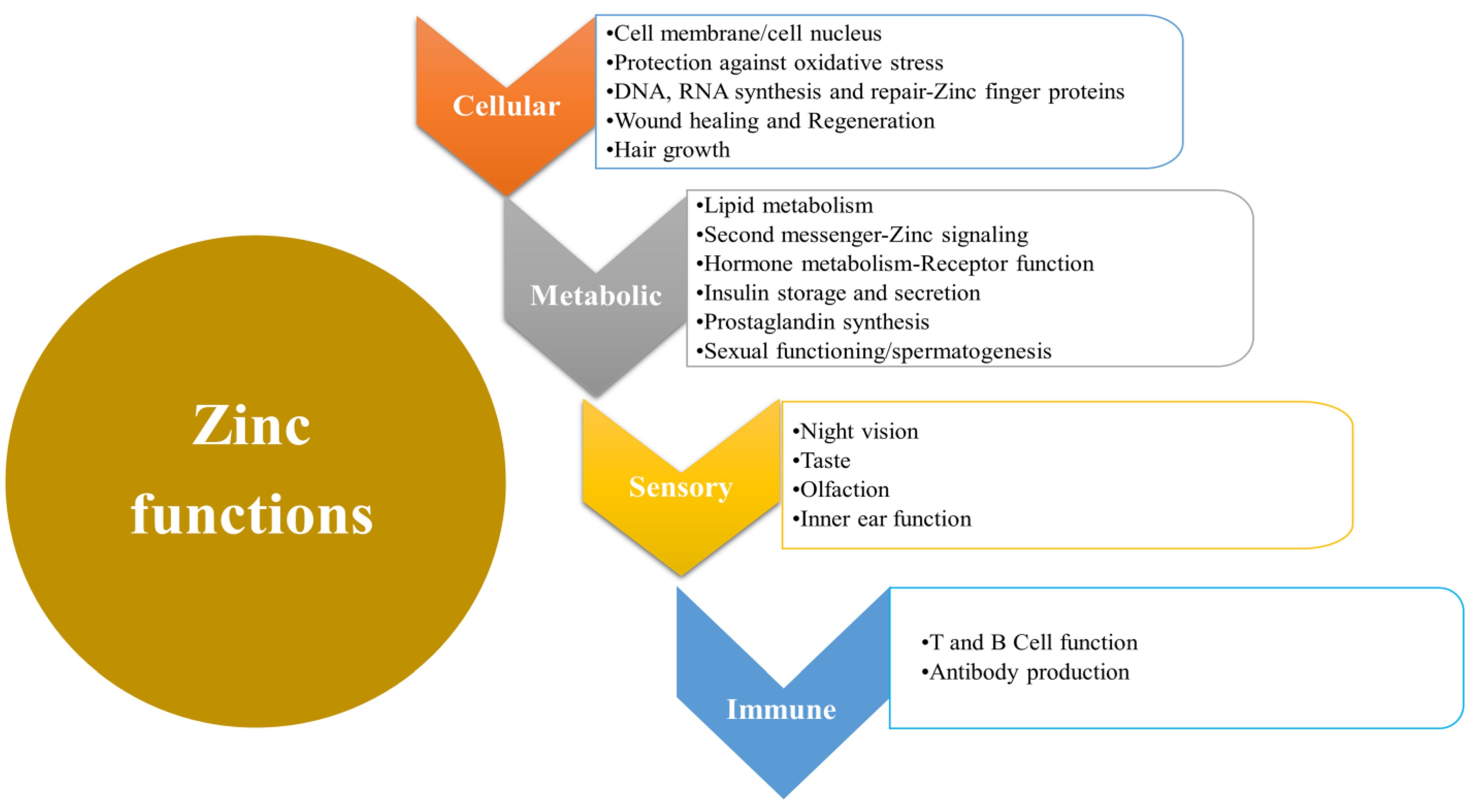
2. Biological Functions of Zinc

Zinc is an essential trace element with enormous biological functions, including cellular, metabolic, and immune functions (Figure 2). Zn functions as anti-inflammatory, antioxidant and apoptotic effects are well described [28]. It is mainly involved in cellular metabolism, specifically protein synthesis. In protein synthesis, Zn is a manipulator in the propagation of cells, diversity, and apoptotic function with dynamic expansion, revival, and cellular restoration. In the human proteome, the enzymatic catalytic domains carry zinc ions and the activity of zinc-binding proteins for more than 300 enzymes [29,30]. It also has a significant role in regulating immune functions at multiple reaction stages. In addition, it mediates its role in cellular growth and differentiation, gene expression, and metabolism [29,31]. The biological functions of Zn in human physiology are widely described, and its deficiency effects are predisposing in almost half of the world population [14]. A World Health Organization (WHO) report stated that Zn insufficiency is the fifth main risk factor for morbidity and mortality in poor or underdeveloped countries. Zn deficiency develops when its normal plasma or serum threshold level decreases from its lower normal limit of 100 µg/dL [32]. The effects of Zn deficiency can impart in different cellular and metabolic errors, including growth and oxidative depression, immune disturbances, and cognitive disorder. Due to its key biological association with protein activity, the enzymes are involved in the metabolism of collagen, consequently impacting the development of fibrosis [29].

3. Clinical Implications of Zinc Deficiency

The deficiency of zinc can lead to a variety of clinical expressions and is multifactorial. In chronic liver disease, changes in carbohydrate and lipid metabolism cause the precipitation of micronutrients and protein, resulting in malnutrition [33]. The clinical features include loss of or poor appetite, a change in taste and smell, body hair loss, delayed wound healing, and testicular dysfunction. It also causes decreased immunity and reduced drug elimination from the body [31].
The mechanism of action of Zn is mainly bound to proteins (albumin and alpha 2-macroglobulin) and acids which is directly proportional to the rate of Zn absorption. In progressive liver disease, the albumin level decreases, which may lead to reduced Zn absorption consequences in hepatic cellular carcinoma [31]. Other factors contributing to Zn deficiency in liver cirrhosis include decreased zinc absorption due to the inflammatory gut by cytokines (interleukin-6) and endotoxemia of the gut. This causes changes in the mucosa of GIT and decreases the absorption of Zn [34]. In addition, some drugs like diuretics increase the excretion of zinc and reduce the Zn binding capacity to albumin, resulting in Zn deficiency in the circulation. Furthermore, cirrhosis is complicated by hepatic encephalopathy (HE), ascites, variceal bleeding, toxicities, infections, and hepatocellular carcinoma (HCC). It characterizes the continuum of chronic liver diseases irrespective of etiology.

Mechanism of Zinc Activity and Effects of Deficiency

Zinc participates in various physiological mechanisms of enzymatic reactions having a catalytic influence on the substrate exchange and maintaining the equilibrium of enzyme assembly. It also employs fundamental activities to control transcription factors and hormonal regulation by acting on the respective receptors of the hormone and for the specific gene function/expression. The role of Zn in metabolism and development has also been well-established [35]. Furthermore, it also entails a vital second messenger role, acts as a signaling ion, and disturbs redox activity. Zinc deficiency is the key source for the development of oxidative stress and cellular apoptosis [35,36]. While Zn2+ has antioxidant proper by having the redox indolent. The defense through-provoking vigorous redox conversion of metals such as iron and copper and the safety of the sulfhydryl protein group from oxidative injury is considered the most significant antioxidant source of Zn. Additionally, this element is significantly involved in the neurotransmitters’ metabolism, development, reproductive, thyroid, and insulin storage [36,37].
Zinc ions are not protein bounded and are required principally in intra-cellular parts of various cells [38]. It acts as a messenger in inter-cellular communication and the regulation of intracellular control [39]. Zn is one of the most abundant intra-cellular micronutrient elements found in all body tissues. The largest concentration of Zn is found in bones and muscles, which is about 85% of Zn stores, and the remaining 11% is present in the skin and liver. In an adult, 70 kg of weight is equivalent to 2–3 g of zinc [40]. In developed nations, the Zn concentration in the diet usually disbursed is normally sufficient to provide this requirement, and meat is a rich source of Zn [31].
Zinc is an essential co-factor for regulating diverse caspases, an enzymatic family comprising cysteine proteases that enterprises cellular apoptosis [41]. In addition, its regulatory role is established in molecular processes, intracellular signaling systems, and activation of growth factors, hormones, and cytokines [42]. Moreover, it is a proficient intra-cellular secondary messenger compared to the calcium regulatory pathway. The hindrance in the free mobility of intracellular ionic Zn2+ applies a critical influence on the progression and development of various diseases [43]. The intracellular homeostasis of Zn is imperiled as a protein regulator for numerous metabolic reactions. The presence of these types of switches accentuates Zn’s importance. Metallothioneins are Zn carrier proteins that act as key constituents of Zn absorption and storage in this pathway [44]. A set of discrete Zn transporters expedite the incursion and efflux of zinc through the cell membrane [45].
In human cells, zip transporters (Zip and SLC39 family) and zinc carriers (ZnT and SLC30 family) are present. ZnT condenses the intra-cellular accessibility of Zn by cellular efflux or into vesicles of the cell. Although, the Zip transporters enhance the intracellular trafficking of Zn by instigating its extracellular counterparts. Moreover, perhaps, it activates the discharge of Zn from the cytoplasmic vesicle [46]. Equally, both transporter types have a precise expression in each tissue with unlikely roles in controlling nutritional zinc insufficiency or superfluity and regulating the biological functions of cytokines and hormones. Additionally, different zinc transporters harmonize the cellular, sub-cellular, and organelle-based translocation of zinc ions [47].
Gene expression of cells is mediated by the proteins targeting the regulatory Zn2+ ions, curtailing the temporary zinc-binding sites on membrane receptors, the activity of enzymes, sensor proteins, and protein–protein interactions. The regulation of Zn through a series of processes relies on specific proteins that cause metal ions to be detached. In cellular activity, these findings support the essential Zn functions and its metalloproteins [44].
The expression of Zn carrier genes (Zip1, mRNA of ZnT) is exceedingly synchronized, which is independent of nutritional Zn [48]. The expression of ZnT mRNA is speckled substantially amid specific transporters. The most expressed transporters are ZnT1, ZnT7, and Zip1. Consequently, the enhanced expression of Zn transporter recycled as a substitute represents the intra-cellular Zn deficit, equivalent to ferritin consumption in iron deficiency. The Zn metabolism aberrations also disturb mitochondrial activities, resulting in DNA damage, developmental disorders, and congenital birth defects. The availability of Zn ions was demonstrated to be higher in an oxidative environment, whereas the reductive environment decreases its availability [46]. It is proposed that the Zn disruption of the antioxidant role in deficient cases may escalate the understanding of factors that cause DNA damage and reduce DNA repair mechanisms [49].

4. Metabolism of Zinc in the Liver

The liver mainly maintains the total body circulating Zn balance [50]. Any parenchymal change in the liver results in Zn deficiency, and zinc releases from hepatocytes are differentially controlled. Throughput discoveries expending that Zn2+ displayed comprehensive interchange of Zn in hepatocytes entails fewer than two days [51]. In lieu of metabolic activity, these constituents activate a momentary metabolic Zn dysregulation with ensuing plasma Zn scarcity. Some mediators and stress factors, identical to pro-inflammatory cytokines or lipo-polysaccharides, can have comparable properties [52]. Alteration in zinc status precisely disturbs the expression of gene/s. Perhaps, the expression levels of mRNA for metallothionein, retinol-binding protein, cholecystokinin, uroguanylin, endothelin, etc., increase or collapse in retort to variations in zinc concentration [53]. Lacking zinc marks the diverse hepatic roles and, having a significant role in metabolism, it also influences the metabolic developments in other organs [54].
There are constricted connections between Zn and metallothionein, an acute-phase protein primarily required for Zn absorption, distribution, and intracellular storage. Additionally, the higher zinc consumption generates a rise in metallothionein production. The adjacent communications between zinc and a cytokine, interleukin-6 (IL-6), have also been recognized, and IL-6 is significantly involved in regulating genes of acute-phase proteins [55,56].
The role of Zn in the liver also produces acute phase proteins, in the gluconeogenesis decree, in the volatile substrates (nitrogen-monoxide) mechanism or hydrophilic radicals, other than microbial growth control enhancing IL-6. IL-6 has a role in the Zip14 transporter in the liver, promoting hypozincemia or low zinc with persistent acute-phase reactions [56]. The valuable characteristics of zinc transporters are also associated with hepatocytes; nevertheless, substantial breaches in indulgence persist. Thus far, IL-6 has leading activity for ZnT5 (a bone growth factor) and has strong evidence for activities for Zip5 and Zip6 transporters (steroid hormones, prostate, and mammary gland).
Zip-14 is multifaceted in the induction of IL-6 of hypozincemia throughout provocative progressions. IL-6 interrelating with Zip-14 shows similar impacts as in serum iron (hypo-ferritinemia) in inflammatory situations by modifying the hepcidin production in the liver [57], and Zip-14 triggers the uptake of both zinc and non-transferrin bound iron (NTBI). In hemochromatosis, the higher NTBI concentration provokes surplus iron in different tissues predisposing to cardiomyopathy, diabetes, hepatocellular carcinoma (HCC), etc. In addition, it is suggested that the HFE gene causes hereditary hemochromatosis; Zip-14 intervened in the transport of iron. The Zip-14 transporter also intercedes zinc consumption through hepatic restoration [57]. In chronic liver disease, Zip-14 mediating activity might epitomize a potential therapeutic target to endorse hepatic revival. Nonetheless, it also shows an intense role in the expansion of lumps, like in HCC. A report observed down-regulation of Zip-14 reducing zinc in cancerous cells in HCC patients that augment tumor progression. It might be concluded that the Zip-14 transporter is involved in the concentrated capacity of tumor cells to yield Zn [58].

5. Deficiency of Zinc in Liver Diseases

The deficiency of Zn has become an increasing medical or health issue worldwide. Zinc insufficiency was primarily recognized in 1960, and it is more frequent in all age groups and equally affects both genders. It may occur due to insufficient nutritional intake, diminished absorption in the small intestine, body requirements, less consumption of Zn and its loss through feces, urine, sweat, etc., and a variety of inherited disorders [59]. It is linked to various disorders, including chronic liver disease, malabsorption syndromes, and red cell pathologies. The most recent research by non-invasive high-throughput techniques [60] to detect nano-mechanical changes in the liver [61] or red blood cells [62], respectively, which may directly affect human pathologies. Various factors are responsible for developing Zn deficiency or can progress to alter the Zn metabolism in liver cirrhosis (Table 1). These factors include insufficient intake alterations in amino acid and protein metabolism, lessened portosystemic shunts, hepatic extraction, impaired absorption due to alcohol influence, and the effects of endotoxins and cytokines, primarily IL-6 [63,64].
There have been reports about severe muscular wastage resulting in considerable loss of Zn in the urine [65]. Due to the complications of ascites in cirrhotic patients, catabolism increases and demonstrates an immense fall in muscle activity. The diuretic treatment in cirrhotic patients and the complication of ascites marks not merely an augmented renal Zn elimination, nevertheless similar to condensing the serum albumin levels and compact volume of albumin to fix zinc [66].
In liver cirrhosis, several clinical characteristics have been linked to Zn deficiency, encompassing body hair loss, testicular atrophy, loss of or poor appetite, abnormal cerebral function, immune abnormalities, altered taste and smell, reduced or altered metabolism of protein, thyroid hormones, and vitamin A, late wound healing, and reduced drug elimination ability [67]. There is a wide variety of conceivable pathomechanisms of Zn deficit in liver cirrhosis. The deficiency of Zn may result in oxidative damage to tissues and/or the controlling influence on specific signaling systems in the liver [68]. The deficit of Zn may also be affecting oxidative pressure and ensuing environments like susceptibility to inflammatory hepatitis, loss of defense of acute phase in hepatitis, and factors for oxidation of lipids. In redox state variation, Zn deficiency supports transcription factors that are oxidatively favorable and can disrupt cellular function, dissemination, and existence [69,70]. This deficiency has variable impacts on liver activity, especially the restoration capability of the liver.
Due to oxidative stress influence, reduced or deficit Zn can lead to cellular and tissue damage by modifying specific signaling cascades with subsequent configurational impairment of enzymes, mitochondria, and ribosomes [71]. Production of oxidative species stimulated by lacking Zn raises inflammatory parenchyma of the liver (hepatitis) and compromises the defense phenomena against viruses and lethal materials. The change in redox potential confines the reductive oxidation of transcription factors responsible for cellular activity diversity [70]. In liver diseases, oxidative tension may endorse gut penetrability with endotoxemia, infections like spontaneous peritonitis, and discharge of hormonal stress [71].
In addition, liver cirrhosis is linked with the reflective involvement of immunological abnormalities or their failures. For example, the reticuloendothelial system failure results in scarcity or low impact of immune reconnaissance characteristics, and decreased liver production of proteins complicates the innate immunity and forms the recognition site, delaying the capability of bacteria for the phagocytosis process [72,73,74,75].

Zinc Deficiency in Liver Cirrhosis Complications

In liver cirrhosis, ascites is one of the most serious and life-threatening impediments. The liver shows a fundamental character in the directive of nourishment by governing the digestion of macro or micro-nutrients, their spreading, and apposite practice. Subsequently, protein-energy malnourishment in advanced liver disease patients characterizes a vital predictive marker for distressing mortality, the success of liver transplantation, and the value of lifespan [76,77,78]. In cirrhosis, the catabolic state has characteristic disproportionate plasma levels of amino acids: a decreasing or diminished branched amino acids (Isoleucine, Leucine, Valine) and excessive level of plasma aromatic amino acids (Phenylalanine, Tyrosine, Tryptophan) [79]. Moreover, amino acid and protein metabolism reorganization appeared by advanced undernourished muscle cessation. They lessened ammonia removal from muscles and the liver and muscles, and cirrhotic patients suffered from metabolism turbulences with better energy disbursement and repeatedly improved hyperactive catabolic rate [80].
Several reports have described that supplementing some basic amino acids or with a zinc mixture might develop hypo-albuminemia and ascites, causing the amplified source of substrate for proteins and the incentive of protein combination [81,82,83]. Albumin has been revealed to be a multi-functional protein with antioxidant, immune modulator, and reclamation purposes [84]. It shows an ultimate character in the metabolites dissemination; Zn and copper ion transition, hormones, and drugs [85]. Albumin is a main transport protein of Zn in the plasma, and about 80% of plasma Zn is transported by this mode [86]. It has been described that liver cirrhosis is linked to massive Zn deficiency, which progresses to abnormalities of nitrogen metabolism [87]. The branched change amino acids are twisted in innumerable biological progressions like stimulus of albumin and glycogenesis, upgrading of insulin confrontation, stimulation of mitochondrial bioenergetics, limitation of ROS production and apoptosis of hepatic cells, and improvement in liver restoration [88]. In randomized clinical trials, it has been presented as a conceivable outcome of branch chain amino acids (BCAAs) in the control of CLD [89]. Two mechanisms have been involved in reducing hypoalbuminemia in regard to the ability of BCAAs rich supplement.
Additionally, another report described that constant nutritional supplementation with BCAAs prompted ribosomal phosphorylation of the S6 protein in the liver of rats with CLD. Therefore, subsequent higher production of albumin BCAAs supplementation can increase an osmotic pressure, which results in extracellular fluid reduction and can lead to ascites progression. The nutritional supply of Zn might affect the depletion of BCAAs in muscle mass. Therefore, the intake of BCAAs might be utilized for albumin synthesis, which reduces serum concentrations of albumin and results in the reduction of ascites degree [89]. Zinc stored in different organs like the liver, muscle, and bone can be replaced independently of the severity of liver cirrhosis and complications such as diabetes mellitus.
In another study, children suffering from malnutrition with chromosomal damage and cytotoxic properties could be restored in vitro with Zn sulfate nutrients. Nonetheless, it should be known that more investigations involving large patient numbers are required to govern the comprehensive application of achieving and maintaining clinical productivity in liver cirrhosis management [90].
Hepatic encephalopathy (HE) is a reversible neuro-psychiatric syndrome that might emerge in serious disorders like acute and chronic liver conditions. Approximately 70% of cirrhotic patients are involved in incidences of HE. While the pathogenesis of HE is diverse and heterogeneous, the higher concentrations of ammonia contribute a critical role in this pathway. Hyperammonemia persuades the inflammation of the astrocyte cells and concomitant deviations that dislocate neuronal communication and decrease the brain’s energy provision. In addition, edema in the astrocytes influences the fundamental brain proteins and activates nitrosative and oxidative pressure [91,92].
Further aspects of excessive ammonia that are complicated in the HE pathogenesis contain contagions, abnormal neutrophil functions, and the properties of pro-inflammatory cytokines [93]. The poor status of Zn spoils the metabolism of nitrogen by tumbling the action of enzymes of the urea cycle in the liver [10] and in the skeletal muscle [79]. Zn deficiency is linked with metabolic nitrogen alterations, in an investigational rat model of cirrhosis and progressive liver disease patients [94]. It is proposed that the diminished Zn may be involved in the HE pathogenesis, as serum Zn levels are curtailed in HE patients and associated inversely with ammonia concentrations in the blood [95]. The mechanism is presented in Figure 3. It is also supported in another report of a patient with severe HE and a Zn deficit [96]. The longer duration of Zn supplementation enhanced the improvement in encephalopathy and eminence of lifespan by depressing the circulating ammonia. Very few controlled studies have commenced scrutinizing Zn proficiency for treating HE cases associated with liver cirrhosis [97]. Overall, some clinical-oriented controlled reports of Zn replacement in HE patients endured minor and inconsistent outcomes. One of the fascinating statements suggests that the exchange of Zn has a valuable result on the muscle spasms commonly agonized by liver cirrhosis patients [98].
Another complication of liver cirrhosis, hepatocellular carcinoma (HCC), is considered the fifth most common type and third most death-associated cancer globally [99]. Liver cirrhosis is the conclusive disease risk factor causing the development of HCC. The mechanism of hepatic carcinogenesis is not clearly understood; however, exclusive details have been explicated. It is understood that the involvement of signal transduction paths alleviated cancer progression and may represent the objective constructions for systemic recovery. Still, there is massive HCC heterogeneity, and the wide-ranging energies are necessary to express the molecular genesis of HCC, evidently [100].
The close relationship between chronic inflammatory effects and cancer expansion has been clearly documented. HCC cases increase due to the genetic variability associated with chronic inflammation exposure. The inflammatory factors may be diverse, comprising viral infections and toxic sources like alcohol and aflatoxin. Moreover, metabolic instabilities or syndromes, like diabetes mellitus, may persuade and disseminate chronic inflammatory responses, stimulating cancer expansion. Many heterogeneous regulatory agents and signal pathways are concerned with the carcinogenesis elicited by infection. In the cellular aging mechanism, chronic inflammation results in early cellular death, telomeres limitation, and genetic uncertainty. The relationship between chronic inflammation exposure and immunological deviations controlled by viruses can be evidently described in hepatitis C predisposition [101].
HCV-induced infections carry immunological variations that devastate the hepatocytes, whereas generating recreating routes at the same point maintains the inflammation, affects the oxidative stress, and may damage the DNA. At this stage, HCC seems to develop obstinate inflammation with progressive fibrosis, ultimately progressing to liver growth [99]. Zinc is one of the essential trace elements with diverse functions, and its low concentrations in plasma are associated with HCC. Plasma zinc levels are exaggerated by anxiety, impurities, disturbances, and malicious routes. The raised serum copper levels are a decree linked with reduced zinc meditations [100], and a connection between high serum copper and possible pathogenesis of HCC progression has been established. Conversely, the zinc concentration was abnormally low in HCC tissue compared to its level in the healthy liver parenchyma. A retrospective analysis of liver cirrhosis patients presented about 18.5% of cases of HCC. Of these patients, about 76.7% displayed zinc deficiency. About 55% of alcohol-induced cirrhosis cases unveiled decreased zinc levels, and zinc deficiency was detected in 95% of chronic hepatitis C [101]. It is not yet clear whether the fluctuations of zinc levels between serum and tissue subsidize the tumor development or are, as an alternative, influencing the malignant transformation of the situation.
Depending on remarks of intra-cellular Zn levels in dissimilar cancers, it has been hypothesized that these aberrations may lead to tumor progress by impressing Zn signal function, which ensures the inhibitory or triggering possessions on a wide variability of molecular assemblies. Furthermore, there are receptors, transcription factors, and caspase kinases, and phosphates. A fundamental part possibly follows abnormalities in the expression of different zinc transporters (Zip, ZnT), as it has been recommended in the initial research on certain measures of pancreatic and mammary carcinoma [51]. A previous study detected a reduction in the Zip14 expression profile, fixed with a lessening of zinc concentrations throughout the change and evolution of HCC [70,102].

6. Conclusions

The biological properties of zinc have been illustrated widely. The roles of zinc are stated in organic compounds metabolism (Glucose, Protein, and Fat), immune functions, and hepatocarcinogenesis. Zinc deficiency reduces the capacity of the liver to metabolize appropriately, resulting in impaired protein metabolism. This phenomenon significantly contributes to the development of chronic liver disease and cirrhosis, proposing the efficiency of zinc supplementation therapy. Advance studies would be necessary to explain whether zinc transporters in hepatocytes have the restraint or prompting influence on the zinc-dependent signal cascades that are substantial in the growth of hepatocellular carcinogenesis. The contribution of zinc to pathologic conditions other than impaired nitrogen metabolism in patients with liver cirrhosis and possibilities for treatment also requires further study. While HCC’s huge heterogeneity may control only a small patient group, it may still represent a precise, thrilling, and esteemed assignment. It would take longer to understand tailored, personalized medicine perception.

Author Contributions

Conceptualization, M.I.U.; investigation, M.I.U., A.A.M.A. and M.U.M.; resources, Z.H.A.-O. and L.B.E.; data curation, M.A. and M.U.M.; writing—original draft preparation, M.I.U., A.A.M.A. and M.A.; writing—review and editing, Z.H.A.-O., L.B.E., M.U.M. and H.E.; supervision, M.I.U. and H.E.; project administration, H.E. All authors have read and agreed to the published version of the manuscript.

Funding

This research received no external funding.

Institutional Review Board Statement

Not Applicable.

Informed Consent Statement

Not Applicable.

Data Availability Statement

All the available data included in this study can be found in the text of the manuscript.

Conflicts of Interest

Authors declare no conflicts of interest.

References

  1. Almaeen, A.H.; Alduraywish, A.A.; Mobasher, M.A.; Almadhi, O.I.M.; Nafeh, H.M.; El-Metwally, T.H. Oxidative stress, immunological and cellular hypoxia biomarkers in hepatitis C treatment-naïve and cirrhotic patients. Arch. Med. Sci. 2021, 17, 368–375. [Google Scholar] [CrossRef]
  2. Vuppalanchi, R.; Ghabril, M.; Chalasani, N.; Juluri, R.; Bell, L.N.; Kamendulis, L.; Klaunig, J.E.; Saxena, R.; Agarwal, D.; Johnson, M.S. Oxidative stress in chronic liver disease: Relationship between peripheral and hepatic measurements. Am. J. Med. Sci. 2011, 342, 314–317. [Google Scholar] [CrossRef] [Green Version]
  3. Amodio, P.; Bemeur, C.; Butterworth, R.; Cordoba, J.; Kato, A.; Montagnese, S.; Uribe, M.; Vilstrup, H.; Morgan, M.Y. The nutritional management of hepatic encephalopathy in patients with cirrhosis: International Society for Hepatic Encephalopathy and Nitrogen Metabolism Consensus. Hepatology 2013, 58, 325–336. [Google Scholar] [CrossRef]
  4. Conde de la Rosa, L.; Goicoechea, L.; Torres, S.; Garcia-Ruiz, C.; Fernandez-Checa, J.C. Role of Oxidative Stress in Liver Disorders. Livers 2022, 2, 283–314. [Google Scholar] [CrossRef]
  5. Kumar, V.; Abbas, A.K.; Fausto, N.; Aster, J.C. Robbins and Cotran Pathologic Basis of Disease, Professional Edition E-Book; Elsevier Health Sciences: Amsterdam, The Netherlands, 2014. [Google Scholar]
  6. Himoto, T.; Masaki, T. Associations between Zinc Deficiency and Metabolic Abnormalities in Patients with Chronic Liver Disease. Nutrients 2018, 10, 88. [Google Scholar] [CrossRef] [Green Version]
  7. Wessels, I.; Maywald, M.; Rink, L. Zinc as a Gatekeeper of Immune Function. Nutrients 2017, 9, 1286. [Google Scholar] [CrossRef] [PubMed] [Green Version]
  8. Zago, M.P.; Oteiza, P.I. The antioxidant properties of zinc: Interactions with iron and antioxidants. Free Radic. Biol. Med. 2001, 31, 266–274. [Google Scholar] [CrossRef] [PubMed]
  9. Schechter, P.; Giroux, E.; Schlienger, J.; Hoenig, V.; Sjoerdsma, A. Distribution of serum zinc between albumin and alpha2-macroglobulin in patients with decompensated hepatic cirrhosis. Eur. J. Clin. Investig. 1976, 6, 147–150. [Google Scholar] [CrossRef]
  10. Karayalcin, S.; Arcasoy, A.; Uzunalimoglu, O. Zinc plasma levels after oral zinc tolerance test in nonalcoholic cirrhosis. Dig. Dis. Sci. 1988, 33, 1096–1102. [Google Scholar] [CrossRef]
  11. Cheemerla, S.; Balakrishnan, M. Global Epidemiology of Chronic Liver Disease. Clin. Liver Dis. 2021, 17, 365–370. [Google Scholar] [CrossRef]
  12. The Global Health Observatory. Global Health Estimates: Leading Causes of Death. 2023. WHO. Available online: https://www.who.int/data/gho/data/themes/mortality-and-global-health-estimates/ghe-leading-causes-of-death (accessed on 20 February 2023).
  13. GBD 2017 Cirrhosis Collaborators. The global, regional, and national burden of cirrhosis by cause in 195 countries and territories, 1990–2017: A systematic analysis for the Global Burden of Disease Study 2017. Lancet Gastroenterol. Hepatol. 2020, 5, 245–266. [Google Scholar] [CrossRef] [Green Version]
  14. Grüngreiff, K.; Reinhold, D. Liver cirrhosis and “liver” diabetes mellitus are linked by zinc deficiency. Med. Hypotheses. 2005, 64, 316–317. [Google Scholar] [CrossRef]
  15. Riggio, O.; Merli, M.; Capocaccia, L.; Caschera, M.; Zullo, A.; Pinto, G.; Gaudio, E.; Franchitto, A.; Spagnoli, R.; D’Aquilino, E.; et al. Zinc supplementation reduces blood ammonia and increases liver ornithine transcarbamylase activity in experimental cirrhosis. Hepatology 1992, 16, 785–789. [Google Scholar] [CrossRef]
  16. Williams, R.; Alexander, G.; Aspinall, R.; Batterham, R.; Bhala, N.; Bosanquet, N.; Severi, K.; Burton, A.; Burton, R.; Cramp, M.E. Gathering momentum for the way ahead: Fifth report of the Lancet Standing Commission on Liver Disease in the UK. Lancet 2018, 392, 2398–2412. [Google Scholar] [CrossRef] [Green Version]
  17. Vega, S.; Neira, J.L.; Marcuello, C.; Lostao, A.; Abian, O.; Velazquez-Campoy, A. NS3 Protease from Hepatitis C Virus: Biophysical Studies on an Intrinsically Disordered Protein Domain. Int. J. Mol. Sci. 2013, 14, 13282–13306. [Google Scholar] [CrossRef] [Green Version]
  18. Williams, R.; Aspinall, R.; Bellis, M.; Camps-Walsh, G.; Cramp, M.; Dhawan, A.; Ferguson, J.; Forton, D.; Foster, G.; Gilmore, I. Addressing liver disease in the UK: A blueprint for attaining excellence in health care and reducing premature mortality from lifestyle issues of excess consumption of alcohol, obesity, and viral hepatitis. Lancet 2014, 384, 1953–1997. [Google Scholar] [CrossRef] [PubMed]
  19. Grüngreiff, K.; Reinhold, D.; Wedemeyer, H. The role of zinc in liver cirrhosis. Ann. Hepatol. 2016, 15, 7–16. [Google Scholar] [CrossRef] [PubMed]
  20. Yoshida, Y.; Higashi, T.; Nouso, K.; Nakatsukasa, H.; Nakamura, S.-I.; Watanabe, A.; Tsuji, T. Effects of zinc deficiency/zinc supplementation on ammonia metabolism in patients with decompensated liver cirrhosis. Acta Med. Okayama 2001, 55, 349–355. [Google Scholar] [PubMed]
  21. McClain, C.J.; Antonow, D.R.; Cohen, D.A.; Shedlofsky, S.I. Zinc metabolism in alcoholic liver disease. Alcohol. Clin. Exp. Res. 1986, 10, 582–589. [Google Scholar] [CrossRef]
  22. El-Serag, H.B. Hepatocellular carcinoma. N. Engl. J. Med. 2011, 365, 1118–1127. [Google Scholar] [CrossRef]
  23. Grüngreiff, K.; Presser, H.; Franke, D.; Lössner, B.; Abicht, K.; Kleine, F. Correlations between zinc, amino acids and ammonia in liver cirrhosis. Z. Gastroenterol. 1989, 27, 731–735. [Google Scholar]
  24. Miatto, O.; Casaril, M.; Gabrielli, G.B.; Nicoli, N.; Bellisola, G.; Corrocher, R. Diagnostic and prognostic value of serum copper and plasma fibrinogen in hepatic carcinoma. Cancer 1985, 55, 774–778. [Google Scholar] [CrossRef]
  25. Zoroddu, M.A.; Aaseth, J.; Crisponi, G.; Medici, S.; Peana, M.; Nurchi, V.M. The essential metals for humans: A brief overview. J. Inorg. Biochem. 2019, 195, 120–129. [Google Scholar] [CrossRef]
  26. Schuppan, D.; Afdhal, N.H. Liver cirrhosis. Lancet 2008, 371, 838–851. [Google Scholar] [CrossRef]
  27. Ullah, M.I.; Alsrhani, A.; Atif, M.; Shaukat, I.; Hussain, S.; Ejaz, H. Estimation of serum iron, serum lipids and serum liver enzymes in celiac disease patients of Saudi Arabia. Pak. J. Med. Sci. 2022, 38, 2101–2106. [Google Scholar] [PubMed]
  28. McClain, C.J.; Marsano, L.; Burk, R.F.; Bacon, B. Trace metals in liver disease. Semin. Liver Dis. 1991, 11, 321–339. [Google Scholar] [CrossRef]
  29. Sengupta, S.; Wroblewski, K.; Aronsohn, A.; Reau, N.; Reddy, K.G.; Jensen, D.; Te, H. Screening for Zinc Deficiency in Patients with Cirrhosis: When Should We Start? Dig. Dis. Sci. 2015, 60, 3130–3135. [Google Scholar] [CrossRef] [PubMed]
  30. Vallee, B.L.; Falchuk, K.H. The biochemical basis of zinc physiology. Physiol. Rev. 1993, 73, 79–118. [Google Scholar] [CrossRef] [PubMed]
  31. Leoni, G.; Rosato, A.; Perozzi, G.; Murgia, C. Zinc proteome interaction network as a model to identify nutrient-affected pathways in human pathologies. Genes. Nutr. 2014, 9, 1–9. [Google Scholar] [CrossRef] [Green Version]
  32. Plum, L.M.; Rink, L.; Haase, H. The essential toxin: Impact of zinc on human health. Int. J. Environ. Res. Public Health 2010, 7, 1342–1365. [Google Scholar] [CrossRef] [Green Version]
  33. Wessells, K.; King, J.; Brown, K. Development of a plasma zinc concentration cutoff to identify individuals with severe zinc deficiency based on results from adults undergoing experimental severe dietary zinc restriction and individuals with acrodermatitis enteropathica. J. Nutr. 2014, 144, 1204–1210. [Google Scholar] [CrossRef] [Green Version]
  34. Maret, W. Zinc and sulfur: A critical biological partnership. Biochemistry 2004, 43, 3301–3309. [Google Scholar] [CrossRef]
  35. Stamoulis, I.; Kouraklis, G.; Theocharis, S. Zinc and the liver: An active interaction. Dig. Dis. Sci. 2007, 52, 1595–1612. [Google Scholar] [CrossRef]
  36. Maret, W.; Krezel, A. Cellular zinc and redox buffering capacity of metallothionein/thionein in health and disease. Mol. Med. 2007, 13, 371–375. [Google Scholar] [CrossRef]
  37. Faure, P.; Roussel, A.; Richard, M.; Foulon, T.; Groslambert, P.; Hadjian, A.; Favier, A. Effect of an acute zinc depletion on rat lipoprotein distribution and peroxidation. Biol. Trace Elem. Res. 1991, 28, 135–146. [Google Scholar] [CrossRef]
  38. Prasad, A.S. Discovery of human zinc deficiency and studies in an experimental human model. Am. J. Clin. Nutr. 1991, 53, 403–412. [Google Scholar] [CrossRef]
  39. Danscher, G.; Stoltenberg, M. Zinc-specific autometallographic in vivo selenium methods: Tracing of zinc-enriched (ZEN) terminals, ZEN pathways, and pools of zinc ions in a multitude of other ZEN cells. J. Histochem. Cytochem. 2005, 53, 141–153. [Google Scholar] [CrossRef] [PubMed] [Green Version]
  40. Fukada, T.; Yamasaki, S.; Nishida, K.; Murakami, M.; Hirano, T. Zinc homeostasis and signaling in health and diseases: Zinc signaling. J. Biol. Inorg. Chem. 2011, 16, 1123–1134. [Google Scholar] [CrossRef] [PubMed] [Green Version]
  41. Muhamed, P.K.; Vadstrup, S. Zinc is the most important trace element. Ugeskr. Laeger 2014, 176, V11120654. (In Danish) [Google Scholar]
  42. Huber, K.L.; Hardy, J.A. Mechanism of zinc-mediated inhibition of caspase-9. Protein Sci. 2012, 21, 1056–1065. [Google Scholar] [CrossRef] [PubMed] [Green Version]
  43. Beyersmann, D.; Haase, H. Functions of zinc in signaling, proliferation and differentiation of mammalian cells. Biometals 2001, 14, 331–341. [Google Scholar] [CrossRef]
  44. Maret, W. Zinc coordination environments in proteins as redox sensors and signal transducers. Antioxid. Redox. Signal. 2006, 8, 1419–1441. [Google Scholar] [CrossRef]
  45. Maret, W. Zinc biochemistry: From a single zinc enzyme to a key element of life. Adv. Nutr. 2013, 4, 82–91. [Google Scholar] [CrossRef] [Green Version]
  46. Temple, V.J.; Masta, A. Zinc in human health. Papua New Guin. Med. J. 2004, 47, 146–158. [Google Scholar]
  47. Liuzzi, J.P.; Cousins, R.J. Mammalian zinc transporters. Annu. Rev. Nutr. 2004, 24, 151. [Google Scholar] [CrossRef] [PubMed]
  48. Lichten, L.A.; Cousins, R.J. Mammalian zinc transporters: Nutritional and physiologic regulation. Annu. Rev. Nutr. 2009, 29, 153–176. [Google Scholar] [CrossRef] [PubMed]
  49. Foster, M.; Hancock, D.; Petocz, P.; Samman, S. Zinc transporter genes are coordinately expressed in men and women independently of dietary or plasma zinc. J. Nutr. 2011, 141, 1195–1201. [Google Scholar] [CrossRef] [PubMed] [Green Version]
  50. Ho, E. Zinc deficiency, DNA damage and cancer risk. J. Nutr. Biochem. 2004, 15, 572–578. [Google Scholar] [CrossRef]
  51. Tuerk, M.J.; Fazel, N. Zinc deficiency. Curr. Opin. Gastroenterol. 2009, 25, 136–143. [Google Scholar] [CrossRef] [PubMed]
  52. Taylor, J.; Simons, T. The mechanism of zinc uptake by cultured rat liver cells. J. Physiol. 1994, 474, 55–64. [Google Scholar] [CrossRef] [Green Version]
  53. Cousins, R.J. Absorption, transport, and hepatic metabolism of copper and zinc: Special reference to metallothionein and ceruloplasmin. Physiol. Rev. 1985, 65, 238–309. [Google Scholar] [CrossRef]
  54. Peng, H.; Wisse, E.; Tian, Z. Liver natural killer cells: Subsets and roles in liver immunity. Cell. Mol. Immunol. 2016, 13, 328–336. [Google Scholar] [CrossRef] [PubMed] [Green Version]
  55. Tom Dieck, H.; Döring, F.; Roth, H.-P.; Daniel, H. Changes in rat hepatic gene expression in response to zinc deficiency as assessed by DNA arrays. J. Nutr. 2003, 133, 1004–1010. [Google Scholar] [CrossRef] [Green Version]
  56. Liuzzi, J.P.; Lichten, L.A.; Rivera, S.; Blanchard, R.K.; Aydemir, T.B.; Knutson, M.D.; Ganz, T.; Cousins, R.J. Interleukin-6 regulates the zinc transporter Zip14 in liver and contributes to the hypozincemia of the acute-phase response. Proc. Natl. Acad. Sci. USA 2005, 102, 6843–6848. [Google Scholar] [CrossRef] [PubMed] [Green Version]
  57. Nemeth, E.; Tuttle, M.S.; Powelson, J.; Vaughn, M.B.; Donovan, A.; Ward, D.M.; Ganz, T.; Kaplan, J. Hepcidin regulates cellular iron efflux by binding to ferroportin and inducing its internalization. Science 2004, 306, 2090–2093. [Google Scholar] [CrossRef] [Green Version]
  58. Hentze, M.W.; Muckenthaler, M.U.; Andrews, N.C. Balancing acts: Molecular control of mammalian iron metabolism. Cell 2004, 117, 285–297. [Google Scholar] [CrossRef] [PubMed] [Green Version]
  59. Franklin, R.B.; Levy, B.A.; Zou, J.; Hanna, N.; Desouki, M.M.; Bagasra, O.; Johnson, L.A.; Costello, L.C. ZIP14 zinc transporter downregulation and zinc depletion in the development and progression of hepatocellular cancer. J. Gastrointest. Cancer 2012, 43, 249–257. [Google Scholar] [CrossRef] [Green Version]
  60. Magazzù, A.; Marcuello, C. Investigation of Soft Matter Nanomechanics by Atomic Force Microscopy and Optical Tweezers: A Comprehensive Review. Nanomaterials 2023, 13, 963. [Google Scholar] [CrossRef]
  61. Islam, M.R.; Virag, J.; Oyen, M.L. Micromechanical poroelastic and viscoelastic properties of ex-vivo soft tissues. J. Biomech. 2020, 113, 110090. [Google Scholar] [CrossRef]
  62. Tognato, R.; Jones, P.H. Ray Optics Model for Optical Trapping of Biconcave Red Blood Cells. Micromachines 2022, 14, 83. [Google Scholar] [CrossRef]
  63. Ullah, M.I.; Alzahrani, B.; Alsrhani, A.; Atif, M.; Alameen, A.A.M.; Ejaz, H. Determination of serum tumor necrosis factor-alpha (TNF-α) levels in metabolic syndrome patients from Saudi population. Pak. J. Med. Sci. 2021, 37, 700–705. [Google Scholar] [CrossRef]
  64. Hameed, T.; Khan, Z.; Imran, M.; Ali, S.; Albegali, A.A.; Ullah, M.I.; Ejaz, H. Associations of transcription factor 7-Like 2 (TCF7L2) gene polymorphism in patients of type 2 diabetes mellitus from Khyber Pakhtunkhwa population of Pakistan. Afr. Health Sci. 2021, 21, 15–22. [Google Scholar] [CrossRef]
  65. Hara, K.; Yonezawa, K.; Weng, Q.P.; Kozlowski, M.T.; Belham, C.; Avruch, J. Amino acid sufficiency and mTOR regulate p70 S6 kinase and eIF-4E BP1 through a common effector mechanism. J. Biol. Chem. 1998, 273, 14484–14494. [Google Scholar] [CrossRef] [PubMed] [Green Version]
  66. Cuthbertson, D.P.; Fell, G.S.; Smith, C.M.; Tilstone, W.J. Metabolism after injury. I. Effects of severity, nutrition, and environmental temperatue on protein potassium, zinc, and creatine. Br. J. Surg. 1972, 59, 926–931. [Google Scholar]
  67. Sattar, A.; Khan, J.; Kakar, N.H.; Butt, M.U.A.; Yousaf, H.; Ullah, M.I. Serum zinc levels and the prevalence of zinc deficiency in patients with liver cirrhosis. Rawal. Med. J. 2021, 46, 37–40. [Google Scholar]
  68. Barry, M.; Keeling, P.W.; Feely, J. Tissue zinc status and drug elimination in patients with chronic liver disease. Clin. Sci. 1990, 78, 547–549. [Google Scholar] [CrossRef] [Green Version]
  69. Oteiza, P.I.; Mackenzie, G.G. Zinc, oxidant-triggered cell signaling, and human health. Mol. Aspects Med. 2005, 26, 245–255. [Google Scholar] [CrossRef]
  70. DiSilvestro, R.A. Zinc in relation to diabetes and oxidative disease. J. Nutr. 2000, 130 (Suppl. S5), 1509s–1511s. [Google Scholar] [CrossRef] [PubMed] [Green Version]
  71. Parsons, S.E.; DiSilvestro, R.A. Effects of mild zinc deficiency, plus or minus an acute-phase response, on galactosamine-induced hepatitis in rats. Br. J. Nutr. 1994, 72, 611–618. [Google Scholar] [CrossRef] [PubMed] [Green Version]
  72. Gaetke, L.M.; McClain, C.J.; Talwalkar, R.T.; Shedlofsky, S.I. Effects of endotoxin on zinc metabolism in human volunteers. Am. J. Physiol. 1997, 272 Pt 1, E952–E956. [Google Scholar] [CrossRef] [PubMed]
  73. Jenne, C.N.; Kubes, P. Immune surveillance by the liver. Nat. Immunol. 2013, 14, 996–1006. [Google Scholar] [CrossRef] [PubMed]
  74. Racanelli, V.; Rehermann, B. The liver as an immunological organ. Hepatology 2006, 43 (Suppl. 1), S54–S62. [Google Scholar] [CrossRef]
  75. Albillos, A.; Lario, M.; Álvarez-Mon, M. Cirrhosis-associated immune dysfunction: Distinctive features and clinical relevance. J. Hepatol. 2014, 61, 1385–1396. [Google Scholar] [CrossRef] [PubMed] [Green Version]
  76. Wiest, R.; Lawson, M.; Geuking, M. Pathological bacterial translocation in liver cirrhosis. J. Hepatol. 2014, 60, 197–209. [Google Scholar] [CrossRef] [PubMed] [Green Version]
  77. Bémeur, C.; Desjardins, P.; Butterworth, R.F. Role of nutrition in the management of hepatic encephalopathy in end-stage liver failure. J. Nutr. Metab. 2010, 2010, 489823. [Google Scholar] [CrossRef] [PubMed] [Green Version]
  78. O’Brien, A.; Williams, R. Nutrition in end-stage liver disease: Principles and practice. Gastroenterology 2008, 134, 1729–1740. [Google Scholar] [CrossRef] [Green Version]
  79. Grüngreiff, K. Branched Amino Acids and Zinc in the Nutrition of Liver Cirrhosis. J. Clin. Exp. Hepatol. 2018, 8, 480–483. [Google Scholar] [CrossRef]
  80. Buchman, A. L Total parenteral nutrition: Challenges and practice in the cirrhotic patient. Transpl. Proc. 2006, 38, 1659–1663. [Google Scholar] [CrossRef]
  81. Bianchi, G.; Marzocchi, R.; Lorusso, C.; Ridolfi, V.; Marchesini, G. Nutritional treatment of chronic liver failure. Hepatol. Res. 2008, 38 (Suppl. 1), S93–S101. [Google Scholar] [CrossRef]
  82. Hayashi, M.; Ikezawa, K.; Ono, A.; Okabayashi, S.; Hayashi, Y.; Shimizu, S.; Mizuno, T.; Maeda, K.; Akasaka, T.; Naito, M.; et al. Evaluation of the effects of combination therapy with branched-chain amino acid and zinc supplements on nitrogen metabolism in liver cirrhosis. Hepatol. Res. 2007, 37, 615–619. [Google Scholar] [CrossRef]
  83. Itou, M.; Kawaguchi, T.; Taniguchi, E.; Oku, Y.; Fukushima, N.; Ando, E.; Oriishi, T.; Uchida, Y.; Otsuka, M.; Tanaka, S.; et al. Branched-chain amino acid supplements reduced ascites and increased the quality of life in a patient with liver cirrhosis: A case repor. Mol. Med. Rep. 2009, 2, 977–981. [Google Scholar] [CrossRef]
  84. Garcia-Martinez, R.; Caraceni, P.; Bernardi, M.; Gines, P.; Arroyo, V.; Jalan, R. Albumin: Pathophysiologic basis of its role in the treatment of cirrhosis and its complications. Hepatology 2013, 58, 1836–1846. [Google Scholar] [CrossRef]
  85. Bal, W.; Sokołowska, M.; Kurowska, E.; Faller, P. Binding of transition metal ions to albumin: Sites, affinities and rates. Biochim. Biophys. Acta 2013, 1830, 5444–5455. [Google Scholar] [CrossRef]
  86. Lu, J.; Stewart, A.J.; Sadler, P.J.; Pinheiro, T.J.; Blindauer, C.A. Albumin as a zinc carrier: Properties of its high-affinity zinc-binding site. Biochem. Soc. Trans. 2008, 36 Pt 6, 1317–1321. [Google Scholar] [CrossRef] [PubMed] [Green Version]
  87. Tajiri, K.; Shimizu, Y. Branched-chain amino acids in liver diseases. World. J. Gastroenterol. 2013, 19, 7620–7629. [Google Scholar] [CrossRef] [PubMed]
  88. Holecek, M. Branched-chain amino acids and ammonia metabolism in liver disease: Therapeutic implications. Nutrition 2013, 29, 1186–1191. [Google Scholar] [CrossRef] [PubMed]
  89. Takuma, Y.; Nouso, K.; Makino, Y.; Hayashi, M.; Takahashi, H. Clinical trial: Oral zinc in hepatic encephalopathy. Aliment. Pharmacol. Ther. 2010, 32, 1080–1090. [Google Scholar] [CrossRef]
  90. Padula, G.; González, H.F.; Varea, A.; Seoane, A.I. Protein energy-malnutrition: Does the in vitro zinc sulfate supplementation improve chromosomal damage repair? Biol. Trace. Elem. Res. 2014, 162, 64–71. [Google Scholar] [CrossRef]
  91. Riggio, O.; Ridola, L.; Pasquale, C. Hepatic encephalopathy therapy: An overview. World J. Gastrointest. Pharmacol. Ther. 2010, 1, 54–63. [Google Scholar] [CrossRef]
  92. Schliess, F.; Görg, B.; Häussinger, D. RNA oxidation and zinc in hepatic encephalopathy and hyperammonemia. Metab. Brain Dis. 2009, 24, 119–134. [Google Scholar] [CrossRef]
  93. Shawcross, D.L.; Shabbir, S.S.; Taylor, N.J.; Hughes, R.D. Ammonia and the neutrophil in the pathogenesis of hepatic encephalopathy in cirrhosis. Hepatology 2010, 51, 1062–1069. [Google Scholar] [CrossRef]
  94. Marchesini, G.; Fabbri, A.; Bianchi, G.; Brizi, M.; Zoli, M. Zinc supplementation and amino acid-nitrogen metabolism in patients with advanced cirrhosis. Hepatology 1996, 23, 1084–1092. [Google Scholar] [CrossRef]
  95. Reding, P.; Duchateau, J.; Bataille, C. Oral zinc supplementation improves hepatic encephalopathy. Results of a randomised controlled trial. Lancet 1984, 2, 493–495. [Google Scholar] [CrossRef] [PubMed]
  96. Van der Rijt, C.C.; Schalm, S.W.; Schat, H.; Foeken, K.; De Jong, G. Overt hepatic encephalopathy precipitated by zinc deficiency. Gastroenterology 1991, 100, 1114–1118. [Google Scholar] [CrossRef]
  97. Riggio, O.; Ariosto, F.; Merli, M.; Caschera, M.; Zullo, A.; Balducci, G.; Ziparo, V.; Pedretti, G.; Fiaccadori, F.; Bottari, E.; et al. Short-term oral zinc supplementation does not improve chronic hepatic encephalopathy. Results of a double-blind crossover trial. Dig. Dis. Sci. 1991, 36, 1204–1208. [Google Scholar] [CrossRef] [PubMed]
  98. Kugelmas, M. Preliminary observation: Oral zinc sulfate replacement is effective in treating muscle cramps in cirrhotic patients. J. Am. Coll. Nutr. 2000, 19, 13–15. [Google Scholar] [CrossRef] [PubMed]
  99. Walker, M.; El-Serag, H.B.; Sada, Y.; Mittal, S.; Ying, J.; Duan, Z.; Richardson, P.; Davila, J.A.; Kanwal, F. Cirrhosis is under-recognised in patients subsequently diagnosed with hepatocellular cancer. Aliment. Pharmacol. Ther. 2016, 43, 621–630. [Google Scholar] [CrossRef] [Green Version]
  100. Grüngreiff, K.; Hebell, T.; Gutensohn, K.; Reinhold, A.; Reinhold, D. Plasma concentrations of zinc, copper, interleukin-6 and interferon-γ, and plasma dipeptidyl peptidase IV activity in chronic hepatitis C. Mol. Med. Rep. 2009, 2, 63–68. [Google Scholar] [CrossRef]
  101. Ebara, T.; Ohno, T.; Nakano, T. Quantitative medical cost-effectiveness analysis of molecular-targeting cancer drugs in Japan. Daru. 2013, 21, 40. [Google Scholar] [CrossRef] [Green Version]
  102. Nishito, Y.; Tsuji, N.; Fujishiro, H.; Takeda, T.A.; Yamazaki, T.; Teranishi, F.; Okazaki, F.; Matsunaga, A.; Tuschl, K.; Rao, R.; et al. Direct Comparison of Manganese Detoxification/Efflux Proteins and Molecular Characterization of ZnT10 Protein as a Manganese Transporter. J. Biol. Chem. 2016, 291, 14773–14787. [Google Scholar] [CrossRef] [Green Version]
Figure 1. Outline of Ammonia Metabolism by Urea and Glutamine cycles in the Human body. The -glutamine cycle is a metabolic pathway that describes the formation of glutamate (reversible reaction-red arrow) from α-ketoglutarate carried out by glutamate synthetase, while branch chain amino acids are converted to branch chain α-keto acid by branch chain dehydrogenase enzyme.
Biomedicines 11 01094 g001
Figure 2. Biological functions of Zinc in the human body.
Biomedicines 11 01094 g002
Figure 3. Zinc deficiency and liver cirrhosis.
Biomedicines 11 01094 g003
Table 1. Zinc deficiency and associated conditions in liver diseases [59].
Causes of Liver DiseaseMechanism
Insufficient nutritional consumptionProtein and amino acids metabolism variabilities
Contracted hepatic abstractionPorto-systemic shunts
Alcohol-induced defective absorptionProduction of cytokines, primarily interleukin-6 (IL-6)
Production of Endotoxins by Biological pathogens
Disclaimer/Publisher’s Note: The statements, opinions and data contained in all publications are solely those of the individual author(s) and contributor(s) and not of MDPI and/or the editor(s). MDPI and/or the editor(s) disclaim responsibility for any injury to people or property resulting from any ideas, methods, instructions or products referred to in the content.

