Abstract
Despite the well-known cutaneous beneficial effect of thermal water on the skin, no data exist regarding the potential biological effect of orally consumed water on healthy skin. Thus, in this single-center, double-blind, randomized controlled clinical trial conducted on age and menstrual cycle timing-matched healthy female volunteers (24 + 24) consuming water A (oligo-mineral) or water B (medium-mineral) for 1 month (T1), the cutaneous lipidomics were compared. Interestingly, only water A consumers had a statistically significant (p < 0.001) change in cutaneous lipidomics, with 66 lipids different (8 decreased and 58 increased). The cutaneous lipidomics of consumers of water A vs. water B were statistically different (p < 0.05). Twenty cutaneous lipids were necessary to predict the water type previously consumed (AUC ~70). Our study suggests that drinking oligo-mineral water may change skin biology and may influence the cutaneous barrier, so future dermatological clinical trials should also account for the water type consumed to avoid potential confounders.
1. Introduction
Ablutions in thermal water are an ancient treatment for several dermatological disorders refractory to the poor topical pharmacopeia that shamans, healers, alchemists, surgeons and doctors worldwide adopted [1,2,3,4,5].
Preliminary in vitro studies on keratinocyte cultures suggested antioxidant [6], anti-angiogenic [7] and anti-inflammatory [6] properties clinically interpreted as a global anti-senescence effect in healthy skin. Furthermore, these characteristics are also beneficial as a complementary contact treatment for pathological cutaneous conditions, ranging from inflammatory (i.e., psoriasis) [4] to allergic (i.e., atopic dermatitis) [8]. Using modelling, Caiazzo et al. recently evaluated the effect of daily ultraviolet cutaneous exposure in HaCaT keratinocytes, finding that oligo-mineral water was protective and limited UVB-related pro-inflammatory effects [9].
Water can also be classified using the fixed residue (fd) of mineralized, oligo-mineral, mineral or medium-mineral, and highly mineralized (see Section 2.1.1. for more details). Fd contributes to the taste of water and, consequently, also to consumer preferences; for example, in Italy, >90% of the water consumed daily belongs to the oligo-mineral and medium-mineral categories.
Interestingly, evidence for the potential cutaneous effects of water consumed orally is widespread and seldom discordant [10,11,12], especially in healthy subjects. Moreover, encouraging results are displayed in patients with atopic dermatitis [13].
Although no linear correlation has been proved between water intake and skin hydration, some studies sustained that proper water intake may also marginally improve skin hydration in healthy patients [14,15]. Remarkably, water is still regarded as an inert drink, not meritorious to be standardized in nutritional, as well as clinical, studies, in plenty contradiction to its effect on vessels [12,16].
Thus, we decided to design a clinical trial to evaluate the effect of different water types on skin biology in healthy patients.
2. Materials and Methods
2.1. Study Design
This is a single-center, double-blind (subject and investigator), randomized controlled clinical trial conducted on healthy volunteers. The study includes age and menstrual cycle timing-matched female subjects who blindly consumed water A or water B, both marketed as food and not supplements. The enrolled subjects were asked to drink 2 L of water of the assigned type (water A or water B) per day.
At the baseline (T0) and after 1 month (T1), patients (a) were clinically evaluated independently by two board-certified dermatologists (>5 years of experience), (b) performed complete blood count (CBC), creatinine and physical (macroscopic) chemical urine tests, and (c) collected a skin stripping with Patch Sebutape®.
The study fulfilled the Helsinki Declaration and the General Data Protection Regulation 2016/679, so it was approved by the Institutional Review Board of San Raphael Hospital (protocol code: 176/INT/2020 and date of approval: 11 November 2020). All participants signed an approved informed consent form.
2.1.1. Mineral Water Evaluation
Waters were classified in accord with the Marotta & Sicca classification of Italian mineral waters that remains the parameters required by Italian law to market mineral waters (see D.Lgs. 25.01.1992 n°105 and D.Lgs. 176/2011) [17]. The parameters evaluated in the algorithm are temperature, fixed residue (fd) and chemical composition. With respect to the temperature, mineral waters can be divided into (a) cold, with a temperature < 20 °C; (b) hypothermal, with a temperature between 20 °C and 30 °C; (c) homeothermal or simply thermal, with a temperature between 30 °C and 40 °C; and (d) hyperthermal, with a temperature > 40 °C. With respect to fd, mineral waters are differentiated by the categories: (a) mineralized, with fd < 50 mg/L; (b) oligo-mineral, with fd < 500 mg/L; (c) mineral or medium-mineral, with fd between 500 and 1000 mg/L; and (d) highly mineralized, with fd > 1500 mg/L. Then, based on the chemical composition of the fixed residue, thermal water derives its name from the element or set of elements from which it is formed. Parameters important in this phase are the main anion (or anions) and main cation; when an ion is present in quantities greater than 20 mEq/L, it gives the name to the water. Based on the prevailing ionic composition, mineral waters are classified into (a) bicarbonate, (b) sauce or chloride-sodic, (c) sulfuric, (d) ferruginous, (e) arsenicated and (f) sulphated.
Notably, chemical analyses of water A (bicarbonate-calcic oligo-mineral, marketed as Rocchetta water) and B (bicarbonate-calcic medium-mineral) are reported in Supplementary S1.
2.2. Inclusion and Exclusion Criteria
We enrolled (a) female subjects, (b) aged between 30 and 50 years, (c) in good health (absence of pathologies that have an International Classification of Diseases (ICD) code in compliance with the definition of the World Health Organization: “a state of complete physical, mental and social well-being and not merely the absence of disease or infirmity.”), (d) omnivores [18,19,20,21,22,23], (e) Italian native speakers, (f) who signed the informed consent form.
Conversely, subjects were excluded if they were/had (a) males; (b) in a different age range (<30 and >50 years of age) or with a Body Mass Index (BMI) < 19 or >30; (c) active or latent inflammatory, oncological and rheumatological diseases; (d) menopausal/gynecological disorders (i.e., dysmenorrhea); (e) chronic dermatoses (i.e., psoriasis or even a transitory dermatosis in the previous 6 months); (f) allergic disorders (i.e., asthma [24], atopic dermatitis [25,26] or multiple chemical sensitivity (MCS) [27]); (g) a different diet than omnivore, included fasting; (h) active renal/urological disorders, or even a history thereof; (i) refused to sign the informed consent form.
2.3. Dermatological Evaluation
Medical histories were carefully collected by two Italian, experienced (>5 years of experience in university hospitals), board-certified dermatologists (G.D. and P.P.). Clinical and dermatoscopic evaluations were performed at T0 and T1.
Dermatologists also administered the Skin Satisfaction Questionnaire (SSQ) at T0 and T1 to the enrolled subjects [28].
2.4. Sample Processing
The cutaneous sample was obtained with Patch Sebutape® (Cantabria Labs, Varese, Italy) placed on the cleansed (micellar water + clorexidin 2%) glabella region for 30 min, and after removal, it was stocked in a 13 mL vial. Then, we added 5 mL dichloromethane:methanol (2:1), and the vial was shook for 15 s with a vortex. We performed a sonication for 5 min and a centrifugation at 4000 rpm for 10 min at 15 °C on the solution of the organic phase, which was transferred to an Eppendorf tube and concentrated under nitrogen flow until the extract was completely dry. The residue was centrifuged for 5 min at 4000 rpm at 15 °C, and the supernatant was collected and finally analyzed. At the same time as the analysis of the samples, QC samples obtained by taking and combining 15 µL of each extracted sample were prepared and analyzed.
2.5. High-Resolution Mass Spectrometry Analysis (UHPLC-HRMS)
All samples were analyzed at UNITECH OMICs (University of Milano, Italy) using an ExionLC™ AD system (SCIEX, AB Sciex Pte. Ltd., Singapore) connected to a TripleTOF™ 6600 System (SCIEX, AB Sciex Pte. Ltd., Singapore) equipped with a Turbo V™ Ion Source with an ESI Probe. The software used was SCIEX OS version 3.1 (AB Sciex Pte. Ltd., Singapore).
Chromatography of the Skin Strips
The chromatographic separation on a Kinetex® EVO C18—2.1 × 100 mm, 1.7 μm (Phenomenex, Torrance, CA, USA) equipped with a precolumn was achieved using, as mobile phase A, water/acetonitrile (60:40) and, as mobile phase B, 2-propanol/acetonitrile (90:10), both containing 10 mM ammonium acetate and 0.1% of formic acid. The flow rate was 0.4 mL/min, and the column temperature was 45 °C. The elution gradient was set as 0–2 min (45% B), 2–12 min (45–97% B), 12–17 min (97% B), 17–17.10 min (97–45% B) and 17.10–20 min (45% B). The sample injection volume was 5 μL. Details are carefully reported in Table 1.

Table 1.
Instrumental details for cutaneous lipidomics analysis.
MS spectra of the skin was collected over an m/z range of 140–2000 Da in positive and negative polarity, operating in IDA mode (Information Dependent Acquisition, SCIEX). The collision energy was set at 35 (CES 15).
2.6. Data Analysis
The False Discovery Rate (FDR) was calculated with Tibshirani and Storey’s q-value, with a = 0.05 or b = 0.10. The True Discovery Rate (TDR) was estimated from 1000 series of simulations in R. The CV (coefficient of variation) for the simulation was set at 0.35. The importance of the single metabolite depends on the ratio between the log ((FC))/CV, in which FC increases or decreases in the single metabolite. We chose an FDR of 0.83, hypothesizing that 200 metabolites of concentrations of at least 1.5 times would truly differ. Thus, our sample size calculation suggested a minimum of 20 subjects for the cases and controls that we increased 20%, arriving at an enrollment of 48 (24 + 24) subjects.
Clinical variables’ distribution was assessed for normality by applying the Kolmogorov–Smirnov test. Data were computed as means ± standard deviations for continuous variables, and they were expressed as percentages in the case of categorical parameters. Student’s t-test for paired samples was applied to compute the mean differences before and after drinking Water type A or B.
All samples were processed with MS-DIAL software ver. 4.24 by setting the LipidBlast integrated database (version 68) (Fiehn Lab, UC Davis, Davis, CA, USA). The processing of the files was set by applying the normalization that supports the LOWESS algorithm and the “Blank Filter” option that filters the peaks present in the “Blank Sebutape” samples. Following this normalization, the program returned a number of identifications (IDs) based on the value of the m/z (parent ion) found and determined in high resolution of these IDs, those who, in the QC samples alone, had a CV% (calculated on the area value of each single ID) lower than 40%, with a number of identifications equal to thirty (n = 30). After this first processing of the samples under analysis, only the certain identifications (IDs) were considered by comparison with the LipidBlast database and with the acquired MS/MS spectrum.
Data for skin were obtained separately and analyzed using R version 4.0.3 (Lucent Technologies, Murray Hill, NJ, USA). Each dataset was cleaned and underwent quality control to ensure there were no outliers. Samples that had an “nd”, signifying not detected, were changed to 0.
Principal component analysis was performed on the datasets using the function prcomp(), with the options “center = TRUE” and “scale = TRUE”, from R package stats version 4.0.3. Plots were generated using R package ggplot2 version 3.3.6, while heatmaps were generated using pheatmap version 1.0.12.
Differential abundance analysis was performed using the wilcox.test() function from R package stats version 4.0.3. Given that we paired samples, Time 0 and Time 1 for each sample pair, in comparisons with paired samples, we used the option “paired = TRUE”; otherwise, comparisons between different groups used “paired = FALSE”. The correction for multiple testing was performed using the p.adjust() function using the Benjamini–Hochberg method. Samples with a PAdj ≤ 0.05 were considered significant; meanwhile, when there was a lack of any significant lipids, a p value ≤ 0.05 was used instead.
Using the cv.glmnet() function with “family = ‘binomial’” from R package glmnet version 4.1-2, a classifier using k-fold cross-validation was built to attempt to differentiate between the different types of water used. The area under the curve was calculated and used as the metric to determine the model that has the highest accuracy.
3. Results
3.1. Clinical Data
In the present clinical trial, 48 (24 + 24) female subjects were enrolled, and their mean age was 44.7 ± 2.6 years of age. No age differences were detected between Water A and Water B drinkers. The average BMI of the enrolled patients was 24.3 ± 2.6 kg/m2 (64.6 ± 8.1 kg), with no difference between the two considered groups (p < 0.05).
SSQ globally improved in both groups, but in a non-statistically significant manner (p > 0.05); interestingly, subjects that consumed Water A manifested a statistically significant decrease of SSQ from T0 to T1.
3.2. Differences in Cutaneous Lipidomics between the Two Groups Drinking Different Waters
Interestingly, after 1 month, only patients that consumed Water A displayed a change in cutaneous lipidomics. In total, 66 lipids registered a statistically significant change (p < 0.001): 54 Glycerolipids (18 Diacylglycerols (DG), 12 EtherDG, 24 Triacylglycerols (TG)), 8 Glycerophospholipids (5 Lysophophatidylcholines (LPC), 3 Phosphatidylcholines (PC)), 3 Sphingolipids, 1 Hexosylceramide hydroxyfatty acid-sphingosines (HexCer_HS), 1 Sphingosine (Sph), 1 Sphinganine (DHSph) and 1 Fatty acyls (1 Acylcarnitine (CAR)). Overall, 8 glycerolipids decreased (6 TG and 2 DG), and the others increased. For further details, see Table 2.

Table 2.
Cutaneous lipids differentially changed after 1 month of Water A.
Subjects who consumed Water B did not display any statistically significant change in cutaneous lipidomics.
Comparing the T1 lipidomics in the two groups, the difference was nearly statistically significant, so we further evaluated the data with Principal Component Analysis.
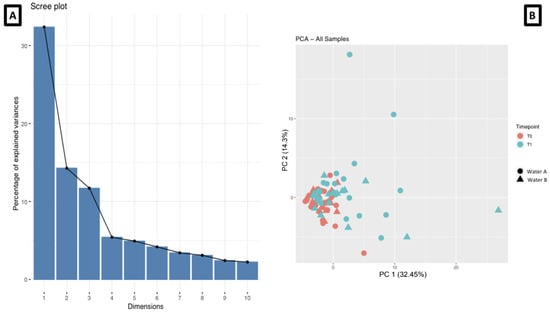
The first two PCAs were more representative of the data variance (Figure 1A), so we adopted and graphically represented it in Figure 1B (PC1 = 32.45% and PC2 = 14.3%). It suggested that a difference in the two groups was present, but linear statistics did not magnify it.

Figure 1.
Cutaneous lipidomics evaluation of total variance (A) and representation of the two main Principal Component Analyses (B).
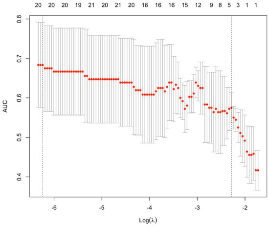
Thus, we decided to apply machine learning to predict which water was consumed, starting from the cutaneous lipids abundance at T1. Surprisingly, only 20 lipids were necessary to predict the water consumed, with an accuracy of almost 70% (Figure 2).

Figure 2.
Accuracy of the predictive algorithm that uses cutaneous lipids to predict the water type consumed. The line composed by red dots depicts the average accuracy of the presented predictive algorithm. AUC: Area under the ROC Curve.
For the list of predictive lipids, see Table 3.

Table 3.
Cutaneous lipids capable to predict the water type consumed.
4. Discussion
In this clinical trial, we demonstrated that water orally consumed had an effect on skin lipidomics; oligo-mineral water has a prominent effect on cutaneous biology if compared with the medium-mineral one.
Some evidence evaluated the biological effect of water consumed orally on dermatologically healthy patients, but it focused mainly on cerebral circulation [16] or on pathological skin [13]. These aspects, together with the biologically relevant effect of oligo-mineral water, suggest that researchers should carefully mention the water type in the inclusion/exclusion criteria in observation studies and clinical trials examining nutrition in dermatology and urology.
Ideally, studies should include the water type orally consumed in the exposome to modify the cutaneous outcomes, especially in complex skin disorders, in which susceptible genes [29,30] interact with exposure to manifest the pathological phenotype (i.e., psoriasis or hidradenitis suppurativa) [31,32]. In fact, exposures may condition the epigenetics that modulate genes expression [33] and even drug response [34].
From this perspective, the water ingested should be regarded as a supplement, or even a drug, that synergically collaborates with drugs to improve the clinical outcome. The present evidence may also sustain, with solid evidence, that the water type orally consumed should be reported in the medical history, or even included as a part of the therapeutical prescription. This concept should also be shared with patients, who often ask their doctor for diet and dietological suggestions to improve their skin quality. Nowadays, our data contributed to the literature to solve doubts regarding the potential modulation of skin biology that water induced; yet, at the same time, the data maintained that water is an active principle and factor to be considered in science and therapy.
In healthy patients, oligo-mineral water displayed a prominent effect in restoring the cutaneous barrier, as previously suggested in atopic dermatitis patients [13]. From this point of view, oligo-mineral water should always be supplemented in the case of barrier dysfunction or even allergic disorders.
At the same time, in line with Caiazzo et al.’s findings, our results could also suggest a direct effect on the skin microbiome capable of positively influencing the skin barrier and its lipidomics. Future efforts should be focused on a better understanding of the biological dynamics occurring between the cutaneous microbiome and keratinocytes biology. To confirm our data, omics and machine learning were able to magnify the water-based changes in cutaneous lipidomics and may project clinicians into the new future led by precision medicine.
Despite the innovative design (omics and machine learning) the present clinical trial also has some limitations, such as the inclusion of solely female subjects between 30 to 50 years of age. Thus, future studies should also validate these results in different ages, males and even diets.
5. Conclusions
Water orally consumed has a remarkable effect on the skin lipidomics in healthy subjects, so especially oligo-mineral water should be regarded as a complementary therapy in dermatological disorders. Furthermore, clinical trials evaluating nutritional supplements or diets in dermatology should account also for the water type consumed. Thus, water has to be regarded as a supplement for its biological effect on skin.
Supplementary Materials
The following supporting information can be downloaded at: https://www.mdpi.com/article/10.3390/biomedicines11041036/s1, Supplementary S1.
Author Contributions
Conceptualization, G.D.; methodology, G.D. and H.A.-S.; software, H.A.-S.; validation, G.D., I.C. and H.A.-S.; formal analysis, H.A.-S.; investigation, G.D., I.C. and H.A.-S.; resources, P.D.M.P.; data curation, P.D.M.P.; writing—original draft preparation, G.D.; writing—review and editing, G.D., I.C., H.A.-S. and P.D.M.P.; visualization, H.A.-S.; supervision, P.D.M.P.; project administration, G.D. and I.C.; funding acquisition, P.D.M.P. All authors have read and agreed to the published version of the manuscript.
Funding
This research was partially funded by CoGeDi International SpA.
Institutional Review Board Statement
The study was conducted according to the guidelines of the Declaration of Helsinki and approved by the Institutional Review Board of San Raphael Hospital (protocol code: 176/INT/2020 and date of approval: 11 November 2020).
Informed Consent Statement
Informed consent was obtained from all subjects involved in the study.
Data Availability Statement
The data are available in a publicly accessible repository.
Acknowledgments
Part of this work was carried out in OMICs, an advanced mass spectrometry platform established by the Università degli Studi di Milano. We thank Fiorenza Fare and Donatella Caruso for their assistance.
Conflicts of Interest
The funders had no role in the design of the study; in the collection, analyses, or interpretation of data; in the writing of the manuscript; or in the decision to publish the results. Paolo Pigatto received a research grant from CoGeDi International SpA and SIDeMaST (Italian Society of Medical, Surgical, Aesthetic Dermatology and Sexually Transmitted Diseases).
References
- Fikri-Benbrahim, K.; Houti, A.; Lalami, A.E.O.; Flouchi, R.; El Hachlafi, N.; Houti, M.; Rachiq, S. Main Therapeutic Uses of Some Moroccan Hot Springs’ Waters. Evid.-Based Complement. Altern. Med. 2021, 2021, 5599269. [Google Scholar] [CrossRef] [PubMed]
- Silva, A.; Oliveira, A.S.; Vaz, C.V.; Correia, S.; Ferreira, R.; Breitenfeld, L.; Martinez-De-Oliveira, J.; Palmeira-De-Oliveira, R.; Pereira, C.M.F.; Cruz, M.T. Anti-inflammatory potential of Portuguese thermal waters. Sci. Rep. 2020, 10, 22313. [Google Scholar] [CrossRef]
- Fillimonova, E.; Kharitonova, N.; Baranovskaya, E.; Maslov, A.; Aseeva, A. Geochemistry and therapeutic properties of Cau-casian mineral waters: A review. Environ. Geochem. Health 2022, 44, 2281–2299. [Google Scholar] [CrossRef]
- Khalilzadeh, S.; Shirbeigi, L.; Naghizadeh, A.; Mehriardestani, M.; Shamohammadi, S.; Tabarrai, M. Use of mineral waters in the treatment of psoriasis: Perspectives of Persian and conventional medicine. Dermatol. Ther. 2019, 32, e12969. [Google Scholar] [CrossRef] [PubMed]
- Kimata, H.; Tai, H.; Nakagawa, K.; Yokoyama, Y.; Nakajima, H.; Ikegami, Y. Improvement of skin symptoms and mineral im-balance by drinking deep sea water in patients with atopic eczema/dermatitis syndrome (AEDS). Acta Med. 2002, 45, 83–84. [Google Scholar]
- Vaz, C.; Oliveira, A.; Silva, A.; Cortes, L.; Correia, S.; Ferreira, R.; Breitenfeld, L.; Martinez-De-Oliveira, J.; Palmeira-De-Oliveira, R.; Pereira, C.; et al. Protective role of Portuguese natural mineral waters on skin aging: In vitro evaluation of anti-senescence and anti-oxidant properties. Int. J. Biometeorol. 2022, 66, 2117–2131. [Google Scholar] [CrossRef]
- Karagülle, M.Z.; Karagülle, M.; Kılıç, S.; Sevinç, H.; Dündar, C.; Türkoğlu, M. In vitro evaluation of natural thermal mineral wa-ters in human keratinocyte cells: A preliminary study. Int. J. Biometeorol. 2018, 62, 1657–1661. [Google Scholar] [CrossRef]
- Lopez, D.J.; Singh, A.; Waidyatillake, N.T.; Su, J.C.; Bui, D.S.; Dharmage, S.C.; Lodge, C.J.; Lowe, A.J. The association between domestic hard water and eczema in adults from the UK Biobank cohort study. Br. J. Dermatol. 2022, 187, 704–712. [Google Scholar] [CrossRef] [PubMed]
- Caiazzo, G.; Parisi, M.; Luciano, M.A.; Di Caprio, R.; Gallo, L.; Cacciapuoti, S.; Quaranta, M.; Fabbrocini, G. Beneficial effects of Roc-chetta® oligomineral water in HaCaT keratinocytes after ultraviolet-B irradiation. Ital. J. Dermatol. Venerol. 2022, 157, 335–341. [Google Scholar]
- Tanaka, A.; Jung, K.; Matsuda, A.; Jang, H.; Kajiwara, N.; Amagai, Y.; Oida, K.; Ahn, G.; Ohmori, K.; Kang, K.G.; et al. Daily in-take of Jeju groundwater improves the skin condition of the model mouse for human atopic dermatitis. J. Dermatol. 2013, 40, 193–200. [Google Scholar] [CrossRef]
- Cacciapuoti, S.; Luciano, M.A.; Megna, M.; Annunziata, M.C.; Napolitano, M.; Patruno, C.; Scala, E.; Colicchio, R.; Pagliuca, C.; Salvatore, P.; et al. The Role of Thermal Water in Chronic Skin Diseases Management: A Review of the Literature. J. Clin. Med. 2020, 9, 3047. [Google Scholar] [CrossRef] [PubMed]
- Fujii, N.; Kataoka, Y.; Lai, Y.-F.; Shirai, N.; Hashimoto, H.; Nishiyasu, T. Ingestion of carbonated water increases middle cerebral artery blood velocity and improves mood states in resting humans exposed to ambient heat stress. Physiol. Behav. 2022, 255, 113942. [Google Scholar] [CrossRef] [PubMed]
- Hataguchi, Y.; Tai, H.; Nakajima, H.; Kimata, H. Drinking deep-sea water restores mineral imbalance in atopic eczema/dermatitis syndrome. Eur. J. Clin. Nutr. 2005, 59, 1093–1096. [Google Scholar] [CrossRef] [PubMed]
- Williams, S.; Krueger, N.; Davids, M.; Kraus, D.; Kerscher, M. Effect of fluid intake on skin physiology: Distinct differences be-tween drinking mineral water and tap water. Int. J. Cosmet. Sci. 2007, 29, 131–138. [Google Scholar] [CrossRef]
- Mac-Mary, S.; Creidi, P.; Marsaut, D.; Courderot-Masuyer, C.; Cochet, V.; Gharbi, T.; Guidicelli-Arranz, D.; Tondu, F.; Humbert, P. Assessment of effects of an additional dietary natural mineral water uptake on skin hydration in healthy subjects by dy-namic barrier function measurements and clinic scoring. Skin Res. Technol. 2006, 12, 199–205. [Google Scholar] [CrossRef]
- Silva, F.; Rodrigues Amorim Adegboye, A.; Lachat, C.; Curioni, C.; Gomes, F.; Collins, G.S.; Kac, G.; de Beyer, J.A.; Cook, J.; Ismail, L.C.; et al. Completeness of Reporting in Diet- and Nutrition-Related Randomized Controlled Trials and Systematic Reviews with Meta-Analysis: Protocol for 2 Independent Meta-Research Studies. JMIR Res. Protoc. 2023, 12, e43537. [Google Scholar] [CrossRef] [PubMed]
- Marotta, D.; Sica, C. Composition and classification of Italian mineral waters Nota II. Ann. Chim. Appl. 1933, 23, 245–257. [Google Scholar]
- Pacifico, A.; Conic, R.R.Z.; Cristaudo, A.; Garbarino, S.; Ardigò, M.; Morrone, A.; Iacovelli, P.; di Gregorio, S.; Pigatto, P.D.M.; Grada, A.; et al. Diet-Related Phototoxic Reactions in Psoriatic Patients Undergoing Photo-therapy: Results from a Multicenter Prospective Study. Nutrients 2021, 13, 2934. [Google Scholar] [CrossRef]
- Bragazzi, N.L.; Trabelsi, K.; Garbarino, S.; Ammar, A.; Chtourou, H.; Pacifico, A.; Malagoli, P.; Kocic, H.; Conic, R.R.Z.; Young Derma-Tologists Italian Network; et al. Can intermittent, time-restricted circadian fasting modulate cutaneous severity of dermatological disorders? Insights from a multicenter, observational, prospective study. Dermatol. Ther. 2021, 34, e14912. [Google Scholar] [CrossRef]
- Damiani, G.; Mahroum, N.; Pigatto, P.D.M.; Pacifico, A.; Malagoli, P.; Tiodorovic, D.; Conic, R.R.; Amital, H.; Bragazzi, N.L.; Watad, A.; et al. The Safety and Impact of a Model of Intermittent, Time-Restricted Circadian Fasting (“Ramadan Fasting”) on Hidradenitis Suppurativa: Insights from a Multicenter, Observational, Cross-Over, Pilot, Exploratory Study. Nutrients 2019, 11, 1781. [Google Scholar] [CrossRef] [PubMed]
- Adawi, M.; Damiani, G.; Bragazzi, N.L.; Bridgewood, C.; Pacifico, A.; Conic, R.R.Z.; Morrone, A.; Malagoli, P.; Pigatto, P.D.M.; Amital, H.; et al. The Impact of Intermittent Fasting (Ramadan Fasting) on Psoriatic Arthritis Disease Activity, Enthesitis, and Dactylitis: A Multicentre Study. Nutrients 2019, 11, 601. [Google Scholar] [CrossRef] [PubMed]
- Damiani, G.; Watad, A.; Bridgewood, C.; Pigatto, P.D.M.; Pacifico, A.; Malagoli, P.; Bragazzi, N.L.; Adawi, M. The Impact of Ramadan Fasting on the Reduction of PASI Score, in Moderate-To-Severe Psoriatic Patients: A Real-Life Multicenter Study. Nutrients 2019, 11, 277. [Google Scholar] [CrossRef]
- Bragazzi, N.L.; Sellami, M.; Salem, I.; Conic, R.Z.; Kimak, M.; Pigatto, P.D.M.; Damiani, G. Fasting and Its Impact on Skin Anatomy, Physiology, and Physiopathology: A Comprehensive Review of the Literature. Nutrients 2019, 11, 249. [Google Scholar] [CrossRef] [PubMed]
- Available online: https://ginasthma.org/wp-content/uploads/2022/07/GINA-Main-Report-2022-FINAL-22-07-01-WMS.pdf (accessed on 15 March 2023).
- Damiani, G.; Calzavara-Pinton, P.; Stingeni, L.; Hansel, K.; Cusano, F.; “Skin Allergy” Group of SIDeMaST; “ADOI” (Associazione Dermatologi Ospedalieri Italiani); “SIDAPA” (Società Italiana di Dermatologia Allergologica, Professionale e Ambientale); Pigatto, P.D.M. Italian guidelines for therapy of atopic dermatitis-Adapted from consensus-based European guidelines for treatment of atopic eczema (atopic dermatitis). Dermatol. Ther. 2019, 32, e13121. [Google Scholar] [CrossRef]
- Stingeni, L.; Bianchi, L.; Hansel, K.; Corazza, M.; Gallo, R.; Guarneri, F.; Patruno, C.; Rigano, L.; Romita, P.; Pigatto, P.D.; et al. Italian Guidelines in Patch Testing—Adapted from the European Society of Contact Dermatitis (ESCD). G. Ital. Dermatol. Venereol. 2019, 154, 227–253. [Google Scholar] [CrossRef]
- Damiani, G.; Alessandrini, M.; Caccamo, D.; Cormano, A.; Guzzi, G.; Mazzatenta, A.; Micarelli, A.; Migliore, A.; Piroli, A.; Bianca, M.; et al. Italian Expert Consensus on Clinical and Therapeutic Management of Multiple Chemical Sensitivity (MCS). Int. J. Environ. Res. Public Health 2021, 18, 11294. [Google Scholar] [CrossRef]
- Grolle, M.; Kupfer, J.; Brosig, B.; Niemeier, V.; Hennighausen, L.; Gieler, U. The Skin Satisfaction Questionnaire—An Instrument to Assess Attitudes toward the Skin in Healthy Persons and Patients. Dermatol. Psychosom. 2003, 4, 14–20. [Google Scholar] [CrossRef]
- Duchatelet, S.; Miskinyte, S.; Delage, M.; Ungeheuer, M.N.; Lam, T.; Benhadou, F.; Del Marmol, V.; Vossen, A.R.J.V.; Prens, E.P.; Cogrel, O.; et al. Low Prevalence of GSC Gene Mu-tations in a Large Cohort of Predominantly Caucasian Patients with Hidradenitis Suppurativa. J. Investig. Dermatol. 2020, 140, 2085–2088.e14. [Google Scholar] [CrossRef]
- Tawfik, N.Z.; Abdallah, H.Y.; Hassan, R.; Hosny, A.; Ghanem, D.E.; Adel, A.; Atwa, M.A. PSORS1 Locus Genotyping Profile in Psoriasis: A Pilot Case-Control Study. Diagnostics 2022, 12, 1035. [Google Scholar] [CrossRef]
- Nguyen, T.V.; Damiani, G.; Orenstein, L.A.; Hamzavi, I.; Jemec, G. Hidradenitis suppurativa: An update on epidemiology, phenotypes, diagnosis, pathogenesis, comorbidities and quality of life. J. Eur. Acad. Dermatol. Venereol. 2021, 35, 50–61. [Google Scholar] [CrossRef] [PubMed]
- Damiani, G.; Bragazzi, N.L.; Aksut, C.K.; Wu, D.; Alicandro, G.; McGonagle, D.; Guo, C.; Dellavalle, R.; Grada, A.; Wong, P.; et al. The Global, Regional, and National Burden of Psoriasis: Results and Insights from the Global Burden of Disease 2019 Study. Front. Med. 2021, 8, 743180. [Google Scholar] [CrossRef] [PubMed]
- Radhakrishna, U.; Ratnamala, U.; Jhala, D.; Vadsaria, N.; Patel, M.; Uppala, L.; Vishweswaraiah, S.; Vedangi, A.; Saiyed, N.; Damiani, G.; et al. Methylated miRNAs may serve as potential biomarkers and therapeutic targets for hidradenitis suppurativa. J. Eur. Acad. Dermatol. Venereol. 2022, 36, 2199–2213. [Google Scholar] [CrossRef] [PubMed]
- Radhakrishna, U.; Ratnamala, U.; Jhala, D.D.; Vadsaria, N.; Patel, M.; Uppala, L.V.; Vedangi, A.; Saiyed, N.; Rawal, R.M.; Damiani, G.; et al. Cytochrome P450 Genes Mediated by DNA Methylation Are Involved in the Resistance to Hidradenitis Suppurativa. J. Investig. Dermatol. 2022, 142, 670–673.e19. [Google Scholar] [CrossRef] [PubMed]
Disclaimer/Publisher’s Note: The statements, opinions and data contained in all publications are solely those of the individual author(s) and contributor(s) and not of MDPI and/or the editor(s). MDPI and/or the editor(s) disclaim responsibility for any injury to people or property resulting from any ideas, methods, instructions or products referred to in the content. |
© 2023 by the authors. Licensee MDPI, Basel, Switzerland. This article is an open access article distributed under the terms and conditions of the Creative Commons Attribution (CC BY) license (https://creativecommons.org/licenses/by/4.0/).