Article

Lactobacillus plantarum Generate Electricity through Flavin Mononucleotide-Mediated Extracellular Electron Transfer to Upregulate Epithelial Type I Collagen Expression and Thereby Promote Microbial Adhesion to Intestine

1
Department of Biomedical Sciences and Engineering, National Central University, Taoyuan City 320317, Taiwan
2
Faculty of Applied Sciences, Ton Duc Thang University, Ho Chi Minh City 700000, Vietnam
3
Department of Chemical and Materials Engineering, National Central University, Taoyuan City 320317, Taiwan
*
Author to whom correspondence should be addressed.
Biomedicines 2023, 11(3), 677; https://doi.org/10.3390/biomedicines11030677
Submission received: 3 January 2023 / Revised: 17 February 2023 / Accepted: 21 February 2023 / Published: 23 February 2023
(This article belongs to the Section Microbiology in Human Health and Disease)

Abstract

:
The mechanism behind how flavin mononucleotide (FMN)-producing bacteria attach to a host intestine remains unclear. In order to address this issue, this study isolated the Gram-positive bacteria Lactobacillus plantarum from Mongolian fermented Airag, named L. plantarum MA. These bacteria were further employed as the model microbes, and their electrogenic properties were first identified by their significant expression of type II NADH-quinone oxidoreductase. This study also demonstrated that the electrical activity of L. plantarum MA can be conducted through flavin mononucleotide (FMN)-based extracellular electron transfer, which is highly dependent on the presence of a carbon source in the medium. Our data show that approximately 15 µM of FMN, one of the key electron donors for the generation of electricity, can be produced from L. plantarum MA, as they were cultured in the presence of lactulose for 24 h. We further demonstrated that the electrical activity of L. plantarum MA can promote microbial adhesion and can thus enhance the colonization effectiveness of Caco-2 cells and mouse cecum. Such enhanced adhesiveness was attributed to the increased expression of type I collagens in the intestinal epithelium after treatment with L. plantarum MA. This study reveals the mechanism behind the electrogenic activity of L. plantarum MA and shows how the bacteria utilize electricity to modulate the protein expression of gut tissue for an enhanced adhesion process.

1. Introduction

Lactobacillus plantarum (L. plantarum) is a facultative Gram-positive lactic acid bacterium that has been used extensively in the dairy industries for its ameliorative properties to the intestinal environment, as well as its benefits to gastroenteric health [1]. Microorganisms such as L. plantarum, which are extracted from Mongolian mare milk-drink (Airag), may also provide increased antioxidant activity when compared to other Lactobacilli strains, as previously reported [2]. L. plantarum has eight-gene locus codes which are utilized for nicotinamide adenine dinucleotide (NADH) dehydrogenase and can generate electricity through extracellular electron transfer (EET); such electrical activity is highly correlated to the level of NAD+ in the surrounding environment [3]. Microbial electrical production using EET is a key survival feature of electrogenic bacteria in the gut environment, however, the host–microbe interaction remains unclear [4].
EET allows electrons to be transported from the microbial cytosol to external conductors such as electrodes and/or minerals, and plays an important role in bacterial physiology including growth [3], colonization [5], and fermentation [6]. For example, it has been reported that both Bacillus cereus DIF1 and Rhodococcus ruber DIF2 can secrete redox mediators such as riboflavin and/or flavin mononucleotide (FMN) to promote their growth [7]. Electricity generated from Clostridium cochlearium in mice guts can enhance bacterial growth and fermentation [6]. Our previous work demonstrated that electricity produced from the Gram-positive lactic acid bacteria Leuconostoc mesenteroides is favorable for bacterial colonization in the intestine [5]. More electrogenic Gram-positive bacteria such as Enterococcus faecalis, Faecalibacterium prausnitzii and Lactococcus lactis which enable flavin-mediated EET were recently discovered by use of a microbial fuel cell chamber (MFCC) [8,9,10].
Colonization of probiotics such as L. plantarum can trigger the immune system and eliminate pathogenic bacteria [11]. However, L. plantarum does not naturally exist in the human body and must be taken exogenously through dietary means [12]. Bacterial adhesion to the human gut is typically carried out through binding with the extracellular matrix (ECM) proteins such as collagen, fibronectin, and/or laminin of the epithelium at the mucus layer [13], while collagen type I (Col-I) is the most prevalent type of ECM protein in the intestinal tissues [14]. Binding with collagen is often the first step for microbes to be colonized on the sub-intestinal mucus [15], and it has been demonstrated that 75% of Lactobacillus isolated from humans and/or animals achieve intestinal adhesion through the use of collagen-binding proteins located on the surface of the bacteria [16]. There are two types of collagen-binding adhesin proteins on the Lactobacillus surface, named the ATP-binding cassette (ABC) transporter system and the S-layer proteins [17,18]. The Col-I binding protein is one of the ABC proteins on the Lactobacillus surface that has a high affinity for Col-I on the intestinal mucosa [19], while expression of the S-layer protein can be modulated via the level of collagen in the colon [20]. Such circumstances indicate that the level of Col-I expression in the intestine plays a crucial role in the colonization of L. plantarum in the gastrointestinal tract. However, the mechanism by which electrogenic bacteria (e.g., L. plantarum) affects collagen expression of intestinal cells needs further elucidation.
In this paper, we aim to investigate the correlation between electricity and the adhesion capability of L. plantarum MA to the intestine, and how microbial electricity is affected by the culture environment. Lactulose, a disaccharide composed of galactose and fructose, is a selective carbon source (prebiotic) for L. plantarum fermentation [21] and can stimulate the growth of Gram-positive bacteria, since it may increase the quantity of short-chain fatty acids (SCFA) including acetic, butyric, and lactic acids [22] and may therefore improve the microflora environment in vivo [23]. In this study, L. plantarum extracted from Mongolian Airag were investigated for their electrical properties and colonization capabilities in the presence and absence of lactulose in vitro and ex vivo.

2. Materials and Methods

2.1. Bacterial Identification and Cultivation

The L. plantarum that were utilized in this study were self-isolated from Mongolian fermented Airag (named L. plantarum MA) and identified/confirmed using the 16S ribosomal RNA sequencing method (BigDye Terminator V3.1 Cycle Sequencing Kit, ThermoFisher Scientific, Waltham, MA, USA). Both L. plantarum MA and Lactobacillus pentosus (L. pentosus; ATCC® 8041, ATCC, Rockville, MD, USA) were regularly cultured in shaken de Man-Rogosa-Sharpe broth (MRS, Hi-Media, Mumbai, India) under aerobic conditions at 37 °C. Staphylococcus epidermidis (S. epidermidis; ATCC® 12228, ATCC) were cultivated in shaken tryptic soy broth (TSB, Sigma, St. Louis, MO, USA) under aerobic conditions at 37 °C. The microbes were cultured with 2% of lactulose or glucose according to the experimental setup. The number of bacteria was evaluated using spectrophotometry at λabs = 600 nm. Sub-cultivation of the bacteria was performed using 1:100 dilution, when the value of the optical density (OD600) was ≥1.0.

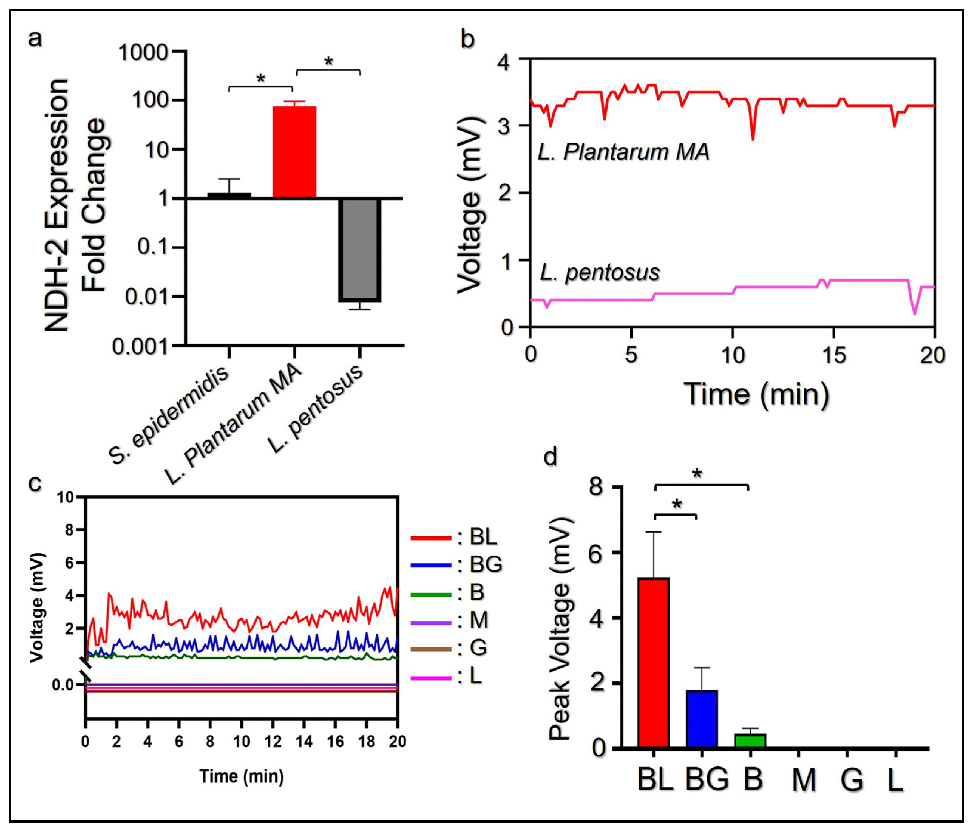
2.2. Quantification of Microbial NDH-2 Expression by RT-qPCR

The expression levels of the type II NADH-quinone oxidoreductase (NDH-2) gene in L. plantarum MA, L. pentosus, and S. epidermidis were quantitatively measured using a RT-qPCR. L. pentosus is the known electrogenic bacteria which can produce electricity without mediators [24] and therefore served as the positive control in this assay. In brief, bacterial RNA from 1 × 10⁸ microbes was extracted using the RNA Mini-Purification Kit (Biokit Biotechnology Inc., Miaoli, Taiwan) followed by DNase treatment to remove DNA contamination, according to the manufacturer’s instructions. The purity of the isolated RNA was assessed via spectrophotometry, where the accepted range of A260/A280 was between 1.8–2.0. Afterward, the total RNA was converted to cDNA using the iScript cDNA Synthesis Kit (Bio-Rad, Hercules, CA, USA) and the products were immediately subjected to qPCR or stored at −20 °C until use.
SYBR Green qPCR was performed to quantitatively detect the expression level of the NDH-2 transcripts in each microbial group. The qPCR sample was prepared by combining 2× qPCR Master Mix (Thermo Fisher Scientific, Waltham, MA, USA), primers (0.4 µM; Table 1), and 2 µL of the cDNA template. The total volume of each reaction was adjusted to 10 µL using nuclease-free water. The qPCR associated with the melting-curve analysis was carried out using the StepOnePlus RT PCR System (Thermo Fisher Scientific) which was incorporated with a fixed thermal-cycling program set at 95 °C for 10 min, 95 °C for 15 s, 60 °C for 60 s, and 72 °C for 30 s, leading to a total of 40 cycles. All primers (Table 1) used in RT-qPCR were designed with an intron-spanning assay through a Primer-Blast tool (https://blast.ncbi.nlm.nih.gov/Blast.cgi/ (accessed on 20 December 2022)) and produced by Sigma. Gene expression was determined using the cycle threshold (2−ΔΔCt) and was quantitatively analyzed against the blank after normalization.

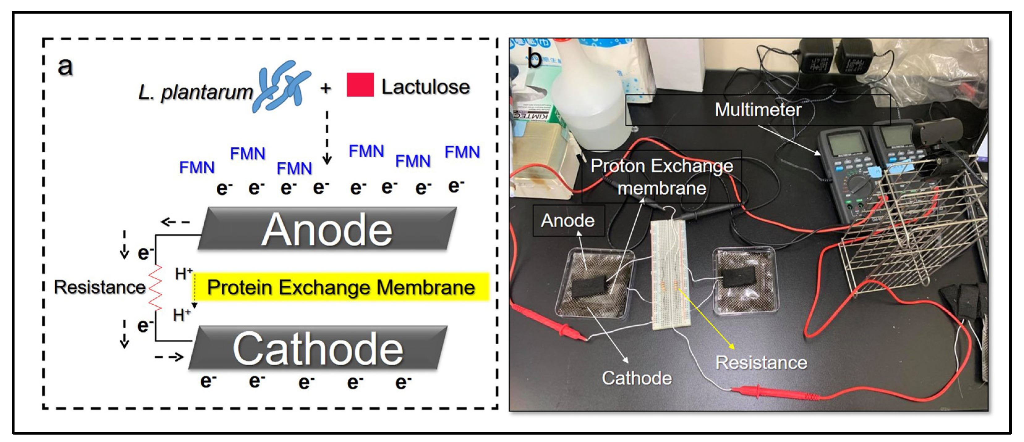
2.3. Detection of Microbial Electricity In Vitro

Electricity generated from the microbials was detected using a microbial fuel cell chamber (MFCC) assembled according to a previous study [25]. In brief, the MFCC was fabricated using carbon cloth (Homy Tech, Taoyuan, Taiwan) and carbon felt (Homy Tech) for the cathode and anode, respectively, with 1000 Ω of electrical impedance placed between the two electrodes. In addition, a NafionTM membrane N117 (Chemours, Wilmington, DE, USA) was set in the middle of the chamber to serve as a proton exchange membrane (PEM). The whole MFCC instrumental setup, along with illustrations, are shown in Figure 1a,b, respectively. To measure bacterial electricity, 107 colony forming units (CFU) per mL of L. plantarum MA or L. pentosus were first incubated in 5 mL of MRS broth in the presence or absence of a carbon source (i.e., 2% of lactulose or glucose) at 37 °C for 24 h. After transferring to the MFCC, the voltage generated from each group was detected continuously for 20 min using a digital multimeter (Lutron, DM-9962SD, Sydney, Australia).

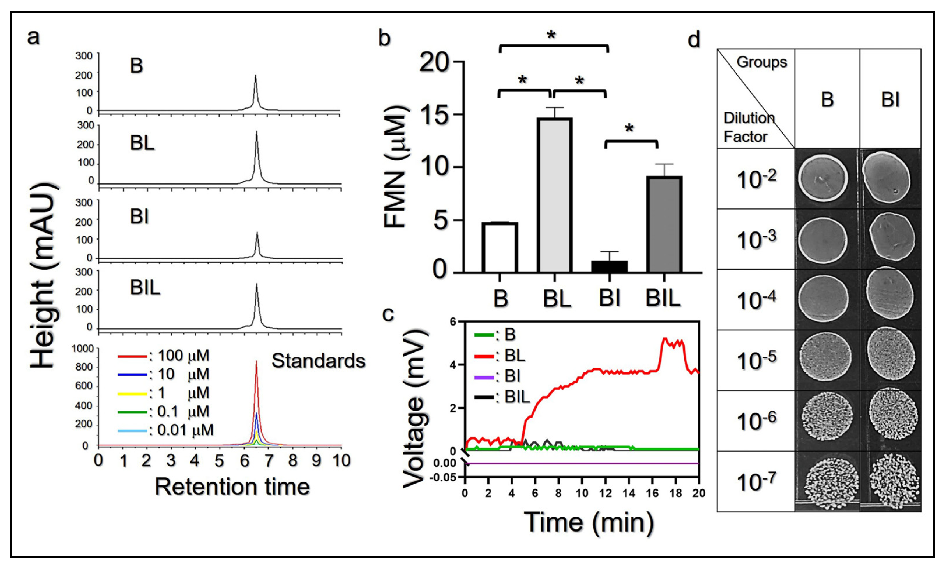
2.4. Detection of Endogenous FMN Yielded from L. plantarum MA

L. plantarum MA (107 CFU/mL) pretreated with 1 µM of roseoflavin, which is a FMN inhibitor, or dimethyl sulfoxide (DMSO) were separately incubated for 24 h in 10 mL of MRS broth, in the presence or absence of 2% lactulose. Afterward, the bacteria were centrifuged at 6000 rpm for 15 min and the supernatants were collected in stages, filtrated using 0.22-μm filters, and stored at 4 °C. The quantity of FMN in each supernatant was analyzed using a high-performance liquid chromatography (HPLC) (Alliance-HPLC e2695 System, Waters Corp. Milford, MA, USA) equipped with a ZORBAX Eclipse XDB-C18 column (250 mm × 4.6 mm ID, 5-μm particle size; Agilent Technologies, Santa Clara, CA, USA). Each supernatant was initially dissolved in 1 mL of HPLC-grade methanol and separated in a mobile phase consisting of 20 mM of ammonium acetate (pH 5.4) and methanol at 25 ± 2 °C under 1 mL/min of flow rate. The concentration of methanol in the mobile phase was first set at 5% in the initial isocratic step for 6 min, then gradually increased to 34.5% for 6 min, 37% for 16 min, and 95% for 10 min, followed by a rapid drop to 5% for 16 min. The eluted signal was measured using the spectrophotometry set at λ = 275 nm and evaluated for FMN quantification using the ChromQuest v3.1.6 System (Thermo Fisher Scientific) in association with OriginPro (2022 SR1, OriginLab, Northampton, MA, USA).

2.5. Cell Culture

The Caco-2 cells (ATCC® HTB-37™, ATCC) were cultured with Dulbecco’s Modified Eagle medium (DMEM) supplemented with 20% fetal bovine serum (FBS) and 100-U/mL penicillin/streptomycin at 37 °C with 5% CO2.

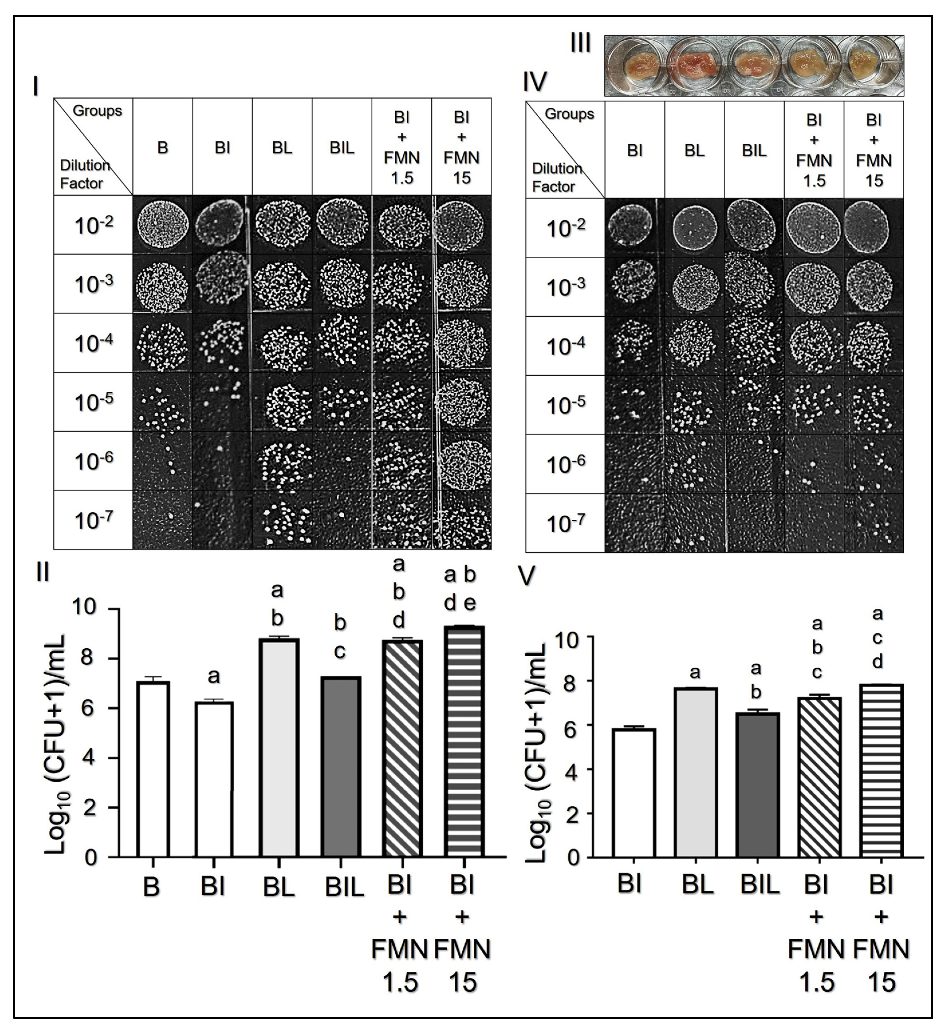
2.6. Microbial Adhesion Assay In Vitro

Caco-2 cells with 105 cells/well in 96-well plates were initially washed 3 times with PBS before the experiment. Next, 200 µL of cell culture medium without antibiotics, containing 107 CFU/mL of L. plantarum MA and designated agents, were added to the Caco-2 cells, as described in Table 2. After co-incubation with the cells for 60 min, the cells were trypsinized and added to 25 µL of hypotonic tissue protein extraction reagent (T-PER) for the collection of adhered bacteria. To quantify the adhesion capability of L. plantarum MA with different treatments, 10 µL of the collected L. plantarum MA was seeded on the MRS agar plates using different dilution factors. After maintenance at 37 °C for 12 h, the in vitro adhesion capability of each group was determined based on the value of the microbial population index (MPI) defined by the logarithm of the CFU (MPI = Log10 ((CFU + 1)/mL)), as previously reported [5].

2.7. Ethics Statement for the Animal Study

A total of 12 Institute of Cancer Research (ICR) mice (8–9 weeks, female) weighing between 25–35 g were employed to build the animal models in this study. All the animal procedures, including the care and operations of the experimental mice, were in accordance with the guidelines approved by the Institutional Animal Care and Use Committee (IACUC) at National Central University (Approval number: NCU-106-016. Approval Date: 19 December 2017).

2.8. Microbial Adhesion Assay Ex Vivo

To evaluate the adhesion capability of L. plantarum MA on the intestine in vivo, an ex vivo microbial adhesion assay was conducted on murine cecal tissue, since cecum is the main site for bacterial fermentation and electron production [26]. After the mice were anesthetized using CO2, the cecum from all mice were collected, washed with 0.9% NaCl to remove the feces, and cut into pieces of approximately equal weight. Afterward, the cecum pieces were separately incubated with L. plantarum MA under various conditions, as described in Table 3. After maintenance at 37 °C for 20 h, each cecum sample was washed 3 time with PBS and incubated with 50-µL T-PER and 3-µL protease inhibitor at 37 °C for 10 min, followed by mashing the cecum with a pestle. In this study, the ex vivo adhesion capability of each group was determined based on the MPI, which was assessed by seeding 10 µL of various dilutions of the homogenized samples on MRS agar plates, as described above.

2.9. Measurement of Col-I Expression of the Cecum after Various Treatments Ex Vivo

The expression level of Col-I of each cecum sample after microbial treatment was evaluated using a Western blot. Briefly, after various microbial treatments, the homogenized cecum were centrifuged using 15,000 rpm at 4 °C for 10 min and the supernatants were subjected to a bicinchoninic acid assay (BCA) to quantify the total protein that was extracted. Next, 20 µg of protein from each cecum was mixed with a 4× Laemmli sample buffer (Bio-Rad, Hercules, CA, USA) and immediately separated via electrophoresis in 10% sodium dodecyl sulfate polyacrylamide gels (SDS-PAGE) using 100 V of electricity. The gels were then blotted onto a polyvinylidene difluoride membrane (Millipore, Temecula, CA, USA) and incubated with primary antibodies of Col-I (1:2000, Cusabio Technology, Houston, TX, USA) or β-actin (1:5000, Cusabio Technology) for 2.5 h, followed by conjugation with the secondary antibody at a dilution of 1:2000 for 60 min. After washing 3 times with PBS-Tween-20 (0.05% (v/v); PBS-T), the membranes were treated with chemiluminescent substrates for 5 min and the level of each Col-I or β-actin band was visualized using the Omega Lum™ C Imaging System (Gel company, San Francisco, CA, USA). The β-actin was employed as the reference gene in all Western blot analyses. The blot intensity of each Col-I band was quantified using the ImageJ software and analyzed after normalization against that of the β-actin.

2.10. Statistical Analysis

All data were acquired from ≥ three independent experiments and are presented as the mean ± standard deviation (SD). Statistical analyses were conducted using the GraphPad Prism software, in which comparisons between groups were performed using the Student’s t-test and 2-way ANOVA, with a significance level of * p < 0.05 throughout the study.

3. Results and Discussion

3.1. Identification and Electrogenic Properties of L. plantarum MA

Supplementary Materials S1 show the 16S rRNA gene sequence of the microbials isolated from the Mongolian Airag (i.e., L. plantarum MA). The result shows that L. plantarum MA shares 99.6% of its genes with the L. plantarum KLDS 1.0728 strain, demonstrating that it is a strain of L. plantarum.
To investigate the electrogenic characteristics of L. plantarum MA, the gene expression of NDH-2, one of the key proteins in the EET mechanism [3], was examined using a RT-qPCR assay. As shown in Figure 2a, L. plantarum MA exhibited significantly higher levels of NDH-2 compared with that gained from S. epidermidis or L. pentosus (p < 0.05 for each). Furthermore, through the detection of electricity using MFCC, we found that L. plantarum MA in the presence of lactulose were able to generate >3-mV of electricity within 20 min, while L. pentosus (a known electrogenic bacteria) produced <1 mV over the same time period (Figure 2b). These data clearly demonstrate that L. plantarum MA is an electrogenic bacteria and that is consistent with the known electrical properties of L. plantarum, as previously reported [3].
To further investigate how carbon source affects the production of electricity of L. plantarum MA, the microbes cultured with lactulose or glucose were placed separately in the MFCC and an in situ voltage detection was performed for 20 min. As shown in Figure 2c, no voltage change was observed in the groups without bacteria (Figure 2c, M, G, and L), while a tiny voltage fluctuation in the range of 0.05–0.31 mV was detected in the group of L. plantarum MA without a carbon source (Figure 2c, B). However, electricity production of L. plantarum MA was markedly enhanced in the presence of a carbon source such as lactulose (Figure 2c, BL) or glucose (Figure 2c, BG), where the peak voltage obtained in the former was approximately 2.8 folds (Figure 2d, p < 0.05) higher than that of the latter within 20-min detection. These results clearly show that the electrogenic activity of L. plantarum MA is highly dependent on the presence of a carbon source, and the lactulose may induce a higher electricity yield compared to glucose with an equal concentration.
In fact, lactulose is a common selective carbon source for L. plantarum fermentation [21] and its fermentative byproducts, including acetic acid and FMN, can serve as electron donors for the generation of electricity, as previously reported [27]. Since L. plantarum with lactulose can produce a higher amount of acetic acid in fermentation compared to that with glucose [28], we reason that the higher electricity led by lactulose, as opposed to glucose, is attributable to increased fermentation byproducts in the medium.

3.2. Effect of FMN on Electricity Generation of L. plantarum MA

We further investigated how the fermentation product FMN was influenced by lactulose. Figure 3a shows the HPLC chromatograms of FMN fermented from L. plantarum MA with various treatments. Based on the spectrophotometric analysis, L. plantarum MA with lactulose generated significantly enhanced FMN compared to the bacteria without a carbon source (Figure 3b, B vs. BL, p < 0.05); the latter was dramatically decreased by ~80% after treatment with roseoflavin for 24 h (Figure 3b, B vs. BI, p < 0.05). However, roseoflavin-mediated FMN impairment in L. plantarum MA can be effectively mitigated by the addition of lactulose, as plotted in Figure 3b. The bioelectrical productivity of the bacterial culture media with the above settings and the subsequent results are presented in Figure 3c. Our data show that the intensity of voltage is ranked by the group + lactulose (Figure 3c, BL) >> + (roseoflavin + lactulose) (Figure 3c, BIL) > no treatment (Figure 3c, B), while the electricity of the group with roseoflavin (Figure 3c, BI) was shown to be at 0 mV throughout the 20-min detection sequence. This outcome is positively correlated with the data of FMN production, as plotted in Figure 3b. On the other hand, FMN expression is independent of the microbial viability and/or growth, since L. plantarum MA with and without roseoflavin displayed similar colony patterns, as shown in Figure 3d.
Pretreatment with a riboflavin kinase inhibitor (i.e., roseoflavin) can reduce the electricity production in Gram-positive bacteria [29]. In this study, we showed that a 24 h pretreatment of L. plantarum MA with roseoflavin significantly reduced the concentration of FMN by approximately 12 folds (Figure 3b) while also decreasing electricity production (Figure 3c), suggesting that the bioelectrical activity of L. plantarum MA was mediated through the FMN-based EET system. On the other hand, it was reported that roseoflavin may target a FMN riboswitch and impair the bacterial growth under concentrations of 100 µM [30]. In this study, roseoflavin was employed with only 1 µM and thus no detrimental effect was found to the growth of L. plantarum MA (Figure 3d). Taken together, these results clearly demonstrate that a carbon source (e.g., lactulose) plays a crucial role in the bioelectrical properties of L. plantarum MA since it can modulate the production of FMN, which is one of the key electron mediators for EET activity [13].

3.3. Effects of FMN-Mediated Electricity on Microbial Adhesion In Vitro and Ex Vivo

To evaluate how electricity influences the interactions between L. plantarum MA and human intestines, the adhesion efficacies of the conditional L. plantarum MA to the intestine in vitro and ex vivo were examined step-by-step. As shown in Figure 4I,II, it can be observed that L. plantarum MA with lactulose exhibited a significantly enhanced adhesiveness to Caco-2 cells, while the L. plantarum MA with roseoflavin showed an opposite result compared to the setting without a carbon source (Figure 4II, p < 0.05). However, the roseoflavin-led reduction in cell adhesion can be ameliorated by simultaneously treating the bacteria with lactulose or exogenous FMN, as displayed in Figure 4I. Based on the CFU analysis, the MPI of the group with roseoflavin + FMN was 43% and 55% (Figure 4II, p < 0.05 for each) higher than the one with roseoflavin alone when the dose of FMN was set as 1.5 and 15 µM, respectively. These outcomes indicate that an electron mediator (i.e., FMN) does play a crucial role in the gut adhesion of L. plantarum MA, and its production is highly dependent on the supply of carbon source (e.g., lactulose) in the medium (Figure 3b).
Similar results can be found in the ex vivo assay (Figure 4III), where lactulose as well as FMN, but not roseoflavin, facilitated microbial adhesion to mouse cecum (Figure 4IV). Based on the quantitative analysis of the CFUs, the MPI of the group concurrently treated with roseoflavin + lactulose, roseoflavin + 1.5 µM of FMN, or roseoflavin + 15 µM of FMN was 10%, 24%, and 35% higher than the one with roseoflavin alone (Figure 4V, p < 0.05 for each), implying that the presence and concentration of FMN is pivotal for the adhesion of L. plantarum MA in vivo. Taken together, we reason that lactulose/FMN-enhanced bacterial adhesion to the intestine results from the enhanced electricity of L. plantarum MA, since lactulose may facilitate the production of electron mediators (e.g., FMN) and increase microbial bioelectric activity [4]. On the contrary, roseoflavin inhibited FMN production and thus diminished microbial electricity, resulting in reduced bacterial colorization in the gut.

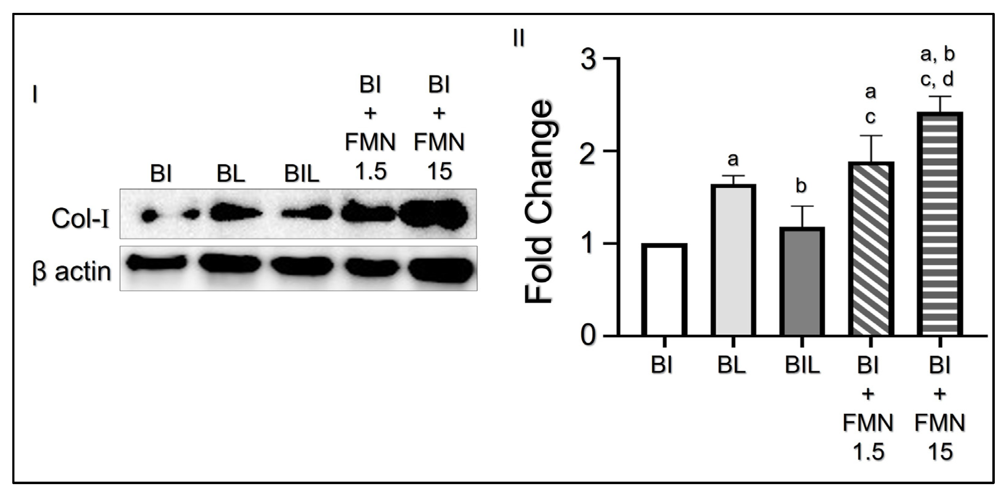
3.4. FMN Upregulates Col-I Collagen Expression on Cecum

To verify the rationale for the proposed hypothesis of enhanced microbial adhesion, we further examined how the mouse cecum responded to FMN/electricity using a Western blot. As shown in Figure 5I, it can be observed that the group with L. plantarum MA and lactulose exhibited a remarkably enhanced Col-I expression compared with that gained from the L. plantarum MA + roseoflavin. Furthermore, addition of lactulose or FMN was indeed able to restore the Col-I expression level suppressed by roseoflavin. Based on the quantitative analyses of the blot intensities (Figure 5II), the expression level of Col-I of the cecum treated with roseoflavin + lactulose, roseoflavin + 1.5 µM of FMN, or roseoflavin + 15 µM of FMN was 14.6%, 28% (p < 0.05), and 40.7% (p < 0.05) higher than the one with roseoflavin alone. Taken together with the results of FMN-mediated EET activity shown in Figure 3c, these outcomes indicate that the Col-I expression of the intestinal tissue can be upregulated using microbial electricity and hence can promote the bacterial adhesion to the gut epithelial cells ex vivo.
It has been widely demonstrated that electron mediators/currents play an important role in bacterial aggregation, colonization, and motility [31]. In this study, we demonstrated that the addition of either lactulose or FMN to the roseoflavin-pretreated L. plantarum MA was indeed able to promote adhesiveness of the bacteria in vitro and ex vivo (Figure 5), and was also able to dramatically enhance the Col-I expression of the attached cecum, as illustrated in Figure 5I and Figure 5II, respectively. However, direct treatment with FMN may provide significantly higher microbial adhesion and Col-I expression compared to bacteria treated with lactulose, suggesting that the FMN-mediated EET is the central mechanism for modulation of their colonization, where L. plantarum MA can swiftly utilize FMN to generate electricity and upregulate collagen expression of the gut tissue for an enhanced microbial adhesion. Nonetheless, further investigation into how molecular signaling of the intestine correlates with microbial electricity is certainly needed and efforts are currently in progress.

4. Conclusions

In this study, we have successfully isolated L plantarum from Mongolian fermented Airag, named L. plantarum MA, and identified their electrogenic property due to significant NDH-2 expression. We demonstrated that a carbon source such as lactulose plays a crucial role in the generation of microbial electricity for L. plantarum MA, where the produced FMN can serve as electron mediators for the activity of extracellular electron transfer. Furthermore, we demonstrated that L. plantarum MA can upregulate the Col-I expression of the intestinal cells as well as tissues in the presence of lactulose or FMN, and thereby enhance the bacterial adhesion in vitro and ex vivo. Since L. plantarum are a type of probiotic which can enhance nutrient digestibility, modulate immune response, and eliminate pathogenic microbes [32], the efforts presented in this study may provide a new insight into the dietary applications of L. plantarum-containing food such as Mongolian fermented Airag.

Supplementary Materials

The following supporting information can be downloaded at: https://www.mdpi.com/article/10.3390/biomedicines11030677/s1, S1: Result of 16sRNA sequence of L. plantarum MA.

Author Contributions

Conceptualization, B.G., C.-M.H. and Y.-H.L.; methodology, E.Z. and S.M.; software, E.Z.; validation, B.G., C.-M.H. and Y.-H.L.; formal analysis, Y.-H.L.; investigation, E.Z. and M.T.P.; resources, C.-M.H.; data curation, B.G., E.Z., M.T.P. and S.M.; writing—original draft preparation, B.G.; writing—review and editing, Y.-H.L.; visualization, B.G.; supervision, C.-M.H. and Y.-H.L.; project administration, B.G.; funding acquisition, C.-M.H. and Y.-H.L. All authors have read and agreed to the published version of the manuscript.

Funding

This research was funded by the National Science and Technology Council, Taiwan. Grant number: NSTC 111-2221-E-008-016-MY2 (Y.-H.L.).

Institutional Review Board Statement

The animal study was conducted according to the guidelines of the Declaration of Helsinki and approved by the Institutional Animal Care and Use Committee (IACUC) of National Central University (Approval number: NCU-106-016. Approval Date: 19 December 2017).

Informed Consent Statement

Not applicable.

Data Availability Statement

The datasets created and/or analyzed in this work are available upon request.

Acknowledgments

The authors appreciate the Gyals Medical Center (Ulaanbaatar, Mongolia) for providing L. plantarum MA and collaboration. In addition, the authors are grateful for Ming-Fa Hsieh in Chung Yuan Christian University (Taiwan) for his technical support of HPLC operation and analysis.

Conflicts of Interest

The authors declare no conflict of interest.

References

  1. Alizadeh Behbahani, B.; Noshad, M.; Falah, F. Inhibition of Escherichia coli adhesion to human intestinal Caco-2 cells by probiotic candidate Lactobacillus plantarum strain L15. Microb. Pathog. 2019, 136, 103677. [Google Scholar] [CrossRef] [PubMed]
  2. Uugantsetseg, E.; Batjargal, B. Antioxidant activity of probiotic lactic acid bacteria isolated from Mongolian airag. Mong. J. Chem. 2014, 15, 73–78. [Google Scholar] [CrossRef] [Green Version]
  3. Light, S.H.; Su, L.; Rivera-Lugo, R.; Cornejo, J.A.; Louie, A.; Iavarone, A.T.; Ajo-Franklin, C.M.; Portnoy, D.A. A flavin-based extracellular electron transfer mechanism in diverse Gram-positive bacteria. Nature 2018, 562, 140–144. [Google Scholar] [CrossRef] [PubMed]
  4. Cahoon, L.A.; Freitag, N.E. The electrifying energy of gut microbes. Nature 2018, 562, 43–44. [Google Scholar] [CrossRef] [PubMed] [Green Version]
  5. Pham, M.T.; Yang, J.J.; Balasubramaniam, A.; Rahim, A.R.; Adi, P.; Do, T.T.M.; Herr, D.R.; Huang, C.M. Leuconostoc mesenteroides mediates an electrogenic pathway to attenuate the accumulation of abdominal fat mass induced by high fat diet. Sci. Rep. 2020, 10, 21916. [Google Scholar] [CrossRef]
  6. Schwab, L.; Rago, L.; Koch, C.; Harnisch, F. Identification of Clostridium cochlearium as an electroactive microorganism from the mouse gut microbiome. Bioelectrochemistry 2019, 130, 107334. [Google Scholar] [CrossRef]
  7. Tian, T.; Fan, X.; Feng, M.; Su, L.; Zhang, W.; Chi, H.; Fu, D. Flavin-mediated extracellular electron transfer in Gram-positive bacteria Bacillus cereus DIF1 and Rhodococcus ruber DIF2. RSC Adv. 2019, 9, 40903–40909. [Google Scholar] [CrossRef] [Green Version]
  8. Zhang, E.; Cai, Y.; Luo, Y.; Piao, Z. Riboflavin-shuttled extracellular electron transfer from Enterococcus faecalis to electrodes in microbial fuel cells. Can. J. Microbiol. 2014, 60, 753–759. [Google Scholar] [CrossRef]
  9. Khan, M.T.; Browne, W.R.; van Dijl, J.M.; Harmsen, H.J. How can Faecalibacterium prausnitzii employ riboflavin for extracellular electron transfer? Antioxid. Redox Signal. 2012, 17, 1433–1440. [Google Scholar] [CrossRef]
  10. Masuda, M.; Freguia, S.; Wang, Y.F.; Tsujimura, S.; Kano, K. Flavins contained in yeast extract are exploited for anodic electron transfer by Lactococcus lactis. Bioelectrochemistry 2010, 78, 173–175. [Google Scholar] [CrossRef] [Green Version]
  11. Arena, M.P.; Silvain, A.; Normanno, G.; Grieco, F.; Drider, D.; Spano, G.; Fiocco, D. Use of Lactobacillus plantarum Strains as a Bio-Control Strategy against Food-Borne Pathogenic Microorganisms. Front. Microbiol. 2016, 7, 464. [Google Scholar] [CrossRef] [PubMed] [Green Version]
  12. Liu, Y.-W.; Liong, M.T.; Tsai, Y.C. New perspectives of Lactobacillus plantarum as a probiotic: The gut-heart-brain axis. J. Microbiol. 2018, 56, 601–613. [Google Scholar] [CrossRef] [PubMed]
  13. Adlerberth, I.; Cerquetti, M.; Poilane, I.; Wold, A.; Collignon, A. Mechanisms of Colonisation and Colonisation Resistance of the Digestive Tract Part 1: Bacteria/host Interactions. Microb. Ecol. Health Dis. 2000, 12, 223–239. [Google Scholar]
  14. Graham, M.F.; Diegelmann, R.F.; Elson, C.O.; Lindblad, W.J.; Gotschalk, N.; Gay, S.; Gay, R. Collagen content and types in the intestinal strictures of Crohn’s disease. Gastroenterology 1988, 94, 257–265. [Google Scholar] [CrossRef] [PubMed]
  15. Wolf, B.; Garleb, K.; Ataya, D.; Casas, I. Safety and tolerance of Lactobacillus reuteri in healthy adult male subjects. Microb. Ecol. Health Dis. 1995, 8, 41–50. [Google Scholar] [CrossRef]
  16. Aleljung, P.; Shen, W.; Rozalska, B.; Hellman, U.; Ljungh, Å.; Wadström, T. Purification of collagen-binding proteins of Lactobacillus reuteri NCIB 11951. Curr. Microbiol. 1994, 28, 231–236. [Google Scholar] [CrossRef]
  17. Toba, T.; Virkola, R.; Westerlund, B.; Bjorkman, Y.; Sillanpaa, J.; Vartio, T.; Kalkkinen, N.; Korhonen, T.K. A collagen-binding S-layer protein in Lactobacillus crispatus. Appl. Environ. Microbiol. 1995, 61, 2467–2471. [Google Scholar] [CrossRef] [Green Version]
  18. Schneitz, C.; Nuotio, L.; Lounatma, K. Adhesion of Lactobacillus acidophilus to avian intestinal epithelial cells mediated by the crystalline bacterial cell surface layer (S-layer). J. Appl. Bacteriol. 1993, 74, 290–294. [Google Scholar] [CrossRef]
  19. Roos, S.; Aleljung, P.; Robert, N.; Lee, B.; Wadström, T.; Lindberg, M.; Jonsson, H. A collagen binding protein from Lactobacillus reuteri is part of an ABC transporter system? FEMS Microbiol. Lett. 1996, 144, 33–38. [Google Scholar] [CrossRef]
  20. Sillanpää, J.; Martínez, B.; Antikainen, J.; Toba, T.; Kalkkinen, N.; Tankka, S.; Lounatmaa, K.; Keränen, J.; Höök, M.; Westerlund-Wikström, B.; et al. Characterization of the collagen-binding S-layer protein CbsA of Lactobacillus crispatus. J. Bacteriol. 2000, 182, 6440–6450. [Google Scholar] [CrossRef] [Green Version]
  21. Mao, B.; Li, D.; Zhao, J.; Liu, X.; Gu, Z.; Chen, Y.Q.; Zhang, H.; Chen, W. In Vitro Fermentation of Lactulose by Human Gut Bacteria. J. Agric. Food Chem. 2014, 62, 10970–10977. [Google Scholar] [CrossRef] [PubMed]
  22. Salminen, S.; Salminen, E. Lactulose, Lactic Acid Bacteria, Intestinal Microecology and Mucosal Protection. Scand. J. Gastroenterol. 1997, 32, 45–48. [Google Scholar] [CrossRef] [PubMed]
  23. Calik, A.; Ergün, A. Effect of lactulose supplementation on growth performance, intestinal histomorphology, cecal microbial population, and short-chain fatty acid composition of broiler chickens. J Poult. Sci. 2015, 94, 2173–2182. [Google Scholar] [CrossRef]
  24. Vilas Boas, J.; Oliveira, V.B.; Marcon, L.R.C.; Simões, M.; Pinto, A.M.F.R. Optimization of a single chamber microbial fuel cell using Lactobacillus pentosus: Influence of design and operating parameters. Sci. Total Environ. 2019, 648, 263–270. [Google Scholar] [CrossRef] [PubMed]
  25. Balasubramaniam, A.; Adi, P.; Do Thi, T.M.; Yang, J.H.; Labibah, A.S.; Huang, C.M. Skin bacteria mediate glycerol fermentation to produce electricity and resist UV-B. Microorganisms 2020, 8, 1092. [Google Scholar] [CrossRef] [PubMed]
  26. Brown, K.; Abbott, D.W.; Uwiera, R.R.E.; Inglis, G.D. Removal of the cecum affects intestinal fermentation, enteric bacterial community structure, and acute colitis in mice. Gut Microbes 2018, 9, 218–235. [Google Scholar] [CrossRef] [PubMed] [Green Version]
  27. Finke, N.; Vandieken, V.; Jørgensen, B.B. Acetate, lactate, propionate, and isobutyrate as electron donors for iron and sulfate reduction in Arctic marine sediments, Svalbard. FEMS Microbiol. Ecol. 2007, 59, 10–22. [Google Scholar] [CrossRef] [Green Version]
  28. Hernandez-Hernandez, O.; Muthaiyan, A.; Moreno, F.J.; Montilla, A.; Sanz, M.L.; Ricke, S.C. Effect of prebiotic carbohydrates on the growth and tolerance of Lactobacillus. Food Microbiol. 2012, 30, 355–361. [Google Scholar] [CrossRef] [Green Version]
  29. Kao, M.-S.; Yang, J.H.; Balasubramaniam, A.; Traisaeng, S.; Jackson Yang, A.; Yang, J.J.; Salamon, B.P.; Herr, D.R.; Huang, C.M. Colonization of nasal cavities by Staphylococcus epidermidis mitigates SARS-CoV-2 nucleocapsid phosphoprotein-induced interleukin (IL)-6 in the lung. Microb. Biotechnol. 2022, 15, 1984–1994. [Google Scholar] [CrossRef]
  30. Mansjö, M.; Johansson, J. The riboflavin analog roseoflavin targets an FMN-riboswitch and blocks Listeria monocytogenes growth, but also stimulates virulence gene-expression and infection. RNA Biol. 2011, 8, 674–680. [Google Scholar] [CrossRef] [Green Version]
  31. Gomez-Carretero, S.; Libberton, B.; Svennersten, K.; Persson, K.; Jager, E.; Berggren, M.; Rhen, M.; Richter-Dahlfors, A. Redox-active conducting polymers modulate Salmonella biofilm formation by controlling availability of electron acceptors. Npj Biofilms Microbiomes 2017, 3, 19. [Google Scholar] [CrossRef] [PubMed] [Green Version]
  32. Al-Tawaha, R.; Meng, C. Potential benefits of Lactobacillus plantarum as probiotic and its advantages in human health and industrial applications: A review. Adv. Environ. Biol. 2018, 12, 16–27. [Google Scholar]
Figure 1. Identification of the bacteria isolated from the Mongolian fermented Airag. (a) Schematic diagram of the MFCC operations. (b) Photograph of the real MFCC device.
Biomedicines 11 00677 g001
Figure 2. Assessment of the electrogenic property of L. plantarum MA. (a) NDH-2 expressions of the S. epidermidis, L. plantarum MA and L. pentosus represented by fold change. (b) The pattern of electricity production of L. plantarum MA and L. pentosus which were treated with 2% lactulose within 20 min. (c) The pattern of electricity production generated from the MRS (B), MRS + 2% glucose (G), and MRS + 2% lactulose (L), or that were generated from L. plantarum MA treated with MRS (B), MRS + 2% lactulose (BL), or MRS + 2% glucose (BG) within 20 min. (d) The average peak voltage of each group shown in (c). Values are the mean ± SD (n = 3). * p < 0.05.
Biomedicines 11 00677 g002
Figure 3. Effect of roseoflavin on the FMN production of L. plantarum MA. (a) HPLC chromatograms of FMN produced from L. plantarum MA treated with 2% lactulose (BL), 1-µM roseoflavin (BI), or 1-µM roseoflavin + 2% lactulose (BIL). Bacteria treated with normal MRS broth (B) was employed as the control. The chromatograms of the standard FMN in various concentrations exhibit that the FMN peak is appeared at approximately the 6.5th min. (b) Quantification of the FMN of each group shown in (a). Values are the mean ± SD (n = 3). * p < 0.05. (c) Patterns of electricity productions of L. plantarum MA under treatment of nothing (i.e., MRS only; B), lactulose (BL), roseoflavin (BI), or roseoflavin + lactulose (BIL) for 20 min. (d) Photographic images of the colonies of L. plantarum MA which were pretreated with nothing (B) or roseoflavin (BI) for 24 h before being subjected to the colony assay. Images in the six rows represent the colony forming conditions made by seeding 102–107-fold bacterial dilutions. All images were photographed using a digital camera after cultivation on the MRS agar plates for 24 h.
Biomedicines 11 00677 g003
Figure 4. Evaluation of the adhesion capability of L. plantarum MA to the intestine in vitro and ex vivo. (I) Photographic images of the colonies of cell-attached L. plantarum MA grown on the MRS agar plates for 24 h. The bacteria were co-cultured with the Caco-2 cells in the presence of nothing (B), roseoflavin (BI), lactulose (BL), roseoflavin + lactulose (BIL), roseoflavin + 1.5 µM FMN (BI + FMN 1.5), or roseoflavin + 15 µM FMN (BI + FMN 15) for 24 h, followed by isolation from the cells for the colony assay. (II) Quantitative analyses of the MPI of L. plantarum MA shown in (I). Values are the mean ± SD (n = 3). a, b, c, d, and e represent p < 0.05 compared to the groups treated with B, BI, BL, BIL, and BI + FMN 1.5, respectively. (III) Photographs of the cecum used in the adhesion assay ex vivo. (IV) Photographic images of the colonies of the cecum-attached L. plantarum MA grown on the MRS agar plates for 24 h. L. plantarum MA were co-cultured with cecum in the presence of roseoflavin (BI), lactulose (BL), roseoflavin + lactulose (BIL), roseoflavin + 1.5 µM FMN (BI + FMN 1.5), or roseoflavin + 15 µM FMN (BI + FMN 15) for 24 h, followed by isolation form the cecum for the colony assay. (V) Quantitative analyses of the MPI of L. plantarum MA shown in (IV). Values are the mean ± SD (n = 3). a, b, c, and d represent p < 0.05 compared to the groups treated with BI, BL, BIL, and BI + FMN 1.5, respectively.
Biomedicines 11 00677 g004
Figure 5. Col-I expressions of the cecum after different treatments. (I) Western blot images of Col-I and β-actin of each mouse cecum after treatment with L. plantarum MA in the presence of roseoflavin (BI), lactulose (BL), roseoflavin + lactulose (BIL), roseoflavin + 1.5 µM FMN (BI + FMN 1.5), or roseoflavin + 15 µM FMN (BI + FMN 15) for 24 h. (II) Quantitative analyses of the Col-I expressions shown in (I). Values are the mean ± SD (n = 3). a, b, c, and d represent p < 0.05 compared to the groups treated with BI, BL, BIL, and BI + FMN 1.5, respectively.
Biomedicines 11 00677 g005
Table 1. Primers of the NDH-2 transcripts for three microbial sets.
Microbial SpeciesPrimers (F: Forward; R: Reverse)
L. plantarum MAF: AACACGCCATCAACTGGATGT
R: TTTAGTCCAGCAGTCCCATAACC
L. pentosusF: GCAGAACTCGACCCTGCAAA
R: TGCGTCCGATTGAGCATGT
S. epidermidisF: TGGACAGCAGGCATACAACC
R: TCTGCTAGTTGAGCACTGGG
Table 2. Experimental design for microbial adhesion assay in vitro.
Microbial SpeciesAdhesion TargetTreatment (Dose)
L. plantarum MACaco-2 Cells* None
Glucose (2% w/w)
Lactulose (2% w/w)
Roseoflavin (1 µM)
Roseoflavin (1 µM) + Lactulose (2% w/w)
Roseoflavin (1 µM) + 1.5 µM FMN
Roseoflavin (1 µM) + 15 µM FMN
* The Caco-2 cells and L. plantarum MA were co-cultured in regular cell growth medium without antibiotics.
Table 3. Experimental design for microbial adhesion assay ex vivo.
Microbial SpeciesAdhesion TargetTreatment (Dose)
L. plantarum MAMice cecumRoseoflavin (1 µM)
Lactulose (2% w/w)
Roseoflavin (1 µM) + Lactulose (2% w/w)
Roseoflavin (1 µM) + 1.5 µM FMN
Roseoflavin (1 µM) + 15 µM FMN
Disclaimer/Publisher’s Note: The statements, opinions and data contained in all publications are solely those of the individual author(s) and contributor(s) and not of MDPI and/or the editor(s). MDPI and/or the editor(s) disclaim responsibility for any injury to people or property resulting from any ideas, methods, instructions or products referred to in the content.

