
Propolis and Their Active Constituents for Chronic Diseases


Abstract
1. Introduction
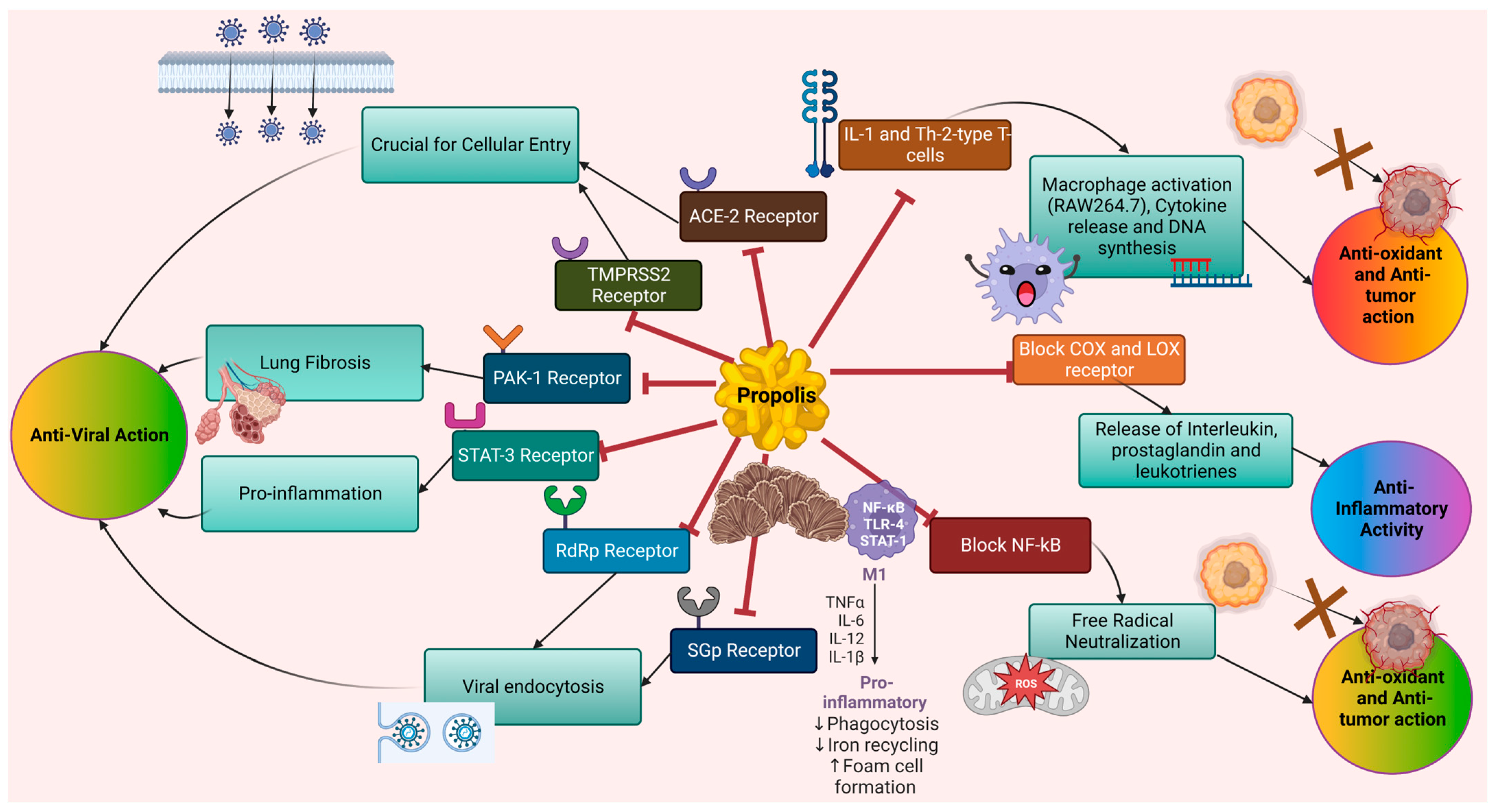
2. Propolis and Molecular Mechanism of Their Active Constituents
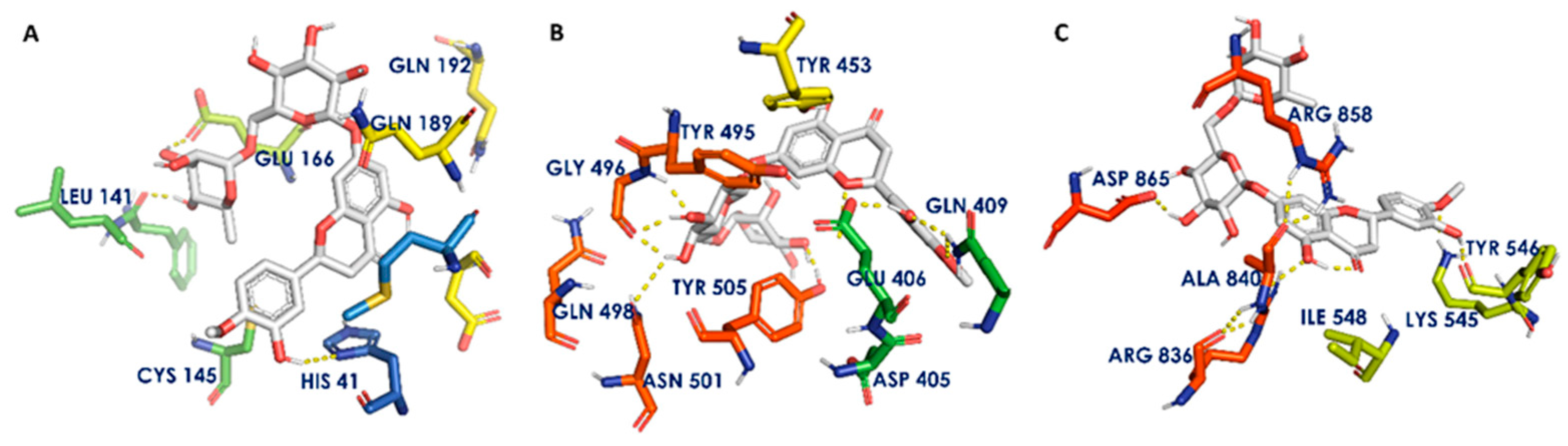
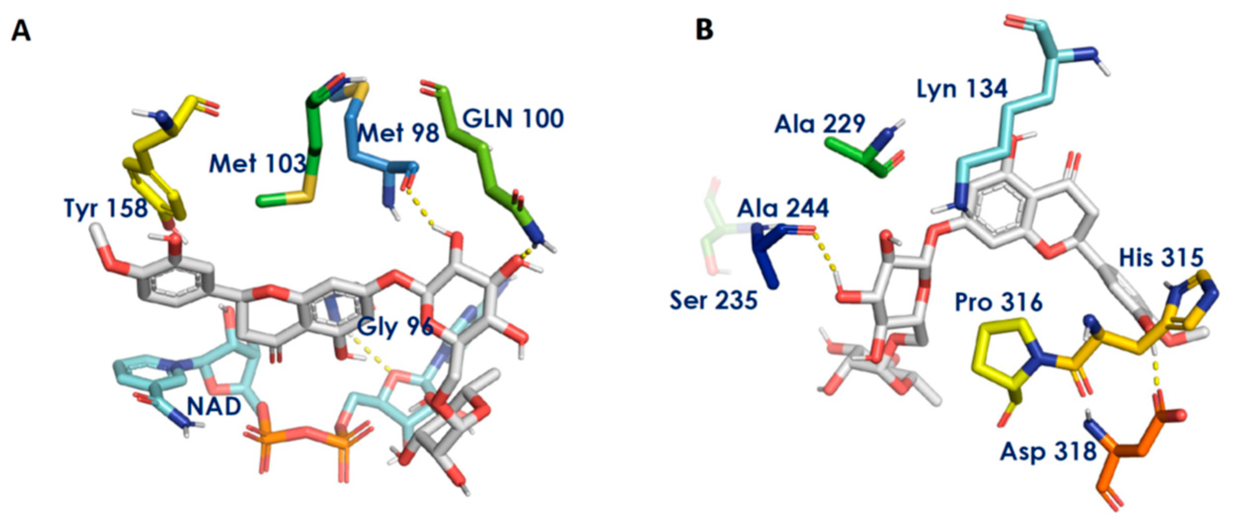
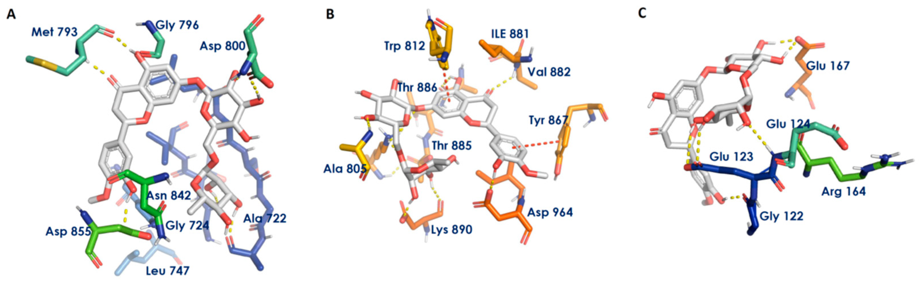
3. Molecular Docking for Propolis Constituents against Chronic Diseases
4. Chronic Diseases with Reported Therapeutic Properties of Propolis and Its Active Constituents
4.1. Chronic Kidney Disease

| Propolis | Study Design | Dose | Measured Outcome | References |
|---|---|---|---|---|
| Brazilian green propolis extract | double-blinded randomized placebo-controlled study on CKD patients | 500 mg per day for 12 months |
| [45] |
| Water-soluble derivative of Croatian propolis | Swiss Albino mice model | 50 mg/kg per day for one week |
| [52] |
| Chrysin, propolis constituent | STZ/HFD-induced T2DM Wistar albino rat model | 40 mg/kg per day for 16 weeks |
| [53] |
| Brazilian propolis and Chinese propolis | STZ-induced diabetic male Sprague Dawley rats | Group A = 100 mg/kg Chinese propolis and Group B = 100 mg/kg Brazilian propolis twice daily for 8 weeks |
| [55] |
| Ethanol extract of Iranian Propolis | STZ-induced diabetic Wistar rats | 100 mg/kg per day and 200 mg/kg per day for 6 weeks |
| [56] |
| Brazilian Red Propolis | 5/6 renal ablation model Wistar rats | 150 mg/kg per day for three months |
| [44] |
| Propolis, unknown source | Methotrexate-induced kidney injury in male Wistar albino rats | 100 mg/kg per day for 15 days |
| [57] |
| Taiwanese green propolis | Aristolochic acids-induced nephropathy model in C57BL/6 mice | 0.2 mg/kg for 12 weeks |
| [58] |
4.2. Rheumatoid Arthritis
4.3. Cancer
4.4. Diabetes
| Propolis Source | Study Design | Dose | Measured Outcome | References |
|---|---|---|---|---|
| Encapsulated propolis, China | T2DM rat model | Low dose =50 mg/kg per day, middle dose =100 mg/kg per day and high dose= 200 mg/kg per day |
| [80] |
| Mexican chihuahua propolis | STZ-diabetic mice model | 0.3 g/kg per day, fifteen days of treatment |
| [81] |
| Brazilian propolis | OLETF rats model | One group with 0.1% w/w and another group with 0.5% w/w propolis diet |
| [82] |
| Taiwanese green propolis ethanolic extract | STZ/HFD-induced T2DM rat model, three different groups: controlled group, 1X group and 5X group | 183.9 mg/kg per day, 919.5 mg/kg per day for 8 weeks. |
| [83] |
| Brazilian propolis ethanolic extract | C57BL/6 male obese mice | 100 mg/kg, two doses within seven days, twelve weeks of treatment |
| [84] |
| Iranian propolis | Human clinical trial: randomized, double-blinded, controlled study on T2DM patients | 500 mg propolis capsule three times per day for 8 weeks |
| [85] |
| Brazilian green propolis | Human clinical trial: randomized, double-blinded, controlled study on T2DM patients | 226.8 mg every day for 8 weeks |
| [87] |
| Brazilian green propolis | Human clinical trial | 900 mg every day for 18 weeks |
| [88] |
| Chinese propolis | Human clinical trial, randomized controlled study | 900 mg every day for 18 weeks |
| [89] |
| Bee propolis peels manufactured by Soren Tech Toos, Mashhad, Iran | Human clinical trial: randomized, double-blinded, controlled study on T2DM patients | 300 mg propolis pills three times per day for 12 weeks |
| [90] |
| Iranian propolis | Human clinical trial: randomized, double-blinded, controlled study on T2DM patients | 500 mg Iranian propolis capsule twice a day for 12 weeks |
| [91] |
4.5. Tuberculosis
4.6. COPD
4.7. Cardiovascular Disease
4.8. Other Chronic Diseases
5. Current Scenario of Preclinical and Clinical Development
6. Conclusions and Future Directions
Author Contributions
Funding
Institutional Review Board Statement
Informed Consent Statement
Data Availability Statement
Conflicts of Interest
Abbreviations
| AAPH | 2,2′-azobis(2-amidinopropane) dihydrochloride |
| ACE-2 | Angiotensin-converting enzyme-2 |
| BGL | Blood glucose level |
| CAPE | Caffeic Acid Phenethyl Ester |
| COPD | Chronic obstructive pulmonary disease |
| CKD | Chronic kidney disease |
| CVD | Cardiovascular disease |
| COVID-19 | Coronavirus disease 2019 |
| DM | Diabetes mellitus |
| DNA | Deoxyribonucleic acid |
| EEP | Ethanolic extract of propolis |
| FBG | Fasting blood glucose |
| GFR | Glomerular filtration rate |
| GPx | Glutathione peroxidase |
| HDL | High-density lipoprotein |
| HFD | High-fat diet |
| HOMA-IR | Homeostasis model assessment-insulin resistance |
| IL-1 | Interleukin-1 |
| IL-6 | Interleukin-6 |
| IL-17 | Interleukin-17 |
| IC50 | Half-maximal inhibitory concentration |
| JNK | C-Jun N-terminal kinases |
| LDL | Low-density lipoprotein |
| LPS | Lipopolysaccharide |
| MCF | Michigan Cancer Foundation |
| Mpro | Main protease |
| Mtb | Mycobacterium tuberculosis |
| NF-κB | Nuclear factor kappa Β |
| OLETF | Otsuka Long-Evans Tokushima fatty |
| PE | Propolis extract |
| PARP | Poly-ADP ribose polymerase |
| RA | Rheumatoid arthritis |
| RCC | Renal cell carcinoma |
| ROS | Reactive oxygen species |
| SARS | Severe acute respiratory syndrome |
| SOD | Serum superoxide dismutase |
| STZ | Streptozotocin |
| T1DM | Type 1 diabetes mellitus |
| T2DM | Type 2 diabetes mellitus |
| TB | Tuberculosis |
| TC | Total cholesterol |
| TGF-β | Transforming growth factor-β |
| TLR4 | Toll-like receptor 4 |
| TNF-α | Tumor necrosis factor alpha |
References
- Castaldo, S.; Capasso, F. Propolis, an Old Remedy Used in Modern Medicine. Fitoterapia 2002, 73, S1–S6. [Google Scholar] [CrossRef]
- Papa, G.; Maier, R.; Durazzo, A.; Lucarini, M.; Karabagias, I.K.; Plutino, M.; Bianchetto, E.; Aromolo, R.; Pignatti, G.; Ambrogio, A.; et al. The Honey Bee Apis Mellifera: An Insect at the Interface between Human and Ecosystem Health. Biology 2022, 11, 233. [Google Scholar] [CrossRef]
- Kasote, D.; Bankova, V.; Viljoen, A.M. Propolis: Chemical Diversity and Challenges in Quality Control. Phytochem. Rev. 2022, 21, 1887–1911. [Google Scholar] [CrossRef] [PubMed]
- Rivera-Yañez, N.; Rivera-Yañez, C.R.; Pozo-Molina, G.; Méndez-Catalá, C.F.; Méndez-Cruz, A.R.; Nieto-Yañez, O. Biomedical Properties of Propolis on Diverse Chronic Diseases and Its Potential Applications and Health Benefits. Nutrients 2020, 13, 78. [Google Scholar] [CrossRef] [PubMed]
- Durazzo, A.; Lucarini, M.; Plutino, M.; Lucini, L.; Aromolo, R.; Martinelli, E.; Souto, E.B.; Santini, A.; Pignatti, G. Bee Products: A Representation of Biodiversity, Sustainability, and Health. Life 2021, 11, 970. [Google Scholar] [CrossRef] [PubMed]
- Hossain, R.; Quispe, C.; Khan, R.A.; Saikat, A.S.M.; Ray, P.; Ongalbek, D.; Yeskaliyeva, B.; Jain, D.; Smeriglio, A.; Trombetta, D.; et al. Propolis: An Update on Its Chemistry and Pharmacological Applications. Chin. Med. 2022, 17, 100. [Google Scholar] [CrossRef]
- Thompson, D.M.; Booth, L.; Moore, D.; Mathers, J. Peer Support for People with Chronic Conditions: A Systematic Review of Reviews. BMC Health Serv. Res. 2022, 22, 427. [Google Scholar] [CrossRef] [PubMed]
- Kim, J.H.; Kismali, G.; Gupta, S.C. Natural Products for the Prevention and Treatment of Chronic Inflammatory Diseases: Integrating Traditional Medicine into Modern Chronic Diseases Care. Evid.-Based Complement. Altern. Med. 2018, 2018, 9837863. [Google Scholar] [CrossRef]
- Flamandita, D.; Lischer, K.; Pratami, D.K.; Aditama, R.; Sahlan, M. Molecular Docking Analysis of Podophyllotoxin Derivatives in Sulawesi Propolis as Potent Inhibitors of Protein Kinases. AIP Conf. Proc. 2020, 2230, 020010. [Google Scholar] [CrossRef]
- Rodrigues, D.M.; Portapilla, G.B.; Silva, G.M.; Duarte, A.; Rotta, C.G.; de Paula da Silva, C.H.T.; de Albuquerque, S.; Bastos, J.K.; Campo, V.L. Synthesis, Antitumor Activity and in Silico Analyses of Amino Acid Derivatives of Artepillin C, Drupanin and Baccharin from Green Propolis. Bioorg. Med. Chem. 2021, 47, 116372. [Google Scholar] [CrossRef]
- Elnakady, Y.A.; Rushdi, A.I.; Franke, R.; Abutaha, N.; Ebaid, H.; Baabbad, M.; Omar, M.O.M.; Al Ghamdi, A.A. Characteristics, Chemical Compositions and Biological Activities of Propolis from Al-Bahah, Saudi Arabia. Sci. Rep. 2017, 7, 41453. [Google Scholar] [CrossRef] [PubMed]
- Bueno-Silva, B.; Rosalen, P.L.; Alencar, S.M.; Mayer, M.P.A. Anti-Inflammatory Mechanisms of Neovestitol from Brazilian Red Propolis in LPS-Activated Macrophages. J. Funct. Foods 2017, 36, 440–447. [Google Scholar] [CrossRef]
- Ansorge, S.; Reinhold, D.; Lendeckel, U. Propolis and Some of Its Constituents Down-Regulate DNA Synthesis and Inflammatory Cytokine Production but Induce TGF-Β1 Production of Human Immune Cells. Z. Für Nat. C 2003, 58, 580–589. [Google Scholar] [CrossRef] [PubMed]
- Zhang, X.; Wang, G.; Gurley, E.C.; Zhou, H. Flavonoid Apigenin Inhibits Lipopolysaccharide-Induced Inflammatory Response through Multiple Mechanisms in Macrophages. PLoS ONE 2014, 9, e107072. [Google Scholar] [CrossRef]
- Missima, F.; Pagliarone, A.C.; Orsatti, C.L.; Araújo, J.P.; Sforcin, J.M. The Effect of Propolis on Th1/Th2 Cytokine Expression and Production by Melanoma-Bearing Mice Submitted to Stress: PROPOLIS EFFECT ON MELANOMA-BEARING MICE SUBMITTED TO STRESS. Phytother. Res. 2010, 24, 1501–1507. [Google Scholar] [CrossRef]
- Uzel, A.; Sorkun, K.; Önçağ, Ö.; Çoğulu, D.; Gençay, Ö.; Sali˙h, B. Chemical Compositions and Antimicrobial Activities of Four Different Anatolian Propolis Samples. Microbiol. Res. 2005, 160, 189–195. [Google Scholar] [CrossRef]
- Araujo, M.A.R.; Libério, S.A.; Guerra, R.N.M.; Ribeiro, M.N.S.; Nascimento, F.R.F. Mechanisms of Action Underlying the Anti-Inflammatory and Immunomodulatory Effects of Propolis: A Brief Review. Rev. Bras. Farmacogn. 2012, 22, 208–219. [Google Scholar] [CrossRef]
- Oršolić, N.; Jazvinšćak Jembrek, M. Molecular and Cellular Mechanisms of Propolis and Its Polyphenolic Compounds against Cancer. IJMS 2022, 23, 10479. [Google Scholar] [CrossRef]
- Wang, L.; Tu, Y.-C.; Lian, T.-W.; Hung, J.-T.; Yen, J.-H.; Wu, M.-J. Distinctive Antioxidant and Antiinflammatory Effects of Flavonols. J. Agric. Food Chem. 2006, 54, 9798–9804. [Google Scholar] [CrossRef]
- Chiu, H.-F.; Han, Y.-C.; Shen, Y.-C.; Golovinskaia, O.; Venkatakrishnan, K.; Wang, C.-K. Chemopreventive and Chemotherapeutic Effect of Propolis and Its Constituents: A Mini-Review. J. Cancer Prev. 2020, 25, 70–78. [Google Scholar] [CrossRef]
- Sies, H.; Belousov, V.V.; Chandel, N.S.; Davies, M.J.; Jones, D.P.; Mann, G.E.; Murphy, M.P.; Yamamoto, M.; Winterbourn, C. Defining Roles of Specific Reactive Oxygen Species (ROS) in Cell Biology and Physiology. Nat. Rev. Mol. Cell Biol. 2022, 23, 499–515. [Google Scholar] [CrossRef]
- Forma, E.; Bryś, M. Anticancer Activity of Propolis and Its Compounds. Nutrients 2021, 13, 2594. [Google Scholar] [CrossRef] [PubMed]
- Zhao, J.-Q.; Wen, Y.-F.; Bhadauria, M.; Nirala, S.K.; Sharma, A.; Shrivastava, S.; Shukla, S.; Agrawal, O.P.; Mathur, R. Protective Effects of Propolis on Inorganic Mercury Induced Oxidative Stress in Mice. Indian J. Exp. Biol. 2009, 47, 264–269. [Google Scholar] [PubMed]
- Galati, G.; O’Brien, P.J. Potential Toxicity of Flavonoids and Other Dietary Phenolics: Significance for Their Chemopreventive and Anticancer Properties. Free Radic. Biol. Med. 2004, 37, 287–303. [Google Scholar] [CrossRef]
- Kabała-Dzik, A.; Rzepecka-Stojko, A.; Kubina, R.; Jastrzębska-Stojko, Ż.; Stojko, R.; Wojtyczka, R.; Stojko, J. Comparison of Two Components of Propolis: Caffeic Acid (CA) and Caffeic Acid Phenethyl Ester (CAPE) Induce Apoptosis and Cell Cycle Arrest of Breast Cancer Cells MDA-MB-231. Molecules 2017, 22, 1554. [Google Scholar] [CrossRef] [PubMed]
- Song, Y.S.; Jin, C.; Jung, K.J.; Park, E.-H. Estrogenic Effects of Ethanol and Ether Extracts of Propolis. J. Ethnopharmacol. 2002, 82, 89–95. [Google Scholar] [CrossRef]
- Eitsuka, T.; Nakagawa, K.; Kato, S.; Ito, J.; Otoki, Y.; Takasu, S.; Shimizu, N.; Takahashi, T.; Miyazawa, T. Modulation of Telomerase Activity in Cancer Cells by Dietary Compounds: A Review. Int. J. Mol. Sci. 2018, 19, 478. [Google Scholar] [CrossRef] [PubMed]
- Amoros, M.; Sauvager, F.; Girre, L.; Cormier, M. In Vitro Antiviral Activity of Propolis. Apidologie 1992, 23, 231–240. [Google Scholar] [CrossRef]
- Kujumgiev, A.; Tsvetkova, I.; Serkedjieva, Y.; Bankova, V.; Christov, R.; Popov, S. Antibacterial, Antifungal and Antiviral Activity of Propolis of Different Geographic Origin. J. Ethnopharmacol. 1999, 64, 235–240. [Google Scholar] [CrossRef]
- Kwon, M.J.; Shin, H.M.; Perumalsamy, H.; Wang, X.; Ahn, Y.-J. Antiviral Effects and Possible Mechanisms of Action of Constituents from Brazilian Propolis and Related Compounds. J. Apic. Res. 2020, 59, 413–425. [Google Scholar] [CrossRef]
- Berretta, A.A.; Silveira, M.A.D.; Cóndor Capcha, J.M.; De Jong, D. Propolis and Its Potential against SARS-CoV-2 Infection Mechanisms and COVID-19 Disease. Biomed. Pharmacother. 2020, 131, 110622. [Google Scholar] [CrossRef] [PubMed]
- Chavda, V.P.; Patel, A.B.; Vihol, D.; Vaghasiya, D.D.; Ahmed, K.M.S.B.; Trivedi, K.U.; Dave, D.J. Herbal Remedies, Nutraceuticals, and Dietary Supplements for COVID-19 Management: An Update. Clin. Complement. Med. Pharmacol. 2022, 2, 100021. [Google Scholar] [CrossRef]
- Costela-Ruiz, V.J.; Illescas-Montes, R.; Puerta-Puerta, J.M.; Ruiz, C.; Melguizo-Rodríguez, L. SARS-CoV-2 Infection: The Role of Cytokines in COVID-19 Disease. Cytokine Growth Factor Rev. 2020, 54, 62–75. [Google Scholar] [CrossRef] [PubMed]
- Shaldam, M.A.; Yahya, G.; Mohamed, N.H.; Abdel-Daim, M.M.; Al Naggar, Y. In Silico Screening of Potent Bioactive Compounds from Honeybee Products against COVID-19 Target Enzymes. Env. Sci. Pollut. Res. Int. 2021, 28, 40507–40514. [Google Scholar] [CrossRef] [PubMed]
- Yosri, N.; Abd El-Wahed, A.A.; Ghonaim, R.; Khattab, O.M.; Sabry, A.; Ibrahim, M.A.A.; Moustafa, M.F.; Guo, Z.; Zou, X.; Algethami, A.F.M.; et al. Anti-Viral and Immunomodulatory Properties of Propolis: Chemical Diversity, Pharmacological Properties, Preclinical and Clinical Applications, and In Silico Potential against SARS-CoV-2. Foods 2021, 10, 1776. [Google Scholar] [CrossRef] [PubMed]
- Dilokthornsakul, W.; Kosiyaporn, R.; Wuttipongwaragon, R.; Dilokthornsakul, P. Potential Effects of Propolis and Honey in COVID-19 Prevention and Treatment: A Systematic Review of in Silico and Clinical Studies. J. Integr. Med. 2022, 20, 114–125. [Google Scholar] [CrossRef]
- Giacco, F.; Brownlee, M. Oxidative Stress and Diabetic Complications. Circ. Res. 2010, 107, 1058–1070. [Google Scholar] [CrossRef]
- Jasprica, I.; Mornar, A.; Debeljak, Ž.; Smolčić-Bubalo, A.; Medić-Šarić, M.; Mayer, L.; Romić, Ž.; Bućan, K.; Balog, T.; Sobočanec, S.; et al. In Vivo Study of Propolis Supplementation Effects on Antioxidative Status and Red Blood Cells. J. Ethnopharmacol. 2007, 110, 548–554. [Google Scholar] [CrossRef]
- Kanbur, M.; Eraslan, G.; Silici, S. Antioxidant Effect of Propolis against Exposure to Propetamphos in Rats. Ecotoxicol. Environ. Saf. 2009, 72, 909–915. [Google Scholar] [CrossRef]
- De Miranda, M.B.; Lanna, M.F.; Nascimento, A.L.B.; de Paula, C.A.; de Souza, M.E.; Felipetto, M.; da Silva Barcelos, L.; de Moura, S.A.L. Hydroalcoholic Extract of Brazilian Green Propolis Modulates Inflammatory Process in Mice Submitted to a Low Protein Diet. Biomed. Pharmacother. 2019, 109, 610–620. [Google Scholar] [CrossRef]
- Mesbah, L.; Samia, A. Bioavailability and Pharmacokinetic of the Algerian Propolis Constituent Naringenin in Rats after Oral Administration. Planta Med. 2011, 77, PA11. [Google Scholar] [CrossRef]
- Bank, R.P.D. RCSB PDB: Homepage. Available online: https://www.rcsb.org/ (accessed on 24 December 2022).
- Schroeder, E.K.; de Souza, N.; Santos, D.S.; Blanchard, J.S.; Basso, L.A. Drugs That Inhibit Mycolic Acid Biosynthesis in Mycobacterium Tuberculosis. Curr. Pharm. Biotechnol. 2002, 3, 197–225. [Google Scholar] [CrossRef] [PubMed]
- Teles, F.; da Silva, T.M.; da Cruz Júnior, F.P.; Honorato, V.H.; de Oliveira Costa, H.; Barbosa, A.P.F.; de Oliveira, S.G.; Porfírio, Z.; Libório, A.B.; Borges, R.L.; et al. Brazilian Red Propolis Attenuates Hypertension and Renal Damage in 5/6 Renal Ablation Model. PLoS ONE 2015, 10, e0116535. [Google Scholar] [CrossRef] [PubMed]
- Silveira, M.A.D.; Teles, F.; Berretta, A.A.; Sanches, T.R.; Rodrigues, C.E.; Seguro, A.C.; Andrade, L. Effects of Brazilian Green Propolis on Proteinuria and Renal Function in Patients with Chronic Kidney Disease: A Randomized, Double-Blind, Placebo-Controlled Trial. BMC Nephrol. 2019, 20, 140. [Google Scholar] [CrossRef]
- Wagh, V.D. Propolis: A Wonder Bees Product and Its Pharmacological Potentials. Adv. Pharm. Sci. 2013, 2013, 308249. [Google Scholar] [CrossRef] [PubMed]
- Elumalai, P.; Muninathan, N.; Megalatha, S.T.; Suresh, A.; Kumar, K.S.; Jhansi, N.; Kalaivani, K.; Krishnamoorthy, G. An Insight into Anticancer Effect of Propolis and Its Constituents: A Review of Molecular Mechanisms. Evid.-Based Complement. Altern. Med. 2022, 2022, 5901191. [Google Scholar] [CrossRef]
- Nattagh-Eshtivani, E.; Pahlavani, N.; Ranjbar, G.; Gholizadeh Navashenaq, J.; Salehi-Sahlabadi, A.; Mahmudiono, T.; Nader Shalaby, M.; Jokar, M.; Nematy, M.; Barghchi, H.; et al. Does Propolis Have Any Effect on Rheumatoid Arthritis? A Review Study. Food Sci. Nutr. 2022, 10, 1003–1020. [Google Scholar] [CrossRef]
- Kovesdy, C.P. Epidemiology of Chronic Kidney Disease: An Update 2022. Kidney Int. Suppl. 2022, 12, 7–11. [Google Scholar] [CrossRef]
- Vaziri, N.D.; Bai, Y.; Ni, Z.; Quiroz, Y.; Pandian, R.; Rodriguez-Iturbe, B. Intra-Renal Angiotensin II/AT 1 Receptor, Oxidative Stress, Inflammation, and Progressive Injury in Renal Mass Reduction. J. Pharm. Exp. Ther. 2007, 323, 85–93. [Google Scholar] [CrossRef]
- Anvarifard, P.; Anbari, M.; Ostadrahimi, A.; Ardalan, M.; Ghoreishi, Z. A Comprehensive Insight into the Molecular and Cellular Mechanisms of the Effects of Propolis on Preserving Renal Function: A Systematic Review. Nutr. Metab. (Lond.) 2022, 19, 6. [Google Scholar] [CrossRef]
- Oršolić, N.; Sirovina, D.; Končić, M.Z.; Lacković, G.; Gregorović, G. Effect of Croatian Propolis on Diabetic Nephropathy and Liver Toxicity in Mice. BMC Complement. Altern. Med. 2012, 12, 117. [Google Scholar] [CrossRef] [PubMed]
- Ahad, A.; Ganai, A.A.; Mujeeb, M.; Siddiqui, W.A. Chrysin, an Anti-Inflammatory Molecule, Abrogates Renal Dysfunction in Type 2 Diabetic Rats. Toxicol. Appl. Pharmacol. 2014, 279, 1–7. [Google Scholar] [CrossRef]
- Granados-Pineda, J.; Uribe-Uribe, N.; García-López, P.; Ramos-Godinez, M.; Rivero-Cruz, J.; Pérez-Rojas, J. Effect of Pinocembrin Isolated from Mexican Brown Propolis on Diabetic Nephropathy. Molecules 2018, 23, 852. [Google Scholar] [CrossRef] [PubMed]
- Zhu, W.; Li, Y.-H.; Chen, M.-L.; Hu, F.-L. Protective Effects of Chinese and Brazilian Propolis Treatment against Hepatorenal Lesion in Diabetic Rats. Hum. Exp. Toxicol. 2011, 30, 1246–1255. [Google Scholar] [CrossRef]
- Sameni, H.R.; Ramhormozi, P.; Bandegi, A.R.; Taherian, A.A.; Mirmohammadkhani, M.; Safari, M. Effects of Ethanol Extract of Propolis on Histopathological Changes and Anti-oxidant Defense of Kidney in a Rat Model for Type 1 Diabetes Mellitus. J. Diabetes Investig. 2016, 7, 506–513. [Google Scholar] [CrossRef] [PubMed]
- Ulusoy, H.B.; Öztürk, İ.; Sönmez, M.F. Protective Effect of Propolis on Methotrexate-Induced Kidney Injury in the Rat. Ren. Fail. 2016, 38, 744–750. [Google Scholar] [CrossRef]
- Chang, J.-F.; Hsieh, C.-Y.; Lu, K.-C.; Chen, Y.-W.; Liang, S.-S.; Lin, C.-C.; Hung, C.-F.; Liou, J.-C.; Wu, M.-S. Therapeutic Targeting of Aristolochic Acid Induced Uremic Toxin Retention, SMAD 2/3 and JNK/ERK Pathways in Tubulointerstitial Fibrosis: Nephroprotective Role of Propolis in Chronic Kidney Disease. Toxins 2020, 12, 364. [Google Scholar] [CrossRef] [PubMed]
- Zhang, J.-M.; An, J. Cytokines, Inflammation, and Pain. Int. Anesthesiol. Clin. 2007, 45, 27–37. [Google Scholar] [CrossRef]
- Phull, A.-R.; Nasir, B.; Haq, I.U.; Kim, S.J. Oxidative Stress, Consequences and ROS Mediated Cellular Signaling in Rheumatoid Arthritis. Chem. Biol. Interact. 2018, 281, 121–136. [Google Scholar] [CrossRef] [PubMed]
- Nattagh-Eshtivani, E.; Jokar, M.; Tabesh, H.; Nematy, M.; Safarian, M.; Pahlavani, N.; Maddahi, M.; Khosravi, M. The Effect of Propolis Supplementation on Inflammatory Factors and Oxidative Status in Women with Rheumatoid Arthritis: Design and Research Protocol of a Double-Blind, Randomized Controlled. Contemp. Clin. Trials Commun. 2021, 23, 100807. [Google Scholar] [CrossRef]
- Park, E.-H.; Kahng, J.-H. Suppressive Effects of Propolis in Rat Adjuvant Arthritis. Arch. Pharm. Res. 1999, 22, 554–558. [Google Scholar] [CrossRef]
- Orsi, R.O.; Sforcin, J.M.; Funari, S.R.C.; Bankova, V. Effects of Brazilian and Bulgarian Propolis on Bactericidal Activity of Macrophages against Salmonella Typhimurium. Int. Immunopharmacol. 2005, 5, 359–368. [Google Scholar] [CrossRef] [PubMed]
- Wolska, K.; Gorska, A.; Antosik, K.; Lugowska, K. Immunomodulatory Effects of Propolis and Its Components on Basic Immune Cell Functions. Pharm. Sci. 2019, 81, 575–588. [Google Scholar] [CrossRef]
- Ramos, A.F.N.; Miranda, J.L. Propolis: A Review of Its Anti-Inflammatory and Healing Actions. J. Venom. Anim. Toxins Incl. Trop. Dis. 2007, 13, 697–710. [Google Scholar] [CrossRef]
- Kurek-Górecka, A.M.; Sobczak, A.; Rzepecka-Stojko, A.; Górecki, M.T.; Wardas, M.; Pawłowska-Góral, K. Antioxidant Activity of Ethanolic Fractions of Polish Propolis. Z. Für Naturforschung. 2012, 67, 545–550. [Google Scholar] [CrossRef] [PubMed]
- Mihai, C.M.; Marghitas, L.A.; Mărghitaş, D.S.; Barnutiu, L. Correlation between Polyphenolic Profile and Antioxidant Activity of Propolis from Transylvania. Sci. Pap. 2011, 44, 100–103. [Google Scholar]
- Tanaka, M.; Okamoto, Y.; Fukui, T.; Masuzawa, T. Suppression of Interleukin 17 Production by Brazilian Propolis in Mice with Collagen-Induced Arthritis. Inflammopharmacology 2012, 20, 19–26. [Google Scholar] [CrossRef] [PubMed]
- Matsumoto, Y.; Takahashi, K.; Sugioka, Y.; Inui, K.; Okano, T.; Mandai, K.; Yamada, Y.; Shintani, A.; Koike, T. Double-Blinded Randomized Controlled Trial to Reveal the Effects of Brazilian Propolis Intake on Rheumatoid Arthritis Disease Activity Index; BeeDAI. PLoS ONE 2021, 16, e0252357. [Google Scholar] [CrossRef]
- Sforcin, J.M.; Bankova, V. Propolis: Is There a Potential for the Development of New Drugs? J. Ethnopharmacol. 2011, 133, 253–260. [Google Scholar] [CrossRef]
- Valente, M.J.; Baltazar, A.F.; Henrique, R.; Estevinho, L.; Carvalho, M. Biological Activities of Portuguese Propolis: Protection against Free Radical-Induced Erythrocyte Damage and Inhibition of Human Renal Cancer Cell Growth in Vitro. Food Chem. Toxicol. 2011, 49, 86–92. [Google Scholar] [CrossRef]
- Watabe, M.; Hishikawa, K.; Takayanagi, A.; Shimizu, N.; Nakaki, T. Caffeic Acid Phenethyl Ester Induces Apoptosis by Inhibition of NFκB and Activation of Fas in Human Breast Cancer MCF-7 Cells*. J. Biol. Chem. 2004, 279, 6017–6026. [Google Scholar] [CrossRef]
- Kamiya, T.; Nishihara, H.; Hara, H.; Adachi, T. Ethanol Extract of Brazilian Red Propolis Induces Apoptosis in Human Breast Cancer MCF-7 Cells through Endoplasmic Reticulum Stress. J. Agric. Food Chem. 2012, 60, 11065–11070. [Google Scholar] [CrossRef]
- Xuan, H.; Li, Z.; Yan, H.; Sang, Q.; Wang, K.; He, Q.; Wang, Y.; Hu, F. Antitumor Activity of Chinese Propolis in Human Breast Cancer MCF-7 and MDA-MB-231 Cells. Evid.-Based Complement. Altern. Med. 2014, 2014, 280120. [Google Scholar] [CrossRef] [PubMed]
- Chang, H.; Wang, Y.; Yin, X.; Liu, X.; Xuan, H. Ethanol Extract of Propolis and Its Constituent Caffeic Acid Phenethyl Ester Inhibit Breast Cancer Cells Proliferation in Inflammatory Microenvironment by Inhibiting TLR4 Signal Pathway and Inducing Apoptosis and Autophagy. BMC Complement. Altern. Med. 2017, 17, 471. [Google Scholar] [CrossRef] [PubMed]
- Frión-Herrera, Y.; Díaz-García, A.; Ruiz-Fuentes, J.L.; Rodríguez-Sánchez, H.; Sforcin, J.M. The Cytotoxic Effects of Propolis on Breast Cancer Cells Involve PI3K/Akt and ERK1/2 Pathways, Mitochondrial Membrane Potential, and Reactive Oxygen Species Generation. Inflammopharmacology 2019, 27, 1081–1089. [Google Scholar] [CrossRef] [PubMed]
- Li, J.; Liu, H.; Liu, X.; Hao, S.; Zhang, Z.; Xuan, H. Chinese Poplar Propolis Inhibits MDA-MB-231 Cell Proliferation in an Inflammatory Microenvironment by Targeting Enzymes of the Glycolytic Pathway. J. Immunol. Res. 2021, 2021, 6641341. [Google Scholar] [CrossRef]
- Popova, M.; Giannopoulou, E.; Skalicka-Woźniak, K.; Graikou, K.; Widelski, J.; Bankova, V.; Kalofonos, H.; Sivolapenko, G.; Gaweł-Bęben, K.; Antosiewicz, B.; et al. Characterization and Biological Evaluation of Propolis from Poland. Molecules 2017, 22, 1159. [Google Scholar] [CrossRef] [PubMed]
- Davì, G.; Santilli, F.; Patrono, C. Nutraceuticals in Diabetes and Metabolic Syndrome: Nutraceuticals in Diabetes and Metabolic Syndrome. Cardiovasc. Ther. 2010, 28, 216–226. [Google Scholar] [CrossRef]
- Li, Y.; Chen, M.; Xuan, H.; Hu, F. Effects of Encapsulated Propolis on Blood Glycemic Control, Lipid Metabolism, and Insulin Resistance in Type 2 Diabetes Mellitus Rats. Evid.-Based Complement. Altern. Med. 2012, 2012, 981896. [Google Scholar] [CrossRef]
- Rivera-Yañez, N.; Rodriguez-Canales, M.; Nieto-Yañez, O.; Jimenez-Estrada, M.; Ibarra-Barajas, M.; Canales-Martinez, M.M.; Rodriguez-Monroy, M.A. Hypoglycaemic and Antioxidant Effects of Propolis of Chihuahua in a Model of Experimental Diabetes. Evid.-Based Complement. Altern. Med. 2018, 2018, 4360356. [Google Scholar] [CrossRef]
- Aoi, W.; Hosogi, S.; Niisato, N.; Yokoyama, N.; Hayata, H.; Miyazaki, H.; Kusuzaki, K.; Fukuda, T.; Fukui, M.; Nakamura, N.; et al. Improvement of Insulin Resistance, Blood Pressure and Interstitial PH in Early Developmental Stage of Insulin Resistance in OLETF Rats by Intake of Propolis Extracts. Biochem. Biophys. Res. Commun. 2013, 432, 650–653. [Google Scholar] [CrossRef] [PubMed]
- Chen, L.-H.; Chien, Y.-W.; Chang, M.-L.; Hou, C.-C.; Chan, C.-H.; Tang, H.-W.; Huang, H.-Y. Taiwanese Green Propolis Ethanol Extract Delays the Progression of Type 2 Diabetes Mellitus in Rats Treated with Streptozotocin/High-Fat Diet. Nutrients 2018, 10, 503. [Google Scholar] [CrossRef] [PubMed]
- Kitamura, H.; Naoe, Y.; Kimura, S.; Miyamoto, T.; Okamoto, S.; Toda, C.; Shimamoto, Y.; Iwanaga, T.; Miyoshi, I. Beneficial Effects of Brazilian Propolis on Type 2 Diabetes in Ob/Ob Mice: Possible Involvement of Immune Cells in Mesenteric Adipose Tissue. Adipocyte 2013, 2, 227–236. [Google Scholar] [CrossRef]
- Afsharpour, F.; Hashemipour, S.; Haghighian, H.K.; Koushan, Y. Effects of Iranian Propolis on Glycemic Status, Inflammatory Factors, and Liver Enzyme Levels in Type 2 Diabetic Patients: A Randomized, Double-Blind, Placebo-Controlled, Clinical Trial. J. Nutr. Sci. Dietet. 2017, 9–14. [Google Scholar]
- Hesami, S.; Hashemipour, S.; Shiri-Shahsavar, M.; Koushan, Y.; Khadem Haghighian, H. Administration of Iranian Propolis Attenuates Oxidative Stress and Blood Glucose in Type II Diabetic Patients: A Randomized, Double-Blind, Placebo-Controlled, Clinical Trial. Casp. J. Intern. Med. 2019, 10, 48–54. [Google Scholar] [CrossRef]
- Fukuda, T.; Fukui, M.; Tanaka, M.; Senmaru, T.; Iwase, H.; Yamazaki, M.; Aoi, W.; Inui, T.; Nakamura, N.; Marunaka, Y. Effect of Brazilian Green Propolis in Patients with Type 2 Diabetes: A Double-Blind Randomized Placebo-Controlled Study. Biomed. Rep. 2015, 3, 355–360. [Google Scholar] [CrossRef]
- Zhao, L.; Pu, L.; Wei, J.; Li, J.; Wu, J.; Xin, Z.; Gao, W.; Guo, C. Brazilian Green Propolis Improves Antioxidant Function in Patients with Type 2 Diabetes Mellitus. Int. J. Environ. Res. Public Health 2016, 13, 498. [Google Scholar] [CrossRef]
- Gao, W.; Pu, L.; Wei, J.; Yao, Z.; Wang, Y.; Shi, T.; Zhao, L.; Jiao, C.; Guo, C. Serum Antioxidant Parameters Are Significantly Increased in Patients with Type 2 Diabetes Mellitus after Consumption of Chinese Propolis: A Randomized Controlled Trial Based on Fasting Serum Glucose Level. Diabetes Ther. 2018, 9, 101–111. [Google Scholar] [CrossRef]
- Samadi, N.; Mozaffari-Khosravi, H.; Rahmanian, M.; Askarishahi, M. Effects of Bee Propolis Supplementation on Glycemic Control, Lipid Profile and Insulin Resistance Indices in Patients with Type 2 Diabetes: A Randomized, Double-Blind Clinical Trial. J. Integr. Med. 2017, 15, 124–134. [Google Scholar] [CrossRef]
- Zakerkish, M.; Jenabi, M.; Zaeemzadeh, N.; Hemmati, A.A.; Neisi, N. The Effect of Iranian Propolis on Glucose Metabolism, Lipid Profile, Insulin Resistance, Renal Function and Inflammatory Biomarkers in Patients with Type 2 Diabetes Mellitus: A Randomized Double-Blind Clinical Trial. Sci. Rep. 2019, 9, 7289. [Google Scholar] [CrossRef]
- Teixeira, É.W.; Message, D.; Meira, R.M.S.A. Methacrylate: An Alternative Fixing Agent for Identifying the Botanical Origin of Propolis. Appl. Plant. Sci. 2019, 7, e11309. [Google Scholar] [CrossRef] [PubMed]
- Zabaiou, N.; Fouache, A.; Trousson, A.; Baron, S.; Zellagui, A.; Lahouel, M.; Lobaccaro, J.-M.A. Biological Properties of Propolis Extracts: Something New from an Ancient Product. Chem. Phys. Lipids 2017, 207, 214–222. [Google Scholar] [CrossRef]
- Scheller, S.; Kawalski, H.; Oklek, K.; Dworniczak, S.; Matsuno, T.; Waldemar-Klimmek, K.; Rajca, M.; Shani, A. Correlation between Virulence of Various Strains of Mycobacteria and Their Susceptibility to Ethanolic Extract of Propolis (EEP). Z. Für Nat. C 1998, 53, 1040–1044. [Google Scholar] [CrossRef]
- Scheller, S.; Dworniczak, S.; Waldemar-Klimmek, K.; Rajca, M.; Tomczyk, A.; Shani, J. Synergism Between Ethanolic Extract of Propolis (EEP) and Anti-Tuberculosis Drugs on Growth of Mycobacteria. Z. Für Nat. C 1999, 54, 549–553. [Google Scholar] [CrossRef]
- de Albuquerque, I.L.; Alves, L.A.; Lemos, T.L.G.; Dorneles, C.A.; de Morais, M.O. Constituents of the Essential Oil of Brazilian Green Propolis from Brazil. J. Essent. Oil Res. 2008, 20, 414–415. [Google Scholar] [CrossRef]
- Quintino, R.L.; Reis, A.C.; Fernandes, C.C.; Martins, C.H.G.; Colli, A.C.; Crotti, A.E.M.; Squarisi, I.S.; Ribeiro, A.B.; Tavares, D.C.; Miranda, M.L.D. Brazilian Green Propolis: Chemical Composition of Essential Oil and Their In Vitro Antioxidant, Antibacterial and Antiproliferative Activities. Braz. Arch. Biol. Technol. 2020, 63, e20190408. [Google Scholar] [CrossRef]
- De Lima, V.H.M.; Almeida, K.d.C.R.; Alves, C.C.F.; Rodrigues, M.L.; Crotti, A.E.M.; de Souza, J.M.; Ribeiro, A.B.; Squarisi, I.S.; Tavares, D.C.; Martins, C.H.G.; et al. Biological Properties of Volatile Oil from Brazilian Brown Propolis. Rev. Bras. Farmacogn. 2019, 29, 807–810. [Google Scholar] [CrossRef]
- Sawicki, R.; Widelski, J.; Okińczyc, P.; Truszkiewicz, W.; Glous, J.; Sieniawska, E. Exposure to Nepalese Propolis Alters the Metabolic State of Mycobacterium Tuberculosis. Front. Microbiol. 2022, 13, 929476. [Google Scholar] [CrossRef] [PubMed]
- The Global Initiative for Chronic Obstructive Lung Disease (GOLD). Global Strategy for Prevention, Diagnosis and Management of Chronic Obstructive Pulmonary Disease. Available online: https://goldcopd.org/wp-content/uploads/2022/12/GOLD-2023-ver-1.1-2Dec2022_WMV.pdf (accessed on 10 January 2023).
- Machado, J.L.; Assunção, A.K.M.; da Silva, M.C.P.; dos Reis, A.S.; Costa, G.C.; Arruda, D.d.S.; Rocha, B.A.; Vaz, M.M.d.O.L.L.; Paes, A.M.d.A.; Guerra, R.N.M.; et al. Brazilian Green Propolis: Anti-Inflammatory Property by an Immunomodulatory Activity. Evid.-Based Complement. Altern. Med. 2012, 2012, 157652. [Google Scholar] [CrossRef] [PubMed]
- Guo, R. Herbal Medicines for the Treatment of COPD: A Systematic Review. Eur. Respir. J. 2006, 28, 330–338. [Google Scholar] [CrossRef]
- Khayyal, M.T.; El-Ghazaly, M.A.; El-Khatib, A.S.; Hatem, A.M.; de Vries, P.J.F.; El-Shafei, S.; Khattab, M.M. A Clinical Pharmacological Study of the Potential Beneficial Effects of a Propolis Food Product as an Adjuvant in Asthmatic Patients. Fundam. Clin. Pharm. 2003, 17, 93–102. [Google Scholar] [CrossRef] [PubMed]
- Kolarov, V.; Stevuljevi, J.K.; Ili, M.; Bogdan, M.; Tušek, B.; Agic, A.; Dugajli, M.; Vereš, K.T.; Kutleši, S.; Zvezdin, B. Factorial Analysis of N-Acetylcysteine and Propolis Treatment Effects on Symptoms, Life Quality and Exacerbations in Patients with Chronic Obstructive Pulmonary Disease (COPD): A Randomized, Double-Blind, Placebo-Controlled Trial. Eur. Rev. Med. Pharmacol. Sci. 2022, 26, 3192–3199. [Google Scholar] [PubMed]
- Buha, I.; Miri, M.; Agi, A.; Simi, M.; Stjepanovi, M.; Milenkovi, B.; Nagorni-Obradovi, L.; Škodri, V.; Ili, B.; Popevi, S.; et al. A Randomized, Double-Blind, Placebo-Controlled Study Evaluating the Efficacy of Propolis and N-Acetylcysteine in Exacerbations of Chronic Obstructive Pulmonary Disease. Eur. Rev. Med. Pharmacol. Sci. 2022, 26, 4809–4815. [Google Scholar] [PubMed]
- Tsao, C.W.; Aday, A.W.; Almarzooq, Z.I.; Alonso, A.; Beaton, A.Z.; Bittencourt, M.S.; Boehme, A.K.; Buxton, A.E.; Carson, A.P.; Commodore-Mensah, Y.; et al. Heart Disease and Stroke Statistics—2022 Update: A Report From the American Heart Association. Circulation 2022, 145, e153–e639. [Google Scholar] [CrossRef] [PubMed]
- Maulana, M.; Andriyani, S. The Relationship between Alcohol Consumption and Cardiovascular Disease in Adults: Meta-Analysis. J. Epidemiol. Public Health 2022, 7, 187–195. [Google Scholar] [CrossRef]
- Majiene, D.; Trumbeckaite, S.; Savickas, A.; Toleikis, A. Influence of Propolis Water Solution on Heart Mitochondrial Function. J. Pharm. Pharmacol. 2010, 58, 709–713. [Google Scholar] [CrossRef] [PubMed]
- Zhang, Y.-X.; Yang, T.-T.; Xia, L.; Zhang, W.-F.; Wang, J.-F.; Wu, Y.-P. Inhibitory Effect of Propolis on Platelet Aggregation In Vitro. J. Healthc. Eng. 2017, 2017, 3050895. [Google Scholar] [CrossRef]
- Wang, H.H.; Zeng, J.; Wang, H.Z.; Jiang, Y.X.; Wang, J.; Zhou, P.P. Effects of Total Flavonoids of Propolis on Apoptosis of Myocardial Cells of Chronic Heart Failure and Its Possible Mechanism in Rats. Chin. J. Appl. Physiol. 2015, 31, 201–206. [Google Scholar]
- Ji, C.; Pan, Y.; Xu, S.; Yu, C.; Ji, J.; Chen, M.; Hu, F. Propolis Ameliorates Restenosis in Hypercholesterolemia Rabbits with Carotid Balloon Injury by Inhibiting Lipid Accumulation, Oxidative Stress, and TLR4/NF-κB Pathway. J. Food Biochem. 2021, 45, e13577. [Google Scholar] [CrossRef]
- Sang, H.; Yuan, N.; Yao, S.; Li, F.; Wang, J.; Fang, Y.; Qin, S. Inhibitory Effect of the Combination Therapy of Simvastatin and Pinocembrin on Atherosclerosis in ApoE-Deficient Mice. Lipids Health Dis. 2012, 11, 166. [Google Scholar] [CrossRef]
- Hsiao, G.; Lee, J.; Lin, K.; Shen, C.; Fong, T.; Chou, D.; Sheu, J. Characterization of a Novel and Potent Collagen Antagonist, Caffeic Acid Phenethyl Ester, in Human Platelets: In Vitro and in Vivo Studies. Cardiovasc. Res. 2007, 75, 782–792. [Google Scholar] [CrossRef] [PubMed]
- He, T.; Sui, X.; Sun, W.; Yang, P.; Sui, D.; Cui, H.; Wang, W.; Wang, X.; Liu, Y.; Sun, G. The Protective Effects and Mechanism of Total Flavonoids of Propolis on Pathological Cardiac Hypertrophy and Heart Failure in Mice. J. Am. Coll. Cardiol. 2018, 72, C161. [Google Scholar] [CrossRef]
- Zulhendri, F.; Perera, C.O.; Chandrasekaran, K.; Ghosh, A.; Tandean, S.; Abdulah, R.; Herman, H.; Lesmana, R. Propolis of Stingless Bees for the Development of Novel Functional Food and Nutraceutical Ingredients: A Systematic Scoping Review of the Experimental Evidence. J. Funct. Foods 2022, 88, 104902. [Google Scholar] [CrossRef]
- Chen, Y.; Wang, J.; Wang, Y.; Wang, P.; Zhou, Z.; Wu, R.; Xu, Q.; You, H.; Liu, Y.; Wang, L.; et al. A Propolis-Derived Small Molecule Ameliorates Metabolic Syndrome in Obese Mice by Targeting the CREB/CRTC2 Transcriptional Complex. Nat. Commun. 2022, 13, 246. [Google Scholar] [CrossRef] [PubMed]
- Refaat, H.; Mady, F.M.; Sarhan, H.A.; Rateb, H.S.; Alaaeldin, E. Optimization and Evaluation of Propolis Liposomes as a Promising Therapeutic Approach for COVID-19. Int. J. Pharm. 2021, 592, 120028. [Google Scholar] [CrossRef]
- Guler, H.I.; Tatar, G.; Yildiz, O.; Belduz, A.O.; Kolayli, S. Investigation of Potential Inhibitor Properties of Ethanolic Propolis Extracts against ACE-II Receptors for COVID-19 Treatment by Molecular Docking Study. Arch. Microbiol. 2021, 203, 3557–3564. [Google Scholar] [CrossRef]
- Khayrani, A.C.; Irdiani, R.; Aditama, R.; Pratami, D.K.; Lischer, K.; Ansari, M.J.; Chinnathambi, A.; Alharbi, S.A.; Almoallim, H.S.; Sahlan, M. Evaluating the Potency of Sulawesi Propolis Compounds as ACE-2 Inhibitors through Molecular Docking for COVID-19 Drug Discovery Preliminary Study. J. King Saud Univ. Sci. 2021, 33, 101297. [Google Scholar] [CrossRef]
- H Elwakil, B.; Shaaban, M.M.; Bekhit, A.A.; El-Naggar, M.Y.; Olama, Z.A. Potential Anti-COVID-19 Activity of Egyptian Propolis Using Computational Modeling. Future Virol. 2021, 16, 107–116. [Google Scholar] [CrossRef]
- Fatriansyah, J.F.; Rizqillah, R.K.; Yandi, M.Y.; Fadilah; Sahlan, M. Molecular Docking and Dynamics Studies on Propolis Sulabiroin—A as a Potential Inhibitor of SARS-CoV-2. J. King Saud Univ. Sci. 2022, 34, 101707. [Google Scholar] [CrossRef]
- Dewi, L.K.; Sahlan, M.; Pratami, D.K.; Agus, A.; Agussalim; Sabir, A. Identifying propolis compounds potential to be covid-19 therapies by targeting sars-cov-2 main protease. Int. J. Appl. Pharm. 2021, 13, 103–110. [Google Scholar] [CrossRef]
- Sahlan, M.; Irdiani, R.; Flamandita, D.; Aditama, R.; Alfarraj, S.; Ansari, M.J.; Khayrani, A.C.; Pratami, D.K.; Lischer, K. Molecular Interaction Analysis of Sulawesi Propolis Compounds with SARS-CoV-2 Main Protease as Preliminary Study for COVID-19 Drug Discovery. J. King Saud Univ. Sci. 2021, 33, 101234. [Google Scholar] [CrossRef]
- Noelker, C.; Bacher, M.; Gocke, P.; Wei, X.; Klockgether, T.; Du, Y.; Dodel, R. The Flavanoide Caffeic Acid Phenethyl Ester Blocks 6-Hydroxydopamine-Induced Neurotoxicity. Neurosci. Lett. 2005, 383, 39–43. [Google Scholar] [CrossRef] [PubMed]
- Miranda, M.M.; Panis, C.; Cataneo, A.H.D.; da Silva, S.S.; Kawakami, N.Y.; Lopes, L.G.d.F.; Morey, A.T.; Yamauchi, L.M.; Andrade, C.G.T.d.J.; Cecchini, R.; et al. Nitric Oxide and Brazilian Propolis Combined Accelerates Tissue Repair by Modulating Cell Migration, Cytokine Production and Collagen Deposition in Experimental Leishmaniasis. PLoS ONE 2015, 10, e0125101. [Google Scholar] [CrossRef] [PubMed]
- De Moura, S.A.L.; Negri, G.; Salatino, A.; Lima, L.D.d.C.; Dourado, L.P.A.; Mendes, J.B.; Andrade, S.P.; Ferreira, M.A.N.D.; Cara, D.C. Aqueous Extract of Brazilian Green Propolis: Primary Components, Evaluation of Inflammation and Wound Healing by Using Subcutaneous Implanted Sponges. Evid.-Based Complement. Altern. Med. 2011, 2011, 748283. [Google Scholar] [CrossRef]
- Ichi, I.; Hori, H.; Takashima, Y.; Adachi, N.; Kataoka, R.; Okihara, K.; Hashimoto, K.; Kojo, S. The Beneficial Effect of Propolis on Fat Accumulation and Lipid Metabolism in Rats Fed a High-Fat Diet. J. Food Sci. 2009, 74, H127–H131. [Google Scholar] [CrossRef] [PubMed]
- Paulino, N.; Abreu, S.R.L.; Uto, Y.; Koyama, D.; Nagasawa, H.; Hori, H.; Dirsch, V.M.; Vollmar, A.M.; Scremin, A.; Bretz, W.A. Anti-Inflammatory Effects of a Bioavailable Compound, Artepillin C, in Brazilian Propolis. Eur. J. Pharmacol. 2008, 587, 296–301. [Google Scholar] [CrossRef] [PubMed]


| Sr. No. | Name of Constituents | Docking Score (kcal/mol) | ||
|---|---|---|---|---|
| Mpro (PDB: 7K40) | Spike-ACE2 RBD (PDB: 6M0J) | RdRp (PDB: 7BV2) | ||
| 1 | Hesperidin | −9.59 | −9.25 | −8.91 |
| 2 | CAPE | −8.84 | −4.33 | −2.07 |
| 3 | Myricetin | −8.24 | −9.51 | −8.59 |
| 4 | Quercetin | −8.20 | −8.90 | −8.65 |
| 5 | Kaempferol | −7.90 | −6.57 | −7.98 |
| 6 | Limonin | −7.12 | −8.78 | −6.42 |
| Sr. No. | Name of Constituents | Docking Score (kcal/mol) | |
|---|---|---|---|
| InhA (PDB: 4TZK) | DprE1 (PDB: 4P8N) | ||
| 1 | Hesperidin | −11.95 | −10.79 |
| 2 | CAPE | −10.12 | −6.02 |
| 3 | Myricetin | −9.64 | −9.56 |
| 4 | Quercetin | −10.89 | −8.95 |
| 5 | Kaempferol | −8.11 | −8.53 |
| 6 | Limonin | −6.82 | −2.99 |
| Sr. No. | Name of Constituents | Docking Score (kcal/mol) | ||
|---|---|---|---|---|
| EGFR (PDB: 3POZ) | PI3K (PDB: 3L54) | Caspase-3 (PDB: 3DEI) | ||
| 1 | Hesperidin | −13.43 | −13.93 | −7.84 |
| 2 | CAPE | −10.13 | −6.76 | −5.51 |
| 3 | Myricetin | −10.59 | −12.35 | −5.20 |
| 4 | Quercetin | −10.12 | −10.49 | −4.07 |
| 5 | Kaempferol | −9.37 | −10.04 | −3.88 |
| 6 | Limonin | −3.44 | −2.96 | −3.13 |
| Propolis Source | Study Design | Dose | Measured Outcome | References |
|---|---|---|---|---|
| Portuguese propolis, Bornes and Fundão regions | RCC model | Hemolytic protection IC50, Bornes = 6.3 ± 0.7 μg/mL Fundão = 10.4 ± 2.7 μg/mL Anticancer activity IC50 Bornes = 56.5 ± 16.7 μg/mL Fundão = 56.1 ± 20.9 μg/mL |
| [71] |
| CAPE | MCF-7 cell line | IC50 = 10 μg/mL |
| [72] |
| Brazilian red propolis, CAPE | MCF-7 tumor cells and human fibroblasts | Propolis = 0.1–20 μg/mL CAPE = 0.1–2 μM |
| [73] |
| Chinese propolis ethanolic extract | MDA- MB-231, MCF -7 tumor cell line | 25, 50, 100, 200 μg/mL |
| [74] |
| Chinese propolis ethanolic extract, CAPE | MDA- MB-231 tumor cell line | Propolis extract = 25 μg/mL (low dose), 50 μg/mL (middle dose) and 100 μg/mL (high dose) CAPE = 25 μg/mL |
| [75] |
| Cuban red propolis | MDA- MB-231 tumor cell line | IC50 = 67.3 μg/mL |
| [76] |
| Chinese poplar propolis | MDA- MB-231 tumor cell line | Low dose = 25 μg/mL, middle = 50 μg/mL and high = 100 μg/mL |
| [77] |
| Propolis Source | Study Design | Dose | Measured Outcome | References |
|---|---|---|---|---|
| Water extract of propolis (CAPE and other flavonoids) | In vitro, platelet-rich plasma | 25 to 300 mg/L |
| [109] |
| Propolis water solution (propolis from Lithuania) | Male Wistar rat model | 63 and 125 μg/mL |
| [108] |
| Total flavonoids of propolis | In vivo, chronic heart failure (CHF) model rats | Low, middle and high doses continuing for 6 weeks of treatment |
| [110] |
| Total flavonoids of propolis | In vivo, mice model | 25 mg/kg per day and 50 mg/kg per day continuing for 7 days of treatment |
| [114] |
| Populus (raw material of propolis, north China) | Hypercholesterolemia rabbit model | 125 mg/kg per day, 250 mg/kg per day, continuing for 6 weeks of treatment |
| [111] |
| Other IDs | Conditions | Phase | Study Design | Number Enrolled | NCT Number |
|---|---|---|---|---|---|
| FJ2011-1.1 | Stable angina pectoris | Phase 3 | Randomized, parallel assignment, double | 478 | NCT01453582 |
| 31099320.6.0000.0049 | COVID-19 Inflammation | Phase 2 Phase 3 | Randomized, parallel assignment, quadruple | 200 | NCT04800224 |
| 2015-c03 | Diabetes mellitus, Type 2 | Not available | Case—control, cross-sectional | 31 | NCT03649243 |
| Denise Mafra8 | Chronic kidney diseases Inflammation | Not applicable | Randomized, crossover Assignment, triple | 60 | NCT04411758 |
| E.6289 | Oral mucositis | Not applicable | Randomized, parallel assignment, open label | 64 | NCT05250661 |
| UFPa-0011 | Dentin sensitivity | Not applicable | Randomized, parallel assignment, triple | 18 | NCT05083052 |
| 19-10-1269’ | Endometriosis; peritoneum Endometriosis | Early phase 1 | Randomized, parallel assignment, open label | 120 | NCT04374006 |
| TabletUFRJPed | Dental plaque | Phase 2 Phase 3 | Randomized, crossover assignment, triple | 30 | NCT03394729 |
| 00001 | SARS-CoV-2 infection | Not applicable | Randomized, parallel assignment, open label | 45 | NCT04916821 |
| UPlymouth | Blood pressure Periodontal diseases | Not applicable | Non-randomized, parallel assignment, quadruple | 45 | NCT04117451 |
| DeniseMafra13 | Chronic kidney diseases Inflammation | Not applicable | Randomized, parallel assignment, triple | 34 | NCT05183737 |
| Propolis in 2 vehicles | Early childhood caries | Phase 2 | Randomized, parallel assignment, single | 60 | NCT03812315 |
| Hospital São Rafael S.A | Chronic kidney disease requiring chronic dialysis | Phase 1 Phase 2 | Randomized, sequential assignment, open label | 40 | NCT04072341 |
| AAkber | Dentine hypersensitivity | Early Phase 1 | Randomized, parallel assignment, investigator | 52 | NCT04754763 |
| 54326916.4.0000.0068 USP Brazil | Chronic kidney diseases | Phase 2 | Randomized, parallel assignment, triple | 32 | NCT02766036 |
| IRB/ KKUCOD/ ETH/2018-19/112 | Dental root sensitivity | Not applicable | Randomized, parallel assignment, double | 75 | NCT05588518 |
| MDSJUZER ISRCTN66816132 | Pain, post-operative | Phase 3 | Randomized, parallel assignment, double | 80 | NCT03723980 |
| MShah | Dentin hypersensitivity | Phase 2 Phase 3 | Randomized, parallel assignment, single | 40 | NCT04819867 |
| VSantos-UFMG | Dental plaque Gingivitis | Phase 2 | Single group Assignment, open label | 25 | NCT01142843 |
| 339888 | Radiation-induced mucositis of oral mucous membranes | Phase 2 | Randomized, parallel assignment, triple | 20 | NCT01375088 |
| 10726612.8.0000.5149 | Streptococcal infections Saliva altered | Phase 1 Phase 2 | Single group assignment, open label | 11 | NCT02052973 |
| DENT-1971 | Diabetes mellitus Periodontitis | Phase 4 | Randomized, parallel assignment, double | 52 | NCT02794506 |
| FSoomro | Post-operative pain | Not applicable | Randomized, parallel assignment, double | 90 | NCT05569590 |
| Propolis-metformin-T2DM | Diabetes mellitus, type 2 | Phase 2 | Randomized, parallel assignment, double | 36 | NCT0341612 |
| 31099320.6.0000.0048 | Covid-19 | Phase 2 Phase 3 | Randomized, parallel assignment, single | 120 | NCT04480593 |
| SSirry | Post-operative pain, chronic Necrotic Pulp | Not applicable | Randomized, parallel assignment, single | 46 | NCT04983524 |
| 1057529 | Candidiasis, oral stomatitis, denture | Phase 3 | Randomized, parallel assignment, open label | 40 | NCT02818803 |
| oral Aphthosis 650/3607 | Oral Mucositis ORAS Oral Infection Oral Ulcer | Early phase 1 | Randomized, parallel assignment, triple | 100 | NCT05413096 |
| oper252215 | Caries, dental risk reduction | Not applicable | Randomized, parallel assignment, quadruple | 64 | NCT03553628 |
| 2022/61 | Oral mucositis | Not applicable | Randomized, parallel assignment, triple | 108 | NCT05400031 |
Disclaimer/Publisher’s Note: The statements, opinions and data contained in all publications are solely those of the individual author(s) and contributor(s) and not of MDPI and/or the editor(s). MDPI and/or the editor(s) disclaim responsibility for any injury to people or property resulting from any ideas, methods, instructions or products referred to in the content. |
© 2023 by the authors. Licensee MDPI, Basel, Switzerland. This article is an open access article distributed under the terms and conditions of the Creative Commons Attribution (CC BY) license (https://creativecommons.org/licenses/by/4.0/).
Share and Cite
Chavda, V.P.; Chaudhari, A.Z.; Teli, D.; Balar, P.; Vora, L. Propolis and Their Active Constituents for Chronic Diseases. Biomedicines 2023, 11, 259. https://doi.org/10.3390/biomedicines11020259
Chavda VP, Chaudhari AZ, Teli D, Balar P, Vora L. Propolis and Their Active Constituents for Chronic Diseases. Biomedicines. 2023; 11(2):259. https://doi.org/10.3390/biomedicines11020259
Chicago/Turabian StyleChavda, Vivek P., Amit Z. Chaudhari, Divya Teli, Pankti Balar, and Lalitkumar Vora. 2023. "Propolis and Their Active Constituents for Chronic Diseases" Biomedicines 11, no. 2: 259. https://doi.org/10.3390/biomedicines11020259
APA StyleChavda, V. P., Chaudhari, A. Z., Teli, D., Balar, P., & Vora, L. (2023). Propolis and Their Active Constituents for Chronic Diseases. Biomedicines, 11(2), 259. https://doi.org/10.3390/biomedicines11020259