
Foot Plantar Pressure Abnormalities in Near Adulthood Patients with Type 1 Diabetes

 ,
,
Abstract
:1. Introduction
2. Material and Methods
- -
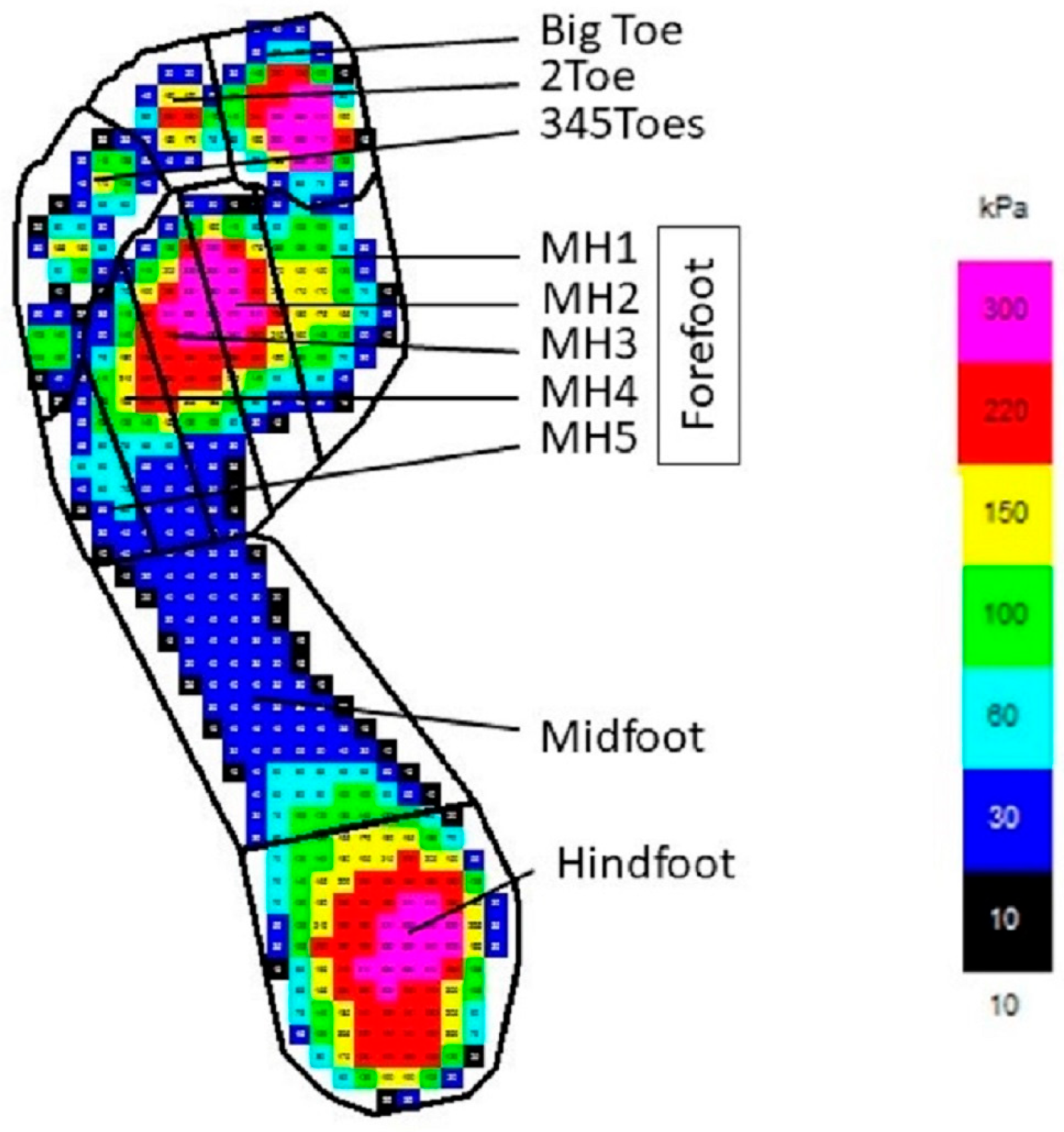
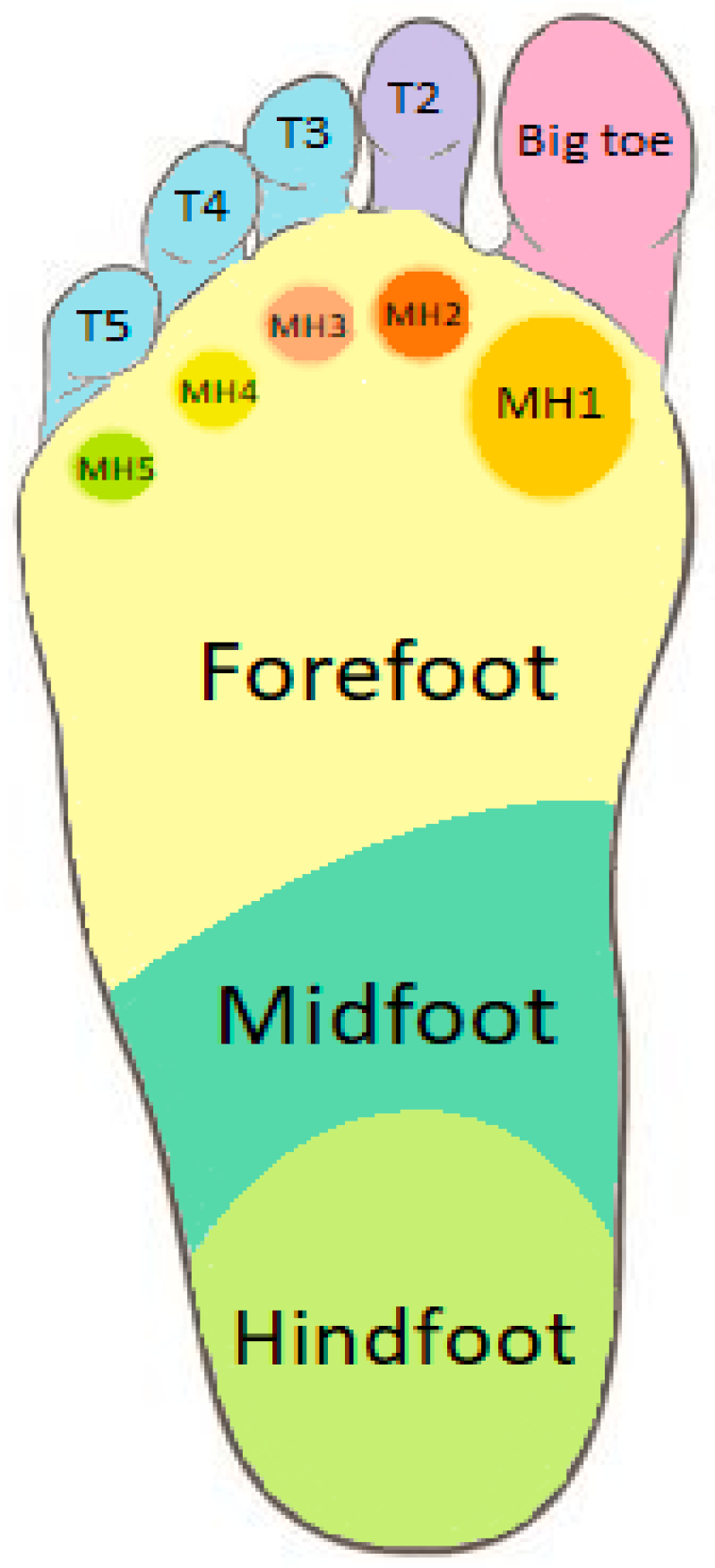
- The separation of straight lines between hindfoot and midfoot and midfoot and forefoot are perpendicular to the straight line that is created using the least square method;
- -
- The boundary between the hindfoot and the midfoot is defined as 73% of the foot length from the top of the footprint;
- -
- The boundary between the midfoot and the forefoot is defined as 45% of the foot length from the top;
- -
- The boundary between the forefoot and toes and also between toes is defined considering the values of peak pressure under the toes and the gradients of pressure around these maximum values;
- -
- The angles that define the metatarsal heads are defined as, respectively, 30%, 17%, 17%, 17%, and 19% of the long plantar angle for MH1, MH2, MH3, MH4, and MH5;
- -
- The boundary between toes and the forefoot is defined as 10% of the foot length from the top.
- -
- Peak pressure (PP) [kPa]—the maximum pressure value on the total foot or area;
- -
- Mean pressure (MP) [kPa]—the ratio of the sum of peak pressures over the sensors to the number of loaded sensors;
- -
- Contact area (CA) [cm2] which is defined as the maximum contact area during stance [16],
- -
- Force time integral [Ns]—the sum of products of force in each frame and the duration of one frame and represents the area under the force curve in time. Force is calculated as a sum of products of averaged pressure beneath the sensor and area of this sensor;
- -
- Force time integral normalized to body weight [Ns/kg] as above but normalized to patient’s body weight;
- -
- Pressure time integral [kPas]—the sum of products of peak pressure in each frame and the duration of one frame and represents the area under the pressure curve in time.
Statistical Analysis
3. Results
4. Discussion
5. Conclusions
Author Contributions
Funding
Institutional Review Board Statement
Informed Consent Statement
Data Availability Statement
Conflicts of Interest
References
- Van Netten, J.J.; Price, P.E.; Lavery, L.A.; Monteiro-Soares, M.; Rasmussen, A.; Jubiz, Y.; Bus, S.A.; on behalf of the International Working Group on the Diabetic Foot (IWGDF). Prevention of foot ulcers in the at-risk patient with diabetes: A systematic review. Diabetes Metab. Res. Rev. 2016, 32, 84–98. [Google Scholar] [CrossRef] [PubMed]
- Rosário, J.L.P. A review of the utilization of baropodometry in postural assessment. J. Bodyw. Mov. Ther. 2014, 18, 215–219. [Google Scholar] [CrossRef]
- Sutkowska, E.; Sutkowski, K.; Sokołowski, M.; Franek, E.; Dragan, S. Distribution of the Highest Plantar Pressure Regions in Patients with Diabetes and Its Association with Peripheral Neuropathy, Gender, Age, and BMI: One Centre Study. J. Diabetes Res. 2019, 2019, 7395769. [Google Scholar] [CrossRef] [PubMed]
- Zulkifli, S.S.; Loh, W.P. A state-of-the-art review of foot pressure. Foot Ankle Surg. 2020, 26, 25–32. [Google Scholar] [CrossRef]
- Lorkowski, J.; Grzegorowska, O. Is pedobarography applicable only in orthopedics? Ostry Dyżur. 2016, 9, 2. [Google Scholar]
- Fang, F.; Wanf, Y.; Gu, W. Pedobarography—A novel screening tool for diabetic peripheral neuropathy? Eur. Rev. Med. Pharmacol. Sci. 2013, 17, 3206–3212. [Google Scholar] [PubMed]
- Gurney, J.K.; Kersting, U.G.; Rosenbaum, D.; Dissanayake, A.; York, S.; Grech, R.; Ng, A.; Milne, B.; Stanley, J.; Sarfati, D. Pedobarography as a clinical tool in the management of diabetic feet in New Zealand: A feasibility study. J. Foot Ankle Res. 2017, 10, 24. [Google Scholar] [CrossRef]
- Lavery, L.A.; Armstrong, D.G.; Wunderlich, R.P.; Tredwell, J.; Boulton, A.J. Predictive Value of Foot Pressure Assessment as Part of a Population-Based Diabetes Disease Management Program. Diabetes Care 2003, 26, 1069–1073. [Google Scholar] [CrossRef]
- Veves, A.; Murray, H.J.; Young, M.J.; Boulton, A.J.M. The risk of foot ulceration in diabetic patients with high foot pressure: A propective study. Diabetologia 1992, 35, 660–663. [Google Scholar]
- Fernando, M.E.; Crowther, R.G.; Pappas, E.; Lazzarini, P.A.; Cunningham, M.; Sangla, K.S.; Buttner, P.; Golledge, J. Plantar Pressure in Diabetic Peripheral Neuropathy Patients with Active Foot Ulceration, Previous Ulceration and No History of Ulceration: A Meta-Analysis of Observational Studies. PLoS ONE 2014, 9, e99050. [Google Scholar] [CrossRef]
- Sharad, P. Pendsey Understanding diabetic foot. Int. J. Diabetes Dev. Ctries. 2010, 30, 75–79. [Google Scholar]
- Grimm, A.; Kästenbauer, T.; Sauseng, S.; Sokol, G.; Irsigler, K. Progression and distribution of plantar pressure in Type 2 diabetic patients. Diabetes Nutr. Metab. 2004, 17, 108–113. [Google Scholar]
- Waldecker, U. Pedographic classification and ulcer detection in the diabetic foot. Foot Ankle Surg. 2012, 18, 42–49. [Google Scholar] [CrossRef] [PubMed]
- Bus, S.A.; Lavery, L.A.; Monteiro-Soares, M.; Rasmussen, A.; Raspovic, A.; Sacco, I.C.; van Netten, J.J.; International Working Group on the Diabetic Foot. Guidelines on the prevention of foot ulcers in persons with diabetes (IWGDF 2019 update). Diabetes Metab. Res. Rev. 2020, 36, e3269. [Google Scholar] [CrossRef]
- ISPAD’s Clinical Practice Consensus Guidelines. Available online: https://www.ispad.org/page/ISPADGuidelines2018 (accessed on 1 January 2020).
- Emed System Manual; v.23; Novel GmbH: Munich, Germany, 2012.
- Fernando, M.E.; Crowther, R.G.; Scott Wearing, S. The Importance of Foot Pressure in Diabetes. In Handbook of Human Motion; Müller, B., Wolf, S.I., Eds.; Springer: Cham, Switzerland, 2018. [Google Scholar] [CrossRef]
- Rahman, M.A.; Aziz, Z.; Acharya, U.R.; Ha, T.P.; Kannathal, N.; Ng, E.Y.; Law, C.; Subramaniam, T.; Shuen, W.Y.; Fang, S.C. Analysis of plantar pressure in diabetic type 2 subjects with and without neuropaty. ITBM-RBM 2006, 27, 46–55. [Google Scholar] [CrossRef]
- Gefen, A. Plantar soft tissue loading under the medial metatarsals in the standing diabetic foot. Med. Eng. Phys. 2003, 25, 491–499. [Google Scholar] [CrossRef] [PubMed]
- Keukenkamp, R.; Busch-Westbroek, T.E.; Barn, R.; Woodburn, J.; Bus, S.A. Foot ulcer recurrence, plantar pressure and footwear adherence in people with diabetes and Charcot midfoot deformity: A cohort analysis. Diabet. Med. 2021, 38, e14438. [Google Scholar] [CrossRef]
- Cowley, M.S.; Boyko, E.J.; Shofer, J.B.; Ahroni, J.H.; Ledoux, W.R. Foot ulcer risk and location in relation to prospective clinical assessment of foot shape and mobility among persons with diabetes. Diabetes Res. Clin. Pract. 2008, 82, 226–232. [Google Scholar] [CrossRef] [PubMed]
- Barn, R.; Waaijman, R.; Nollet, F.; Woodburn, J.; Bus, S.A. Predictors of Barefoot Plantar Pressure during Walking in Patients with Diabetes, Peripheral Neuropathy and a History of Ulceration. PLoS ONE 2015, 10, e0117443. [Google Scholar] [CrossRef]
- Boulton, A. The diabetic foot: Epidemiology, risk factors and the status of care. Diabetes Voice 2005, 50, 57. [Google Scholar]
- Armstrong, D.G.; Boulton, A.J.M.; Bus, S.A. Diabetic Foot Ulcers and Their Recurrence. N. Engl. J. Med. 2017, 376, 2367–2375. [Google Scholar] [CrossRef] [PubMed]
- Bus, S.; Valk, G.; van Deursen, R. The effectiveness of footwear and offloading interventions to prevent and heal foot ulcers and reduce plantar pressure in diabetes: A systematic review. Diabetes Metab. Res. Rev. 2008, 24 (Suppl. 1), S162–S180. [Google Scholar]
- Lázaro-Martínez, J.L.; Aragón-Sánchez, J.; Álvaro-Afonso, F.J.; García-Morales, E.; García-Álvarez, Y.; Molines-Barroso, R.J. The best way to reduce reulcerations: If you understand biomechanics of the diabetic foot, you can do it. Int. J. Low. Extrem. Wounds 2014, 13, 294. [Google Scholar] [CrossRef]
- Bus, S.A. Foot structure and footwear prescription in diabetes mellitus. Diabetes/Metab. Res. Rev. 2008, 24, S90–S95. [Google Scholar] [CrossRef]
- Bus, S.A.; van Deursen, R.; Armstrong, D.G.; Lewis, J.E.A.; Caravaggi, C.F.; Cavanagh, P.R.; on behalf of the International Working Group on the Diabetic Foot (IWGDF). Footwear and offloading interventions to prevent and heal foot ulcers and reduce plantar pressure in patients with diabetes: A systematic review. Diabetes/Metab. Res. Rev. 2016, 32, 99–118. [Google Scholar] [CrossRef]
- Healy, A.; Naemi, R.; Chockalingam, N. The effectiveness of footwear as an intervention to prevent or to reduce biomechanical risk factors associated with diabetic foot ulceration: A systematic review. J. Diabetes Its Complicat. 2013, 27, 391–400. [Google Scholar] [CrossRef] [PubMed]
- Skopljak, A.; Muft, M.; Sukalo, A. Pedobarography in Diagnosis and Clinical Application. Acta Inform. Med. 2014, 22, 374–378. [Google Scholar] [CrossRef] [PubMed]
- Deschamps, K.; Matricali, A.; Roosen, P. Classification of Forefoot Plantar Pressure Distribution in Persons with Diabetes: A Novel Perspective for the Mechanical Management of Diabetic Foot? PLoS ONE 2013, 8, e79924. [Google Scholar]
- Zhang, P.; Lu, J.; Jing, Y.; Tang, S.; Zhu, D.; Bi, Y. Global epidemiology of diabetic foot ulceration: A systematic review and meta-analysis. Ann. Med. 2017, 49, 106–116. [Google Scholar] [CrossRef]
- Pataky, Z.; Vischer, U. Diabetic foot disease in the elderly. Diabetes Metab. 2007, 33 (Suppl. 1), S56–S65. [Google Scholar]
- Feka, K.; Brusa, J.; Cannata, R. Is bodyweight affecting plantar pressure distribution in children?: An observational study. Medicine 2020, 4, e21968. [Google Scholar]
- Murray, H.J.; Young, M.J.; Hollis, S.; Boulton, A.J. The association between callus formation, high pressures and neuropathy in diabetic foot ulceration. Diabet Med. 1996, 13, 979–982. [Google Scholar] [CrossRef]
- Abouaesha, F.; van Schie, C.; Griffths, G. Plantar Tissue Thickness Is Related to Peak Plantar Pressure in the High-Risk Diabetic Foot. Diabetes Care 2001, 24, 1270–1274. [Google Scholar] [PubMed]
- Chatwin, K.; Abbott, C.; Boulton, A. The role of foot pressure measurement in the prediction and prevention of diabetic foot ulceration—A comprehensive review. Diabetes Metab. Res Rev. 2020, 36, e3258. [Google Scholar] [PubMed]
- Demirbüken, İ.; Özgül, B.; Timurtaş, E.; Yurdalan, S.U.; Çekin, M.D.; Polat, M.G. Gender and age impact on plantar pressure distribution in early adolescence. Acta Orthop. Traumatol. Turc. 2019, 53, 215–220. [Google Scholar]

| Mean ± SD | Min–Max | |
|---|---|---|
| Age (years) | 17.8 ± 0.14 | 17.3–18.6 |
| Age at diabetes onset | 10.3 ± 4.2 | 1.8–17.0 |
| Diabetes duration | 7.36 ± 4.2 | 0.8–16.1 |
| HbA1c for whole diabetes duration | 8.1 ± 1.2 | 6.2–12.1 |
| BMI | 23.3 ± 3.3 | 17.3–32.7 |
| Peak Pressure [kPa] LEFT FOOT | Peak Pressure [kPa] RIGHT FOOT | ||||||||||
|---|---|---|---|---|---|---|---|---|---|---|---|
| Foot Area | Result | ±SD | p-Value | References | ±SD | Foot Area | Result | ±SD | p-Value | References | ±SD |
| Totalfoot | 595.16 | 203.0 | 0.25 | 548.9 | 195.1 | Totalfoot | 577.53 | 195.3 | 0.35 | 539.9 | 118.1 |
| Hindfoot | 345.79 | 74.7 | 0.45 | 345.1 | 92.2 | Hindfoot | 339.8 | 65.7 | 0.2 | 322.5 | 1.5 |
| Midfoot | 124.52 | 44.1 | 0.23 | 113.6 | 45.7 | Midfoot | 128.19 | 38.9 | 0.13 | 115.6 | 80.3 |
| Forefoot | 459.91 | 180.5 | 0.75 | 448.40 | 150.0 | Forefoot | 427.71 | 155.8 | 0.54 | 447.4 | 1.2 |
| MH1 | 265.13 | 126.9 | 0.39 | 242.3 | 139.0 | MH1 | 256.62 | 115.9 | 0.9 | 250.7 | 121.8 |
| MH2 | 361.06 | 141.7 | 0.23 | 348.7 | 103.8 | MH2 | 349.59 | 105.8 | 0.2 | 379.6 | 1.0 |
| MH3 | 336.49 | 93.3 | 0.7 | 341.5 | 103.3 | MH3 | 334.7 | 105.8 | 0.3567 | 355.5 | 225.6 |
| MH4 | 260.32 | 91.9 | 0.49 | 273.4 | 90.9 | MH4 | 270.38 | 118.8 | 0.2 | 250 | 1.8 |
| MH5 | 237.40 | 167.3 | 0.58 | 254.6 | 178.8 | MH5 | 226.72 | 146.7 | 0.52 | 207 | 83.3 |
| BigToe | 449.61 | 225.3 | 0.14 | 380.5 | 245.2 | BigToe | 443.82 | 235.6 | 0.6 | 420 | 1.3 |
| 2Toe | 179.30 | 95.8 | 0.83 | 174.1 | 92.4 | 2 Toe | 184.31 | 99.6 | 0.43 | 168.9 | 68.3 |
| 345Toes | 121.44 | 65.4 | 0.33 | 129.8 | 82.4 | 345 Toes | 134.85 | 92.5 | 0.3 | 116.1 | 2.6 |
| Mean Pressure [kPa] LEFT FOOT | Mean Pressure [kPa] RIGHT FOOT | ||||||||||
| Foot Area | Result | ±SD | p-Value | References | ±SD | Foot Area | Result | ±SD | p-Value | References | ±SD |
| Totalfoot | 133.34 | 14.4 | 0 | 117.88 | 10.7 | Totalfoot | 132.58 | 14.2 | 0 | 118.66 | 10.7 |
| Hindfoot | 183.37 | 27.5 | 0 | 159.7 | 20.7 | Hindfoot | 181.16 | 24.3 | 0 | 159.14 | 21.5 |
| Midfoot | 57.77 | 18.0 | 0.03 | 50.15 | 18.3 | Midfoot | 59.17 | 17.9 | 0.019 | 50.4 | 17.7 |
| Forefoot | 146.25 | 21.8 | 0.11 | 139.18 | 19.0 | Forefoot | 145.43 | 22.6 | 0.07 | 137.52 | 16.9 |
| MH1 | 124.62 | 34.0 | 0.0028 | 101.72 | 43.5 | MH1 | 123.62 | 32.0 | 0.029 | 108.38 | 38.0 |
| MH2 | 181.18 | 37.8 | 0.0945 | 168.06 | 36.9 | MH2 | 178.64 | 34.2 | 0.3 | 178.16 | 42.2 |
| MH3 | 171.56 | 34.4 | 0.25 | 167.37 | 34.5 | MH3 | 171.59 | 34.4 | 0.17 | 167.25 | 34.0 |
| MH4 | 129.51 | 36.0 | 0.28 | 137.71 | 39.7 | MH4 | 132.63 | 35.7 | 0.9 | 124.76 | 34.0 |
| MH5 | 99.86 | 43.4 | 0.45 | 106.98 | 49.9 | MH5 | 98.42 | 39.3 | 0.32 | 90.69 | 39.2 |
| BigToe | 142.75 | 42.9 | 0.0136 | 120.07 | 47.1 | BigToe | 143.02 | 45.7 | 0.29 | 133.16 | 45.1 |
| 2Toe | 73.34 | 24.2 | 0.05 | 69.83 | 28.7 | 2 Toe | 75.09 | 25.6 | 0.41 | 70.69 | 26.8 |
| 345Toes | 47.50 | 17.1 | 0.48 | 48.69 | 22.3 | 345 Toes | 49.82 | 20.3 | 0.23 | 44.84 | 18.4 |
| Contact Area [cm2] LEFT FOOT | Contact Area [cm2] RIGHT FOOT | ||||||||||
| Foot Area | Result | ±SD | p-Value | References | ±SD | Foot Area | Result | ±SD | p-Value | References | ±SD |
| Totalfoot | 126.42 | 17.7 | 0.01 | 135 | 14.9 | Totalfoot | 128.00 | 18.3 | 0.05 | 135.03 | 14.2 |
| Hindfoot | 33.12 | 4.4 | 0.33 | 33.97 | 4.0 | Hindfoot | 33.16 | 4.4 | 0.14 | 34.37 | 3.8 |
| Midfoot | 24.21 | 7.5 | 0.09 | 27.25 | 4.1 | Midfoot | 24.6 | 7.3 | 0.06 | 27.13 | 3.6 |
| Forefoot | 49.10 | 6.2 | 0.25 | 50.57 | 6.3 | Forefoot | 49.46 | 6.1 | 0.28 | 50.79 | 5.4 |
| MH1 | 12.83 | 2.2 | 0.28 | 12.48 | 2.4 | MH1 | 12.87 | 1.9 | 0.87 | 12.99 | 2.1 |
| MH2 | 10.13 | 1.6 | 0.52 | 10.45 | 1.8 | MH2 | 10.26 | 1.6 | 0.75 | 10.3 | 1.4 |
| MH3 | 11.13 | 1.4 | 0.12 | 11.67 | 1.6 | MH3 | 11.03 | 1.5 | 0.37 | 11.5 | 1.5 |
| MH4 | 9.29 | 1.2 | 0.14 | 9.61 | 1.3 | MH4 | 9.3 | 1.2 | 0.77 | 9.68 | 1.2 |
| MH5 | 5.72 | 0.9 | 0.0004 | 6.35 | 1.1 | MH5 | 5.79 | 0.9 | 0.0086 | 6.3 | 1.0 |
| BigToe | 10.45 | 1.8 | 0.1681 | 10.95 | 1.7 | BigToe | 10.54 | 2.2 | 0.074 | 11.33 | 1.8 |
| 2Toe | 3.53 | 0.9 | 0 | 4.42 | 1.2 | 2 Toe | 3.67 | 1.0 | 0.008 | 4.25 | 1.3 |
| 345Toes | 5.94 | 2.4 | 0.0005 | 7.68 | 2.2 | 345 Toes | 6.48 | 2.7 | 0.26 | 7.11 | 2.6 |
| Force Time Integral [Ns] LEFT FOOT | Force Time Integral [Ns] RIGHT FOOT | ||||||||||
|---|---|---|---|---|---|---|---|---|---|---|---|
| Foot Area | Result | ±SD | p-Value | References | ±SD | Foot Area | Result | ±SD | p-Value | References | ±SD |
| Totalfoot | 390.9 | 75.2 | 0 | 490.1 | 86.2 | Totalfoot | 391.2 | 75.1 | 0 | 485.1 | 88.9 |
| Hindfoot | 113.7 | 30.0 | 0 | 152.0 | 37.8 | Hindfoot | 106.2 | 23.0 | 0 | 147.3 | 33.9 |
| Midfoot | 33.7 | 20.3 | 0.1840 | 38.2 | 20.9 | Midfoot | 34.8 | 20.3 | 0.3410 | 36.8 | 17.9 |
| Forefoot | 203.7 | 45.5 | 0.0002 | 246.0 | 60.2 | Forefoot | 208.5 | 47.9 | 0.0022 | 242.8 | 50.8 |
| MH1 | 43.9 | 17.8 | 0.4412 | 44.6 | 26.1 | MH1 | 44.2 | 16.5 | 0.1655 | 48.5 | 24.8 |
| MH2 | 50.5 | 13.1 | 0.0160 | 61.0 | 19.4 | MH2 | 51.4 | 11.9 | 0.0001 | 63.8 | 19.3 |
| MH3 | 55.1 | 14.1 | 0.0001 | 69.6 | 21.8 | MH3 | 57.0 | 15.0 | 0.0023 | 68.1 | 19.2 |
| MH4 | 37.1 | 12. 5 | 0.0008 | 47.8 | 18.5 | MH4 | 38.7 | 13.4 | 0.0896 | 43.2 | 14.9 |
| MH5 | 17.1 | 8.8 | 0.0101 | 22.9 | 15.3 | MH5 | 17.2 | 9.1 | 0.1950 | 19.2 | 11.4 |
| BigToe | 29.5 | 14.4 | 0.0695 | 35.2 | 21.3 | BigToe | 30.2 | 15.5 | 0.0055 | 41.8 | 29.5 |
| 2Toe | 4.8 | 2.5 | 0 | 7.9 | 5.3 | 2 Toe | 5.1 | 2.9 | 0.0008 | 7.7 | 5.0 |
| 345Toes | 5.5 | 4.3 | 0.0001 | 10.7 | 10.0 | 345 Toes | 6.4 | 5.8 | 0.0624 | 8,7 | 7,5 |
| Force Time Integral Normalized to Body Weight[Ns/kg] LEFT FOOT | Force Time Integral Normalized to Body Weight[Ns/kg] RIGHT FOOT | ||||||||||
| Foot Area | Result | ±SD | p-Value | References | ±SD | Foot Area | Result | ±SD | p-Value | References | ±SD |
| Totalfoot | 57.7 | 4.4 | 0 | 75.6 | 9.9 | Totalfoot | 57.7 | 4.5 | 0 | 74.5 | 10.3 |
| Hindfoot | 16.9 | 3.7 | 0 | 23.5 | 5.4 | Hindfoot | 15.8 | 2.9 | 0 | 22.6 | 4.5 |
| Midfoot | 4.8 | 2.6 | 0.0468 | 5.9 | 3.0 | Midfoot | 5.0 | 2.5 | 0.1315 | 5.7 | 2.8 |
| Forefoot | 30.0 | 4.2 | 0 | 37.9 | 7.7 | Forefoot | 30.7 | 4.4 | 0 | 37.3 | 6.8 |
| MH1 | 6.5 | 2.3 | 0.2660 | 6.9 | 3.9 | MH1 | 6.5 | 2.1 | 0.0450 | 7.5 | 3.6 |
| MH2 | 7.5 | 1.6 | 0 | 9.4 | 2.6 | MH2 | 7.6 | 1.3 | 0 | 9.8 | 2.9 |
| MH3 | 8.1 | 1.6 | 0 | 10.7 | 2.9 | MH3 | 8.4 | 1.6 | 0 | 10.5 | 2.8 |
| MH4 | 5.5 | 1.5 | 0 | 7.4 | 2.7 | MH4 | 5.7 | 1.6 | 0.0154 | 6.6 | 2.1 |
| MH5 | 2.5 | 1.1 | 0.0009 | 3.6 | 2.5 | MH5 | 2.5 | 1.2 | 0.0605 | 3.0 | 1.8 |
| BigToe | 4.4 | 2.1 | 0.0378 | 5.4 | 3.1 | BigToe | 4.4 | 2.2 | 0.0015 | 6.3 | 4.0 |
| 2Toe | 0.7 | 0.4 | 0.0001 | 1.2 | 0.9 | 2 Toe | 0.8 | 0.5 | 0.0020 | 1.2 | 0.8 |
| 345Toes | 0.8 | 0.7 | 0.0001 | 1.7 | 1.8 | 345 Toes | 1.0 | 1.0 | 0.1226 | 1.3 | 1.3 |
| Pressure Time Integral [kPas] LEFT FOOT | Pressure Time Integral [kPas] RIGHT FOOT | ||||||||||
| Foot Area | Result | ±SD | p-Value | References | ±SD | Foot Area | Result | ±SD | p-Value | References | ±SD |
| Totalfoot | 219.16 | 54.16 | 0.0001 | 274.99 | 81.16 | Totalfoot | 212.90 | 52.27 | 0 | 269.70 | 80.90 |
| Hindfoot | 79.44 | 19.29 | 0 | 108.63 | 28.52 | Hindfoot | 74.04 | 14.78 | 0 | 101.09 | 24.27 |
| Midfoot | 37.79 | 14.26 | 0.0599 | 43.59 | 19.34 | Midfoot | 38.40 | 13.58 | 0.1858 | 41.51 | 17.20 |
| Forefoot | 133.92 | 44.82 | 0.0011 | 171.74 | 70.31 | Forefoot | 131.30 | 47.25 | 0.0110 | 169.46 | 63.16 |
| MH1 | 76.49 | 33.67 | 0.0890 | 89.28 | 58.79 | MH1 | 76.00 | 33.97 | 0.0535 | 91.09 | 55.23 |
| MH2 | 101.24 | 27.08 | 0.0007 | 124.86 | 39.64 | MH2 | 100.96 | 24.00 | 0 | 135.72 | 50.60 |
| MH3 | 101.42 | 28.81 | 0.0003 | 128.37 | 41.72 | MH3 | 103.18 | 32.88 | 0.0003 | 134.12 | 47.60 |
| MH4 | 83.23 | 30.27 | 0.0028 | 105.36 | 41.09 | MH4 | 88.11 | 38.48 | 0.2219 | 95.15 | 31.94 |
| MH5 | 69.83 | 41.00 | 0.0414 | 90.27 | 74.78 | MH5 | 69.66 | 41.73 | 0.3659 | 73.28 | 50.12 |
| BigToe | 101.46 | 59.67 | 0.1886 | 115.51 | 88.11 | BigToe | 99.89 | 57.79 | 0.0326 | 129.14 | 92.21 |
| 2Toe | 36.47 | 18.41 | 0.1230 | 47.94 | 30.02 | 2 Toe | 37.50 | 21.49 | 0.0506 | 46.76 | 29.70 |
| 345Toes | 27.81 | 17.25 | 0.0077 | 40.60 | 36.06 | 345 Toes | 30.34 | 23.15 | 0.1928 | 35.41 | 27.40 |
Disclaimer/Publisher’s Note: The statements, opinions and data contained in all publications are solely those of the individual author(s) and contributor(s) and not of MDPI and/or the editor(s). MDPI and/or the editor(s) disclaim responsibility for any injury to people or property resulting from any ideas, methods, instructions or products referred to in the content. |
© 2023 by the authors. Licensee MDPI, Basel, Switzerland. This article is an open access article distributed under the terms and conditions of the Creative Commons Attribution (CC BY) license (https://creativecommons.org/licenses/by/4.0/).
Share and Cite
Wysocka-Mincewicz, M.; Szczerbik, E.; Mazur, M.; Grabik, M.; Kalinowska, M.; Syczewska, M. Foot Plantar Pressure Abnormalities in Near Adulthood Patients with Type 1 Diabetes. Biomedicines 2023, 11, 2901. https://doi.org/10.3390/biomedicines11112901
Wysocka-Mincewicz M, Szczerbik E, Mazur M, Grabik M, Kalinowska M, Syczewska M. Foot Plantar Pressure Abnormalities in Near Adulthood Patients with Type 1 Diabetes. Biomedicines. 2023; 11(11):2901. https://doi.org/10.3390/biomedicines11112901
Chicago/Turabian StyleWysocka-Mincewicz, Marta, Ewa Szczerbik, Maria Mazur, Magdalena Grabik, Małgorzata Kalinowska, and Małgorzata Syczewska. 2023. "Foot Plantar Pressure Abnormalities in Near Adulthood Patients with Type 1 Diabetes" Biomedicines 11, no. 11: 2901. https://doi.org/10.3390/biomedicines11112901
APA StyleWysocka-Mincewicz, M., Szczerbik, E., Mazur, M., Grabik, M., Kalinowska, M., & Syczewska, M. (2023). Foot Plantar Pressure Abnormalities in Near Adulthood Patients with Type 1 Diabetes. Biomedicines, 11(11), 2901. https://doi.org/10.3390/biomedicines11112901