Article

Prevalence and Virulence of Commensal Pseudomonas Aeruginosa Isolates from Healthy Individuals in Southern Vietnam (2018–2020)

by
Nguyen Bao Vy Tran
1,
Quang Minh Truong
1,
Lam Que Anh Nguyen
1,
Ngoc My Huong Nguyen
1,
Quang Hung Tran
1,
Thi Tuyet Phuong Dinh
1,
Vinh Son Hua
1,
Van Dung Nguyen
1,
Peter A. Lambert
2 and
Thi Thu Hoai Nguyen
1,3,*
1
School of Biotechnology, International University, Vietnam National University, Ho Chi Minh City 700000, Vietnam
2
College of Health and Life Sciences, Aston University, Birmingham B4 7ET, UK
3
Research Center for Infectious Diseases, International University, Vietnam National University, Ho Chi Minh City 700000, Vietnam
*
Author to whom correspondence should be addressed.
Biomedicines 2023, 11(1), 54; https://doi.org/10.3390/biomedicines11010054
Submission received: 5 December 2022 / Revised: 20 December 2022 / Accepted: 21 December 2022 / Published: 26 December 2022
(This article belongs to the Special Issue Microbial Ecology in Health and Disease 2.0)

Abstract

:
Understanding the colonization of Pseudomonas aeruginosa (P. aeruginosa) in healthy humans is useful for future prevention and treatment of P. aeruginosa infection. This study aimed to investigate the prevalence and risk factors of of P. aeruginosa colonization in healthy humans. At the same time, the virulence of the isolated P. aeruginosa was also studied. In the study, 609 Vietnamese volunteers (310 females and 299 males, age range of 2 to 73 years), who had no acute infection or disease symptoms participated at the time of sample collection. Samples were taken from the throat, nostrils, and outer ears. P. aeruginosa was found in 19 participants (3.12%, 95% CI: 0.017–0.045), mainly from the throat (11/19, 57.89%). Participants with a history of sinusitis were 11.57 times more likely to be colonized with P. aeruginosa than participants without a history of sinusitis (OR: 11.57, 95% CI: 4.08–32.76, p-value < 0.0001, Fisher’s Exact test). Age and sex were not significantly associated with P. aeruginosa colonization. Among 16 P. aeruginosa isolates used in virulence tests, 100% (16/16) were positive for the synthesis of biofilm, pyocyanin, and siderophores; 93.75% (15/16) isolates were positive for the synthesis of gelatinase and protease; and 50% (8/16) isolates were positive for lipase. There were no differences in the pattern and range of virulence factors of P. aeruginosa isolates taken from participants with and without sinusitis history. P. aeruginosa colonized 3.12% of participants, and its presence was associated with sinusitis history.

1. Introduction

P. aeruginosa is a Gram-negative bacterium popular in soil, water, and moist areas. It is also a part of the normal flora of humans, which is isolated on the skin (0–2%), in the throat (0–3.3%), and in the stool (2.6–24%) [1]. Under continuous pressure from the human immune system, commensal P. aeruginosa can transform into a virulent pathogen [2]. P. aeruginosa is classified as an opportunistic pathogen that can cause serious infections, particularly in immunocompromised patients, such as in cystic fibrosis, and patients with severe burns [3]. With numerous extracellular enzymes, this bacterium easily adapts to persist, replicate, and attack the host [4]. Extracellular enzymes such as protease, gelatinase, and lipase damage the proteins of host cells, interfere with the immune response, delay the wound-healing process, and are associated with motility, biofilm architecture, and rhamnolipid production [5,6,7,8].
Besides extracellular enzymes, this bacterium has many other virulence factors, such as biofilm, pyocyanin, siderophores, protease, gelatinase, and lipase [9,10]. Biofilm is a complex structure that contains many planktonic cells sticking together by extracellular polymeric substances [11]. This structure can form on virtually any moist surface, such as living tissues, medical devices, water pipes, and so on. Furthermore, biofilm contributes to the persistence of P. aeruginosa infection by protecting this pathogen against the host defenses and antimicrobial strategies [12]. Cells within the biofilm grow slowly, which makes antibiotics work ineffectively [13]. Pyocyanin is a blue phenazine pigment secreted by P. aeruginosa [14]. It damages the host by involving in oxidation-reduction reactions. The oxidants from pyocyanin cause cell respiration dysfunction, calcium homeostasis disruption, and so on [15]. Moreover, pyocyanin possesses antimicrobial properties, so P. aeruginosa can use it as a weapon to kill other microorganisms and become predominant at the site of the infection [16]. Siderophores are low molecular-weight peptides that have the ability to chelate and deliver iron to bacterial cells. Although iron is crucial for all living organisms, including bacteria, the insoluble iron (Fe3+) is abundant in nature. This makes the uptake of iron by P. aeruginosa difficult [17]. Besides scavenging iron, they also stimulate biofilm formation [18]. In addition, siderophore-iron complexes can induce inflammation and oxidative damage, which increase the virulence of P. aeruginosa [19].
The colonization of commensal bacteria like P. aeruginosa on healthy humans deserves special attention because it can be a potential factor for subsequent disease [20]. Because it is opportunistic, it can cause an outbreak by being transmitted among healthy individuals before being detected [21]. So far, studies on P. aeruginosa colonization and its virulence, especially in the Vietnamese population, are limited. It is unclear whether sex, age, or any medical history affect the presence of P. aeruginosa in our body. Our study was the first study to analyze the prevalence of commensal P. aeruginosa in Southern Vietnam, its ability to produce common virulence factors, and the associated factors with P. aeruginosa colonization.

2. Materials and Methods

2.1. Commensal P. aeruginosa Isolation

From 2018 to 2020, throat, naris, and outer ear swab samples of Vietnamese people living in the Southeast area were collected and cultured on Pseudomonas selective Cetrimide media. This study was approved by the Ethics Committee of Vietnam National University of Ho Chi Minh City (Date 6 June 2019, No 1007/DHQG-KHCN).
These areas were chosen for sample collection because of their humidity and sampling convenience with less discomfort for the participants. All volunteers or volunteers’ guardians gave their signed informed consent. Only people with no declared current acute infection or disease symptoms were included in the study. Colonies obtained on Cetrimide agar were further characterized using a Gram-staining, polymerase chain reaction (PCR) with oprL primers [22] as a screening step, and 16S rRNA sequencing for confirmation. For further experiments, all the P. aeruginosa isolates were stored in Luria Bertani broth containing 30% glycerol at −80 °C.

2.2. P. aeruginosa Identification

oprL—Specific Polymerase Chain Reaction and 16S rRNA Sequencing

The specific oprL primers were used to primary detect P. aeruginosa (forward: 5′-ATGGAAATGCTGAAATTCGGC-3′ and reverse: 5′-CTTCTTCAGCTCGACGCGACG-3′) (PHUSA GENOMICS, Can Tho City, Vietnam) and followed the previous study [22]. All positive isolates for oprL were sent to NAM KHOA Biotek Company (Ho Chi Minh City, Vietnam) for 16S rRNA sequencing. The confirmed P. aeruginosa isolates were used for further experiments.

2.3. Virulence Testing
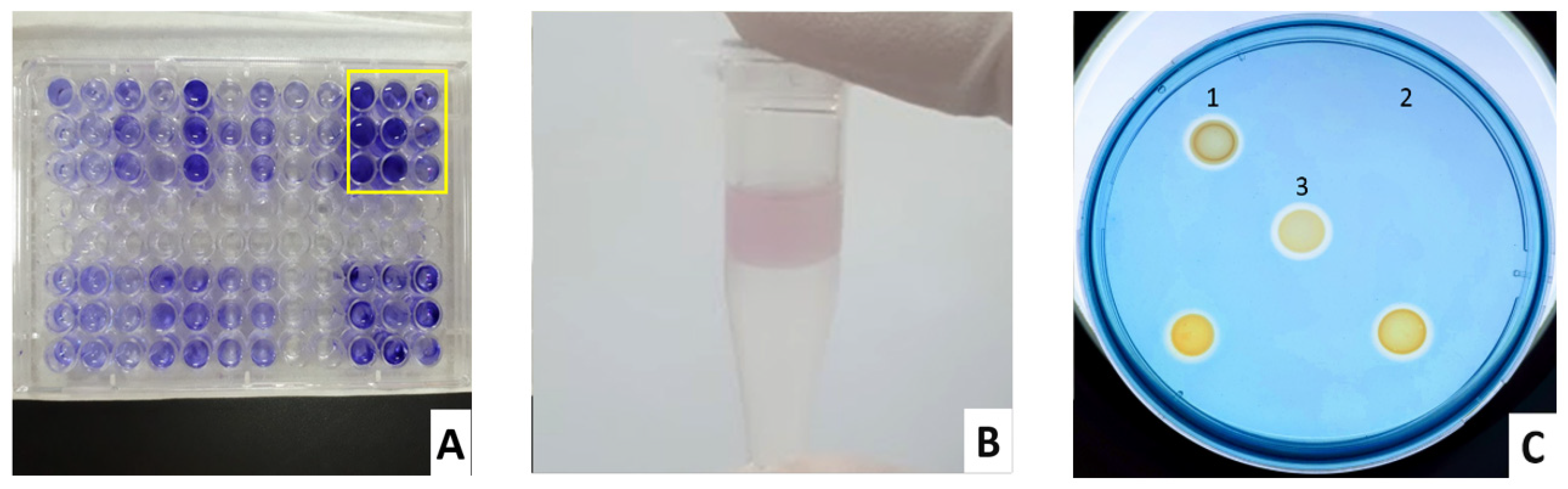
Biofilm, pyocyanin, siderophores, lipase, protease, and gelatinase of the isolated P. aeruginosa isolates were analyzed. P. aeruginosa ATCC 9027 (American Type Culture Collection, Manassas, VA, USA) was used as a positive control for all the producing virulence tests because it was a non-virulent strain isolated from the human outer ear by the American Type Culture Collection and it has ability to produce tested virulence factors in this study [23,24,25,26].

2.3.1. Biofilm

Biofilm production of commensal P. aeruginosa isolates was tested by the crystal violet (HiMedia Laboratories, Kennett Square, PA, USA) staining method. The optimal density (OD) was measured at 550 nm by BioTek Synergy HTX Multimode Reader (Agilent, Santa Clara, CA, USA) [18]. Commensal isolates were classified into negative, weak, moderate and strong biofilm producers by following this category: OD ≤ ODc (negative); ODc < OD ≤ 2 × ODc (weak); 2 × ODc < OD ≤ 4 × ODc (moderate); and OD > 4 × ODc (strong), in which ODc was the cut-off value (ODc = OD negative control + 3 × its standard deviation). OD was the optimal density of P. aeruginosa isolates [27].

2.3.2. Pyocyanin

Pyocyanin was extracted and measured using the chloroform method [20]. P. aeruginosa isolate was cultured in glycerol-alanine (Gly-Ala) (HiMedia Laboratories, Kennett Square, PA, USA; Xilong Scientific Co., Ltd., Shantou, China) broth to maximize the yield of pyocyanin production in commensal isolates. When the OD600 nm bacteria reached 0.08–0.1, 1% of the culture was inoculated in 5 mL of Gly-Ala broth at 37 °C for 24 h with shaking condition. After that, the supernatant was collected (centrifuge 6000 rpm for 15 m). A quantity of 3 mL of supernatant was mixed vigorously with 1.8 mL of chloroform (VN-CHEMSOL Co., Ltd., Ho Chi Minh City, Vietnam). Then, blue layer was collected and mixed with 0.2 N HCl (2:1). The top red color was collected and measured at 520 nm using BioTek Synergy HTX Multimode Reader (Agilent, Santa Clara, United States). The concentration of pyocyanin (μg/mL) was estimated by multiplying the OD at 520 nm by 17.072 (the molar extinction coefficient of pyocyanin 520 nm) [28,29].

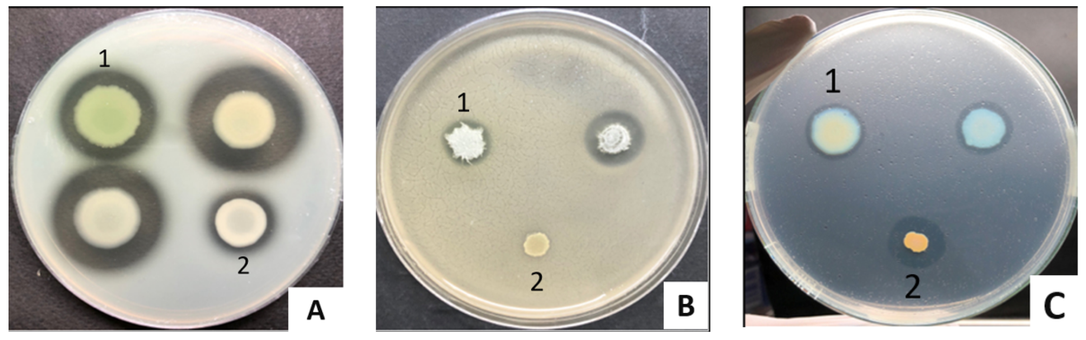
2.3.3. Siderophores, Lipase, Protease, and Gelatinase

Before agar plate test, all tested isolates and controls were cultured in Luria-Bertani broth for 24 h at 37 °C. After the OD600 nm reached the range 0.08–0.1, 5 µL of the cultures spotted on CAS blue agar [30], tributyrin agar (1% tributyrin), skim milk (3% skim milk, Brain Heart Infusion (BHI) medium), or gelatin agar (8% gelatin, BHI) for detecting siderophores, lipase, protease, and gelatinase, respectively. The enzymatic activity (EA) was estimated by measuring the halo zone size: EA = (D − d)/2, where D was the diameter of clear zone (mm) and d was the diameter of a colony (mm). It was categorized as negative (0 mm), weak (<2 mm), moderate (2–4 mm), and strong (>4 mm) activity [31]. Besides P. aeruginosa ATCC 9027, Staphylococcus aureus (S. aureus) was used a positive control for protease and lipase tests, while Vibrio cholerae (V. cholerae) was used for gelatinase. In case of siderophores, E. coli was also used as a positive control, while S. aureus was a negative control because it was Gram-positive and could not survive on CAS blue agar [30]. All commercial materials for these tests were purchased from Sigma-Aldrich, St. Louis, Missouri, United States; HiMedia Laboratories, Kennett Square, United States; Xilong Scientific Co., Ltd., Shantou, China.

2.4. Data Analysis

Each experiment was performed in triplicate. IBM® SPSS® Statistics 20.0 (IBM, Armonk, New York, NY, USA) was used to analyze the data. Chi-square, Fisher’s Exact and Ordinal Regression tests were used to determine the risk factors for P. aeruginosa colonization. In addition, an ANOVA analysis was used to show the association between sinusitis history and commensal P. aeruginosa virulence. The p-value was set to be <0.05.

3. Results and Discussion

3.1. Prevalence of Commensal P. aeruginosa Isolates in Vietnamese Population

There were 612 volunteers who signed informed consent and provided samples. Three were excluded due to their declared health condition and 609 participants were finally included in the study. The number of male and female participants was similar, 299 males (49.10%) versus 310 (50.90%) females. Because the age of participants was not variety, it could only be grouped into three categories: young persons (0–17), adult (18–59), and older persons (≥60). It is well-noted that having sinusitis is a common health problem in the population. More than 1/5 of healthy declaring people experienced sinusitis. The summary of all participants and P. aeruginosa carrier characteristics was described in Table 1.
From 609 participants, 35 P. aeruginosa-like isolates were obtained. These isolates grew on cetrimide selective media, being Gram-negative rod shaped and positive for oprL. The oprL gene encodes the outer membrane peptidoglycan-associated lipoprotein, which has an important role in the interaction between bacteria and environment, is a common and useful target for Pseudomonas detection [22,32,33].
A total of 20 out of 35 isolates positive for oprL (57.14%) were confirmed as P. aeruginosa via 16S rRNA sequencing (Supplementary Table S1). Among 20 P. aeruginosa isolates, two isolates came from one volunteer (from throat and nostrils) and 18 others came from a single site on each participant. In summary, 19 out of 609 participants (3.12%, 95% CI: 0.017–0.045) were colonized with P. aeruginosa (Supplementary Table S2). This was in agreement with previous data showing that P. aeruginosa was not commonly detected in healthy people, but under conditions of antibiotic exposure or hospitalization its prevalence, mainly in the throat and stool, was increased [34,35]. For example, in the case of bronchiectasis, which is a chronic respiratory disease associated with P. aeruginosa, the prevalence of P. aeruginosa colonization in bronchiectasis patients ranged from 9% to 33% [36]. Additionally, Casetta et al. also found that 17.5% of pregnant women were colonized by P. aeruginosa after more than 48 h hospital admission [37]. Importantly, colonization with P. aeruginosa represents a risk of infection: Gómez-Zorrilla et al. reported that 43% of colonized patients in their study developed infection [38].
From our findings, P. aeruginosa isolates were likely obtained from the throat (11/20 P. aeruginosa isolates), while P. stutzeri isolates were rather from the nostril area (9/12 P. stutzeri isolates). Furthermore, the throat was also primarily colonized by P. aeruginosa (11/13 throat-derived isolates from the throat, 84.62%). Only one P. azelaica, one P. nitroreducens isolate and no P. stutzeri isolate were found in this area (Supplementary Table S3). It was consistent with previous studies in which P. aeruginosa is likely found in a mucoid and humid area and is the most common Pseudomonas causing infection in human [39,40].

3.2. Relationship between Sex and Age to P. aeruginosa Colonization

Among 19 commensal P. aeruginosa carriers, 63.16% (12/19) were female and 36.84% (7/19) were male. However, there were no associations between sex and P. aeruginosa colonization of healthy humans (OR: 1.68, 95% CI: 0.65–4.33, p-value = 0.29 (Fisher’s Exact Test) and Chi-Square: p-value = 0.278). To our knowledge, there is still a lack of information about the effect of sex on P. aeruginosa colonization in healthy humans. In contrast to our finding, the study of P. aeruginosa colonization from wounds and burns swabs of patients showed the higher rate of P. aeruginosa among the male than female samples, 55.6% and 44.4%, respectively [41]. However, the presence of P. aeruginosa was associated with a higher morbidity rate in females with respiratory dysfunction, such as cystic fibrosis and bronchiectasis. The estrogen in females could regulate the conversion of P. aeruginosa from non-mucoid to mucoid forms, which are believed to have higher virulence [42]. The relationship between sex and P. aeruginosa colonization is described in Table 2.
Although the group 18–59 years had the highest number of carriers (84.21%, 16/19 P. aeruginosa carriers), age was not a significant factor for P. aeruginosa colonization (0–17: 65 participants, 18–59: 514 participants, and ≥60: 30 participants) (p-value = 0.448, Ordinal Regression). It was because the 95% CI include number 1 in both adult (3.11%, OR: 2.02, 95% CI: 0.26–15.43, p-value < 0.05, Fisher’s Exact test) and older person (6.67%, OR: 6.75, 95% CI: 0.59–77.55, p-value > 0.05, Fisher’s Exact test) groups. Currently, the association between age and P. aeruginosa colonization in healthy humans is unclear, but age > 55 years is considered to be an independent predictor for P. aeruginosa colonization in bronchiectasis patients [43]. The incidence of infections caused by opportunistic pathogens seems to increase significantly in older ages [44] and colonization could be a risk factor for infection [45].

3.3. Relationship of Sinusitis History to P. aeruginosa Colonization

Our data indicated that sinusitis is quite common in the population with 129/609 (21.18%) participants having sinusitis history (Table 1). Interestingly, participants with sinusitis history are more likely to be colonized with P. aeruginosa (14/129, 10.85%) compared to the ones without sinusitis history (5/480, 1.04%). It is estimated that the participants with sinusitis history have a risk of P. aeruginosa colonization 11.57 times higher than that of participants without sinusitis history (OR: 11.57, 95% CI: 4.08–32.76, p-value < 0.0001, Fisher’s Exact test, Table 2). Our data were somewhat in agreement with previous studies. For example, Niederman et al. indicated that the upper and lower respiratory tract was not colonized by Gram-negative bacteria under normal health conditions, but these sites could be harbored by these pathogens when illness developed [46]. In the case of cystic fibrosis patients, Shapiro et al. found that P. aeruginosa was detected in 38% of sinusitis-cystic fibrosis patients [47] while Kasper Aanæs et al., reported that the presence of P. aeruginosa in sinuses was associated with lung infection in cystic fibrosis patients [48]. However, data were also quite controversial as in some studies, P. aeruginosa was not a common pathogen in bacterial flora of chronic sinusitis patients, only present for 1% to 5% of cases [47,48,49].

3.4. Commensal P. aeruginosa Virulence

In this study, 16 out of 20 P. aeruginosa isolates were used for testing the production of key virulence factors, including biofilm, pyocyanin, siderophores, lipase, protease, and gelatinase (Supplementary Tables S4–S6). A total of 100% (16/16 P. aeruginosa isolates) of isolates were positive for biofilm, pyocyanin, siderophores and hemolysin; 93.75% (15/16) of isolates were positive for gelatinase and protease; and 50% (8/16) of isolates were positive for lipase (Table 3).
All tested isolates showed the ability to produce biofilm (16/16). Among them, there were 25% (4/16) of weak, 56.25% (9/16) of moderate, and 18.75% (3/16) of strong biofilm producers. In addition, the biofilm production of commensal P. aeruginosa isolates was weaker than the P. aeruginosa ATCC 9027 (positive control). Some studies showed less dominance of strong biofilm producers in the clinical isolates [50] and isolates from cystic fibrosis patients [51]. Biofilm production did not associate with poor clinical outcomes [52]. Moreover, it was reported that P. aeruginosa often grew planktonically and did not form biofilm under laboratory conditions [53].
All tested commensal P. aeruginosa isolates had the ability to secrete phenazine pyocyanin and siderophores (Figure 1, Table 3). High pyocyanin producers (>18 µg/mL) were positively correlated with septic shock [54]. In our study, the average pyocyanin concentration from commensal P. aeruginosa isolates was 0.61 µg/mL. For siderophore production, the ability of the isolates varied following the isolation sites depending on the availability of free iron. Clinical P. aeruginosa isolates from urinary tract infection produced more siderophores compared with burn skin [55]. In our study, the commensal isolates were unsurprisingly weak siderophore producers (Table 3).
While 93.75% of the tested commensal P. aeruginosa produced protease and gelatinase, their average enzymatic activity was 0.28 ± 0.09 (mm) and 0.29 ± 0.06 (mm), respectively. Only 50% of tested isolates were positive for lipase, for which the average halo size was 0.18 ± 0.04 (mm) (Figure 2, Table 3). Our data are comparable with previous studies on clinical isolates from chronic leg ulcers where 91.67% produced protease, 66.67% produced gelatinase, and 83.33% produced lipase [56], indicating the common presence of these extracellular enzymes in all P. aeruginosa isolates.
There were no significant differences in virulence between P. aeruginosa isolates from carriers with and without sinusitis history (p-value > 0.05, ANNOVA test) (Supplementary Table S7). Sharna’s study showed that P. aeruginosa isolates from cystic fibrosis lung increased biofilm, alginate, and some virulence gene expression [57]. Furthermore, elastase was associated with chronic rhinosinusitis severity [58]. So far, there was no evidence on the increased virulence of commensal P. aeruginosa isolated from people with sinusitis history. However, people with a sinusitis history were more likely to be P. aeruginosa carriers than others, which is a potential risk of subsequent infection with P. aeruginosa [59], and P. aeruginosa infections were known to be particularly dangerous for patients with respiratory diseases [60,61].

4. Conclusions

For the first time, we revealed that the prevalence of commensal P. aeruginosa in the Southern Vietnamese population was 3.12%. Sinusitis could be a potential factor contributing to the colonization of P. aeruginosa. The commensal P. aeruginosa isolates can produce multiple virulence factors, including biofilm, pyocyanin, siderophores, lipase, protease, gelatinase and lipase. Further studies on the transition of P. aeruginosa from commensal to pathogenic state and its persistence in the carriers could give us deeper understanding on the host-pathogen interaction, which would be useful for prevention and treatment of P. aeruginosa infections.

Supplementary Materials

The following supporting information can be downloaded at: https://www.mdpi.com/article/10.3390/biomedicines11010054/s1, Table S1: Pseudomonas species were isolated from three body sites (1-throat, 2-naris, 3-outer ear). They were conforming by 16S rRNA sequencing. The bold fonts indicate P. aeruginosa isolates; Table S2: List of 19 P. aeruginosa carriers; Table S3: oprL—positive Pseudomonas isolates and their colonization sites: Table S4: 16 commensal P. aeruginosa isolates were used in virulence tests; Table S5: Biofilm formation, pyocyanin, and siderophores in commensal P. aeruginosa isolates after inoculation at 37 °C for 24 h. The values were expressed as mean ± standard deviation; Table S6: Lipase, protease, and gelatinase values in commensal P. aeruginosa isolates after incubation at 37 °C for 24 h; Table S7: The production of tested virulence factors in commensal P. aeruginosa isolates from participants with and without sinusitis history. Average ± standard deviation.

Author Contributions

Conceptualization, T.T.H.N.; Methodology, N.B.V.T., Q.M.T., L.Q.A.N., N.M.H.N., T.T.P.D. and V.S.H.; Validation, N.B.V.T., Q.M.T., L.Q.A.N., N.M.H.N., T.T.P.D. and V.S.H.; Formal Analysis, N.B.V.T.; Investigation, N.B.V.T., Q.M.T., L.Q.A.N., N.M.H.N., T.T.P.D. and V.S.H.; Resources, T.T.H.N.; Writing—Original Draft Preparation, N.B.V.T.; Writing—Review & Editing, N.B.V.T., Q.M.T., L.Q.A.N., N.M.H.N., T.T.P.D., V.S.H., V.D.N., Q.H.T., T.T.H.N. and P.A.L.; Visualization, N.B.V.T., Q.M.T., L.Q.A.N., N.M.H.N. and Q.H.T.; Supervision, T.T.H.N.; Project Administration, T.T.H.N.; Funding Acquisition, T.T.H.N. All authors have read and agreed to the published version of the manuscript.

Funding

This research is funded by Vietnam National University of Ho Chi Minh City under grant number C2020-28-03.

Institutional Review Board Statement

The study was conducted in accordance with the Declaration of Helsinki, and approved by the Ethics Committee of Vietnam National University of Ho Chi Minh City (Date 6 June 2019, No 1007/DHQG-KHCN).

Informed Consent Statement

Informed consent was obtained from all subjects involved in the study.

Data Availability Statement

All data generated or analyzed during this study are included in this published article and its supplementary information files.

Acknowledgments

We would like to thank our colleagues at the International University for their support.

Conflicts of Interest

The authors declare no conflict of interest.

Abbreviations

P. aeruginosa: Pseudomonas aeruginosa; P. stutzeri: Pseudomonas stutzeri; P. azelaica: Pseudomonas azelaica; P. nitroreducens: Pseudomonas nitroreducens; E. coli: Escherichia coli; S. aureus: Staphylococcus aureus; ATCC, American Type Culture Collection; PCR: polymerase chain reaction; BHI: brain heart infusion; OR: odd ratio; OD: optical density; CAS: chrome azurol S; HDTMA: hexadecyltrimethylammonium bromide; SPSS: Statistical Package for the Social Sciences; ANOVA: Analysis of Variance.

References

  1. Bergmans, D.; Bonten, M. Colonization and Infection with Pseudomonas aeruginosa in Intensive Care: Endogenous or Exogenous Origin? In Yearbook of Intensive Care and Emergency Medicine 1999; Vincent, J.-L., Ed.; Springer: Berlin/Heidelberg, Germany, 1999; pp. 131–140. [Google Scholar]
  2. Proença, J.T.; Barral, D.C.; Gordo, I. Commensal-to-Pathogen Transition: One-Single Transposon Insertion Results in Two Pathoadaptive Traits in Escherichia Coli -Macrophage Interaction. Sci. Rep. 2017, 7, 4504. [Google Scholar] [CrossRef] [PubMed]
  3. Lyczak, J.B.; Cannon, C.L.; Pier, G.B. Establishment of Pseudomonas aeruginosa Infection: Lessons from a Versatile Opportunist. Microbes Infect. 2000, 2, 1051–1060. [Google Scholar] [CrossRef] [PubMed]
  4. Pereira, S.G.; Rosa, A.C.; Ferreira, A.S.; Moreira, L.M.; Proença, D.N.; Morais, P.V.; Cardoso, O. Virulence Factors and Infection Ability of Pseudomonas aeruginosa Isolates from a Hydropathic Facility and Respiratory Infections. J. Appl. Microbiol. 2014, 116, 1359–1368. [Google Scholar] [CrossRef] [PubMed] [Green Version]
  5. Ołdak, E.; Trafny, E.A. Secretion of Proteases by Pseudomonas aeruginosa Biofilms Exposed to Ciprofloxacin. Antimicrob. Agents Chemother. 2005, 49, 3281–3288. [Google Scholar] [CrossRef] [PubMed] [Green Version]
  6. Prasad, A.S.B.; Shruptha, P.; Prabhu, V.; Srujan, C.; Nayak, U.Y.; Anuradha, C.K.R.; Ramachandra, L.; Keerthana, P.; Joshi, M.B.; Murali, T.S.; et al. Pseudomonas aeruginosa Virulence Proteins Pseudolysin and Protease IV Impede Cutaneous Wound Healing. Lab Investig. 2020, 100, 1532–1550. [Google Scholar] [CrossRef] [PubMed]
  7. Park, M.; Do, E.; Jung, W.H. Lipolytic Enzymes Involved in the Virulence of Human Pathogenic Fungi. Mycobiology 2013, 41, 67–72. [Google Scholar] [CrossRef] [PubMed] [Green Version]
  8. Rosenau, F.; Isenhardt, S.; Gdynia, A.; Tielker, D.; Schmidt, E.; Tielen, P.; Schobert, M.; Jahn, D.; Wilhelm, S.; Jaeger, K.-E. Lipase LipC Affects Motility, Biofilm Formation and Rhamnolipid Production in Pseudomonas Aeruginosa. FEMS Microbiol. Lett. 2010, 309, 25–34. [Google Scholar] [CrossRef]
  9. Liao, C.; Huang, X.; Wang, Q.; Yao, D.; Lu, W. Virulence Factors of Pseudomonas aeruginosa and Antivirulence Strategies to Combat Its Drug Resistance. Front. Cell Infect. Microbiol. 2022, 12, 926758. [Google Scholar] [CrossRef]
  10. Pinna, A.; Usai, D.; Sechi, L.A.; Molicotti, P.; Zanetti, S.; Carta, A. Detection of Virulence Factors in Pseudomonas aeruginosa Strains Isolated from Contact Lens-Associated Corneal Ulcers. Cornea 2008, 27, 320–326. [Google Scholar] [CrossRef]
  11. Hall-Stoodley, L.; Costerton, J.W.; Stoodley, P. Bacterial Biofilms: From the Natural Environment to Infectious Diseases. Nat. Rev. Microbiol. 2004, 2, 95–108. [Google Scholar] [CrossRef]
  12. Mah, T.F.; O’Toole, G.A. Mechanisms of Biofilm Resistance to Antimicrobial Agents. Trends. Microbiol. 2001, 9, 34–39. [Google Scholar] [CrossRef] [PubMed]
  13. Stewart, P.S.; Franklin, M.J. Physiological Heterogeneity in Biofilms. Nat. Rev. Microbiol. 2008, 6, 199–210. [Google Scholar] [CrossRef] [PubMed]
  14. Gonçalves, T.; Vasconcelos, U. Colour Me Blue: The History and the Biotechnological Potential of Pyocyanin. Molecules 2021, 26, 927. [Google Scholar] [CrossRef] [PubMed]
  15. Lau, G.W.; Hassett, D.J.; Ran, H.; Kong, F. The Role of Pyocyanin in Pseudomonas aeruginosa Infection. Trends Mol. Med. 2004, 10, 599–606. [Google Scholar] [CrossRef]
  16. Hassan, H.M.; Fridovich, I. Mechanism of the Antibiotic Action Pyocyanine. J. Bacteriol. 1980, 141, 156–163. [Google Scholar] [CrossRef] [PubMed] [Green Version]
  17. Andrews, S.C.; Robinson, A.K.; Rodríguez-Quiñones, F. Bacterial Iron Homeostasis. FEMS Microbiol. Rev. 2003, 27, 215–237. [Google Scholar] [CrossRef] [Green Version]
  18. Banin, E.; Vasil, M.L.; Greenberg, E.P. Iron and Pseudomonas aeruginosa Biofilm Formation. Proc. Natl. Acad. Sci. USA 2005, 102, 11076–11081. [Google Scholar] [CrossRef] [Green Version]
  19. Coffman, T.J.; Cox, C.D.; Edeker, B.L.; Britigan, B.E. Possible Role of Bacterial Siderophores in Inflammation. Iron Bound to the Pseudomonas Siderophore Pyochelin Can Function as a Hydroxyl Radical Catalyst. J. Clin. Investig. 1990, 86, 1030–1037. [Google Scholar] [CrossRef] [Green Version]
  20. Kluytmans, J.; van Belkum, A.; Verbrugh, H. Nasal Carriage of Staphylococcus aureus: Epidemiology, Underlying Mechanisms, and Associated Risks. Clin. Microbiol. Rev. 1997, 10, 505–520. [Google Scholar] [CrossRef]
  21. Nicolas-Chanoine, M.-H.; Blanco, J.; Leflon-Guibout, V.; Demarty, R.; Alonso, M.P.; Caniça, M.M.; Park, Y.-J.; Lavigne, J.-P.; Pitout, J.; Johnson, J.R. Intercontinental Emergence of Escherichia Coli Clone O25:H4-ST131 Producing CTX-M-15. J. Antimicrob. Chemother. 2008, 61, 273–281. [Google Scholar] [CrossRef]
  22. De Vos, D.; Lim, A.; Pirnay, J.P.; Struelens, M.; Vandenvelde, C.; Duinslaeger, L.; Vanderkelen, A.; Cornelis, P. Direct Detection and Identification of Pseudomonas aeruginosa in Clinical Samples Such as Skin Biopsy Specimens and Expectorations by Multiplex PCR Based on Two Outer Membrane Lipoprotein Genes, OprI and OprL. J. Clin. Microbiol. 1997, 35, 1295–1299. [Google Scholar] [CrossRef] [PubMed] [Green Version]
  23. Enhanced Synergistic Effects of Xylitol and Isothiazolones for Inhibition of Initial Biofilm Formation by Pseudomonas Aeruginosa ATCC 9027 and Staphylococcus aureus ATCC 6538-PubMed. Available online: https://pubmed.ncbi.nlm.nih.gov/31217374/ (accessed on 14 November 2022).
  24. Pseudomonas Aeruginosa (Schroeter) Migula-9027|ATCC. Available online: https://www.atcc.org/products/9027 (accessed on 14 November 2022).
  25. Schulte, G.; Bohne, L.; Winkler, U. Glycogen and Various Other Polysaccharides Stimulate the Formation of Exolipase by Pseudomonas aeruginosa. Can. J. Microbiol. 1982, 28, 636–642. [Google Scholar] [CrossRef] [PubMed]
  26. Vfr or CyaB Promote the Expression of the Pore-Forming Toxin ExlBA Operon in Pseudomonas Aeruginosa ATCC 9027 without Increasing Its Virulence in Mice|Microbiology Society. Available online: https://www.microbiologyresearch.org/content/journal/micro/10.1099/mic.0.001083 (accessed on 14 November 2022).
  27. Stepanović, S.; Vuković, D.; Hola, V.; Di Bonaventura, G.; Djukić, S.; Cirković, I.; Ruzicka, F. Quantification of Biofilm in Microtiter Plates: Overview of Testing Conditions and Practical Recommendations for Assessment of Biofilm Production by Staphylococci. APMIS 2007, 115, 891–899. [Google Scholar] [CrossRef]
  28. Essar, D.W.; Eberly, L.; Hadero, A.; Crawford, I.P. Identification and Characterization of Genes for a Second Anthranilate Synthase in Pseudomonas aeruginosa: Interchangeability of the Two Anthranilate Synthases and Evolutionary Implications. J. Bacteriol. 1990, 172, 884–900. [Google Scholar] [CrossRef] [PubMed] [Green Version]
  29. Gahlout, M.; Prajapati, H.; Chauhan, P.; Patel, N.; Solanki, D. Isolation and Screening of Pyocyanin Producing Pseudomonas Spp. from Soil. Int. J. Adv. Res. Biol. Sci. 2017, 4, 147–152. [Google Scholar] [CrossRef]
  30. Louden, B.C.; Haarmann, D.; Lynne, A.M. Use of Blue Agar CAS Assay for Siderophore Detection. J. Microbiol. Biol. Educ. 2011, 12, 51–53. [Google Scholar] [CrossRef] [Green Version]
  31. Manjunatha, H.; Naik, M.; Rangeshwaran, R. Identification of Fluorescent Pseudomonas Isolates with Potential Biocontrol Activity from the Rhizosphere of Crops. J. Pure Appl. Microbio. 2017, 11, 1487–1495. [Google Scholar] [CrossRef]
  32. Matthijs, S.; Coorevits, A.; Gebrekidan, T.T.; Tricot, C.; Wauven, C.V.; Pirnay, J.-P.; De Vos, P.; Cornelis, P. Evaluation of OprI and OprL Genes as Molecular Markers for the Genus Pseudomonas and Their Use in Studying the Biodiversity of a Small Belgian River. Res. Microbiol. 2013, 164, 254–261. [Google Scholar] [CrossRef]
  33. Abdullahi, R.; Lihan, S.; Carlos, B.; Maurice Bilung, L.; Michelle, K.; Collick, F. Detection of OprL Gene and Antibiotic Resistance of Pseudomonas Aeruginosa from Aquaculture Environment. Eur. J. Exp. Biol. 2013, 3, 148–152. [Google Scholar]
  34. Hoang, S.; Georget, A.; Asselineau, J.; Venier, A.-G.; Leroyer, C.; Rogues, A.M.; Thiébaut, R. Risk Factors for Colonization and Infection by Pseudomonas aeruginosa in Patients Hospitalized in Intensive Care Units in France. PLoS ONE 2018, 13, e0193300. [Google Scholar] [CrossRef]
  35. Berthelot, P.; Grattard, F.; Mahul, P.; Pain, P.; Jospé, R.; Venet, C.; Carricajo, A.; Aubert, G.; Ros, A.; Dumont, A.; et al. Prospective Study of Nosocomial Colonization and Infection Due to Pseudomonas aeruginosa in Mechanically Ventilated Patients. Intensive Care Med. 2001, 27, 503–512. [Google Scholar] [CrossRef] [PubMed]
  36. Pasteur, M.C.; Helliwell, S.M.; Houghton, S.J.; Webb, S.C.; Foweraker, J.E.; Coulden, R.A.; Flower, C.D.; Bilton, D.; Keogan, M.T. An Investigation into Causative Factors in Patients with Bronchiectasis. Am. J. Respir. Crit. Care Med. 2000, 162, 1277–1284. [Google Scholar] [CrossRef] [PubMed] [Green Version]
  37. Casetta, A.; Audibert, F.; Brivet, F.; Boutros, N.; Boithias, C.; Lebrun, L. Emergence of Nosocomial Pseudomonas aeruginosa Colonization/Infection in Pregnant Women with Preterm Premature Rupture of Membranes and in Their Neonates. J. Hosp. Infect. 2003, 54, 158–160. [Google Scholar] [CrossRef]
  38. Gómez-Zorrilla, S.; Camoez, M.; Tubau, F.; Cañizares, R.; Periche, E.; Dominguez, M.A.; Ariza, J.; Peña, C. Prospective Observational Study of Prior Rectal Colonization Status as a Predictor for Subsequent Development of Pseudomonas aeruginosa Clinical Infections. Antimicrob. Agents Chemother. 2015, 59, 5213–5219. [Google Scholar] [CrossRef] [Green Version]
  39. Iglewski, B.H. Pseudomonas. In Medical Microbiology; Baron, S., Ed.; University of Texas Medical Branch at Galveston: Galveston, TX, USA, 1996; ISBN 978-0-9631172-1-2. [Google Scholar]
  40. Rodríguez-Martínez, J.-M.; Poirel, L.; al Naiemi, N.; Debets-Ossenkopp, Y.J.; Nordmann, P. Characterization of Fluoroquinolone Resistance in a Clinical Isolate of Pseudomonas stutzeri. J. Antimicrob. Chemother. 2010, 65, 366–367. [Google Scholar] [CrossRef] [PubMed] [Green Version]
  41. Al-Zaidi, J.R. Antibiotic Susceptibility Patterns of Pseudomonas aeruginosa Isolated from Clinical and Hospital Environmental Samples in Nasiriyah, Iraq. Afr. J. Microbiol. Res. 2016, 10, 844–849. [Google Scholar] [CrossRef] [Green Version]
  42. Chotirmall, S.H.; Smith, S.G.; Gunaratnam, C.; Cosgrove, S.; Dimitrov, B.D.; O’Neill, S.J.; Harvey, B.J.; Greene, C.M.; McElvaney, N.G. Effect of Estrogen on Pseudomonas Mucoidy and Exacerbations in Cystic Fibrosis. N. Engl. J. Med. 2012, 366, 1978–1986. [Google Scholar] [CrossRef]
  43. Pieters, A.; Bakker, M.; Hoek, R.A.S.; Altenburg, J.; van Westreenen, M.; Aerts, J.G.J.V.; van der Eerden, M.M. Predicting Factors for Chronic Colonization of Pseudomonas aeruginosa in Bronchiectasis. Eur. J. Clin. Microbiol. Infect. Dis. 2019, 38, 2299–2304. [Google Scholar] [CrossRef] [Green Version]
  44. Baig, U.; Laxmi, V.; Ojha, A.; Watve, M. Geriatric Infections: Decreased Immunity or Evolved Opportunists? J. Biosci. 2020, 45, 57. [Google Scholar] [CrossRef] [Green Version]
  45. Paling, F.P.; Wolkewitz, M.; Depuydt, P.; de Bus, L.; Sifakis, F.; Bonten, M.J.M.; Kluytmans, J.A.J.W. P. Aeruginosa Colonization at ICU Admission as a Risk Factor for Developing P. Aeruginosa ICU Pneumonia. Antimicrob. Resist. Infect. Control 2017, 6, 38. [Google Scholar] [CrossRef] [Green Version]
  46. Niederman, M.S. Gram-Negative Colonization of the Respiratory Tract: Pathogenesis and Clinical Consequences. Semin. Respir. Infect 1990, 5, 173–184. [Google Scholar] [PubMed]
  47. Shapiro, E.D.; Milmoe, G.J.; Wald, E.R.; Rodnan, J.B.; Bowen, A.D. Bacteriology of the Maxillary Sinuses in Patients with Cystic Fibrosis. J. Infect. Dis. 1982, 146, 589–593. [Google Scholar] [CrossRef] [PubMed]
  48. Aanæs, K. Bacterial Sinusitis Can Be a Focus for Initial Lung Colonisation and Chronic Lung Infection in Patients with Cystic Fibrosis. J. Cyst. Fibros. 2013, 12, S1–S20. [Google Scholar] [CrossRef] [Green Version]
  49. Frederick, J.; Braude, A.I. Anaerobic Infection of the Paranasal Sinuses. N. Engl. J. Med. 1974, 290, 135–137. [Google Scholar] [CrossRef] [PubMed]
  50. Lima, J.L.D.C.; Alves, L.R.; Da Paz, J.N.P.; Rabelo, M.A.; Maciel, M.A.V.; De Morais, M.M.C. Analysis of Biofilm Production by Clinical Isolates of Pseudomonas aeruginosa from Patients with Ventilator-Associated Pneumonia. Rev. Bras Ter. Intensiva. 2017, 29, 310–316. [Google Scholar] [CrossRef]
  51. Perez, L.R.R.; Costa, M.C.N.; Freitas, A.L.P.; Barth, A.L. Evaluation of Biofilm Production by Pseudomonas aeruginosa Isolates Recovered from Cystic Fibrosis and Non-Cystic Fibrosis Patients. Braz. J. Microbiol. 2011, 42, 476. [Google Scholar] [CrossRef] [Green Version]
  52. Guembe, M.; Alonso, B.; Lucio, J.; Pérez-Granda, M.J.; Cruces, R.; Sánchez-Carrillo, C.; Fernández-Cruz, A.; Bouza, E. Biofilm Production Is Not Associated with Poor Clinical Outcome in 485 Patients with Staphylococcus aureus Bacteraemia. Clin. Microbiol. Infect. 2018, 24, 659.e1–659.e3. [Google Scholar] [CrossRef] [Green Version]
  53. Jefferson, K.K. What Drives Bacteria to Produce a Biofilm? FEMS Microbiol. Lett. 2004, 236, 163–173. [Google Scholar] [CrossRef]
  54. Gupte, A.; Jyot, J.; Ravi, M.; Ramphal, R. High Pyocyanin Production and Non-Motility of Pseudomonas aeruginosa Isolates Are Correlated with Septic Shock or Death in Bacteremic Patients. PLoS ONE 2021, 16, e0253259. [Google Scholar] [CrossRef]
  55. Sajeed Ali, S.; Vidhale, N. Evaluation of Siderophore Produced by Different Clinical Isolate Pseudomonas aeruginosa. Int. J. Microbiol. Res. 2011, 3, 131–135. [Google Scholar] [CrossRef] [Green Version]
  56. Georgescu, M.; Gheorghe, I.; Curutiu, C.; Lazar, V.; Bleotu, C.; Chifiriuc, M.-C. Virulence and Resistance Features of Pseudomonas aeruginosa Strains Isolated from Chronic Leg Ulcers. BMC Infect. Dis. 2016, 16, 92. [Google Scholar] [CrossRef] [PubMed]
  57. Naughton, S.; Parker, D.; Seemann, T.; Thomas, T.; Turnbull, L.; Rose, B.; Bye, P.; Cordwell, S.; Whitchurch, C.; Manos, J. Pseudomonas aeruginosa AES-1 Exhibits Increased Virulence Gene Expression during Chronic Infection of Cystic Fibrosis Lung. PLoS ONE 2011, 6, e24526. [Google Scholar] [CrossRef] [PubMed] [Green Version]
  58. Li, J.; Ramezanpour, M.; Fong, S.A.; Cooksley, C.; Murphy, J.; Suzuki, M.; Psaltis, A.J.; Wormald, P.J.; Vreugde, S. Pseudomonas aeruginosa Exoprotein-Induced Barrier Disruption Correlates with Elastase Activity and Marks Chronic Rhinosinusitis Severity. Front. Cell. Infect. Microbiol. 2019, 9, 38. [Google Scholar] [CrossRef] [Green Version]
  59. Liu, C.M.; Price, L.B.; Hungate, B.A.; Abraham, A.G.; Larsen, L.A.; Christensen, K.; Stegger, M.; Skov, R.; Andersen, P.S. Staphylococcus aureus and the Ecology of the Nasal Microbiome. Sci. Adv. 2015, 1, e1400216. [Google Scholar] [CrossRef] [Green Version]
  60. Davies, G.; Wells, A.U.; Doffman, S.; Watanabe, S.; Wilson, R. The Effect of Pseudomonas aeruginosa on Pulmonary Function in Patients with Bronchiectasis. Eur. Respir. J. 2006, 28, 974–979. [Google Scholar] [CrossRef] [PubMed] [Green Version]
  61. Martínez-Solano, L.; Macia, M.D.; Fajardo, A.; Oliver, A.; Martinez, J.L. Chronic Pseudomonas aeruginosa Infection in Chronic Obstructive Pulmonary Disease. Clin. Infect. Dis. 2008, 47, 1526–1533. [Google Scholar] [CrossRef] [PubMed]
Figure 1. The production of biofilm (A), pyocyanin (B) red layer in 0.2 N HCl, and (C) siderophores of commensal P. aeruginosa isolates. (A): yellow rectangle—P. aeruginosa ATCC 9027; others—P. aeruginosa isolates; C1: E. coli, C2: S. aureus, C3: P. aeruginosa ATCC 9027; others—P. aeruginosa isolates. HDTMA in CAS agar was toxic to Gram-positive bacteria, so S. aureus was inhibited and did not grow at the C2 position.
Biomedicines 11 00054 g001
Figure 2. The production of protease (A), gelatinase (B), and lipase (C) of commensal P. aeruginosa isolates. A1, B1, C1: P. aeruginosa ATCC 9027; A2, B2: S. aureus; C2: V. cholerae; the others: P. aeruginosa isolates.
Biomedicines 11 00054 g002
Table 1. Characteristics of 609 participants in the study.
CharacteristicsNumber
Overall (n = 609)
SexMale299 (49.10%)
Female310 (50.90%)
Age0–1765 (10.67%)
18-59514 (84.40%)
≥6030 (4.93%)
Health statusWith sinusitis history129 (21.18%)
Without sinusitis history480 (78.82%)
Table 2. Odd ratio (OR) for sex, age, and health status differences in P. aeruginosa carriers.
CharacteristicsP. aeruginosa CarriersNon-P. aeruginosa CarriersORp-Value
(Fisher’s Exact Test)
SexFemale (n = 310)12 (3.87%)298 (96.13%)1.680.29
Male (n = 299)7 (2.34%)292 (97.66%)1.00
Health statusWith Sinusitis history (n = 129)14 (10.85%)115 (89.15%)11.57<0.0001
Without sinusitis history (n = 480)5 (1.04%)475 (98.96%)1.00
Age groups, years0–17 (n = 65)1 (1.56%)64 (98.46%)1.00
18–59 (n = 514)16 (3.11%)498 (96.89%)2.020.0496
≥60 (n = 30)2 (6.67%)28 (93.33%)6.750.125
Male, age group 0–17, and without sinusitis history were the reference category, hence their OR = 1.
Table 3. Production of biofilm, pyocyanin, siderophores, lipase, protease, and gelatinase in commensal P. aeruginosa isolates and P. aeruginosa ATCC 9027.
Virulence FactorsAverage Virulence Value ± Standard Deviation
Commensal P. aeruginosa IsolatesP. aeruginosa ATCC 9027
Biofilm (OD550 nm)0.11 ± 0.060.72 ± 0.10
Pyocyanin (µg/mL)0.61 ± 0.400.60 ± 0.23
Siderophores (mm)1.31 ± 0.341.33 ± 0.29
Lipase (mm)0.18 ± 0.040.17 ± 0.03
Protease (mm)0.28 ± 0.090.17 ± 0.03
Gelatinase (mm)0.29 ± 0.060.22 ± 0.03
Disclaimer/Publisher’s Note: The statements, opinions and data contained in all publications are solely those of the individual author(s) and contributor(s) and not of MDPI and/or the editor(s). MDPI and/or the editor(s) disclaim responsibility for any injury to people or property resulting from any ideas, methods, instructions or products referred to in the content.

