

Combination Strategies of Different Antimicrobials: An Efficient and Alternative Tool for Pathogen Inactivation


Abstract
1. Introduction
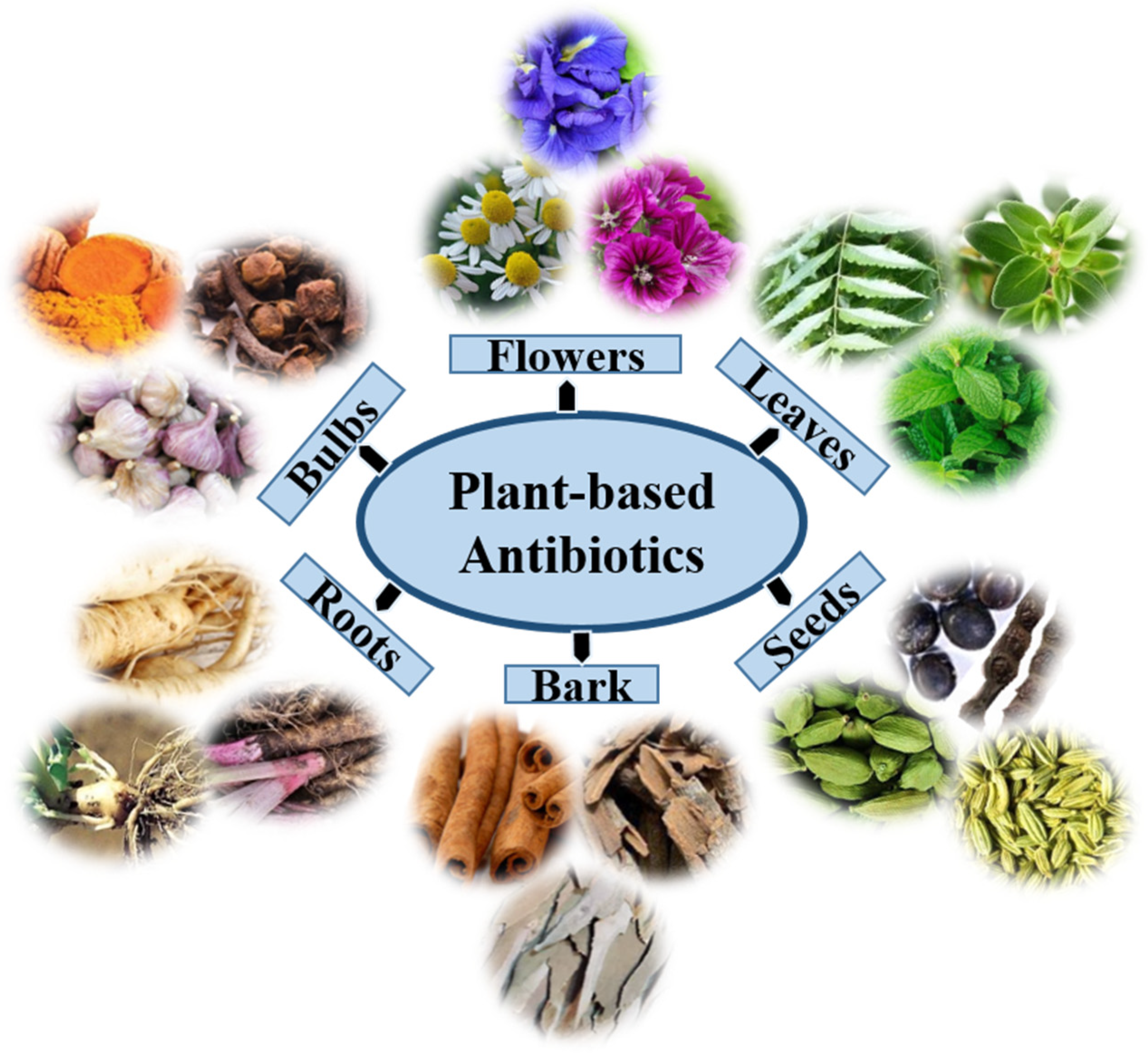
2. Antibacterial Activities of Plant-Derived Compounds
2.1. Terpenoids
2.2. Phenolics
2.3. Alkaloids
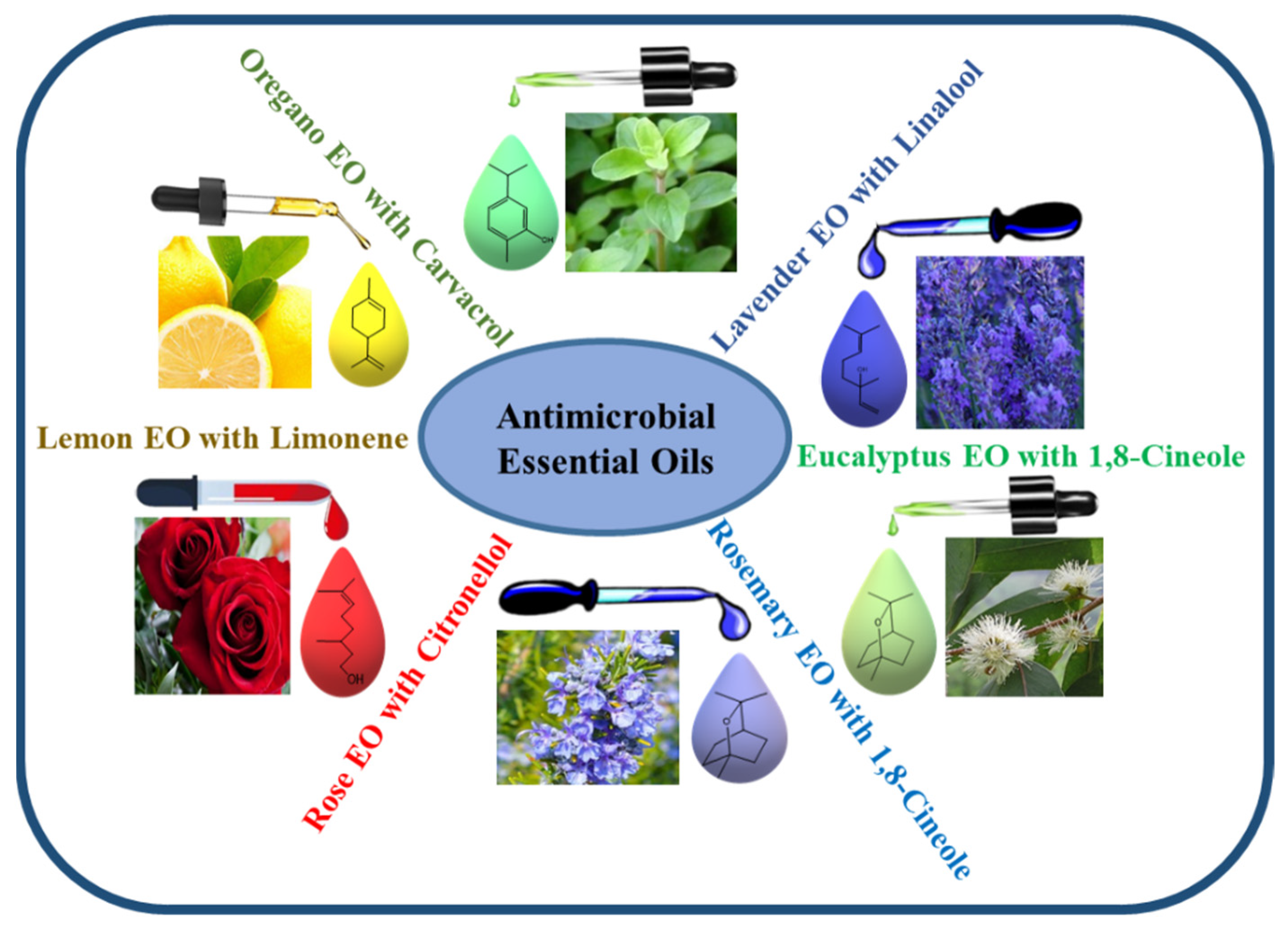
3. Antimicrobial Efficacy of EOs
4. Antimicrobial Nanomaterials
5. Synergistic Antimicrobial Activity of Plant Extracts, EOs, and Nanomaterials
6. Currently Available Conventional Antibiotics
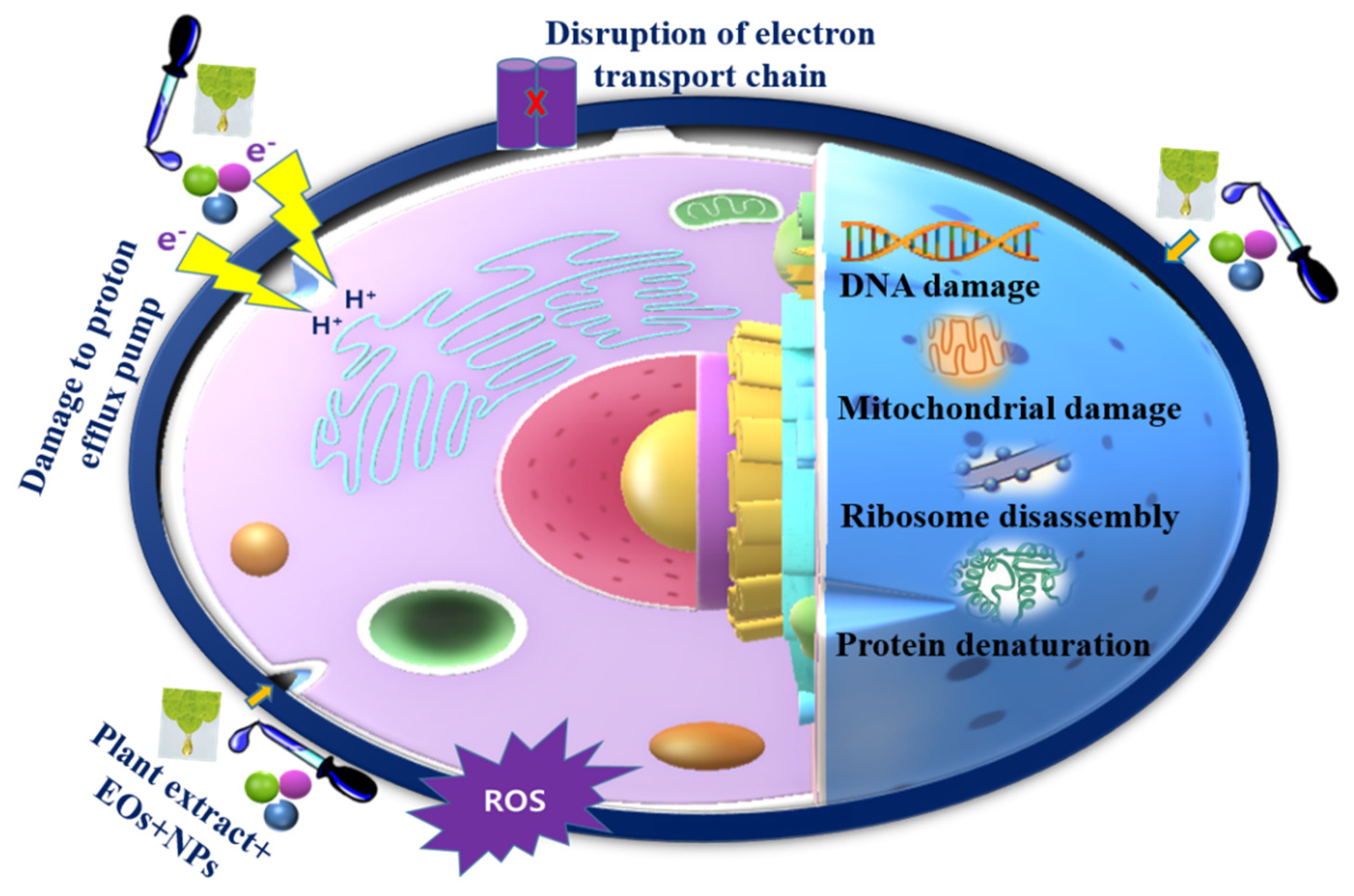
7. Antibacterial Mechanisms of Plant Extracts, EOs, and Nanomaterials
8. Concluding Remarks and Prospects for Future Research
Author Contributions
Funding
Institutional Review Board Statement
Informed Consent Statement
Data Availability Statement
Acknowledgments
Conflicts of Interest
References
- Pang, X.; Xiao, Q.; Cheng, Y.; Ren, E.; Lian, L.; Zhang, Y.; Gao, H.; Wang, X.; Leung, W.; Chen, X. Bacteria-responsive nanoliposomes as smart sonotheranostics for multidrug resistant bacterial infections. ACS Nano 2019, 13, 2427–2438. [Google Scholar] [CrossRef] [PubMed]
- Baym, M.; Stone, L.K.; Kishony, R. Multidrug evolutionary strategies to reverse antibiotic resistance. Science 2016, 351, aad3292. [Google Scholar] [CrossRef]
- Sun, Y.; Ye, J.; Hou, Y.; Chen, H.; Cao, J.; Zhou, T. Predation efficacy of Bdellovibrio bacteriovorus on multidrug-resistant clinical pathogens and their corresponding biofilms. Jpn. J. Infect. Dis. 2017, 70, 485–489. [Google Scholar] [CrossRef] [PubMed]
- Dong, P.; Mohammad, H.; Hui, J.; Leanse, L.G.; Li, J.; Liang, L.; Dai, T.; Seleem, M.N.; Cheng, J. Photolysis of Staphyloxanthin in methicillin-resistant Staphylococcus aureus potentiates killing by reactive oxygen species. Adv. Sci. 2019, 6, 1900030. [Google Scholar] [CrossRef]
- Jacqueline, C.; Caillon, J. Impact of bacterial biofilm on the treatment of prosthetic joint infections. J. Antimicrob. Chemother. 2014, 69, i37–i40. [Google Scholar] [CrossRef]
- Dincer, S.; Uslu, F.M.; Delik, A. Antibiotic resistance in biofilm. In Bacterial Biofilms; IntechOpen: London, UK, 2020; Available online: https://www.intechopen.com/chapters/72109 (accessed on 30 July 2022). [CrossRef]
- Beyth, N.; Houri-Haddad, Y.; Domb, A.; Khan, W.; Hazan, R. Alternative Antimicrobial Approach: Nano-Antimicrobial Materials. Evid.-Based Complement. Altern. Med. 2015, 2015, 246012. [Google Scholar] [CrossRef]
- Dostert, M.; Belanger, C.R.; Hancock, R.E.W. Design and assessment of anti-biofilm peptides: Steps toward clinical application. J. Innate Immun. 2019, 11, 193–204. [Google Scholar] [CrossRef] [PubMed]
- Kaur, I. Novel strategies to combat antimicrobial resistance. J. Infect. Dis. Ther. 2016, 4, 292. [Google Scholar] [CrossRef]
- Bozic, I.; Reiter, J.G.; Allen, B.; Antal, T.; Chatterjee, K.; Shah, P.; Moon, Y.S.; Yaqubie, A.; Kelly, N.; Le, D.T. Evolutionary dynamics of cancer in response to targeted combination therapy. Elife 2013, 2, e00747. [Google Scholar] [CrossRef] [PubMed]
- Nascimento, G.G.F.; Locatelli, J.; Freitas, P.C.; Silva, G.L. Antibacterial activity of plant extracts and phytochemicals on antibiotic-resistant bacteria. Braz. J. Microbiol. 2000, 31, 247–256. [Google Scholar] [CrossRef]
- Ncube, B.; Finnie, J.F.; Van Staden, J. In vitro antimicrobial synergism within plant extract combinations from three South African medicinal bulbs. J. Ethnopharmacol. 2012, 139, 81–89. [Google Scholar] [CrossRef] [PubMed]
- Aggarwal, B.; Lamba, H.S.; Ajeet, P.S. Various pharmacological aspects of Cocos nucifera—A review. Am. J. Pharmacol. Sci. 2017, 5, 25–30. [Google Scholar]
- Olajuyigbe, O.O.; Afolayan, A.J. Evaluation of combination effects of ethanolic extract of Ziziphus mucronata Willd. subsp. mucronata Willd. and antibiotics against clinically important bacteria. Sci. World J. 2013, 2013, 769594. [Google Scholar] [CrossRef]
- León-Buitimea, A.; Garza-Cárdenas, C.R.; Garza-Cervantes, J.A.; Lerma-Escalera, J.A.; Morones-Ramírez, J.R. The demand for new antibiotics: Antimicrobial peptides, nanoparticles, and combinatorial therapies as future strategies in antibacterial agent design. Front. Microbiol. 2020, 11, 1669. [Google Scholar] [CrossRef]
- Cheesman, M.J.; Ilanko, A.; Blonk, B.; Cock, I.E. Developing new antimicrobial therapies: Are synergistic combinations of plant extracts/compounds with conventional antibiotics the solution? Pharmacogn. Rev. 2017, 11, 57. [Google Scholar]
- Reda, F.M.; El-Zawahry, Y.A.; Omar, A.R. Synergistic effect of combined antibiotic and methanol extract of Eucalyptus camaldulensis leaf against Staphylococcus aureus and Pseudomonas aeruginosa. Int. J. Appl. Sci. Biotechnol. 2017, 5, 486–497. [Google Scholar] [CrossRef][Green Version]
- Basavegowda, N.; Baek, K.-H. Synergistic Antioxidant and Antibacterial Advantages of Essential Oils for Food Packaging Applications. Biomolecules 2021, 11, 1267. [Google Scholar] [CrossRef]
- Horváth, G.; Bencsik, T.; Ács, K.; Kocsis, B. Sensitivity of ESBL-Producing Gram-Negative Bacteria to Essential Oils, Plant Extracts, and Their Isolated Compounds; Academic Press: Amsterdam, The Netherlands, 2016; pp. 239–269. [Google Scholar]
- Aleksic, V.; Mimica-Dukic, N.; Simin, N.; Nedeljkovic, N.S.; Knezevic, P. Synergistic effect of Myrtus communis L. essential oils and conventional antibiotics against multi-drug resistant Acinetobacter baumannii wound isolates. Phytomedicine 2014, 21, 1666–1674. [Google Scholar] [CrossRef]
- Dikshit, P.K.; Kumar, J.; Das, A.K.; Sadhu, S.; Sharma, S.; Singh, S.; Gupta, P.K.; Kim, B.S. Green synthesis of metallic nanoparticles: Applications and limitations. Catalysts 2021, 11, 902. [Google Scholar] [CrossRef]
- Basavegowda, N.; Patra, J.K.; Baek, K.-H. Essential oils and mono/Bi/tri-metallic nanocomposites as alternative sources of antimicrobial agents to combat multidrug-resistant pathogenic microorganisms: An overview. Molecules 2020, 25, 1058. [Google Scholar] [CrossRef]
- Basavegowda, N.; Baek, K.-H. Multimetallic nanoparticles as alternative antimicrobial agents: Challenges and perspectives. Molecules 2021, 26, 912. [Google Scholar] [CrossRef]
- Jenkins, M.; Timoshyna, A.; Cornthwaite, M. Wild at Home: Exploring the global harvest, trade and use of wild plant ingredients. Retrieved Jan. 2018, 26, 2019. [Google Scholar]
- Manzo, L.M.; Moussa, I.; Ikhiri, K.; Yu, L. Toxicity studies of Acacia nilotica (L.): A review of the published scientific literature. J. Herbmed Pharmacol. 2019, 8, 163–172. [Google Scholar] [CrossRef]
- Pott, D.M.; Osorio, S.; Vallarino, J.G. From central to specialized metabolism: An overview of some secondary compounds derived from the primary metabolism for their role in conferring nutritional and organoleptic characteristics to fruit. Front. Plant Sci. 2019, 10, 835. [Google Scholar] [CrossRef]
- Eljounaidi, K.; Lichman, B.R. Nature’s chemists: The discovery and engineering of phytochemical biosynthesis. Front. Chem. 2020, 8, 1041. [Google Scholar] [CrossRef]
- Kusakizako, T.; Miyauchi, H.; Ishitani, R.; Nureki, O. Structural biology of the multidrug and toxic compound extrusion superfamily transporters. Biochim. Biophys. Acta BBA-Biomembr. 2020, 1862, 183154. [Google Scholar] [CrossRef]
- Dreier, J.; Ruggerone, P. Interaction of antibacterial compounds with RND efflux pumps in Pseudomonas aeruginosa. Front. Microbiol. 2015, 6, 660. [Google Scholar] [CrossRef] [PubMed]
- Zacchino, S.A.; Butassi, E.; Cordisco, E.; Svetaz, L.A. Hybrid combinations containing natural products and antimicrobial drugs that interfere with bacterial and fungal biofilms. Phytomedicine 2017, 37, 14–26. [Google Scholar] [CrossRef]
- Batista, O.; Duarte, A.; Nascimento, J.; Simões, M.F.; de la Torre, M.C.; Rodríguez, B. Structure and antimicrobial activity of diterpenes from the roots of Plectranthus hereroensis. J. Nat. Prod. 1994, 57, 858–861. [Google Scholar] [CrossRef]
- Cox-Georgian, D.; Ramadoss, N.; Dona, C.; Basu, C. Therapeutic and medicinal uses of terpenes. In Medicinal Plants; Springer: Berlin/Heidelberg, Germany, 2019; pp. 333–359. [Google Scholar]
- Pan, N.; Sun, G. Functional Textiles for Improved Performance, Protection and Health; Elsevier: Amsterdam, The Netherlands, 2011; ISBN 0857092871. [Google Scholar]
- Gomes, F.I.A.; Teixeira, P.; Azeredo, J.; Oliveira, R. Effect of farnesol on planktonic and biofilm cells of Staphylococcus epidermidis. Curr. Microbiol. 2009, 59, 118–122. [Google Scholar] [CrossRef]
- Gyawali, R.; Ibrahim, S.A. Natural products as antimicrobial agents. Food Control 2014, 46, 412–429. [Google Scholar] [CrossRef]
- Cowan, M.M. Plant products as antimicrobial agents. Clin. Microbiol. Rev. 1999, 12, 564–582. [Google Scholar] [CrossRef]
- Thawabteh, A.; Juma, S.; Bader, M.; Karaman, D.; Scrano, L.; Bufo, S.A.; Karaman, R. The biological activity of natural alkaloids against herbivores, cancerous cells and pathogens. Toxins 2019, 11, 656. [Google Scholar] [CrossRef]
- Khan, I.A.; Mirza, Z.M.; Kumar, A.; Verma, V.; Qazi, G.N. Piperine, a phytochemical potentiator of ciprofloxacin against Staphylococcus aureus. Antimicrob. Agents Chemother. 2006, 50, 810–812. [Google Scholar] [CrossRef] [PubMed]
- Lagha, R.; Ben Abdallah, F.; Al-Sarhan, B.O.; Al-Sodany, Y. Antibacterial and biofilm inhibitory activity of medicinal plant essential oils against Escherichia coli isolated from UTI patients. Molecules 2019, 24, 1161. [Google Scholar] [CrossRef]
- Gomes, F.; Dias, M.I.; Lima, Â.; Barros, L.; Rodrigues, M.E.; Ferreira, I.C.F.R.; Henriques, M. Satureja montana L. and Origanum majorana L. decoctions: Antimicrobial activity, mode of action and phenolic characterization. Antibiotics 2020, 9, 294. [Google Scholar] [CrossRef]
- Sampath Kumar, N.S.; Sarbon, N.M.; Rana, S.S.; Chintagunta, A.D.; Prathibha, S.; Ingilala, S.K.; Jeevan Kumar, S.P.; Sai Anvesh, B.; Dirisala, V.R. Extraction of bioactive compounds from Psidium guajava leaves and its utilization in preparation of jellies. AMB Express 2021, 11, 1–9. [Google Scholar] [CrossRef] [PubMed]
- Agyare, C.; Bempah, S.B.; Boakye, Y.D.; Ayande, P.G.; Adarkwa-Yiadom, M.; Mensah, K.B. Evaluation of antimicrobial and wound healing potential of Justicia flava and Lannea welwitschii. Evid.-Based Complement. Altern. Med. 2013, 2013, 632927. [Google Scholar] [CrossRef] [PubMed]
- Ismail, R.M.; Saleh, A.H.A.; Ali, K.S. GC-MS analysis and antibacterial activity of garlic extract with antibiotic. J. Med. Plants Stud 2020, 8, 26–30. [Google Scholar]
- Sabo, V.A.; Knezevic, P. Antimicrobial activity of Eucalyptus camaldulensis Dehn. plant extracts and essential oils: A review. Ind. Crops Prod. 2019, 132, 413–429. [Google Scholar] [CrossRef]
- Mahdavi, B.; Ghorat, F.; Nasrollahzadeh, M.S.; Hosseyni-Tabar, M.; Rezaei-Seresht, H. Chemical composition, antioxidant, antibacterial, cytotoxicity, and hemolyses activity of essential oils from flower of Matricaria chamomilla var. chamomilla. Anti-Infect. Agents 2020, 18, 224–232. [Google Scholar] [CrossRef]
- Antolak, H.; Czyżowska, A.; Kręgiel, D. Activity of Mentha piperita L. Ethanol Extract against Acetic Acid Bacteria Asaia spp. Foods 2018, 7, 171. [Google Scholar] [CrossRef]
- Al-Hadid, K.J. Quantitative analysis of antimicrobial activity of’Foeniculum vulgare’: A review. Plant Omics 2017, 10, 28–36. [Google Scholar] [CrossRef]
- Abdel-Naime, W.A.; Fahim, J.R.; Fouad, M.A.; Kamel, M.S. Antibacterial, antifungal, and GC–MS studies of Melissa officinalis. S. Afr. J. Bot. 2019, 124, 228–234. [Google Scholar] [CrossRef]
- Petkova, N.; Hambarlyiska, I.; Tumbarski, Y.; Vrancheva, R.; Raeva, M.; Ivanov, I. Phytochemical Composition and Antimicrobial Properties of Burdock (Arctium lappa L.) Roots Extracts. Biointerface Res. Appl. Chem. 2021, 12, 2826–2842. [Google Scholar]
- Fathi, M.; Ghane, M.; Pishkar, L. Phytochemical Composition, Antibacterial, and Antibiofilm Activity of Malva sylvestris Against Human Pathogenic Bacteria. Jundishapur J. Nat. Pharm. Prod. 2022, 17, e114164. [Google Scholar] [CrossRef]
- Nadia, Z.; Rachid, M. Antioxidant and antibacterial activities of Thymus vulgaris L. Med. Aromat. Plant Res. J. 2013, 1, 5–11. [Google Scholar]
- Mejía-Argueta, E.L.; Santillán-Benítez, J.G.; Canales-Martinez, M.M.; Mendoza-Medellín, A. Antimicrobial activity of Syzygium aromaticum L. essential oil on extended-spectrum beta-lactamases-producing Escherichia coli. Bull. Natl. Res. Cent. 2020, 44, 1–7. [Google Scholar] [CrossRef]
- Tian, C.; Chang, Y.; Zhang, Z.; Wang, H.; Xiao, S.; Cui, C.; Liu, M. Extraction technology, component analysis, antioxidant, antibacterial, analgesic and anti-inflammatory activities of flavonoids fraction from Tribulus terrestris L. leaves. Heliyon 2019, 5, e02234. [Google Scholar] [CrossRef]
- Salma, U.; Saha, S.K.; Sultana, S.; Ahmed, S.M.; Haque, S.D.; Mostaqim, S. The Antibacterial Activity of Ethanolic Extract of Cinnamon (Cinnamomum zeylanicum) against two Food Borne Pathogens: Staphylococcus aureus and Escherichia coli. Mymensingh Med. J. MMJ 2019, 28, 767–772. [Google Scholar] [PubMed]
- Wang, X.; Shen, Y.; Thakur, K.; Han, J.; Zhang, J.-G.; Hu, F.; Wei, Z.-J. Antibacterial activity and mechanism of ginger essential oil against Escherichia coli and Staphylococcus aureus. Molecules 2020, 25, 3955. [Google Scholar] [CrossRef]
- Czernicka, L.; Grzegorczyk, A.; Marzec, Z.; Antosiewicz, B.; Malm, A.; Kukula-Koch, W. Antimicrobial potential of single metabolites of Curcuma longa assessed in the total extract by thin-layer chromatography-based bioautography and image analysis. Int. J. Mol. Sci. 2019, 20, 898. [Google Scholar] [CrossRef]
- Lingaraju, D.P.; Sudarshana, M.S.; Mahendra, C.; Rao, K.P. Phytochemical screening and antimicrobial activity of leaf extracts of Eryngium foetidum L.(Apiaceae). Indo Am. J. Pharm. Res. 2016, 6, 4339–4344. [Google Scholar]
- Ojah, E.O.; Oladele, E.O.; Chukwuemeka, P. Phytochemical and antibacterial properties of root extracts from Portulaca oleracea Linn. (Purslane) utilised in the management of diseases in Nigeria. J. Med. Plants Econ. Dev. 2021, 5, 103. [Google Scholar] [CrossRef]
- Ahmed, Z.; Noor, A.A. Antibacterial activity of Momordica charantia L. and Citrus limon L. on gram positive and gram negative bacteria. Pure Appl. Biol. 2020, 9, 207–218. [Google Scholar] [CrossRef]
- Nigussie, D.; Davey, G.; Legesse, B.A.; Fekadu, A.; Makonnen, E. Antibacterial activity of methanol extracts of the leaves of three medicinal plants against selected bacteria isolated from wounds of lymphoedema patients. BMC Complement. Med. Ther. 2021, 21, 1–10. [Google Scholar] [CrossRef] [PubMed]
- Srikacha, N.; Ratananikom, K. Antibacterial activity of plant extracts in different solvents against pathogenic bacteria: An in vitro experiment. J. Acute Dis. 2020, 9, 223. [Google Scholar]
- Ribeiro, J.; Silva, V.; Aires, A.; Carvalho, R.; Igrejas, G.; Poeta, P. Antimicrobial activity of phenolic compounds extracted from Platanus hybrida: Exploring alternative therapies for a post-antibiotic era. Proceedings 2020, 66, 18. [Google Scholar] [CrossRef]
- Álvarez-Martínez, F.J.; Rodríguez, J.C.; Borrás-Rocher, F.; Barrajón-Catalán, E.; Micol, V. The antimicrobial capacity of Cistus salviifolius and Punica granatum plant extracts against clinical pathogens is related to their polyphenolic composition. Sci. Rep. 2021, 11, 1–12. [Google Scholar] [CrossRef] [PubMed]
- Lubis, R.R.; Marlisa, D.D.W. Antibacterial activity of betle leaf (Piper betle L.) extract on inhibiting Staphylococcus aureus in conjunctivitis patient. Am. J. Clin. Exp. Immunol. 2020, 9, 1. [Google Scholar] [PubMed]
- El-Beltagi, H.S.; Mohamed, H.I.; Abdelazeem, A.S.; Youssef, R.; Safwat, G. GC-MS Analysis, Antioxidant, Antimicrobial and Anticancer Activities of Extracts from Ficus Sycomorus Fruits and Leaves; Academic Press: Cambridge, MA, USA, 2019. [Google Scholar]
- Aabed, K.; Mohammed, A.E.; Benabdelkamel, H.; Masood, A.; Alfadda, A.A.; Alanazi, I.O.; Alnehmi, E.A. Antimicrobial Mechanism and Identification of the Proteins Mediated by Extracts from Asphaltum punjabianum and Myrtus communis. ACS Omega 2020, 5, 31019–31035. [Google Scholar] [CrossRef]
- Dib, K.; Cherrah, Y.; Rida, S.; Filali-Maltouf, A.; Ennibi, O. In Vitro Antibacterial Activity of Myrtus communis L. and Marrubium vulgare L. Leaves against Aggregatibacter actinomycetemcomitans and Eikenella corrodens. Evid.-Based Complement. Altern. Med. 2021, 2021, 8351332. [Google Scholar] [CrossRef]
- Fahmy, A.H.; Alam, I.P.; Salim, H.M. Bactericidal effects of Extract Bacil Leaves in in-vitro study of Pesudomonas aeruginosa. In Proceedings of the Proceeding Surabaya International Health Conference 2019, Surabaya, Indonesia, 15–30 May 2019; Volume 1, pp. 24–28. [Google Scholar]
- Setiawan, T.; Lay, B.W. Clitoria Ternatea ethanolic extract prevents dental caries via inhibiting streptococcus mutans growth and quorum sensing. Food Res. 2021, 5, 492–497. [Google Scholar]
- Souissi, M.; Azelmat, J.; Chaieb, K.; Grenier, D. Antibacterial and anti-inflammatory activities of cardamom (Elettaria cardamomum) extracts: Potential therapeutic benefits for periodontal infections. Anaerobe 2020, 61, 102089. [Google Scholar] [CrossRef] [PubMed]
- Antika, L.D.; Triana, D.; Ernawati, T. Antimicrobial activity of quinine derivatives against human pathogenic bacteria. In Proceedings of the IOP Conference Series: Earth and Environmental Science, Tangerang, Indonesia, 23–24 October 2019; IOP Publishing: Bristol, UK, 2020; Volume 462, p. 12006. [Google Scholar] [CrossRef]
- Na, S.; Kim, J.-H.; Rhee, Y.K.; Oh, S.-W. Enhancing the antimicrobial activity of ginseng against Bacillus cereus and Staphylococcus aureus by heat treatment. Food Sci. Biotechnol. 2018, 27, 203–210. [Google Scholar] [CrossRef]
- Stephane, F.F.Y.; Jules, B.K.J. Terpenoids as important bioactive constituents of essential oils, essential oils. In Essential Oils-Bioactive Compounds, New Perspectives and Applications; IntechOpen: London, UK, 2020. [Google Scholar]
- Korinek, M.; Handoussa, H.; Tsai, Y.-H.; Chen, Y.-Y.; Chen, M.-H.; Chiou, Z.-W.; Fang, Y.; Chang, F.-R.; Yen, C.-H.; Hsieh, C.-F. Anti-inflammatory and antimicrobial volatile oils: Fennel and cumin inhibit neutrophilic inflammation via regulating calcium and MAPKs. Front. Pharmacol. 2021, 12, 674095. [Google Scholar] [CrossRef] [PubMed]
- Mahanta, B.P.; Bora, P.K.; Kemprai, P.; Borah, G.; Lal, M.; Haldar, S. Thermolabile essential oils, aromas and flavours: Degradation pathways, effect of thermal processing and alteration of sensory quality. Food Res. Int. 2021, 145, 110404. [Google Scholar] [CrossRef]
- Chouhan, S.; Sharma, K.; Guleria, S. Antimicrobial activity of some essential oils—Present status and future perspectives. Medicines 2017, 4, 58. [Google Scholar] [CrossRef] [PubMed]
- Swamy, M.K.; Akhtar, M.S.; Sinniah, U.R. Antimicrobial properties of plant essential oils against human pathogens and their mode of action: An updated review. Evid.-Based Complement. Altern. Med. 2016, 2016, 3012462. [Google Scholar] [CrossRef]
- Teixeira, B.; Marques, A.; Ramos, C.; Neng, N.R.; Nogueira, J.M.F.; Saraiva, J.A.; Nunes, M.L. Chemical composition and antibacterial and antioxidant properties of commercial essential oils. Ind. Crops Prod. 2013, 43, 587–595. [Google Scholar] [CrossRef]
- Damtie, D.; Mekonnen, Y. Antibacterial activity of essential oils from Ethiopian thyme (Thymus serrulatus and Thymus schimperi) against tooth decay bacteria. PLoS ONE 2020, 15, e0239775. [Google Scholar] [CrossRef] [PubMed]
- Sohilait, H.J.; Kainama, H.; Nindatu, M. Chemical composition and antibacterial activity of the essential oils from different parts of Eugenia caryophylata, Thunb grown in Amboina Island. Int. J. Org. Chem. 2018, 8, 229–239. [Google Scholar] [CrossRef][Green Version]
- Xu, Y.; Wei, J.; Wei, Y.; Han, P.; Dai, K.; Zou, X.; Jiang, S.; Xu, F.; Wang, H.; Sun, J. Tea tree oil controls brown rot in peaches by damaging the cell membrane of Monilinia fructicola. Postharvest Biol. Technol. 2021, 175, 111474. [Google Scholar] [CrossRef]
- Hossain, S.; Heo, H.; De Silva, B.C.J.; Wimalasena, S.; Pathirana, H.; Heo, G.-J. Antibacterial activity of essential oil from lavender (Lavandula angustifolia) against pet turtle-borne pathogenic bacteria. Lab. Anim. Res. 2017, 33, 195–201. [Google Scholar] [CrossRef]
- Nikolić, M.; Jovanović, K.K.; Marković, T.; Marković, D.; Gligorijević, N.; Radulović, S.; Soković, M. Chemical composition, antimicrobial, and cytotoxic properties of five Lamiaceae essential oils. Ind. Crops Prod. 2014, 61, 225–232. [Google Scholar] [CrossRef]
- Andrade-Ochoa, S.; Chacón-Vargas, K.F.; Sánchez-Torres, L.E.; Rivera-Chavira, B.E.; Nogueda-Torres, B.; Nevárez-Moorillón, G.V. Differential antimicrobial effect of essential oils and their main components: Insights based on the cell membrane and external structure. Membranes 2021, 11, 405. [Google Scholar] [CrossRef] [PubMed]
- Helal, I.M.; El-Bessoumy, A.; Al-Bataineh, E.; Joseph, M.R.P.; Rajagopalan, P.; Chandramoorthy, H.C.; Ben Hadj Ahmed, S. Antimicrobial efficiency of essential oils from traditional medicinal plants of Asir region, Saudi Arabia, over drug resistant isolates. Biomed Res. Int. 2019, 2019, 8928306. [Google Scholar] [CrossRef]
- Mahboubi, M.; Heidarytabar, R.; Mahdizadeh, E.; Hosseini, H. Antimicrobial activity and chemical composition of Thymus species and Zataria multiflora essential oils. Agric. Nat. Resour. 2017, 51, 395–401. [Google Scholar] [CrossRef]
- Radünz, M.; da Trindade, M.L.M.; Camargo, T.M.; Radünz, A.L.; Borges, C.D.; Gandra, E.A.; Helbig, E. Antimicrobial and antioxidant activity of unencapsulated and encapsulated clove (Syzygium aromaticum, L.) essential oil. Food Chem. 2019, 276, 180–186. [Google Scholar] [CrossRef]
- Alizadeh Behbahani, B.; Falah, F.; Lavi Arab, F.; Vasiee, M.; Tabatabaee Yazdi, F. Chemical composition and antioxidant, antimicrobial, and antiproliferative activities of Cinnamomum zeylanicum bark essential oil. Evid.-Based Complement. Altern. Med. 2020, 2020, 5190603. [Google Scholar] [CrossRef]
- Brun, P.; Bernabè, G.; Filippini, R.; Piovan, A. In vitro antimicrobial activities of commercially available tea tree (Melaleuca alternifolia) essential oils. Curr. Microbiol. 2019, 76, 108–116. [Google Scholar] [CrossRef]
- Lorenzo-Leal, A.C.; Palou, E.; López-Malo, A.; Bach, H. Antimicrobial, cytotoxic, and anti-inflammatory activities of Pimenta dioica and Rosmarinus officinalis essential oils. Biomed Res. Int. 2019, 2019, 1639726. [Google Scholar] [CrossRef]
- Mutlu-Ingok, A.; Tasir, S.; Seven, A.; Akgun, N.; Karbancioglu-Guler, F. Evaluation of the single and combined antibacterial efficiency of essential oils for controlling Campylobacter coli, Campylobacter jejuni, Escherichia coli, Staphylococcus aureus, and mixed cultures. Flavour Fragr. J. 2019, 34, 280–287. [Google Scholar] [CrossRef]
- Gishen, N.Z.; Taddese, S.; Zenebe, T.; Dires, K.; Tedla, A.; Mengiste, B.; Shenkute, D.; Tesema, A.; Shiferaw, Y.; Lulekal, E. In vitro antimicrobial activity of six Ethiopian medicinal plants against Staphylococcus aureus, Escherichia coli and Candida albicans. Eur. J. Integr. Med. 2020, 36, 101121. [Google Scholar] [CrossRef]
- Tu, X.-F.; Hu, F.; Thakur, K.; Li, X.-L.; Zhang, Y.-S.; Wei, Z.-J. Comparison of antibacterial effects and fumigant toxicity of essential oils extracted from different plants. Ind. Crops Prod. 2018, 124, 192–200. [Google Scholar] [CrossRef]
- Zhang, J.; Ye, K.-P.; Zhang, X.; Pan, D.-D.; Sun, Y.-Y.; Cao, J.-X. Antibacterial activity and mechanism of action of black pepper essential oil on meat-borne Escherichia coli. Front. Microbiol. 2017, 7, 2094. [Google Scholar] [CrossRef]
- Moumni, S.; Elaissi, A.; Trabelsi, A.; Merghni, A.; Chraief, I.; Jelassi, B.; Chemli, R.; Ferchichi, S. Correlation between chemical composition and antibacterial activity of some Lamiaceae species essential oils from Tunisia. BMC Complement. Med. Ther. 2020, 20, 1–15. [Google Scholar] [CrossRef]
- Reyes-Jurado, F.; Cervantes-Rincón, T.; Bach, H.; López-Malo, A.; Palou, E. Antimicrobial activity of Mexican oregano (Lippia berlandieri), thyme (Thymus vulgaris), and mustard (Brassica nigra) essential oils in gaseous phase. Ind. Crops Prod. 2019, 131, 90–95. [Google Scholar] [CrossRef]
- Li, Z.-H.; Cai, M.; Liu, Y.-S.; Sun, P.-L.; Luo, S.-L. Antibacterial activity and mechanisms of essential oil from Citrus medica L. var. sarcodactylis. Molecules 2019, 24, 1577. [Google Scholar] [CrossRef]
- Clavijo-Romero, A.; Quintanilla-Carvajal, M.X.; Ruiz, Y. Stability and antimicrobial activity of eucalyptus essential oil emulsions. Food Sci. Technol. Int. 2019, 25, 24–37. [Google Scholar] [CrossRef]
- Ghasemian, A.; Al-Marzoqi, A.-H.; Mostafavi, S.K.S.; Alghanimi, Y.K.; Teimouri, M. Chemical composition and antimicrobial and cytotoxic activities of Foeniculum vulgare Mill essential oils. J. Gastrointest. Cancer 2020, 51, 260–266. [Google Scholar] [CrossRef] [PubMed]
- Marinkovic, J.; Markovic, T.; Nikolic, B.; Soldatovic, I.; Ivanov, M.; Ciric, A.; Sokovic, M.; Markovic, D. Antibacterial and Antibiofilm Potential of Leptospermum petersonii FM Bailey, Eucalyptus citriodora Hook., Pelargonium graveolens L’Hér. and Pelargonium roseum (Andrews) DC. Essential Oils Against Selected Dental Isolates. J. Essent. Oil Bear. Plants 2021, 24, 304–316. [Google Scholar] [CrossRef]
- Khalil, N.; Ashour, M.; Fikry, S.; Singab, A.N.; Salama, O. Chemical composition and antimicrobial activity of the essential oils of selected Apiaceous fruits. Futur. J. Pharm. Sci. 2018, 4, 88–92. [Google Scholar] [CrossRef]
- Kebede, B.H.; Forsido, S.F.; Tola, Y.B.; Astatkie, T. Free radical scavenging capacity, antibacterial activity and essential oil composition of turmeric (Curcuma domestica) varieties grown in Ethiopia. Heliyon 2021, 7, e06239. [Google Scholar] [CrossRef]
- Santamarta, S.; Aldavero, A.C.; Rojo, M. Antibacterial Properties of Cymbopogon martinii essential Oil against Bacillus subtillis food industry pathogen. Proceedings 2020, 66, 1. [Google Scholar] [CrossRef]
- Hojjati, M. Chemical constituents and antibacterial activity of dill (Anethum graveolens) essential oil. In Proceedings of the 15th ASEAN Conference on Food Science and Technology, Singapore, 14–16 November 2017; pp. 14–17. [Google Scholar]
- Amor, G.; Caputo, L.; La Storia, A.; De Feo, V.; Mauriello, G.; Fechtali, T. Chemical composition and antimicrobial activity of Artemisia herba-alba and Origanum majorana essential oils from Morocco. Molecules 2019, 24, 4021. [Google Scholar] [CrossRef] [PubMed]
- Fidan, H.; Stefanova, G.; Kostova, I.; Stankov, S.; Damyanova, S.; Stoyanova, A.; Zheljazkov, V.D. Chemical composition and antimicrobial activity of Laurus nobilis L. essential oils from Bulgaria. Molecules 2019, 24, 804. [Google Scholar] [CrossRef]
- Bączek, K.B.; Kosakowska, O.; Przybył, J.L.; Pióro-Jabrucka, E.; Costa, R.; Mondello, L.; Gniewosz, M.; Synowiec, A.; Węglarz, Z. Antibacterial and antioxidant activity of essential oils and extracts from costmary (Tanacetum balsamita L.) and tansy (Tanacetum vulgare L.). Ind. Crops Prod. 2017, 102, 154–163. [Google Scholar] [CrossRef]
- Soliman, F.M.; Fathy, M.M.; Salama, M.M.; Saber, F.R. Comparative study of the volatile oil content and antimicrobial activity of Psidium guajava L. and Psidium cattleianum Sabine leaves. Bull. Fac. Pharm. Cairo Univ. 2016, 54, 219–225. [Google Scholar] [CrossRef]
- Erdogan, A.; Ozkan, A. Investigatıon of antioxıdative, cytotoxic, membrane-damaging and membrane-protective effects of the essentıal oil of Origanum majorana and its oxygenated monoterpene component linalool in human-derived Hep G2 cell line. Iran. J. Pharm. Res. IJPR 2017, 16, 24. [Google Scholar] [PubMed]
- Simirgiotis, M.J.; Burton, D.; Parra, F.; López, J.; Muñoz, P.; Escobar, H.; Parra, C. Antioxidant and antibacterial capacities of Origanum vulgare L. essential oil from the arid Andean Region of Chile and its chemical characterization by GC-MS. Metabolites 2020, 10, 414. [Google Scholar] [CrossRef]
- Middaugh, J.; Hamel, R.; Jean-Baptiste, G.; Beriault, R.; Chenier, D.; Appanna, V.D. Aluminum triggers decreased aconitase activity via Fe-S cluster disruption and the overexpression of isocitrate dehydrogenase and isocitrate lyase: A metabolic network mediating cellular survival. J. Biol. Chem. 2005, 280, 3159–3165. [Google Scholar] [CrossRef]
- Macomber, L.; Elsey, S.P.; Hausinger, R.P. Fructose-1, 6-bisphosphate aldolase (class II) is the primary site of nickel toxicity in Escherichia coli. Mol. Microbiol. 2011, 82, 1291–1300. [Google Scholar] [CrossRef]
- Seil, J.T.; Webster, T.J. Antimicrobial applications of nanotechnology: Methods and literature. Int. J. Nanomed. 2012, 7, 2767. [Google Scholar]
- Lemire, J.A.; Harrison, J.J.; Turner, R.J. Antimicrobial activity of metals: Mechanisms, molecular targets and applications. Nat. Rev. Microbiol. 2013, 11, 371–384. [Google Scholar] [CrossRef]
- Malarkodi, C.; Rajeshkumar, S.; Paulkumar, K.; Vanaja, M.; Gnanajobitha, G.; Annadurai, G. Biosynthesis and antimicrobial activity of semiconductor nanoparticles against oral pathogens. Bioinorg. Chem. Appl. 2014, 2014, 347167. [Google Scholar] [CrossRef] [PubMed]
- Biswal, A.K.; Misra, P.K. Biosynthesis and characterization of silver nanoparticles for prospective application in food packaging and biomedical fields. Mater. Chem. Phys. 2020, 250, 123014. [Google Scholar] [CrossRef]
- Yaqoob, A.A.; Ahmad, H.; Parveen, T.; Ahmad, A.; Oves, M.; Ismail, I.M.I.; Qari, H.A.; Umar, K.; Mohamad Ibrahim, M.N. Recent Advances in Metal Decorated Nanomaterials and Their Various Biological Applications: A Review. Front. Chem. 2020, 8, 1–23. [Google Scholar] [CrossRef] [PubMed]
- Díez-Pascual, A.M. Antibacterial activity of nanomaterials. Nanomaterials 2018, 8, 359. [Google Scholar] [CrossRef]
- Pardhi, D.M.; Karaman, D.Ş.; Timonen, J.; Wu, W.; Zhang, Q.; Satija, S.; Mehta, M.; Charbe, N.; McCarron, P.A.; Tambuwala, M.M. Anti-bacterial activity of inorganic nanomaterials and their antimicrobial peptide conjugates against resistant and non-resistant pathogens. Int. J. Pharm. 2020, 586, 119531. [Google Scholar] [CrossRef]
- Egger, S.; Lehmann, R.P.; Height, M.J.; Loessner, M.J.; Schuppler, M. Antimicrobial properties of a novel silver-silica nanocomposite material. Appl. Environ. Microbiol. 2009, 75, 2973–2976. [Google Scholar] [CrossRef]
- Yuan, P.; Ding, X.; Yang, Y.Y.; Xu, Q. Metal nanoparticles for diagnosis and therapy of bacterial infection. Adv. Healthc. Mater. 2018, 7, 1701392. [Google Scholar] [CrossRef]
- Hsueh, Y.-H.; Tsai, P.-H.; Lin, K.-S. Ph-dependent antimicrobial properties of copper oxide nanoparticles in staphylococcus aureus. Int. J. Mol. Sci. 2017, 18, 793. [Google Scholar] [CrossRef] [PubMed]
- Yadav, H.M.; Kolekar, T.V.; Pawar, S.H.; Kim, J.-S. Enhanced photocatalytic inactivation of bacteria on Fe-containing TiO2 nanoparticles under fluorescent light. J. Mater. Sci. Mater. Med. 2016, 27, 1–9. [Google Scholar] [CrossRef]
- Moongraksathum, B.; Chen, Y.-W. Anatase TiO2 co-doped with silver and ceria for antibacterial application. Catal. Today 2018, 310, 68–74. [Google Scholar] [CrossRef]
- Şen Karaman, D.; Manner, S.; Rosenholm, J.M. Mesoporous silica nanoparticles as diagnostic and therapeutic tools: How can they combat bacterial infection? Ther. Deliv. 2018, 9, 241–244. [Google Scholar] [CrossRef]
- Mishra, K.; Basavegowda, N.; Lee, Y.R. Biosynthesis of Fe, Pd, and Fe-Pd bimetallic nanoparticles and their application as recyclable catalysts for [3 + 2] cycloaddition reaction: A comparative approach. Catal. Sci. Technol. 2015, 5, 2612–2621. [Google Scholar] [CrossRef]
- Zhang, J.; Ma, J.; Fan, X.; Peng, W.; Zhang, G.; Zhang, F.; Li, Y. Graphene supported Au-Pd-Fe3O4 alloy trimetallic nanoparticles with peroxidase-like activities as mimic enzyme. Catal. Commun. 2017, 89, 148–151. [Google Scholar] [CrossRef]
- Deepak, F.L.; Mayoral, A.; Arenal, R. Advanced Transmission Electron Microscopy: Applications to Nanomaterials; Springer: Berlin/Heidelberg, Germany, 2015; ISBN 3319151770. [Google Scholar]
- Merugu, R.; Gothalwal, R.; Deshpande, P.K.; De Mandal, S.; Padala, G.; Chitturi, K.L. Synthesis of Ag/Cu and Cu/Zn bimetallic nanoparticles using toddy palm: Investigations of their antitumor, antioxidant and antibacterial activities. Mater. Today Proc. 2021, 44, 99–105. [Google Scholar] [CrossRef]
- Vaseghi, Z.; Tavakoli, O.; Nematollahzadeh, A. Rapid biosynthesis of novel Cu/Cr/Ni trimetallic oxide nanoparticles with antimicrobial activity. J. Environ. Chem. Eng. 2018, 6, 1898–1911. [Google Scholar] [CrossRef]
- Paul, D.; Mangla, S.; Neogi, S. Antibacterial study of CuO-NiO-ZnO trimetallic oxide nanoparticle. Mater. Lett. 2020, 271, 127740. [Google Scholar] [CrossRef]
- Dong, Y.; Zhu, H.; Shen, Y.; Zhang, W.; Zhang, L. Antibacterial activity of silver nanoparticles of different particle size against Vibrio Natriegens. PLoS ONE 2019, 14, e0222322. [Google Scholar] [CrossRef] [PubMed]
- Manikandan, V.; Velmurugan, P.; Park, J.-H.; Chang, W.-S.; Park, Y.-J.; Jayanthi, P.; Cho, M.; Oh, B.-T. Green synthesis of silver oxide nanoparticles and its antibacterial activity against dental pathogens. 3 Biotech 2017, 7, 1–9. [Google Scholar] [CrossRef] [PubMed]
- Liu, S.; Wang, C.; Hou, J.; Wang, P.; Miao, L.; Li, T. Effects of silver sulfide nanoparticles on the microbial community structure and biological activity of freshwater biofilms. Environ. Sci. Nano 2018, 5, 2899–2908. [Google Scholar] [CrossRef]
- Zhang, S.S.; Wang, X.; Su, H.F.; Feng, L.; Wang, Z.; Ding, W.Q.; Blatov, V.A.; Kurmoo, M.; Tung, C.H.; Sun, D.; et al. A Water-Stable Cl@Ag14 Cluster Based Metal-Organic Open Framework for Dichromate Trapping and Bacterial Inhibition. Inorg. Chem. 2017, 56, 11891–11899. [Google Scholar] [CrossRef]
- Suryavanshi, P.; Pandit, R.; Gade, A.; Derita, M.; Zachino, S.; Rai, M. Colletotrichum sp.-mediated synthesis of sulphur and aluminium oxide nanoparticles and its in vitro activity against selected food-borne pathogens. LWT-Food Sci. Technol. 2017, 81, 188–194. [Google Scholar] [CrossRef]
- Ortiz-Benítez, E.A.; Velázquez-Guadarrama, N.; Durán Figueroa, N.V.; Quezada, H.; Olivares-Trejo, J.d.J. Antibacterial mechanism of gold nanoparticles on Streptococcus pneumoniae. Metallomics 2019, 11, 1265–1276. [Google Scholar] [CrossRef]
- Rieznichenko, L.S.; Gruzina, T.G.; Dybkova, S.M.; Ushkalov, V.O.; Ulberg, Z.R. Investigation of bismuth nanoparticles antimicrobial activity against high pathogen microorganisms. Am. J. Bioterror. Biosecur. Biodef 2015, 2, 1004. [Google Scholar]
- Keihan, A.H.; Veisi, H.; Veasi, H. Green synthesis and characterization of spherical copper nanoparticles as organometallic antibacterial agent. Appl. Organomet. Chem. 2017, 31, e3642. [Google Scholar] [CrossRef]
- Marquis, G.; Ramasamy, B.; Banwarilal, S.; Munusamy, A.P. Evaluation of antibacterial activity of plant mediated CaO nanoparticles using Cissus quadrangularis extract. J. Photochem. Photobiol. B Biol. 2016, 155, 28–33. [Google Scholar] [CrossRef]
- Qamar, H.; Rehman, S.; Chauhan, D.K.; Tiwari, A.K.; Upmanyu, V. Green synthesis, characterization and antimicrobial activity of copper oxide nanomaterial derived from Momordica charantia. Int. J. Nanomed. 2020, 15, 2541. [Google Scholar] [CrossRef] [PubMed]
- Pop, O.L.; Mesaros, A.; Vodnar, D.C.; Suharoschi, R.; Tăbăran, F.; Magerușan, L.; Tódor, I.S.; Diaconeasa, Z.; Balint, A.; Ciontea, L. Cerium oxide nanoparticles and their efficient antibacterial application in vitro against gram-positive and gram-negative pathogens. Nanomaterials 2020, 10, 1614. [Google Scholar] [CrossRef] [PubMed]
- Katata-Seru, L.; Moremedi, T.; Aremu, O.S.; Bahadur, I. Green synthesis of iron nanoparticles using Moringa oleifera extracts and their applications: Removal of nitrate from water and antibacterial activity against Escherichia coli. J. Mol. Liq. 2018, 256, 296–304. [Google Scholar] [CrossRef]
- Saqib, S.; Munis, M.F.H.; Zaman, W.; Ullah, F.; Shah, S.N.; Ayaz, A.; Farooq, M.; Bahadur, S. Synthesis, characterization and use of iron oxide nano particles for antibacterial activity. Microsc. Res. Tech. 2019, 82, 415–420. [Google Scholar] [CrossRef] [PubMed]
- Argueta-Figueroa, L.; Torres-Gómez, N.; García-Contreras, R.; Vilchis-Nestor, A.R.; Martínez-Alvarez, O.; Acosta-Torres, L.S.; Arenas-Arrocena, M.C. Hydrothermal synthesis of pyrrhotite (Fex-1S) nanoplates and their antibacterial, cytotoxic activity study. Prog. Nat. Sci. Mater. Int. 2018, 28, 447–455. [Google Scholar] [CrossRef]
- Narayanasamy, P.; Switzer, B.L.; Britigan, B.E. Prolonged-acting, multi-targeting gallium nanoparticles potently inhibit growth of both HIV and mycobacteria in co-infected human macrophages. Sci. Rep. 2015, 5, 1–7. [Google Scholar] [CrossRef]
- Kamran, U.; Bhatti, H.N.; Iqbal, M.; Jamil, S.; Zahid, M. Biogenic synthesis, characterization and investigation of photocatalytic and antimicrobial activity of manganese nanoparticles synthesized from Cinnamomum verum bark extract. J. Mol. Struct. 2019, 1179, 532–539. [Google Scholar] [CrossRef]
- Maji, J.; Pandey, S.; Basu, S. Synthesis and evaluation of antibacterial properties of magnesium oxide nanoparticles. Bull. Mater. Sci. 2020, 43, 1–10. [Google Scholar] [CrossRef]
- Kumar, G.S.; Venkataramana, B.; Reddy, S.A.; Maseed, H.; Nagireddy, R.R. Hydrothermal synthesis of Mn3O4 nanoparticles by evaluation of pH effect on particle size formation and its antibacterial activity. Adv. Nat. Sci. Nanosci. Nanotechnol. 2020, 11, 35006. [Google Scholar] [CrossRef]
- André, V.; da Silva, A.R.F.; Fernandes, A.; Frade, R.; Garcia, C.; Rijo, P.; Antunes, A.M.M.; Rocha, J.; Duarte, M.T. Mg-and Mn-MOFs boost the antibiotic activity of nalidixic acid. ACS Appl. Bio Mater. 2019, 2, 2347–2354. [Google Scholar] [CrossRef]
- Din, M.I.; Nabi, A.G.; Rani, A.; Aihetasham, A.; Mukhtar, M. Single step green synthesis of stable nickel and nickel oxide nanoparticles from Calotropis gigantea: Catalytic and antimicrobial potentials. Environ. Nanotechnol. Monit. Manag. 2018, 9, 29–36. [Google Scholar] [CrossRef]
- Behera, N.; Arakha, M.; Priyadarshinee, M.; Pattanayak, B.S.; Soren, S.; Jha, S.; Mallick, B.C. Oxidative stress generated at nickel oxide nanoparticle interface results in bacterial membrane damage leading to cell death. RSC Adv. 2019, 9, 24888–24894. [Google Scholar] [CrossRef] [PubMed]
- Mohana, S.; Sumathi, S. Multi-functional biological effects of palladium nanoparticles synthesized using Agaricus bisporus. J. Clust. Sci. 2020, 31, 391–400. [Google Scholar] [CrossRef]
- Ahmed, K.B.A.; Raman, T.; Anbazhagan, V. Platinum nanoparticles inhibit bacteria proliferation and rescue zebrafish from bacterial infection. Rsc. Adv. 2016, 6, 44415–44424. [Google Scholar] [CrossRef]
- Geoffrion, L.D.; Hesabizadeh, T.; Medina-Cruz, D.; Kusper, M.; Taylor, P.; Vernet-Crua, A.; Chen, J.; Ajo, A.; Webster, T.J.; Guisbiers, G. Naked selenium nanoparticles for antibacterial and anticancer treatments. ACS Omega 2020, 5, 2660–2669. [Google Scholar] [CrossRef]
- Smirnov, N.A.; Kudryashov, S.I.; Nastulyavichus, A.A.; Rudenko, A.A.; Saraeva, I.N.; Tolordava, E.R.; Gonchukov, S.A.; Romanova, Y.M.; Ionin, A.A.; Zayarny, D.A. Antibacterial properties of silicon nanoparticles. Laser Phys. Lett. 2018, 15, 105602. [Google Scholar] [CrossRef]
- Eisa, N.E.; Almansour, S.; Alnaim, I.A.; Ali, A.M.; Algrafy, E.; Ortashi, K.M.; Awad, M.A.; Virk, P.; Hendi, A.A.; Eissa, F.Z. Eco-synthesis and characterization of titanium nanoparticles: Testing its cytotoxicity and antibacterial effects. Green Process. Synth. 2020, 9, 462–468. [Google Scholar] [CrossRef]
- Tiwari, V.; Mishra, N.; Gadani, K.; Solanki, P.S.; Shah, N.A.; Tiwari, M. Mechanism of anti-bacterial activity of zinc oxide nanoparticle against carbapenem-resistant Acinetobacter baumannii. Front. Microbiol. 2018, 9, 1218. [Google Scholar] [CrossRef]
- Khan, M.; Shaik, M.R.; Khan, S.T.; Adil, S.F.; Kuniyil, M.; Khan, M.; Al-Warthan, A.A.; Siddiqui, M.R.H.; Nawaz Tahir, M. Enhanced antimicrobial activity of biofunctionalized zirconia nanoparticles. ACS Omega 2020, 5, 1987–1996. [Google Scholar] [CrossRef]
- Pezeshkpour, V.; Khosravani, S.A.; Ghaedi, M.; Dashtian, K.; Zare, F.; Sharifi, A.; Jannesar, R.; Zoladl, M. Ultrasound assisted extraction of phenolic acids from broccoli vegetable and using sonochemistry for preparation of MOF-5 nanocubes: Comparative study based on micro-dilution broth and plate count method for synergism antibacterial effect. Ultrason. Sonochem. 2018, 40, 1031–1038. [Google Scholar] [CrossRef]
- Andrade, G.R.S.; Nascimento, C.C.; Lima, Z.M.; Teixeira-Neto, E.; Costa, L.P.; Gimenez, I.F. Star-shaped ZnO/Ag hybrid nanostructures for enhanced photocatalysis and antibacterial activity. Appl. Surf. Sci. 2017, 399, 573–582. [Google Scholar] [CrossRef]
- Addae, E.; Dong, X.; McCoy, E.; Yang, C.; Chen, W.; Yang, L. Investigation of antimicrobial activity of photothermal therapeutic gold/copper sulfide core/shell nanoparticles to bacterial spores and cells. J. Biol. Eng. 2014, 8, 1–11. [Google Scholar] [CrossRef]
- Lozhkomoev, A.S.; Bakina, O.V.; Pervikov, A.V.; Kazantsev, S.O.; Glazkova, E.A. Synthesis of CuO–ZnO composite nanoparticles by electrical explosion of wires and their antibacterial activities. J. Mater. Sci. Mater. Electron. 2019, 30, 13209–13216. [Google Scholar] [CrossRef]
- Singh, S.; Barick, K.C.; Bahadur, D. Inactivation of bacterial pathogens under magnetic hyperthermia using Fe3O4–ZnO nanocomposite. Powder Technol. 2015, 269, 513–519. [Google Scholar] [CrossRef]
- Yadav, N.; Jaiswal, A.K.; Dey, K.K.; Yadav, V.B.; Nath, G.; Srivastava, A.K.; Yadav, R.R. Trimetallic Au/Pt/Ag based nanofluid for enhanced antibacterial response. Mater. Chem. Phys. 2018, 218, 10–17. [Google Scholar] [CrossRef]
- Alzahrani, K.E.; Niazy, A.A.; Alswieleh, A.M.; Wahab, R.; El-Toni, A.M.; Alghamdi, H.S. Antibacterial activity of trimetal (CuZnFe) oxide nanoparticles. Int. J. Nanomed. 2018, 13, 77. [Google Scholar] [CrossRef] [PubMed]
- Rajendiran, K.; Zhao, Z.; Pei, D.-S.; Fu, A. Antimicrobial activity and mechanism of functionalized quantum dots. Polymers 2019, 11, 1670. [Google Scholar] [CrossRef]
- Kumari, A.; Thakur, N.; Vashishtt, J.; Singh, R.R. Structural, luminescent and antimicrobial properties of ZnS and CdSe/ZnS quantum dot structures originated by precursors. Spectrochim. Acta Part A Mol. Biomol. Spectrosc. 2020, 229, 117962. [Google Scholar] [CrossRef] [PubMed]
- Sabah, A.; Tasleem, S.; Murtaza, M.; Nazir, M.; Rashid, F. Effect of polymer capping on photonic multi-core–shell quantum dots CdSe/CdS/ZnS: Impact of sunlight and antibacterial activity. J. Phys. Chem. C 2020, 124, 9009–9020. [Google Scholar] [CrossRef]
- Shariati, M.R.; Samadi-Maybodi, A.; Colagar, A.H. Dual cocatalyst loaded reverse type-I core/shell quantum dots for photocatalytic antibacterial applications. J. Mater. Chem. A 2018, 6, 20433–20443. [Google Scholar] [CrossRef]
- Mir, I.A.; Alam, H.; Priyadarshini, E.; Meena, R.; Rawat, K.; Rajamani, P.; Rizvi, M.S.; Bohidar, H.B. Antimicrobial and biocompatibility of highly fluorescent ZnSe core and ZnSe@ ZnS core-shell quantum dots. J. Nanoparticle Res. 2018, 20, 1–11. [Google Scholar] [CrossRef]
- Li, W.; Song, P.; Xin, Y.; Kuang, Z.; Liu, Q.; Ge, F.; Zhu, L.; Zhang, X.; Tao, Y. The effects of luminescent CdSe quantum dot-functionalized antimicrobial peptides nanoparticles on antibacterial activity and molecular mechanism. Int. J. Nanomed. 2021, 16, 1849. [Google Scholar] [CrossRef] [PubMed]
- Chai, S.; Zhou, L.; Pei, S.; Zhu, Z.; Chen, B. P-Doped Carbon Quantum Dots with Antibacterial Activity. Micromachines 2021, 12, 1116. [Google Scholar] [CrossRef] [PubMed]
- Elyamny, S.; Eltarahony, M.; Abu-Serie, M.; Nabil, M.M.; Kashyout, A.E.-H.B. One-pot fabrication of Ag@ Ag2O core–shell nanostructures for biosafe antimicrobial and antibiofilm applications. Sci. Rep. 2021, 11, 1–13. [Google Scholar] [CrossRef] [PubMed]
- Radhi, A.; Mohamad, D.; Rahman, F.S.A.; Abdullah, A.M.; Hasan, H. Mechanism and factors influence of graphene-based nanomaterials antimicrobial activities and application in dentistry. J. Mater. Res. Technol. 2021, 11, 1290–1307. [Google Scholar] [CrossRef]
- Raja, A.; Selvakumar, K.; Rajasekaran, P.; Arunpandian, M.; Ashokkumar, S.; Kaviyarasu, K.; Bahadur, S.A.; Swaminathan, M. Visible active reduced graphene oxide loaded titania for photodecomposition of ciprofloxacin and its antibacterial activity. Colloids Surf. A Physicochem. Eng. Asp. 2019, 564, 23–30. [Google Scholar] [CrossRef]
- Trinh, L.T.; Quynh, L.A.B.; Hieu, N.H. Synthesis of zinc oxide/graphene oxide nanocomposite material for antibacterial application. Int. J. Nanotechnol. 2018, 15, 108–117. [Google Scholar] [CrossRef]
- Yang, Z.; Hao, X.; Chen, S.; Ma, Z.; Wang, W.; Wang, C.; Yue, L.; Sun, H.; Shao, Q.; Murugadoss, V. Long-term antibacterial stable reduced graphene oxide nanocomposites loaded with cuprous oxide nanoparticles. J. Colloid Interface Sci. 2019, 533, 13–23. [Google Scholar] [CrossRef]
- Tu, Q.; Zhang, Q.; Wang, Y.; Jiao, Y.; Xiao, J.; Peng, T.; Wang, J. Antibacterial properties of poly (dimethylsiloxane) surfaces modified with graphene oxide-catechol composite. Prog. Org. Coat. 2019, 129, 247–253. [Google Scholar] [CrossRef]
- Hassani, M.; Tahghighi, A.; Rohani, M.; Hekmati, M.; Ahmadian, M.; Ahmadvand, H. Robust antibacterial activity of functionalized carbon nanotube-levofloxacine conjugate based on in vitro and in vivo studies. Sci. Rep. 2022, 12, 1–19. [Google Scholar] [CrossRef] [PubMed]
- Abo-Neima, S.E.; Motaweh, H.A.; Elsehly, E.M. Antimicrobial activity of functionalised carbon nanotubes against pathogenic microorganisms. IET Nanobiotechnol. 2020, 14, 457–464. [Google Scholar] [CrossRef]
- Mohammed, M.K.A.; Ahmed, D.S.; Mohammad, M.R. Studying antimicrobial activity of carbon nanotubes decorated with metal-doped ZnO hybrid materials. Mater. Res. Express 2019, 6, 55404. [Google Scholar] [CrossRef]
- Alfei, S.; Schito, A.M.; Zuccari, G. Considerable Improvement of Ursolic Acid Water Solubility by its Encapsulation in Dendrimer Nanoparticles: Design, Synthesis and Physicochemical Characterization. Nanomaterials 2021, 11, 2196. [Google Scholar] [CrossRef] [PubMed]
- Alfei, S.; Brullo, C.; Caviglia, D.; Zuccari, G. Preparation and Physicochemical Characterization of Water-Soluble Pyrazole-Based Nanoparticles by Dendrimer Encapsulation of an Insoluble Bioactive Pyrazole Derivative. Nanomaterials 2021, 11, 2662. [Google Scholar] [CrossRef]
- Jiang, G.; Liu, S.; Yu, T.; Wu, R.; Ren, Y.; van der Mei, H.C.; Liu, J.; Busscher, H.J. PAMAM dendrimers with dual-conjugated vancomycin and Ag-nanoparticles do not induce bacterial resistance and kill vancomycin-resistant Staphylococci. Acta Biomater. 2021, 123, 230–243. [Google Scholar] [CrossRef] [PubMed]
- Alfei, S.; Schito, A.M. Positively charged polymers as promising devices against multidrug resistant gram-negative bacteria: A Review. Polymers 2020, 12, 1195. [Google Scholar] [CrossRef]
- Mazumder, A.; Davis, J.; Rangari, V.; Curry, M. Synthesis, characterization, and applications of dendrimer-encapsulated zero-valent Ni nanoparticles as antimicrobial agents. Int. Sch. Res. Not. 2013, 2013, 843709. [Google Scholar] [CrossRef]
- Gholami, M.; Mohammadi, R.; Arzanlou, M.; Akbari Dourbash, F.; Kouhsari, E.; Majidi, G.; Mohseni, S.M.; Nazari, S. In vitro antibacterial activity of poly (amidoamine)-G7 dendrimer. BMC Infect. Dis. 2017, 17, 1–11. [Google Scholar] [CrossRef] [PubMed]
- Schito, A.M.; Alfei, S. Antibacterial activity of non-cytotoxic, amino acid-modified polycationic dendrimers against Pseudomonas aeruginosa and other non-fermenting gram-negative bacteria. Polymers 2020, 12, 1818. [Google Scholar] [CrossRef]
- Tauanov, Z.; Zakiruly, O.; Baimenova, Z.; Baimenov, A.; Akimbekov, N.S.; Berillo, D. Antimicrobial Properties of the Triclosan-Loaded Polymeric Composite Based on Unsaturated Polyester Resin: Synthesis, Characterization and Activity. Polymers 2022, 14, 676. [Google Scholar] [CrossRef]
- Jaramillo, A.F.; Riquelme, S.A.; Sánchez-Sanhueza, G.; Medina, C.; Solís-Pomar, F.; Rojas, D.; Montalba, C.; Melendrez, M.F.; Pérez-Tijerina, E. Comparative study of the antimicrobial effect of nanocomposites and composite based on poly (butylene adipate-co-terephthalate) using Cu and Cu/Cu2O nanoparticles and CuSO4. Nanoscale Res. Lett. 2019, 14, 1–17. [Google Scholar] [CrossRef]
- Jalageri, M.D.; Puttaiahgowda, Y.M. Design and antimicrobial activity of piperazine polymer nanocomposite. Mater. Today Proc. 2019, 15, 262–267. [Google Scholar] [CrossRef]
- Gautam, S.; Sharma, S.; Sharma, B.; Jain, P. Antibacterial efficacy of poly (vinyl alcohol) nanocomposites reinforced with graphene oxide and silver nanoparticles for packaging applications. Polym. Compos. 2021, 42, 2829–2837. [Google Scholar] [CrossRef]
- Aiyegoro, O.A.; Afolayan, A.J.; Okoh, A.I. Synergistic interaction of Helichrysum pedunculatum leaf extracts with antibiotics against wound infection associated bacteria. Biol. Res. 2009, 42, 327–338. [Google Scholar] [CrossRef]
- Iseppi, R.; Mariani, M.; Condò, C.; Sabia, C.; Messi, P. Essential oils: A natural weapon against antibiotic-resistant bacteria responsible for nosocomial infections. Antibiotics 2021, 10, 417. [Google Scholar] [CrossRef]
- Bassolé, I.H.N.; Lamien-Meda, A.; Bayala, B.; Tirogo, S.; Franz, C.; Novak, J.; Nebié, R.C.; Dicko, M.H. Composition and antimicrobial activities of Lippia multiflora Moldenke, Mentha x piperita L. and Ocimum basilicum L. essential oils and their major monoterpene alcohols alone and in combination. Molecules 2010, 15, 7825–7839. [Google Scholar] [CrossRef]
- Soltanzadeh, M.; Peighambardoust, S.H.; Ghanbarzadeh, B.; Mohammadi, M.; Lorenzo, J.M. Chitosan nanoparticles encapsulating lemongrass (Cymbopogon commutatus) essential oil: Physicochemical, structural, antimicrobial and in-vitro release properties. Int. J. Biol. Macromol. 2021, 192, 1084–1097. [Google Scholar] [CrossRef]
- Saquib, S.A.; AlQahtani, N.A.; Ahmad, I.; Kader, M.A.; Al Shahrani, S.S.; Asiri, E.A. Evaluation and comparison of antibacterial efficacy of herbal extracts in combination with antibiotics on periodontal pathobionts: An in vitro microbiological study. Antibiotics 2019, 8, 89. [Google Scholar] [CrossRef]
- Bahmani, M.; Taherikalani, M.; Khaksarian, M.; Rafieian-Kopaei, M.; Ashrafi, B.; Nazer, M.; Soroush, S.; Abbasi, N.; Rashidipour, M. The synergistic effect of hydroalcoholic extracts of Origanum vulgare, Hypericum perforatum and their active components carvacrol and hypericin against Staphylococcus aureus. Futur. Sci. OA 2019, 5, FSO371. [Google Scholar] [CrossRef] [PubMed]
- Musimun, C.; Papiernik, D.; Permpoonpattana, P.; Chumkaew, P.; Srisawat, T. Synergy of green-synthesized silver nanoparticles and Vatica diospyroides fruit extract in inhibiting Gram-positive bacteria by inducing membrane and intracellular disruption. J. Exp. Nanosci. 2022, 17, 420–438. [Google Scholar] [CrossRef]
- Aabed, K.; Mohammed, A.E. Synergistic and antagonistic effects of biogenic silver nanoparticles in combination with antibiotics against some pathogenic microbes. Front. Bioeng. Biotechnol. 2021, 9, 652362. [Google Scholar] [CrossRef]
- Lee, N.L.S.; Yuen, K.Y.; Kumana, C.R. β-Lactam antibiotic and β-lactamase inhibitor combinations. JAMA 2001, 285, 386–388. [Google Scholar] [CrossRef] [PubMed]
- Rakholiya, K.D.; Kaneria, M.J.; Chanda, S.V. Medicinal plants as alternative sources of therapeutics against multidrug-resistant pathogenic microorganisms based on their antimicrobial potential and synergistic properties. In Fighting Multidrug Resistance with Herbal Extracts, essential Oils and Their Components; Academic Press: Cambridge, MA, USA, 2013; pp. 165–179. [Google Scholar] [CrossRef]
- Rachuonyo, H.; Ogola, P.; Arika, W.; Wambani, J.; Gatheri, G. Combined effect of crude leaf extracts of selected medicinal plants against selected enteric bacterial pathogens and Candida albicans. J. Antimicrob Agents 2016, 2, 1212–2472. [Google Scholar]
- Obuekwe, I.S.; Okoyomo, E.P.; Anka, U.S. Effect of Plant Extract Combinations on Some Bacterial Pathogens. J. Appl. Sci. Environ. Manag. 2020, 24, 627–632. [Google Scholar] [CrossRef]
- Gadisa, E.; Usman, H. Evaluation of Antibacterial Activity of Essential Oils and Their Combination against Multidrug-Resistant Bacteria Isolated from Skin Ulcer. Int. J. Microbiol. 2021, 2021, 6680668. [Google Scholar] [CrossRef]
- Purkait, S.; Bhattacharya, A.; Bag, A.; Chattopadhyay, R.R. Synergistic antibacterial, antifungal and antioxidant efficacy of cinnamon and clove essential oils in combination. Arch. Microbiol. 2020, 202, 1439–1448. [Google Scholar] [CrossRef] [PubMed]
- Garza-Cervantes, J.A.; Chávez-Reyes, A.; Castillo, E.C.; García-Rivas, G.; Antonio Ortega-Rivera, O.; Salinas, E.; Ortiz-Martínez, M.; Gómez-Flores, S.L.; Peña-Martínez, J.A.; Pepi-Molina, A. Synergistic antimicrobial effects of silver/transition-metal combinatorial treatments. Sci. Rep. 2017, 7, 1–16. [Google Scholar] [CrossRef]
- Alfei, S.; Schito, A.M. β-Lactam Antibiotics and β-Lactamase Enzymes Inhibitors, Part 2: Our Limited Resources. Pharmaceuticals 2022, 15, 476. [Google Scholar] [CrossRef] [PubMed]
- Alfei, S.; Zuccari, G. Recommendations to Synthetize Old and New β-Lactamases Inhibitors: A Review to Encourage Further Production. Pharmaceuticals 2022, 15, 384. [Google Scholar] [CrossRef] [PubMed]
- World Health Organization. 2019 Antibacterial Agents in Clinical Development: An Analysis of the Antibacterial Clinical Development Pipeline; World Health Organization: Geneva, Switzerland, 2019. [Google Scholar]
- Das, B.; Verma, J.; Kumar, P.; Ghosh, A.; Ramamurthy, T. Antibiotic resistance in Vibrio cholerae: Understanding the ecology of resistance genes and mechanisms. Vaccine 2020, 38, A83–A92. [Google Scholar] [CrossRef]
- Peterson, E.; Kaur, P. Antibiotic resistance mechanisms in bacteria: Relationships between resistance determinants of antibiotic producers, environmental bacteria, and clinical pathogens. Front. Microbiol. 2018, 9, 2928. [Google Scholar] [CrossRef]
- Sultan, I.; Rahman, S.; Jan, A.T.; Siddiqui, M.T.; Mondal, A.H.; Haq, Q.M.R. Antibiotics, resistome and resistance mechanisms: A bacterial perspective. Front. Microbiol. 2018, 9, 2066. [Google Scholar] [CrossRef]
- Kapoor, G.; Saigal, S.; Elongavan, A. Action and resistance mechanisms of antibiotics: A guide for clinicians. J. Anaesthesiol. Clin. Pharmacol. 2017, 33, 300. [Google Scholar] [CrossRef] [PubMed]
- Reygaert, W.C. An overview of the antimicrobial resistance mechanisms of bacteria. AIMS Microbiol. 2018, 4, 482. [Google Scholar] [CrossRef] [PubMed]
- Álvarez-Martínez, F.J.; Barrajón-Catalán, E.; Herranz-López, M.; Micol, V. Antibacterial plant compounds, extracts and essential oils: An updated review on their effects and putative mechanisms of action. Phytomedicine 2021, 90, 153626. [Google Scholar] [CrossRef]
- Mostafa, A.A.; Al-Askar, A.A.; Almaary, K.S.; Dawoud, T.M.; Sholkamy, E.N.; Bakri, M.M. Antimicrobial activity of some plant extracts against bacterial strains causing food poisoning diseases. Saudi J. Biol. Sci. 2018, 25, 361–366. [Google Scholar] [CrossRef] [PubMed]
- Nazzaro, F.; Fratianni, F.; De Martino, L.; Coppola, R.; De Feo, V. Effect of essential oils on pathogenic bacteria. Pharmaceuticals 2013, 6, 1451–1474. [Google Scholar] [CrossRef]
- Wang, L.; Hu, C.; Shao, L. The antimicrobial activity of nanoparticles: Present situation and prospects for the future. Int. J. Nanomed. 2017, 12, 1227. [Google Scholar] [CrossRef]


| Plants | Parts | Pathogens | Mechanism | Ref. |
|---|---|---|---|---|
| Alchornea cordifolia | flower | E. coli | damage of cell wall | [39] |
| Origanum majorana | leaves | S. aureus, K. pneumoniae | membrane damage | [40] |
| Psidium guajava | leaves | B. subtilis, S. aureus | cell wall damage | [41] |
| Justicia flava | leaves | E. coli, P. aeruginosa | changes in internal pH | [42] |
| Allium sativum | bulbs | P. aeruginosa, S. aureus | cell membrane integrity | [43] |
| Lannea welwitschii | leaves | E. coli, P. aeruginosa | cell wall integrity | [42] |
| Eucalyptus camaldulensis | leaves, bark | S. aureus, B. subtilis | leakage of cell constituents | [44] |
| Matricaria chamomilla | flowers | S. aureus, P. aeruginosa | cell wall degradation | [45] |
| Mentha piperita | leaves | S. aureus, B. subtilis | damage of cytoplasmic membranes | [46] |
| Foeniculum vulgare | seeds | A. flavus, C. albicans | cellular DNA damages | [47] |
| Melissa officinalis | leaves | S. aureus, P. aeruginosa | disrupt the membrane structure | [48] |
| Arctium lappa | roots | P. aeruginosa, S. aureus | damage by oxidative stress | [49] |
| Malva sylvestris | flower, leaves | S. aureus, E. faecalis | damaging the membrane | [50] |
| Thymus vulgaris | leaves | E. coli, S. aureus | chemical affinity for membrane lipids | [51] |
| Syzygium aromaticum | buds | E. coli | membrane damage and intracellular content leakage | [52] |
| Tribulus terrestris | leaves | Escherichia coli, Salmonella | membrane damage and leakage of cellular materials | [53] |
| Cinnamomum zeylanicum | bark | S. aureus, E. coli | inhibiting of various cellular enzymes | [54] |
| Zingiber officinale | rhizome | E. coli, S. aureus | damage to cell membrane | [55] |
| Curcuma longa | rhizome | S. aureus, B. subtilis | loss of membrane integrity | [56] |
| Eryngium foetidum | leaves | P. aeruginosa, C. albicans | disruption of the cell membrane | [57] |
| Portulaca oleracea | roots | E. cloacae, B. subtilis | inhibiting the efflux pumps | [58] |
| Momordica charantia | peels | S. aureus, B. cereus | disintegrates the membrane | [59] |
| Lawsonia inermis | leaves | S. aureus, E. coli | inactivating microbial adhesions | [60] |
| Azadirachta indica | leaves | S. pyogenes | inactivating microbial enzymes | [60] |
| Achyranthes aspera | leaves | S. pyogenes | inhibiting energy metabolism | [60] |
| Acacia nilotica | seeds | S. aureus | cell membrane permeability | [61] |
| Platanus hybrida | fruits | E. faecalis, E. faecium | Inhibiting the biofilm production | [62] |
| Cistus salviifolius | aerial parts | S. aureus | cell wall alterations | [63] |
| Punica granatum | peels | S. aureus | cell wall alterations | [63] |
| Piper betle | leaves | S. aureus | destruction of the bacteria cell wall | [64] |
| Ficus sycomorus | leaves, fruits | E. coli, S. aureus | permeability of the cell membranes | [65] |
| Myrtus communis | leaves | E. coli | proteins in the outer membrane specifically involved | [66] |
| Asphaltum punjabianum | mineral resin | E. coli | proteins involved specifically in the outer membrane | [66] |
| Marrubium vulgare | leaves | A. actinomycetemcomitans, E. corrodens | affect cytoplasmic membrane | [67] |
| Ocimum basilicum | leaves | P. aeruginosa | bacterial cells will lose cations and macromolecules | [68] |
| Clitoria ternatea | flowers | Streptococcus mutans | quorum sensing inhibition | [69] |
| Elettaria cardamomum | Seeds | P. gingivalis | cell membrane disrupted | [70] |
| Cinchona officinalis | bark | E. coli, P. aeruginosa | structural damage of bacterial cells | [71] |
| Panax ginseng | roots | B. cereus, S. aureus | changes in the membrane potential | [72] |
| Essential Oils | Plant Source | Major Components | Pathogens | Modes of Action | Ref. |
|---|---|---|---|---|---|
| Basil | Ocimum basilicum | linalool | S. aureus | disrupt the permeability barrier | [85] |
| Thyme | Thymus vulgaris | thymol | P. aeruginosa, A. niger | interferes with membrane functions | [86] |
| Clove | Syzygium aromaticum | eugenol | S. aureus, S. Typhimurium | sensitivity to eugenol | [87] |
| Cinnamon | Cinnamomum zeylanicum | cinnamaldehyde | E. coli, L. innocua | facilitate intracellular compounds leakage | [88] |
| Tea tree | Melaleuca alternifolia | terpinen-4-ol | P. aeruginosa, C. glabrata | alterations of the biological membrane | [89] |
| Rosemary | Rosmarinus officinalis | α-pinene | C. albicans | rupture of the membranes and cell wall | [90] |
| Dill | Anethum graveolens | carvone | S. aureus, E. coli | lesion in the plasma membrane | [91] |
| Cumin | Cuminum cyminum | p-mentha-1,3-dien-7-al | S. aureus, E. coli | deformation of the cell membrane | [91] |
| Cardamom | Elettaria cardamomum | α-terpinly acetate | E. coli, S. aureus | damage the cell membrane | [91] |
| Peppermint | Mentha piperita | menthol | E. coli, S. aureus | lysis and loss of membrane integrity | [92] |
| Anise | Pimpinella anisum | anethole | S. aureus, B. subtilis | alter the cell membrane permeability | [93] |
| Black pepper | Piper nigrum | α-pinene | E. coli | leakage, disorder, and death by breaking cell membrane | [94] |
| Sage | Salvia officinalis | α-thujone | P. aeruginosa | changed the cell membrane permeability | [95] |
| Lavender | Lavandula angustifolia | linalool | S. aureus, E. coli, C. albicans | damaging the cell wall and membrane | [92] |
| Mustard | Brassica nigra | allyl isothiocyanate | A. fumigatus, A. nomius | disrupt the cell wall thus causing cell lysis | [96] |
| Citron | Citrus medica | limonene | S. aureus, E. coli | destruction of the cell membrane | [97] |
| Eucalyptus | Eucalyptus globulus | 1,8-cineole | E. coli, S. aureus | penetrate the membrane and damage cell organelles | [98] |
| Fennel | Foeniculum vulgare | trans-anethole | S. aureus, E. coli | cell deformation and integrity of cell membranes | [99] |
| Rose geranium | Pelargonium roseum | citronellol | S. salivarius | interaction with nitrogen in proteins and nucleic acids | [100] |
| Caraway | Carum carvi | carvone | E. coli, B. bronchiseptica | alteration in the structure of cell wall | [101] |
| Coriander | Coriandrum sativum | linalool | S. tyhimurium, E. coli | cell wall damage by over expression of genes | [101] |
| Turmeric | Curcuma longa | α-turmerone | S. aureus | inducing leakage of ions and important cell contents | [102] |
| Palmarosa | Cymbopogon martinii | geraniol | B. subtillis | alteration in cytoplasm and swelling | [103] |
| Dill | Anethum graveolens | α-phellandrene | S. aureus | disrupt the permeability barrier | [104] |
| Armoise | Artemisia herba-alba | thujone | S. aureus, S. Typhimurium | changing the membrane potential | [105] |
| Laurel | Laurus nobilis | 1,8-cineole | S. aureus. P. aeruginosa | disrupt cellular membranes and increase membrane permeability | [106] |
| Ginger | Zingiber officinale | zingiberene | S. aureus, E. coli | destroy membrane structure, increase cell membrane permeability | [55] |
| Costmary | Tanacetum balsamita | β-thujone | L. monocytogenes, S. sonnei | damage to the cellular membranes | [107] |
| Guava | Psidium cattleianum Sabine | α-pinene | S. aureus, N. gonorrhoeae | propagate through cell membranes and cause the death | [108] |
| Marjoram | Origanum majorana | terpinen-4-ol | S. aureus, K. clocae | exhibited membrane and DNA damaging effects | [109] |
| Oregano | Origanum vulgare | thymol | S. aureus, S. enterica | alteration of the bacterial plasma membrane | [110] |
| NPs | Size (nm) | Bacteria | Modes of Action | Ref. |
|---|---|---|---|---|
| Ag | 10 | V. natriegens | rupture of cell membrane and DNA damage | [132] |
| Ag2O | 10 | L. acidophilus, S. mutans | prevents the growth of pathogen | [133] |
| Ag2S | 65 | Phormidium spp. | cell membrane inhibition | [134] |
| Ag-MOF | - | S. aureus | stable in water and the existence of Ag+ ions | [135] |
| Al2O3 | 30 | S. typhi, F. oxysporum | disintegration of outer membrane by ROS | [136] |
| Au | 20 | S. pneumoniae | cellular disruption | [137] |
| Bi | 40 | M. arginini, E. coli | inhibits protein synthesis | [138] |
| Cu | 15 | B. subtilis, S. aureus | synergistic effects of functional groups | [139] |
| CaO | 58 | S. aureus, E. coli | destruction of the cell membrane | [140] |
| CuO | 60 | B. cereus | damage of several biochemical processes | [141] |
| CeO2 | 5 | B. cereus, E. coli | oxidative stress induced by the pro-oxidants | [142] |
| CdS | 25 | S. aureus, Lactobacillus sp. | CdS NPs impregnated and surrounded by the bacterial cell | [115] |
| Fe | 474 | E. coli | strong affinity between positively charged NPs and negatively charged cell membrane | [143] |
| Fe3O4 | 25 | E. coli, S. aureus | plasma membrane disruption | [144] |
| FeS | 35 | E. coli, S. aureus | internalization of nanomaterials on cell membrane | [145] |
| Ga | 305 | M. tuberculosis | reduction of mycobacterium growth rate | [146] |
| Mn | 50 | E. coli, S. aureus | protein inactivation and membrane permeability decreases | [147] |
| MgO | 27 | E. coli, Bacillus sp. | loss of membrane integrity and leakage of intracellular molecules | [148] |
| Mn3O4 | 130 | P. aeruginosa, K. pneumonia | disrupting bacterial cell membrane | [149] |
| Mg-MOF | - | E. coli, S. aureus | peptide–nalidixic acid conjugation formed | [150] |
| Mn-MOF | - | E. faecalis, P. aeruginosa | peptide–nalidixic acid conjugation formed | [150] |
| Ni | 60 | P. aeruginosa | destruction of cell membrane | [151] |
| NiO | 40 | E. coli, B. subtilis | oxidative stress generated at the NPs interface resulted in membrane damage | [152] |
| Pd | 13 | S. pyrogens, B. subtilis | cell membrane damage and apoptosis | [153] |
| Pt | 2 | A. hydrophila, E. coli | generation of ROS and decrease cell viability | [154] |
| Se | 85 | S. aureus, E. coli | ROS causing cell membrane damage | [155] |
| Si | 90 | P. aeruginosa, S. aureus | direct mechanical damage to the cell membrane | [156] |
| TiO2 | 9.2 | E. coli | outer cell membrane damaged by attacking hydroxyl radicals and ROS | [157] |
| ZnO | 30 | A. baumannii | production of ROS increases | [158] |
| ZrO2 | 2.5 | S. mitis, S. mutans, R. dentocariosa | NPs enhance the interaction with bacterial constituents | [159] |
| Zn-MOF | - | P. aeruginosa | causing cell damage by interaction with hydroxyl group of peptidoglycan | [160] |
| Ag/ZnO | 43 | P. aeruginosa, S. aureus | leaching of silver as Ag+ | [161] |
| Au/CuS | 2 | B. anthracis | cell membrane damage | [162] |
| CuO/ZnO | 50 and 82 | S. aureus, E. coli | membrane depolarization caused due to lectrostatic interaction of NPs | [163] |
| Fe3O4/ZnO | 200 | E. coli, S. aureus | plasm membrane disruption includes oxidative stress | [164] |
| Au/Pt/Ag | 20 | E. faecalis, E. coli | ROS production | [165] |
| Cu/Zn/Fe | 42 | E. coli, E. faecalis | cell disruption by released ions | [166] |
| NPs | Size (nm) | Bacteria | Modes of Action | Ref. |
|---|---|---|---|---|
| ZnS and CdSe/ZnS quantum dot | 1.9 | E. coli, B. subtilis | toxic composition of CdSe QDs demonstrating antimicrobial behavior | [168] |
| CdSe/CdS/ZnS multi-core–shell quantum dots | 12–38 | K. pneumoniae, P. aeruginosa | rupturing of the membrane wall and cause of the decay of bacteria | [169] |
| Ag-PdS/ZnS/CdS core–shell quantum dots | 8 | S. saprophyticus, E. coli | establishment of the catalyst–microorganism complex and a catalyst-related ROS | [170] |
| ZnSe@ZnS core–shell quantum dots | 3.6 and 4.8 | E. coli, S. aureus | high affinity towards the thiol groups of bacterial cell surface proteins | [171] |
| Peptide-loaded CdSe quantum dot | 9 and 14 | E. coli, S. aureus | AP loaded on CdSe NPs had a higher water solubility and bioavailability | [172] |
| P-doped carbon quantum dots | 2.75–4.25 | E. coli, S. aureus | cell walls wrinkled and broken | [173] |
| Ag@Ag2O core–shell | 19–60 | P. aeruginosa, S. aureus | blockage of DNA replication and repair processes | [174] |
| NPs | Size (nm) | Bacteria | Modes of Action | Ref. |
|---|---|---|---|---|
| rGO-TiO2 | 32 | E. coli, S. aureus | improve the contact between TiO2 surface and bacteria | [176] |
| GO-ZnO | 14–26 | E. coli | induces ROS to kill the bacteria | [177] |
| GO-Cu2O | 30 | E. coli, S. aureus | copper ions react with cytoplasmic constituents | [178] |
| DMS-GO-DMA | -- | E. coli, S. aureus | GO induces membrane stress on contact by disrupting and damaging cell membranes | [179] |
| MWCNT-LVX | -- | S. aureus, P. aeruginosa | inhibition of bacterial DNA replication | [180] |
| F-MWNTs | -- | E. coli, S. aureus | smaller diameter of MWNTs can endorse damage to cell membrane through the cell–surface interaction | [181] |
| Ag-doped ZnO on SWCNTs | 12–15 | E. coli, S. aureus | production of ROS on the interaction samples with bacterial membrane | [182] |
| Au-doped ZnO on MWCNTs | 12–18 | E. coli, S. aureus | the toxicity of carbon nanotube is mainly affected by diameter, length, and surface functional group | [182] |
| NPs | Size (nm) | Bacteria | Modes of Action | Ref. |
|---|---|---|---|---|
| Van-PAMAM-AgNP dendrimers | -- | S. aureus | heterofunctionalized Van-PAMAM-AgNP dendrimers for intra-cellular entry through the cell wall and bacterial killing | [185] |
| G4-PAMAM dendrimer | 10 | E. coli, B. subtilis | disrupting of the cell membrane function and inhibiting cell wall synthesis, nucleic acid synthesis, and protein synthesis | [187] |
| PAMAM-G7 dendrimer | 20 | P. mirabilis, S. aureus | dendrimers are mediated by disrupting the bacterial outer and inner membrane by terminal amine groups | [188] |
| Amino-acid-modified polycationic dendrimers | -- | P. aeruginosa | loss of membrane potential, inhibition of biosynthetic pathways, and free radical production | [189] |
| Triclosan-loaded polymeric composite | -- | S. aureus, K. pneumoniae | at high concentrations, triclosan destroys the bacterial membrane, leading to its death | [190] |
| PBAT/Cu-NPs | 100–200 | A. baumannii, E. faecalis | polymer and metal nanocomposites increase the number of ions released from the nanoparticles into the polymer matrix | [191] |
| Piperazine polymer nanocomposite | 559.7 | E. coli, S. aureus | nanoparticles are distributed within the suitable polymer matrix | [192] |
| PVA/GO/Ag nanocomposites | -- | E. coli, S. aureus | physical interactions of the bacterial cell with the nanoparticle | [193] |
| Antimicrobial Agents | Combinations | Pathogens | Ref. |
|---|---|---|---|
| EOs/EOs | Melaleuca alternifolia/Cupressus sempervirens | E. coli | [195] |
| EOs/antibiotics | Eucalyptus globulus/oxacillin | S. aureus | [195] |
| EOs/NPs | Lemongrass/chitosan NP | E. coli, S. aureus | [197] |
| Plant extract/antibiotics | Salvadora persica/amoxicillin | P. gingivalis, T. forsythia | [198] |
| Plant extract/EOs | Origanum vulgare/carvacrol | S. aureus | [199] |
| Plant extract/NPs | Vatica diospyroides/Ag NPs | S. aureus, B. subtilis | [200] |
| NPs/antibiotics | AgNPs/fluconazole | S. aureus, E. coli | [201] |
| β-Lactam/β-lactamase inhibitor | amoxicillin/potassium clavulanate | S. aureus | [202] |
| Antibiotics | Specific Drug | Modes of Action | Resistance Profiles | Target Bacteria | Ref. |
|---|---|---|---|---|---|
| β-Lactams | Penicillin G, amoxicillin, cephalosporin C | Cell wall synthesis inhibition | Hydrolysis, efflux, altered target, reduced permeability | S. aureus, P. aeruginosa | [212] |
| Aminoglycosides | Streptomycin, gentamicin | Inhibition of translation and cell membrane synthesis | Modifying enzyme inactivation by phosphorylation | P. aeruginosa, V. cholerae | [213] |
| Tetracyclines | Minocycline, doxycycline | 30S ribosomal subunit | Monooxygenation, ribosomal modification | Staphylococci, Streptococci | [214] |
| Glycopeptides | Vancomycin, teicoplanin | Peptidoglycan biosynthesis | Altered target | S. haemolyticus, E. faecium | [214] |
| Macrolides | Erythromycin, azithromycin | Inhibition of protein synthesis | Glycosylation, efflux, methylation | Streptococci, Staphylococci | [215] |
| Phenicols | Chloramphenicol | Inhibition of protein synthesis | Acetylation by chloramphenicol acetyltransferase | B. subtilis, S. pneumoniae | [216] |
| Rifamycin | Rifampin | Inhibition of nucleic acid synthesis | ADP-ribosylation, efflux | V. cholerae, E. coli | [213] |
| Quinolone | Ciprofloxacin, levofloxacin | Inhibitors of DNA synthesis | Altered DNA gyrase | S. aureus, P. aeruginosa | [212] |
| Cationic peptides | Polymyxin B, colistin | Disrupt membranes | Altered target, efflux | E. coli, S. typhimurium | [213] |
Publisher’s Note: MDPI stays neutral with regard to jurisdictional claims in published maps and institutional affiliations. |
© 2022 by the authors. Licensee MDPI, Basel, Switzerland. This article is an open access article distributed under the terms and conditions of the Creative Commons Attribution (CC BY) license (https://creativecommons.org/licenses/by/4.0/).
Share and Cite
Basavegowda, N.; Baek, K.-H. Combination Strategies of Different Antimicrobials: An Efficient and Alternative Tool for Pathogen Inactivation. Biomedicines 2022, 10, 2219. https://doi.org/10.3390/biomedicines10092219
Basavegowda N, Baek K-H. Combination Strategies of Different Antimicrobials: An Efficient and Alternative Tool for Pathogen Inactivation. Biomedicines. 2022; 10(9):2219. https://doi.org/10.3390/biomedicines10092219
Chicago/Turabian StyleBasavegowda, Nagaraj, and Kwang-Hyun Baek. 2022. "Combination Strategies of Different Antimicrobials: An Efficient and Alternative Tool for Pathogen Inactivation" Biomedicines 10, no. 9: 2219. https://doi.org/10.3390/biomedicines10092219
APA StyleBasavegowda, N., & Baek, K.-H. (2022). Combination Strategies of Different Antimicrobials: An Efficient and Alternative Tool for Pathogen Inactivation. Biomedicines, 10(9), 2219. https://doi.org/10.3390/biomedicines10092219