Article

Xylitol Fluoride Varnish: In Vitro Effect Analysis on Enamel by Atomic Force Microscopy

by
Catalina Iulia Saveanu
1,†,
Oana Dragos
2,*,
Daniela Anistoroaei
1,*,
Livia Ionela Bobu
1,†,
Alexandra Ecaterina Saveanu
3,†,
Adina Armencia
1,†,
Sorina Mihaela Solomon
4,† and
Oana Tanculescu
4
1
Surgical Department, Faculty of Dental Medicine, University of Medicine and Pharmacy Grigore T. Popa, 700115 Iasi, Romania
2
National Institute of Research-Development for Technical Physics—IFT, 700115 Iasi, Romania
3
Faculty of Dental Medicine, University of Medicine and Pharmacy Grigore T. Popa, 700115 Iasi, Romania
4
Department of Odontology-Periodontology and Fixed Prosthodontics Faculty of Dental Medicine, University of Medicine and Pharmacy, 700115 Iasi, Romania
*
Authors to whom correspondence should be addressed.
These authors contributed equally to this work.
Biomedicines 2022, 10(8), 1900; https://doi.org/10.3390/biomedicines10081900
Submission received: 11 July 2022 / Revised: 30 July 2022 / Accepted: 4 August 2022 / Published: 5 August 2022
(This article belongs to the Section Biomedical Engineering and Materials)

Abstract

:
(1) Background: Numerous studies have shown the beneficial role of fluoride in the primary prevention of dental caries. The aim of the present study was to put into evidence the change in the enamel structure immediately after the application of a fluoride varnish. (2) Methods: A xylitol–fluoride varnish was evaluated. The enamel specimens (n = 10) were analyzed by atomic force microscopy on enamel surface and treatment with fluoride varnish applied. The dimensional topographic analysis was performed by 2D and 3D analysis software. Statistical analysis was performed using SPSS Version 26.00 (IBM, Armonk, NY, USA). A one-sample statistics test was used to identify significant differences (p < 0.05). (3) Results: Surface roughness (Ra) measurements ranged from Ra = 0.039 μm (±0.048), to Ra = 0.049 μm (±0.031), respectively (p < 0.05), with an increase in the surface roughness passing from the intact enamel to the enamel exposed to fluoride varnish. When comparing Ra values of the nonfluorinated enamel and fluorinated enamel, significant differences (p < 0.05) were found, suggesting that the varnish had a protective effect on the enamel surface. (4) Conclusions: Xylitol–fluoride varnish, even in one single short-time application, is effective in reducing the surface roughness of enamel structure exposed to abrasion, thus increasing resistance to dental caries.

1. Introduction

The oral environment has a significant influence on the hard dental tissues in the initiation and evolution of carious and non-carious lesions. Tooth enamel is a hard tissue, which contains 95% mineral substances in its composition. The mineral substances in the enamel are found in the form of calcium phosphate, HA (hydroxyapatite), FA (fluorapatite), carbonates, and silicates. When its structure is affected by various destructive processes, the enamel does not have the possibility of spontaneous regeneration [1].
For this reason, in the case of carious lesions, primary prevention is very important, and may include multiple methods. Of these, food hygiene and oral hygiene are aspects that are strictly under patient control. The application of professional fluoridation agents, however, is mainly related to the dentist.
The beneficial role of fluoride in the primary prevention of dental caries has been demonstrated by numerous studies. The main mechanisms by which fluoride fulfills this role are based on inhibiting demineralization and promoting the remineralization of dental hard tissues, as well as on the antibacterial effect of inhibiting enolase, the enzyme necessary for bacteria to metabolize carbohydrates [2,3,4,5,6]. It has been shown that, by this mechanism, fluoride varnishes can reduce the number of cariogenic bacteria (S. mutans) by more than ten times [7].
Fluoride can reduce coronary carious disease by up to 60% [8]. Fluoride varnishes have the advantage that they can be applied exactly in retentive areas or areas affected by demineralization. The incorporation of resins with very low viscosity in the structure of the varnish ensures its increased adhesion. Due to their content in antiseptic substances (thymol, chlorhexidine, and xylitol), the varnishes also have an effect against bacterial plaque development. In addition, another clinical advantage of applying these products is that they do not require the removal of plaque from the dental surface, which means that they can be applied very easily in a very short time. They dry very quickly and adhere to the tooth surface even in the presence of saliva. Thus, the amount of fluoride ingested is very small or even negligible, and is not a risk factor for fluorosis, a fact highlighted in specialized studies [9].
The role of fluoride varnish in carious lesions prevention was highlighted in numerous in vivo studies [10,11,12]. This method of local fluoridation, as well as gel fluoridation, is mainly recommended to children at high risk of caries [13,14,15,16]. The fluoride varnish is recommended to be applied two to four times a year, by a dentist, dental hygienist, or other health care professional, as a type of topical fluoride therapy [17]. It is also recommended in cases of dentinal hypersensitivity, in patients wearing orthodontic appliances, or to prevent cavities on exposed root surfaces [18,19].
Fluoride can cause rapid changes in the structure of the enamel when applied topically in high concentrations of 5% NaF (sodium fluoride), or 22,600 ppm fluoride [20]. Thus, calcium and phosphorus ions in the enamel, which are continuously lost during the demineralization stages, are subsequently recovered in the form of fluorapatite. Fluoride applied topically can penetrate hard dental tissues to a depth of 30 microns [21].
Enamel demineralization begins at a critical pH of 5.5. The remineralization of incipient lesions is a widely discussed process that is not yet fully known. One of the factors in-volved in remineralization is saliva, which, in addition to other important functions, such as acid neutralization, also has the function of remineralization [22]. The remineralization capacity of saliva is ensured by its content in calcium, fluoride, and phosphate ions that are naturally found in it. In the absence of ions necessary for remineralization, or as a result of repeated acid attacks (when the buffer capacity of the saliva is exceeded), the demineralization process takes place. Thus, in order to improve the remineralization capacity, the application of fluoride products becomes necessary.
One of the questions that is asked about the application of these products is what happens to the enamel and dentin and whether they can really change their structure in a positive way after a single application, given that fluoride varnishes are recommended to be applied at least once every 6 months. Profluorid varnish has a content of 5% NaF (22,600 ppm F). The fluoride ion, along with the calcium ions from the tooth structure, will precipitate calcium fluoride to form fluorapatite which is more resistant than hydroxyapatite to future acid attacks. The product also contains xylitol with an antibacterial effect. The presentation in the form of single doses makes the application very easy and eliminates the risk of overdose. The application of fluoride varnish is ideal for the prevention of dental caries, as it is resistant to moisture, is aesthetic, and comes with a high availability of flavors. Its application is also indicated in the treatment of cervical lesions and in cases of molar hypomineralization.
The application of fluoride on the enamel surface leads to minerals replacement. Specialized studies have shown that Profluorid Varnish leads to an immediate remineralization compared to other fluoride products. Once applied, the material begins to set in the presence of saliva and hardens to the tooth surface in less than 30 s. The present study aimed to investigate the result of the application of fluoride varnish product in the enamel, by analyzing the topographic structure of the enamel using atomic force microscopy (AFM) before and after the application of a varnish containing fluoride and xylitol. AFM is a technique that provides details of the analyzed surface, at the micrometer and nanometer levels. The null hypothesis is that there are no differences in the structure of the enamel, before and after fluoridation with varnish. The test hypothesis is that there are differences in the structure of the enamel, before and after fluoridation with varnish.

2. Materials and Methods

2.1. Materials

A xylitol fluoride varnish was evaluated: Profluorid Varnish (Voco) (VOCO GmbH, Cuxhaven, Germany) single dose 0.4 mL NaF 5% LOT 2109843.

2.2. Specimen Preparation

The enamel samples (N = 10) were prepared from freshly extracted human molars for orthodontic reasons. After extraction the teeth were disinfected in 5.25% sodium hypochlorite solution for one hour and then stored in saline solution. The specimens were sectioned longitudinally buccal–oral at the coronary level, with a high-speed tapered diamond cutter with water-air spray. Specimens with cracks, stains, or white spot lesions were excluded. The selected teeth were stored in hydrogen peroxide 1%, (pH = 5.5) at 4 °C prior to the experiment. The included specimens were embedded in a silicon cylinder with the enamel surface exposed. The specimens were ground under running water using a polishing machine with 320, 600, and 1200-grit silicone–carbide papers [23]. Thereafter, the specimens were cleaned in an ultrasonic device with deionized water for five minutes. The specimens were covered with two layers of acid-resistant nail varnish, leaving an exposed window of enamel, approximately 1 mm × 1 mm, in the center of each occlusal surface. Baseline root mean square roughness (Rrms) was measured for all the specimens before beginning the experiment, and it was observed that Rrms values of the specimens were comparable.

2.3. Mineralization

Treatment Protocols

The enamel specimens were analyzed (N = 10) based on the type of treatment used as follows: before fluoridation—intact enamel; after fluoridation—intact enamel with Profluorid varnish applied. The fluoride varnish (Profluorid Varnish) was applied in a thin layer using an applicator, according to the manufacturer’s instructions. The specimens were stored in artificial saliva at 25 °C for six hours [23,24]. The varnishes were then carefully removed with acetone solution (1:1 water) and a plastic scaler to avoid touching the enamel surface [25]. For this study it was decided to apply the varnish for 1 min to highlight the effect at a short application time.

2.4. Observations on AFM

The specimens were analyzed before and after the application of the fluoride varnish by AFM (Park SYSTEMS XE-100, Mannheim, Germany). The roughness was analyzed for each specimen for a size of 10 μm × 10 μm, with a scan rate of 0.5 Hz and a resolution of 256 × 256 pixels with 300 points in a row. The average surface roughness of each sample was measured with a profilometer (Mar Surf PS 1, MahrGmbH, Esslingen, Germany) for Rpv (maximum profile height and distance between the highest peak and the average roughness profile line), Rq (average deviation quadratic of the evaluated profile and represents the standard deviation of the profile height distribution), Ra (arithmetic mean deviation of the evaluated profile), and Rz (deviation on the Z axis). The dimensional topographic analysis was performed by analysis software (XEI—Image Processing and Analysis, v.1.8.0, Park Systems, Suwon, Korea) and the 3D topographic data was analyzed by using 3D non-contact optical profilometry with data analysis software (Nano Scope III, Version 5.12r2, Digital Instruments, Santa Barbara, CA, USA). The analyzes were performed at the Institute for Physical Research in Iasi, Romania.

2.5. Statistical Analysis

Statistical analysis was performed using SPSS for Windows, Version 26.00 (IBM, Armonk, NY, USA). A one-sample statistics test was used to identify significant differences in Rrms among the two groups. The level of significance was set at p < 0.05.

3. Results

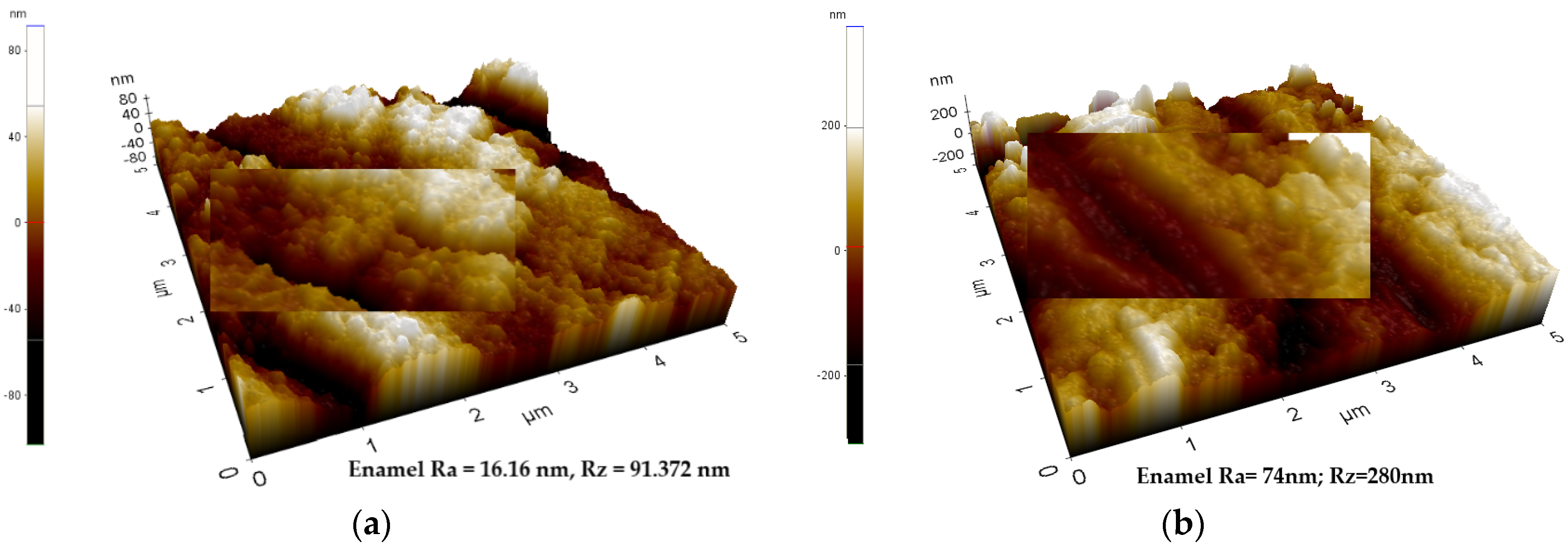
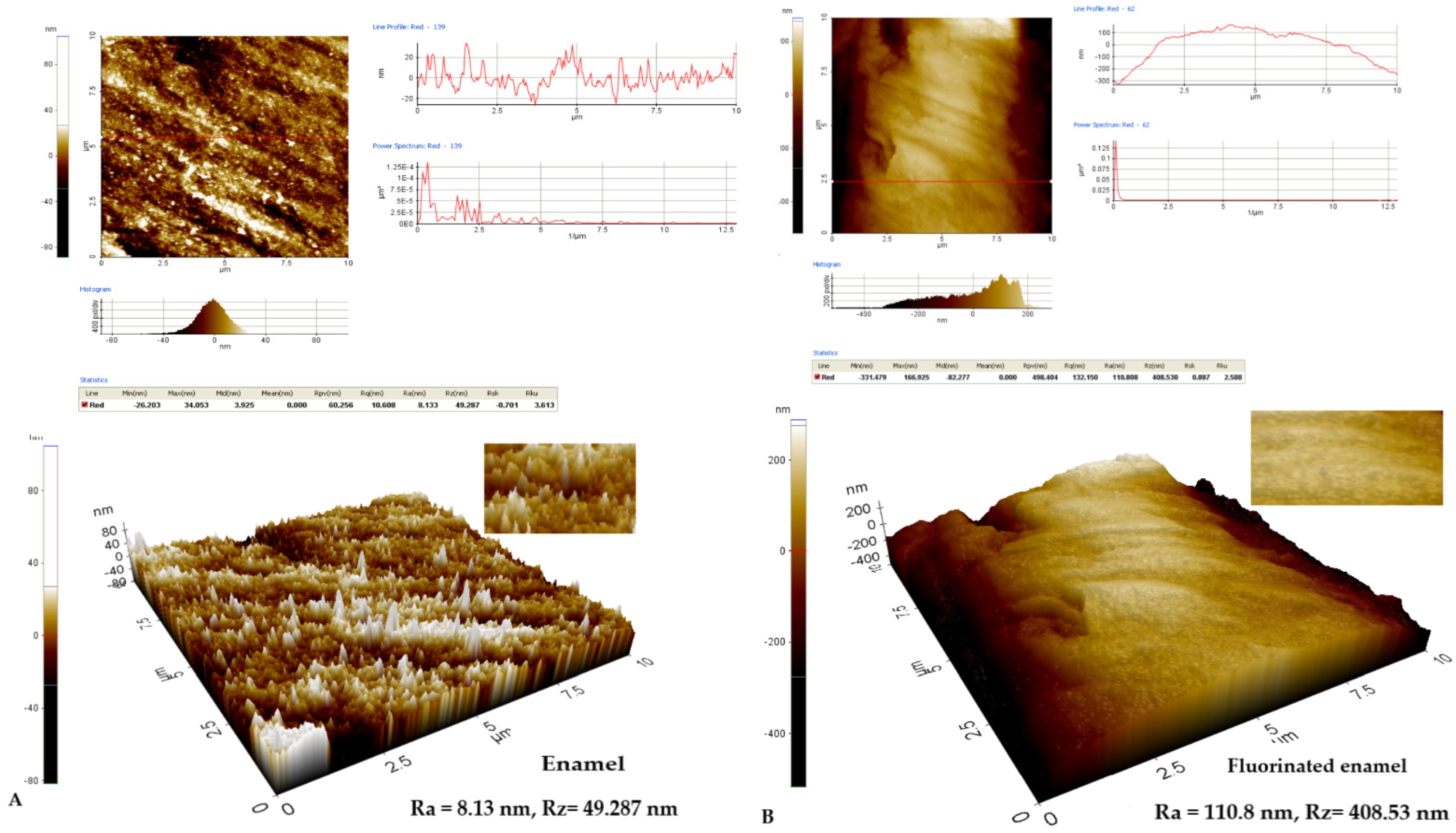
Analysis of the Structure of the Enamel by AFM

The analysis of the enamel structure highlights differences in its topographic characteristics. Therefore, it is possible to follow different aspects of the enamel structure depending on the scanning area and on the scanned micro zone Figure 1, Figure 2 and Figure 3. Differences can be observed between the roughness values at the enamel level as follows: on a scanning area X Scan Size 10 μm, Y Scan Size 10 μm (Figure 1); X Scan Size 5 μm, Y Scan Size 5 μm (Figure 2); and X Scan Size 1 micrometer, Y Scan Size 1 micrometer (Figure 3).
Surface roughness (Ra) measurements ranged from Ra = 0.039 μm (±0.048), Rz = 0.213 μm (±0.232), to Ra = 0.049 μm (±0.031), Rz = 0.341 μm (±0.274), respectively (p < 0.05), with an increase in the surface roughness passing from the intact enamel to the enamel exposed to fluoride varnish, which suggests a remineralizing effect for the varnish. Comparing Ra and Rz values of the nonfluorinated enamel and fluorinated enamel, significant differences (p < 0.05) were found, suggesting that the varnish had a protective effect on the enamel surface (Table 1).
The differences in values between non-fluorinated and fluorinated enamel surfaces are statistically significant (p < 0.05) with an increase in surface roughness from intact enamel to enamel exposed to fluoride varnish Table 2.
The roughness of the enamel structure varies in the direction of its increase in the fluoridated region at Ra = 110.8 nm and Rz = 408.53 nm from Ra = 8.13 nm and Rz = 49.287 nm before fluoridation. It can be seen that line red profile is more uniform after the application of the fluoride varnish (Figure 4B) than before its application (Figure 4A). Additionally, the fluorinated enamel surface has a less fractured appearance (Figure 4B) compared to the structural image of the initially analyzed enamel (Figure 4A). This suggests the effect on the structural change of the enamel after the application of fluorine varnish.
The same aspect can be visualized at a smaller scanning area, respectively, of 5/5 μm, where a roughness of Ra = 7.99 nm and Rz = 44.30 nm can be observed. The enamel has a fractured contour with multiple irregularities, an aspect highlighted in Figure 5A. After applying the fluorine varnish, an increase in the average roughness Ra = 49.60 nm and Rz = 158.368 nm can be observed, but an attenuation of the red profile line is seen in Figure 5B. The attenuation of the structural aspect can also be observed (Figure 5B).
Roughness analysis at a smaller scan area of 1/1 μm shows a decrease in the fluorinated surface roughness from initial values of Ra = 4.5 nm and Rz = 19.256 nm to fluorinated surface values of Ra = 3.46 nm and Rz = 14.71 nm, respectively (Figure 6A,B). This suggests a minimal modification of the line red profile without attenuating it, as found in the larger scanning areas of 5/5 and 10/10 μm. Additionally, the three-dimensional images show an attenuation of the structural morphology of the enamel after fluoridation (Figure 6B) compared to the initial structure of the enamel (Figure 6A).

4. Discussion

The enamel is the only hard dental tissue exposed intraorally. A decrease in the pH of the oral cavity below the critical value of 5.5 is followed by the beginning of hydroxyapatite dissolution. In the presence of fluoride, this produces the formation of fluorapatite crystals with lower solubility than that of hydroxyapatite, which reduces the process of enamel demineralization [21]. In this way, the ions of calcium and the phosphorus of the enamel that are lost during the demineralization process are recovered by remineralization as fluorapatite [26,27].
The presence of fluoride deposited as calcium fluoride on the dental surface in-creases the availability of this ion in the fluid of the biofilm. When the pH drops to a critical level, the demineralization process begins and allows the fluoride to precipitate and, thus, to restore the tooth structure [28].
In order to prevent the demineralization of the enamel and to increase its resistance against erosive attacks, topical applications of highly concentrated fluorides have been proposed in various forms of presentation: oral rinses, gels, or varnishes [24].
Compared to other fluoride vehicles, varnishes are easy to apply, safe, and well tolerated by young children and uncooperative patients. In addition, they ensure the formation of a mechanical barrier. Due to their common clinical use, the high amount of fluoride contained, and the slow release of components, varnishes can be considered the ideal products to prevent the loss of minerals from hard dental tissues [29].
In the present study, the varnish was applied only once and then completely re-moved after 6 h, to simulate the real clinical conditions, in which the varnish is subject to the risk of being removed by toothbrushing and chewing. In this way, the emphasis was more on the chemical effect and less on the mechanical protection [30].
The results of the present study showed that the local application of fluoride in the form of a NaF-containing varnish was effective in reducing the roughness of the enamel structure. Therefore, the use of this type of varnish can be a good alternative to ensure protection against changing enamel roughness and loss of hard dental structures [31].
The effectiveness of sodium fluoride in protecting the enamel against erosive attack has been demonstrated by previous studies [24,31].
This can be explained by the fact that the application of fluoride varnishes is followed by the formation of a protective layer of CaF2 on the surface of hard dental structures. It acts as a physical barrier, inhibiting the contact of the acid with the enamel. At the same time, this layer is also involved in the demineralization and remineralization processes: once the formation of fluoride reservoirs takes place, it participates in the inhibition of the demineralization process and in the precipitation of fluor(hydroxy)apatite [32].
Xylitol is a non-acidogenic sweetener that can form complexes with calcium ions. Due to this property, it is believed that xylitol may increase remineralization and may inhibit the dissolution of calcium and/or phosphate ions from the enamel structure. However, the role of xylitol as a preventive or remineralizing agent has not yet been sufficiently demonstrated [32,33].
The product used in the present study is a varnish containing xylitol/NaF, which produced a reduction in the roughness of the enamel and the loss of hard dental tissue after its abrasion. The values obtained in terms of surface roughness indicate that the varnishes containing this combination of xylitol and NaF are able to achieve surface protection [32].
The ability of conventional fluoride products, such as NaF varnish, to prevent erosive/abrasive processes has been described in the literature [34,35]. Regarding the presence of xylitol, it probably did not increase the protection capacity of the product against mineral losses. The results of a study conducted by Alexandria [32] indicated that fluoride- and xylitol-containing varnish reduced the enamel demineralization but not to a greater extent than xylitol-free fluoride-containing varnish.
In the present study, AFM was used as the method of analysis. AFM is a nanoindentation technique capable of obtaining images with atomic resolution after a minimum sample preparation, which is one of the main advantages of AFM compared to other analysis techniques [36]. AFM can also be used on conductive and insulating surfaces and can be performed in ambient conditions but also in air, liquids, or vacuum. Therefore, there is no risk of damage to fragile samples by harsh preparation techniques, such as coating, dehydration, or vacuum exposure, thus avoiding the artifacts associated with such techniques. In addition, AFM is a very accurate technique, which allows quantitative data to be obtained [24].
The results of the present study showed a modification in the roughness of the enamel structure after fluoridation with varnish, which means that the null hypothesis was rejected, and the test hypothesis was accepted.
These results are consistent with the results of other studies [37,38,39,40,41,42] which have shown that varnishes containing sodium fluoride are effective in modifying the roughness of the enamel surface. Other studies that have used the AFM analysis technique [42,43,44] have found that the hardness of dental hard tissues decreases after the use of fluoride varnishes. Additionally, fluoride varnish is effective in reducing enamel demineralization [45,46].
The limitations of the present study are related, in particular, to the in vitro protocol, by the inability to adequately simulate the complex biological processes that take place at the level of the teeth in the oral cavity. Another limitation is the low number of samples analyzed. Performing more tests on a larger number of samples will be able to provide more conclusive data on the analyzed aspects.

5. Conclusions

Xylitol–fluoride varnish, even in one single short-time application, modifies the surface roughness of exposed enamel structure. The effect of the action of fluoride applied in the form of varnish influences the superficial structure of the enamel, a fact highlighted illustratively, this being an expression of the mineralization of the structure. Even if the average surface roughness was higher after fluoridation with varnish, a fact highlighted by scanning 10/10 or 5/5 μm, where it was observed that when scanning areas smaller than 1/1 micrometer, the average surface roughness decreases. All the samples analyzed and regardless of the scanned area, the improvement of the roughness profile of the red profile was highlighted, which suggests an attenuation of the micro-roughness of the enamel.

Author Contributions

Conceptualization, C.I.S.; Data curation, C.I.S., O.D. and A.E.S.; Formal analysis, C.I.S. and A.E.S.; Investigation, O.D.; Methodology, C.I.S. and O.D.; Resources, O.D.; Software, O.D.; Visualization, D.A., L.I.B., A.A., S.M.S. and O.T.; Writing—original draft, C.I.S., D.A., A.E.S. and O.T.; Writing—review and editing, C.I.S., L.I.B., A.A. and S.M.S. All authors have read and agreed to the published version of the manuscript.

Funding

This research received no external funding.

Institutional Review Board Statement

Ethical acceptance for this study was given in No. 166/21.03.2022. The study was conducted in accordance with the Declaration of Helsinki and approved by the Institutional Review Board (or Ethics Committee) of the University of Medicine and Pharmacy Grigore T. Popa Iasi (protocol code 166 and 21.03.2022) for studies involving humans. The legislation in force regarding the ownership, use and protection of personal data—GDPR (Regulation 679/2016) has been complied with. From an ethical point of view, this study was carried out in accordance with Research Law no. 206 of 27 May 2004 on good conduct in scientific research, technological development and innovation, the Integrity Guide in Scientific Research (published by the National Council for Ethics in Scientific Research, Technological Development and Innovation on 12 November 2020) and current European legislation.

Informed Consent Statement

Informed consent was obtained from all subjects involved in the study.

Data Availability Statement

The data that support the findings of this study are available on request from the corresponding author.

Conflicts of Interest

The authors declare no conflict of interest.

References

  1. Oshiro, M.; Yamaguchi, K.; Takamizawa, T.; Inage, H.; Watanabe, T.; Irokawa, A.; Ando, S.; Miyazaki, M. Effect of CPP-ACP paste on tooth mineralization: An FE-SEM study. J. Oral Sci. 2007, 49, 115–120. [Google Scholar] [CrossRef] [PubMed]
  2. Skinner, J.; Dimitropoulos, Y.; Sohn, W.; Holden, A.; Rambaldini, B.; Spallek, H.; Ummer-Christian, R.; Marshall, S.; Raymond, K.; Ao, T.C.; et al. Child Fluoride Varnish Programs Implementation: A Consensus Workshop and Actions to Increase Scale-Up in Australia. Healthcare 2021, 9, 1029. [Google Scholar] [CrossRef] [PubMed]
  3. Skinner, J.; Dimitropoulos, Y.; Rambaldini, B.; Calma, T.; Raymond, K.; Ummer-Christian, R.; Orr, N.; Gwynne, K. Costing the Scale-Up of a National Primary School-Based Fluoride Varnish Program for Aboriginal Children Using Dental Assistants in Australia. Int. J. Environ. Res. Public Health 2020, 17, 8774. [Google Scholar] [CrossRef] [PubMed]
  4. Geissler, K.H.; Dick, A.W.; Goff, S.L.; Whaley, C.; Kranz, A.M. Dental Fluoride Varnish Application during Medical Visits Among Children Who Are Privately Insured. JAMA Netw. Open 2021, 4, e2122953. [Google Scholar] [CrossRef]
  5. Poornima, P.; Krithikadatta, J.; Ponraj, R.R.; Velmurugan, N.; Kishen, A. Biofilm formation following chitosan-based varnish or chlorhexidine-fluoride varnish application in patients undergoing fixed orthodontic treatment: A double blinded randomized controlled trial. BMC Oral Health 2021, 21, 465. [Google Scholar] [CrossRef]
  6. Chokshi, K. An in vitro Comparative Evaluation of Three Remineralizing Agents using Confocal Microscopy. J. Clin. Diagn. Res. 2016, 10, ZC39–ZC42. [Google Scholar] [CrossRef]
  7. Jeevarathan, J.; Deepti, A.; Muthu, M.S.; Ratha Prabhu, V.; Chamundeeswari, G.S. Effect of fluoride varnish on Streptococcus mutans counts in plaque of caries-free children using Dentocult SM strip mutans test: A randomized controlled triple blind study. J. Indian Soc. Pedod. Prev. Dent. 2007, 25, 157–163. [Google Scholar] [CrossRef]
  8. Baik, A.; Alamoudi, N.; El-Housseiny, A.; Altuwirqi, A. Fluoride Varnishes for Preventing Occlusal Dental Caries: A Review. Dent. J. 2021, 9, 64. [Google Scholar] [CrossRef]
  9. Fluoride Varnish Guide. Available online: webcache.googleusercontent.com (accessed on 7 December 2020).
  10. Jabir, E.; McGrade, C.; Quinn, G.; McGarry, J.; Nic Iomhair, A.; Kelly, N.; Srinivasan, M.; Watson, S.; McKenna, G.J. Evaluating the effectiveness of fluoride varnish in preventing caries amongst Long-Term Care Facility Residents. Gerodontology, 2021; advance online publication. [Google Scholar] [CrossRef]
  11. Poza-Pascual, A.; Serna-Muñoz, C.; Pérez-Silva, A.; Martínez-Beneyto, Y.; Cabello, I.; Ortiz-Ruiz, A.J. Effects of Fluoride and Calcium Phosphate-Based Varnishes in Children at High Risk of Tooth Decay: A Randomized Clinical Trial. Int. J. Environ. Res. Public Health 2021, 18, 10049. [Google Scholar] [CrossRef]
  12. Lin, P.Y.; Wang, J.; Chuang, T.Y.; Chang, Y.M.; Chang, H.J.; Chi, L.Y. Association between population-based fluoride varnish application services and dental caries experience among schoolchildren in Taiwan. J. Formos. Med. Assoc. Taiwanizhi 2022, 121, 986–994. [Google Scholar] [CrossRef]
  13. Jullien, S. Prophylaxis of caries with fluoride for children under five years. BMC Pediatr. 2021, 21 (Suppl. S1), 351. [Google Scholar] [CrossRef]
  14. Schiffner, U. Verwendung von Fluoriden zur Kariesprävention [Use of fluorides for caries prevention]. Bundesgesundheitsblatt Gesundh. Gesundh. 2021, 64, 830–837. [Google Scholar] [CrossRef]
  15. Sathiyakumar, T.; Vasireddy, D.; Mondal, S. Impact of Sociodemographic Factors on Dental Caries in Children and Availing Fluoride Treatment: A Study Based on National Survey of Children’s Health (NSCH) Data 2016–2019. Cureus 2021, 13, e18395. [Google Scholar] [CrossRef]
  16. Alencar, C.M.; Ribeiro, M.; Zaniboni, J.F.; Leandrin, T.P.; Silva, A.M.; Campos, E.A. Anti-erosive profile of an experimental 5% SnCl2 varnish containing different concentrations of NaF. Braz. Dent. J. 2022, 33, 68–76. [Google Scholar] [CrossRef]
  17. Weintraub, J.; Ramos-Gomez, F.; Jue, B.; Shain, S.; Hoover, C.; Featherstone, J.; Gansky, S. Fluoride Varnish Efficacy in Preventing Early Childhood Caries. J. Dent. Res. 2006, 85, 172–176. Available online: https://www.ncbi.nlm.nih.gov/pmc/articles/PMC2257982 (accessed on 17 May 2022). [CrossRef]
  18. Petersson, L.G. The role of fluoride in the preventive management of dentin hypersensitivity and root caries. Clin. Oral Investig. 2013, 17 (Suppl. S1), S63–S71. [Google Scholar] [CrossRef]
  19. Tan, H.P.; Lo, E.C.; Dyson, J.E.; Luo, Y.; Corbet, E.F. A randomized trial on root caries prevention in elders. J. Dent. Res. 2010, 89, 1086–1090. [Google Scholar] [CrossRef]
  20. Siqueira, V.L.; Barreto, G.S.; Silva, E.; Silva, T.; Nascimento, D.; Veronezi, A.; Rodrigues, M.C.; Buzalaf, M.; Cardoso, C. Effect of xylitol varnishes on enamel remineralization of immature teeth: In vitro and in situ studies. Braz. Oral Res. 2021, 35, e137. [Google Scholar] [CrossRef]
  21. Philip, N. State of the Art Enamel Remineralization Systems: The Next Frontier in Caries Management. Caries Res. 2019, 53, 284–295. [Google Scholar] [CrossRef]
  22. Lussi, A.; Hellwig, E.; Zero, D.; Jaeggi, T. Erosive tooth wear: Diagnosis, risk factors and prevention. Am. J. Dent. 2006, 19, 319–325. [Google Scholar]
  23. Comar, L.P.; Cardoso, C.; Charone, S.; Grizzo, L.T.; Buzalaf, M.A.; Magalhães, A.C. TiF4 and NaF varnishes as anti-erosive agents on enamel and dentin erosion progression in vitro. J. Appl. Oral Sci. 2015, 23, 14–18. [Google Scholar] [CrossRef]
  24. Bayrak, S.; Tuloglu, N.; Bicer, H.; Sen Tunc, E. Effect of Fluoride Varnish Containing CPP-ACP on Preventing Enamel Erosion. Scanning 2017, 2017, 1897825. [Google Scholar] [CrossRef]
  25. Shellis, R.P.; Ganss, C.; Ren, Y.; Zero, D.T.; Lussi, A. Methodology and models in erosion research: Discussion and conclusions. Caries Res. 2011, 45 (Suppl. S1), 69–77. [Google Scholar] [CrossRef]
  26. Yu, O.Y.; Mei, M.L.; Zhao, I.S.; Lo, E.C.M.; Chu, C.H. Effects of fluoride on two chemical models of enamel demineralization. Materials 2017, 10, 1245. [Google Scholar] [CrossRef]
  27. Grohe, B.; Mittler, S. Advanced non-fluoride approaches to dental enamel remineralization: The next level in enamel repair management. Biomater. Biosyst. 2021, 4, 100029. [Google Scholar] [CrossRef]
  28. Cury, J.A.; Tenuta, L.M.A. How to Maintain a Cariostatic Fluoride Concentration in the Oral Environment. Adv. Dent. Res. 2008, 20, 13–16. [Google Scholar] [CrossRef]
  29. Alexandria, A.K.; Valença, A.M.G.; Cabral, L.M.; Maia, L.C. Fluoride Varnishes against Dental Erosion Caused by Soft Drink Combined with Pediatric Liquid Medicine. Braz. Dent. J. 2017, 28, 482–488. [Google Scholar] [CrossRef]
  30. Alexandria, A.K.; Vieira, T.I.; Pithon, M.M.; Fidalgo, T.K.S.; Fonseca-Gonçalves, A.; Valença, A.M.G.; Cabral, L.M.; Maia, L.C. In vitro enamel erosion and abrasion-inhibiting effect of different fluoride varnishes. Arch. Oral Biol. 2017, 77, 39–43. [Google Scholar] [CrossRef]
  31. Walczak, M.; Turska-Szybka, A. The efficacy of fluoride varnishes containing different calcium phosphate compounds. Fluoride 2017, 50 Pt 2, 151–160. Available online: https://www.proquest.com/scholarly-journals/efficacy-fluoride-varnishes-containing-different/docview/1942213151/se-2 (accessed on 30 May 2022).
  32. Alexandria, A.; Valença, A.M.G.; Cabral, L.M.; Maia, L.C. Comparative Effects of CPP-ACP and Xylitol F-Varnishes on the Reduction of Tooth Erosion and Its Progression. Braz. Dent. J. 2020, 31, 664–672. [Google Scholar] [CrossRef]
  33. Tuncer, D.; Onen, A.; Yazici, A.R. Effect of chewing gums with xylitol, sorbitol and xylitol-sorbitol on the remineralization and hardness of initial enamel lesions in situ. Dent. Res. J. 2014, 11, 537–543. [Google Scholar]
  34. Bijle, M.N.; Ekambaram, M.; Lo, E.C.M.; You, C.K.Y. The enamel remineralization potential of fluoride varnishes containing arginine. J. Dent. 2020, 99, 103411. [Google Scholar] [CrossRef] [PubMed]
  35. Mantilla, T.F.; Turssi, C.P.; Ramos-Oliveira, T.M.; da Silva, C.V.; Suzuki, L.C.; de Freitas, P.M. The In Situ Effect of Titanium Tetrafluoride Gel on Erosion/Abrasion Progression in Human Dentin. Braz. Dent. J. 2017, 28, 337–345. [Google Scholar] [CrossRef] [PubMed]
  36. Barbour, M.E.; Rees, J.S. The laboratory assessment of enamel erosion: A review. J. Dent. 2004, 32, 591–602. [Google Scholar] [CrossRef] [PubMed]
  37. Soares, L.E.S.; De Carvalho Filho, A.C. Protective effect of fluoride varnish and fluoride gel on enamel erosion: Roughness, SEM-EDS, and µ-EDXRF studies. Microsc. Res. Tech. 2015, 78, 240–248. [Google Scholar] [CrossRef]
  38. Epple, M.; Enax, J.; Meyer, F. Prevention of Caries and Dental Erosion by Fluorides—A Critical Discussion Based on Physio-Chemical Data and Principles. Dent. J. 2022, 10, 6. [Google Scholar] [CrossRef]
  39. Gokkaya, B.; Ozbek, N.; Guler, Z.; Akman, S.; Sarac, A.S.; Kargul, B. Effect of a Single Application of CPP-ACPF Varnish on the Prevention of Erosive Tooth Wear: An AAS, AFM and SMH Study. Oral Health Prev. Dent. 2020, 18, 311–318. [Google Scholar] [CrossRef]
  40. Metwally, N.I.; Niazy, M.A.; El-Malt, M.A. Remineralization of Early Carious Lesions using Biomimetic Self-assembling Peptides Versus Fluoride agent (In vitro and In vivo study). Al-Azhar Dent. J. Girls 2017, 4, 179–188. [Google Scholar] [CrossRef]
  41. Świetlicka, I.; Kuc, D.; Świetlicki, M.; Arczewska, M.; Muszynski, S.; Tomaszewska, E.; Prószynski, A.; Gołacki, K.; Błaszczak, J.; Cieślak, K.; et al. Near-Surface Studies of the Changes to the Structure and Mechanical Properties of Human Enamel under the Action of Fluoride Varnish Containing CPP-ACP Compound. Biomolecules 2020, 10, 765. [Google Scholar] [CrossRef]
  42. Khoubrouypak, Z.; Abbasi, M.; Ahmadi, E.; Rafeie, N.; Behroozibakhsh, M. Effect of Cold Atmospheric Pressure Plasma Coupled with Resin-Containing and Xylitol-Containing Fluoride Varnishes on Enamel Erosion. Int. J. Dent. 2021, 2021, 3298515. [Google Scholar] [CrossRef]
  43. Lippert, F.; Parker, D.M.; Jandt, K.D. In vitro demineralization/remineralization cycles at human tooth enamel surfaces investigated by AFM and nanoindentation. J. Colloid Interface Sci. 2004, 280, 442–448. [Google Scholar] [CrossRef]
  44. Medeiros, I.C.; Brasil, V.L.; Carlo, H.L.; Santos, R.L.; De Lima, B.A.; De Carvalho, F.G. In vitro effect of calcium nano phosphate and high-concentrated fluoride agents on enamel erosion: An AFM study. Int. J. Pediatr. Dent. 2014, 24, 168–174. [Google Scholar] [CrossRef]
  45. Abufarwa, M.; Noureldin, A.; Dziak, R.; Covell, D. Efficacy of CPP-ACP fluoride varnish applied with and without acid etching in preventing enamel demineralization compared to light-curable fluoride varnish. Angle Orthod. 2022, 92, 213–219. [Google Scholar] [CrossRef]
  46. Dhillon, S.N.; Deshpande, A.N.; Macwan, C.; Patel, K.S.; Shah, Y.S.; Jain, A.A. Comparative Evaluation of Microhardness and Enamel Solubility of Treated Surface Enamel with Resin Infiltrant, Fluoride Varnish, and Casein Phosphopeptide-amorphous Calcium Phosphate: An In Vitro Study. Int. J. Clin. Pediatr. Dent. 2020, 13 (Suppl. S1), S14–S25. [Google Scholar] [CrossRef]
Figure 1. AFM image—Three-dimensional appearance of the enamel structure. The variability of the roughness profile can be observed at the enamel level. (a) X Scan Size 10 μm, Y Scan Size 10 μm, Ra = 31.48 nm, Rz = 134.135 nm; (b) X Scan Size 10 μm, Y Scan Size 10 μm, Ra = 150.52 nm, Rz = 279.315 nm.
Biomedicines 10 01900 g001
Figure 2. AFM image—Three-dimensional appearance of the enamel structure. The variability of the roughness profile can be observed at the level of enamel. (a) X Scan Size 5 μm, Y Scan Size 5 μm, Ra = 16.16 nm, Rz = 91.372 nm; (b) X Scan Size 5 μm, Y Scan Size 5 μm, Ra = 74 nm, Rz = 280 nm.
Biomedicines 10 01900 g002
Figure 3. AFM image—Three-dimensional appearance of the enamel structure. The variability of the roughness profile can be observed at the enamel level. (a) X Scan Size 1 micrometer, Y Scan Size 1 micrometer, Ra = 7.19 nm, Rz = 20.22 nm; (b) X Scan Size 5 μm, Y Scan Size 5 μm, Ra = 10.7 nm, Rz = 37.188 nm.
Biomedicines 10 01900 g003
Figure 4. AFM images from the enamel surface layer on the X Scan layer 10 μm/Y Scan layer 10 μm: (A) Two-dimensional and three-dimensional AFM image of the enamel structure in the enamel without fluoridation Ra = 8.13 nm and Rz = 49.287 nm; (B) Two-dimensional and three-dimensional AFM image of the enamel structure in the enamel after fluoridation with varnish Ra = 110.8 nm and Rz = 408.53 nm.
Biomedicines 10 01900 g004
Figure 5. AFM images from the enamel surface layer on the X Scan layer 5 μm/Y Scan layer 5 μm: (A) Two-dimensional and three-dimensional AFM image of the enamel structure in the enamel without fluoridation Ra = 7.99 nm and Rz = 44.30 nm; (B) Two-dimensional and three-dimensional AFM image of the enamel structure in the enamel after fluoridation with varnish Ra = 49.608 nm and Rz = 158.368 nm.
Biomedicines 10 01900 g005
Figure 6. AFM images from the enamel surface layer on the X Scan layer 1 micrometer/Y Scan layer 1 micrometer: (A) Two-dimensional and three-dimensional AFM image of the enamel structure in the enamel without fluoridation Ra = 4.5 nm and Rz = 19.256 nm; (B) Two-dimensional and three-dimensional AFM image of the enamel structure in the enamel after fluoridation with varnish Ra = 3.46 nm and Rz = 14.71 nm.
Biomedicines 10 01900 g006
Table 1. Descriptive Statistics for samples scanned on an area of X Scan size 10 μm on Y Scan size 10 μm before fluoridation and after fluoridation. The values are presented in micrometers.
Descriptive Statistics
NMinimumMaximumMeanStd. Deviation
Ra before100.0080.1500.0390.048
Ra after100.0070.1110.0490.031
Rz before100.0370.8300.2130.232
Ra after100.0030.9500.3410.274
Valid N (listwise)10
Table 2. One-sample test for samples scanned on an area of X Scan size 10 μm and Y Scan size 10 μm before fluoridation and after fluoridation. The values are presented in micrometers.
One-Sample Test
Test Value = 0
tdfSig. (2-Tailed)Mean Difference95% Confidence Interval of the Difference
LowerUpper
Ra before2.5929.0000.029 *0.0390.0050.074
Ra after4.9959.0000.001 *0.0490.0270.072
Rz before2.9049.0000.017 *0.2130.0470.379
Ra after3.9469.0000.003 *0.3410.1460.537
* refers to Significance level.
Publisher’s Note: MDPI stays neutral with regard to jurisdictional claims in published maps and institutional affiliations.

