
Cellular Mechanisms in Acute and Chronic Wounds after PDT Therapy: An Update


Abstract
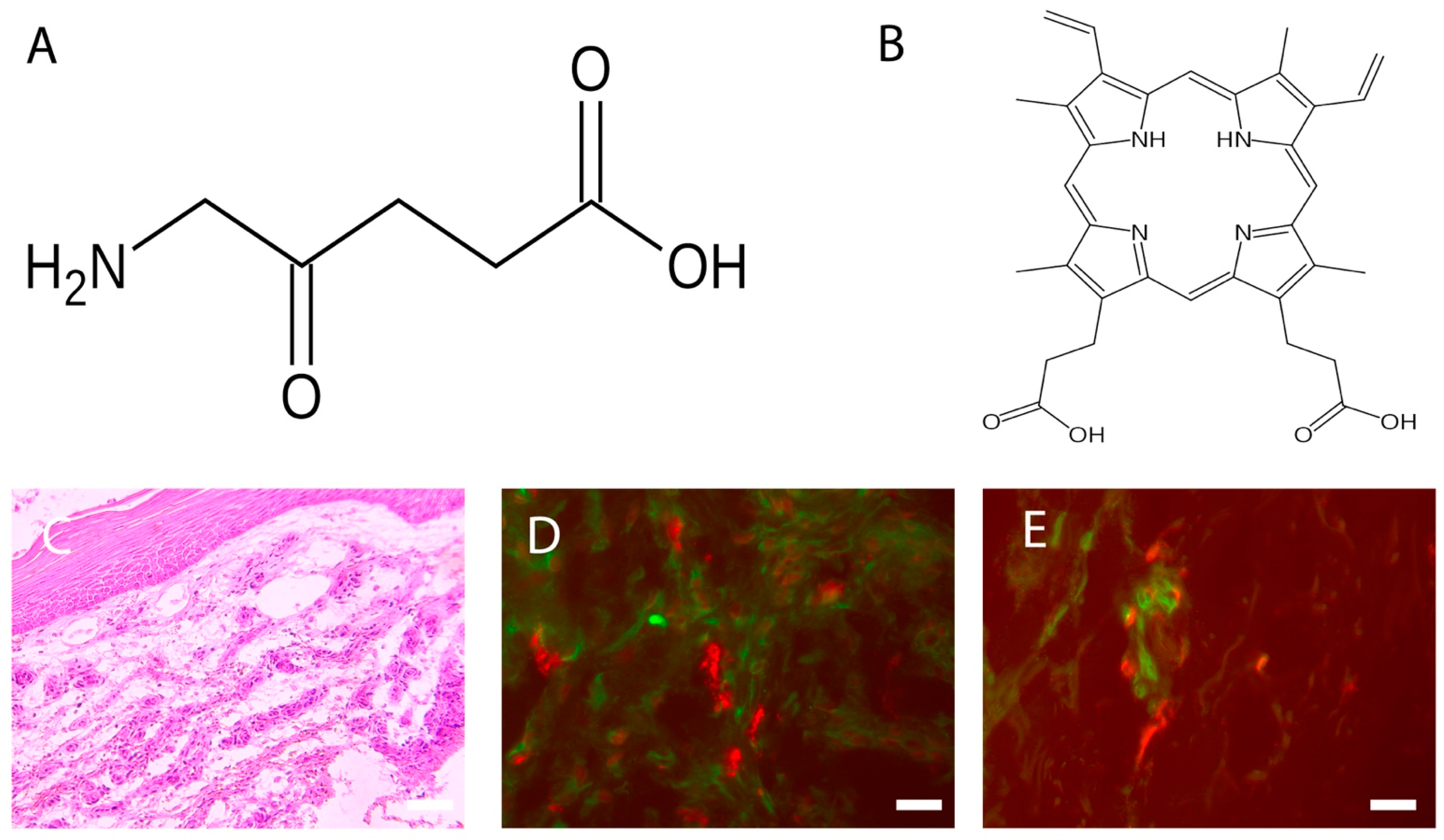
:1. The Photodynamic Therapy
2. Photosensitizers
3. Wound Healing
4. PDT and Wound Healing
5. Chronic Wounds
6. PDT and Chronic Wounds
6.1. The Response of Cellular Infiltrate
6.2. Neuroimmunomodulation
6.3. Future Perspective
7. Current Limitations
Author Contributions
Funding
Institutional Review Board Statement
Informed Consent Statement
Data Availability Statement
Acknowledgments
Conflicts of Interest
Abbreviations
| Acronym | Denomination |
| PDT | Photodynamic therapy |
| ROS | Reactive Oxygen Species |
| ALA | 5-aminolevulinic acid |
| Matrix Metalloproteinases | MMPs |
| MCs | Mast Cells |
| Interleukin | IL |
| TGF | Transforming growth factor |
| ERK/AP1 | ERK-associated changes of AP1 |
| DCs | Dendritic cells |
| TNF | Tumor necrosis factor |
| BDCA | Blood dendritic cell antigen |
| HSP | Heat shock protein |
| FGF | Fibroblast growth factor |
| UEA | Ulex Europaeus Agglutinin |
| NGF | Nerve Growth Factor |
| MAP | Mitogen-activated protein |
| TRP | Transient receptor potential channel |
| P2X | ATP-gated P2X receptor cation channel family |
| DAMP | Damp-associated molecular pattern receptors |
| ATP | Adenosine triphosphate |
| CGRP | Calcitonin Gene Related Peptide |
| NKA | Neurokinin A |
| NPY | Neuropeptide Y |
| SP | Substance P |
| PGP 9.5 | Protein Gene Product 9.5 |
| VIP | Vasoactive intestinal peptide |
| iNOs | Inducible isoform of nitric oxide synthase |
References
- Szeimies, R.M.; Drager, J.; Abels, C.; Landthaler, M. History of photodynamic therapy in dermatology. In Photodynamic Therapy and Fluorescence Diagnosis in Therapy; Calzavara-Pinton, P., Rolf-Markus, S., Ortel, B., Eds.; Elsevier Science: Amsterdam, The Netherlands, 2001; Volume 2, pp. 3–15. [Google Scholar]
- Tampa, M.; Sarbu, M.; Matei, C.; Mitran, C.; Mitran, M.; Caruntu, C.; Georgescu, S. Photodynamic therapy: A hot topic in dermato-oncology. Oncol. Lett. 2019, 17, 4085–4093. [Google Scholar] [CrossRef] [Green Version]
- Niculescu, A.G.; Grumezescu, A.M. Photodynamic Therapy—An up-to-date review. Appl. Sci. 2021, 11, 3626. [Google Scholar] [CrossRef]
- Kwiatkowski, S.; Knap, B.; Przystupski, D.; Saczko, J.; Kedzierska, E.; Knap-Czop, K.; Kotlinska, J.; Michel, O.; Kotowski, K.; Kulbacka, J. Photodynamic therapy—mechanisms, photosensitizers and combinations. Biomed. Pharm. 2018, 106, 1098–1107. [Google Scholar] [CrossRef]
- Grandi, V.; Sessa, M.; Pisano, L.; Rossi, R.; Galvan, A.; Gattai, R.; Mori, M.; Tiradritti, L.; Bacci, S.; Zuccati, G.; et al. Photodynamic therapy with topical photosensitizers in mucosal and semimucosal areas: Review from a dermatologic perspective. Photodiagnosis Photodyn. Ther. 2018, 23, 119–131. [Google Scholar] [CrossRef]
- National Cancer Institute. Available online: https://www.cancer.gov/publications/dictionaries/cancer-terms/def/reactive-oxygen-species (accessed on 28 June 2022).
- Donnelly, R.F.; McCarron, P.A.; Woolfson, A.D. Derivatives of 5-aminolevulinic acid for photodynamic therapy. Perspect. Med. Chem. 2007, 1, 49–63. [Google Scholar] [CrossRef]
- Wang, B.C.; Fu, C.; Qin, L.; Zeng, X.Y.; Liu, Q. Photodynamic therapy with methyl-5-aminolevulinate for basal cell carcinoma: A systematic review and meta-analysis. Photodiagnosis Photodyn. Ther. 2020, 29, 101667–101679. [Google Scholar] [CrossRef]
- Tedesco, A.; Jesus, P. Low level energy photodynamic therapy for skin processes and regeneration. In Photomedicine. Advances in Clinical Practice; Yohey, T., Ed.; Intech Open: London, UK, 2017. [Google Scholar]
- Lecci, P.P.; Corsi, A.; Cappugi, P.P.; Bacci, S. La terapia fotodinamica nel trattamento delle lesioni cutanee croniche. In Evidenze Cliniche e Pratica Sperimentale; Aracne Editrice: Rome, Italy, 2013; pp. 1–64. [Google Scholar]
- Goodpaster, T.; Legesse-Miller, A.; Hameed, M.R.; Aisner, S.C.; Randolph-Habecker, J.; Coller, H.A. An immunohistochemical method for identifying fibroblasts in formalin-fixed, paraffin-embedded tissue. J. Histochem. Cytochem. 2008, 56, 347–358. [Google Scholar] [CrossRef] [Green Version]
- Bergstresser, P.R.; Tigelaar, R.E.; Tharp, M.D. Conjugated avidin identifies cutaneous rodent and human mast cells. J. Investig. Derm. 1984, 83, 214–218. [Google Scholar] [CrossRef] [Green Version]
- ten Broeke, T.; Wubbolts, R.; Stoorvogel, W. MHC class II antigen presentation by dendritic cells regulated through endosomal sorting. Cold Spring Harb. Perspect. Biol. 2013, 5, a016873. [Google Scholar] [CrossRef] [Green Version]
- Bacci, S.; Bani, D. The epidermis in microgravity and unloading conditions and their effects on wound healing. Front. Bioeng. Biotechnol. 2022, 10, 666434. [Google Scholar] [CrossRef]
- Martin, P.; Nunan, R. Cellular and molecular mechanisms of repair in acute and chronic wound healing. Br. J. Derm. 2015, 173, 370–378. [Google Scholar] [CrossRef]
- Gonzalez, A.C.; Costa, T.F.; Andrade, Z.A.; Medrado, A.R. Wound healing—A literature review. An. Bras. Dermatol. 2016, 91, 614–620. [Google Scholar] [CrossRef] [PubMed] [Green Version]
- Sorg, H.; Tilkorn, D.J.; Hager, S.; Hauser, J.; Mirastschijski, U. Skin wound healing: An update on the current knowledge and concepts. Eur. Surg. Res. 2017, 58, 81–94. [Google Scholar] [CrossRef] [PubMed]
- Cañedo-Dorantes, L.; Cañedo-Ayala, M. Skin acute wound healing: A comprehensive review. Int. J. Inflam 2019, 2019, 3706315–3706329. [Google Scholar] [CrossRef] [PubMed]
- Visha, M.G.; Karunagaran, M. A review on wound healing. Int. J. Clin. Correl. 2019, 3, 50–59. [Google Scholar]
- Tottoli, E.M.; Dorati, R.; Genta, I.; Chiesa, E.; Pisani, S.; Conti, B. Skin wound healing process and new emerging technologies for skin wound care and regeneration. Pharmaceutics 2020, 12, 735. [Google Scholar] [CrossRef]
- Wilkinson, H.N.; Hardman, M.J. Wound healing: Cellular mechanisms and pathological outcomes. Open Biol. 2020, 10, 200223–200236. [Google Scholar] [CrossRef]
- Raziyeva, K.; Kim, Y.; Zharkinbekov, Z.; Kassymbek, K.; Jimi, S.; Saparov, A. Immunology of acute and chronic wound healing. Biomolecules 2021, 11, 700. [Google Scholar] [CrossRef]
- Douahiher, J.; Succar, J.; Lancerotto, L.; Gurish, M.F.; Orgill, D.P.; Hamilton, M.J.; Krilis, S.A.; Stevens, R.L. Development of mast cells and importance of their tryptase and chymase serine proteases in inflammation and wound healing. Adv. Immunol. 2014, 122, 211–252. [Google Scholar]
- Bacci, S. Fine regulation during wound healing by mast cells, a physiological role not yet clarified. Int. J. Mol. Sci. 2022, 23, 1820. [Google Scholar] [CrossRef]
- Zhang, Z.; Kurashima, Y. Two sides of the coin: Mast cells as a key regulator of allergy and acute/chronic inflammation. Cells 2021, 10, 1615. [Google Scholar] [CrossRef]
- Nesi-Reis, V.; Lera-Nonose, S.V.; Oyama, J.; Ramos-Milaré, Á.; Demarchi, I.; Alessi-Aristides, S.; Vieira-Teixeira, J.J.; Verzignassi Silveira, T.G.; Campana-Lonardoni, M.V. Contribution of photodynamic therapy in wound healing: A systematic review. Photodiagnosis Photodyn. Ther. 2018, 30, 294–305. [Google Scholar] [CrossRef]
- Oyama, J.; Ramos-Milaré, Á.; Lera-Nonose, S.V.; Nesi-Reis, V.; Demarchi, I.; Alessi-Aristides, S.; Vieira-Teixeira, J.J.; Verzignassi Silveira, T.G.; Campana-Lonardoni, M.V. Photodynamic therapy in wound healing in vivo, a systematic review. Photodiagnosis Photodyn. Ther. 2020, 10, 101682. [Google Scholar] [CrossRef]
- Reginato, E.; Wolf, P.; Hamblin, M.R. Immune response after photodynamic therapy increases anti-cancer and anti-bacterial effects. World J. Immunol. 2014, 4, 1–11. [Google Scholar] [CrossRef]
- Corsi, A.; Lecci, P.P.; Bacci, S.; Cappugi, P. Chronic wounds treated with photodynamic therapy: Analysis of cellular response and preliminary results. Acta Vulnol. 2013, 11, 23–33. [Google Scholar]
- Corsi, A.; Lecci, P.P.; Bacci, S.; Cappugi, P.; Pimpinelli, N. Early activation of fibroblasts during PDT treatment in leg ulcers. G Ital. Derm. Venereol. 2016, 151, 223–229. [Google Scholar]
- Yang, Z.; Hu, X.; Zhou, L.; He, Y.; Zhang, X.; Yang, J.; Ju, Z.; Liou, Y.C.; Shen, H.M.; Luo, G.; et al. Photodynamic therapy accelerates skin wound healing through promoting re-epithelialization. Burn. Trauma 2021, 9, tkab008. [Google Scholar] [CrossRef]
- Grandi, V.; Bacci, S.; Corsi, A.; Sessa, M.; Puliti, E.; Murciano, N.; Scavone, F.; Cappugi, P.; Pimpinelli, N. ALA-PDT exerts beneficial effects on chronic venous ulcers by inducing changes in inflammatory microenvironment, especially through increased TGF-beta release: A pilot clinical and translational study. Photodiagnosis Photodyn. Ther. 2018, 21, 252–256. [Google Scholar] [CrossRef]
- Harding, K.G.; Morris, H.L.; Patel, G.K. Healing chronic wounds. Br. Med. J. 2002, 324, 160–163. [Google Scholar] [CrossRef]
- Toporcer, T.; Lakyová, L.; Radonak, J. Venous ulcer-present view on aetiology, diagnostics and therapy. Cas. Lek. Ceskych 2008, 147, 199–205. [Google Scholar]
- Han, G.; Ceilley, R. Chronic wound healing: A review of current management and treatments. Adv. Ther. 2017, 34, 599–610. [Google Scholar] [CrossRef] [PubMed] [Green Version]
- Sen, C.K. Human wounds and its burden: An updated compendium of estimates. Adv. Wound Care 2019, 8, 39–48. [Google Scholar] [CrossRef] [PubMed] [Green Version]
- Kyaw, B.M.; Järbrink, K.; Martinengo, L.; Car, J.; Harding, K.; Schmidtchen, A. Need for improved definition of chronic wounds in clinical studies. Acta Derm. Venereol. 2018, 12, 157–158. [Google Scholar] [CrossRef] [PubMed] [Green Version]
- Zhao, R.; Liang, H.; Clarke, E.; Jackson, C.; Xue, M. Inflammation in chronic wounds. Int. J. Mol. Sci. 2016, 17, 2085. [Google Scholar] [CrossRef]
- Komi, D.E.A.; Khomtchouk, K.; Santa Maria, P.L. A review of the contribution of mast cells in wound healing: Involved molecular and cellular mechanisms. Clin. Rev. Allergy Immunol. 2020, 58, 298–312. [Google Scholar] [CrossRef]
- Yang, T.; Tan, Y.; Zhang, W.; Yang, W.; Luo, J.; Chen, L.; Liu, H.; Yang, G.; Lei, X. Effects of ALA-PDT on the healing of mouse skin wounds infected with Pseudomonas aeruginosa and its related mechanisms. Front. Cell Dev. Biol. 2020, 8, 585132. [Google Scholar] [CrossRef]
- Haensel, D.; Dai, X. Epithelial-to-mesenchymal transition in cutaneous wound healing: Where we are and where we are heading. Dev. Dyn 2018, 247, 473–480. [Google Scholar] [CrossRef] [Green Version]
- Kushwah, R.; Hu, J. Role of dendritic cells in the induction of regulatory T cells. Cell Biosci. 2011, 1, 20. [Google Scholar] [CrossRef] [Green Version]
- Murciano, N.; University of Florence, Florence, Italy. Personal communication, 2016.
- Frangogiannis, N. Transforming growth factor-β in tissue fibrosis. J. Exp. Med. 2020, 217, e20190103. [Google Scholar] [CrossRef]
- Krystel-Whittemore, M.; Dileepan, K.N.; Wood, J.G. Mast cell: A multi-functional master cell. Front. Immunol. 2016, 6, 620. [Google Scholar] [CrossRef] [Green Version]
- Khorsandi, K.; Fekrazad, R.; Hamblin, M.R. Low-dose photodynamic therapy effect on closure of scratch wounds of normal and diabetic fibroblast cells: An in vitro study. J. Biophotonics 2021, 14, e202100005. [Google Scholar] [CrossRef] [PubMed]
- Bacci, S.; Pimpinelli, N.; Romagnoli, P. Contacts between mast cells and dendritic cells in the human skin. Ital. J. Anat. Embryol. 2010, 11, 25–30. [Google Scholar]
- Gri, G.; Frossi, B.; D’Inca, F.; Danelli, L.; Betto, E.; Mion, F.; Sibilano, R.; Pucillo, C. Mast cell: An emerging partner in immune interaction. Front. Immunol. 2012, 25, 120. [Google Scholar] [CrossRef] [PubMed] [Green Version]
- Bacci, S.; Defraia, B.; Cinci, L.; Calosi, L.; Guasti, D.; Pieri, L.; Lotti, V.; Bonelli, A.; Romagnoli, P. Immunohistochemical analysis of dendritic cells in skin lesions: Correlations with survival time. Forensic Sci. Int. 2014, 244, 179–185. [Google Scholar] [CrossRef]
- Brazil, J.C.; Quiros, M.; Nusrat, A.; Parkos, C.A. Innate immune cell-epithelial crosstalk during wound repair. J. Clin. Investig. 2019, 129, 2983–2993. [Google Scholar] [CrossRef] [Green Version]
- Yamazaki, T.; Mukouyama, Y.S. Tissue specific origin, development, and pathological perspectives of pericytes. Front. Cardiovasc. Med. 2018, 27, 78. [Google Scholar] [CrossRef] [Green Version]
- Gaber, M.A.; Seliet, I.A.; Ehsan, N.A.; Megahed, M.A. Mast cells and angiogenesis in wound healing. Anal. Quant. Cytopathol. Histpathol. 2014, 36, 32–40. [Google Scholar]
- Steinmann, L. Elaborate interactions between the immune and nervous system. Nat. Immunol. 2004, 5, 575–581. [Google Scholar] [CrossRef]
- Ashrafi, M.; Baguneid, M.; Bayat, A. The role of neuromediators and innervation in cutaneous wound healing. Acta Derm. Venereol. 2016, 96, 587–594. [Google Scholar] [CrossRef] [Green Version]
- Laverdet, B.; Danigo, A.; Girard, D.; Magy, L.; Demiot, C.; Desmoulière, A. Skin innervation: Important roles during normal and pathological cutaneous repair. Histol. Histopathol. 2015, 30, 875–892. [Google Scholar] [CrossRef]
- Chiu, I.M.; von Hehn, C.A.; Woolf, C.J. Neurogenic inflammation and the peripheral nervous system in host defense and immunopathology. Nat. Neurosci. 2012, 15, 1063–1067. [Google Scholar] [CrossRef]
- Siiskonen, H.; Harvima, I. Mast cells and sensory nerves contribute to neurogenic inflammation and pruritus in chronic skin inflammation. Front. Cell Neurosci. 2019, 13, 422. [Google Scholar] [CrossRef] [PubMed]
- Forsythe, P. Mast cells in neuroimmune interactions. Trends Neurosci. 2019, 42, 43–55. [Google Scholar] [CrossRef] [PubMed]
- Grandi, V.; Paroli, G.; Puliti, E.; Bacci, S.; Pimpinelli, N. Single ALA-PDT irradiation induces increase in mast cells degranulation and neuropeptide acute response in chronic venous ulcers: A pilot study. Photodiagnosis Photodyn. Ther. 2021, 34, 102222. [Google Scholar] [CrossRef]
- Streilein, J.V.; Alard, P.; Nizzeki, H. A new concept of skin-associated lymphoid tissue (SALT): UVB light impaired cutaneous immunity reveal a preminent role for cutaneous nerves. Kejo J. Med. 1999, 48, 22–27. [Google Scholar] [CrossRef] [Green Version]
- Lee, M.; Rey, K.; Besler, K.; Wang, C.; Choy, J. Immunobiology of nitric oxide and regulation of inducible nitric oxide synthase. Results Probl. Cell Differ. 2017, 62, 181–207. [Google Scholar] [CrossRef] [PubMed]
- Rossi, F. Neuroimmunomodulation in Chronic Wounds Healing after Treatment with Photodynamic Therapy: The Role of iNOs. Bachelor’s Thesis, University of Florence, Florence, Italy, 11 November 2021. [Google Scholar]
- Bacci, S. Cellular mechanisms and therapies in wound healing: Looking toward the future. Biomedicines 2021, 9, 1611. [Google Scholar] [CrossRef]
- Sun, Y.; Ogawa, R.; Xiao, B.H.; Feng, Y.X.; Wu, Y.; Chen, L.H.; Gao, X.H.; Chen, H.D. Antimicrobial photodynamic therapy in skin wound healing: A systematic review of animal studies. Int. Wound J. 2020, 17, 285–299. [Google Scholar] [CrossRef]


Publisher’s Note: MDPI stays neutral with regard to jurisdictional claims in published maps and institutional affiliations. |
© 2022 by the authors. Licensee MDPI, Basel, Switzerland. This article is an open access article distributed under the terms and conditions of the Creative Commons Attribution (CC BY) license (https://creativecommons.org/licenses/by/4.0/).
Share and Cite
Grandi, V.; Corsi, A.; Pimpinelli, N.; Bacci, S. Cellular Mechanisms in Acute and Chronic Wounds after PDT Therapy: An Update. Biomedicines 2022, 10, 1624. https://doi.org/10.3390/biomedicines10071624
Grandi V, Corsi A, Pimpinelli N, Bacci S. Cellular Mechanisms in Acute and Chronic Wounds after PDT Therapy: An Update. Biomedicines. 2022; 10(7):1624. https://doi.org/10.3390/biomedicines10071624
Chicago/Turabian StyleGrandi, Vieri, Alessandro Corsi, Nicola Pimpinelli, and Stefano Bacci. 2022. "Cellular Mechanisms in Acute and Chronic Wounds after PDT Therapy: An Update" Biomedicines 10, no. 7: 1624. https://doi.org/10.3390/biomedicines10071624
APA StyleGrandi, V., Corsi, A., Pimpinelli, N., & Bacci, S. (2022). Cellular Mechanisms in Acute and Chronic Wounds after PDT Therapy: An Update. Biomedicines, 10(7), 1624. https://doi.org/10.3390/biomedicines10071624