Review

Treatment Response Predictors of Neoadjuvant Therapy for Locally Advanced Gastric Cancer: Current Status and Future Perspectives

1
Department of Community Medicine for Gastroenterology and Oncology, Tokushima University Graduate School of Biomedical Sciences, Tokushima 770-8503, Japan
2
Department of Gastroenterology and Oncology, Tokushima University Graduate School of Biomedical Sciences, Tokushima 770-8503, Japan
*
Author to whom correspondence should be addressed.
Biomedicines 2022, 10(7), 1614; https://doi.org/10.3390/biomedicines10071614
Submission received: 6 June 2022 / Revised: 1 July 2022 / Accepted: 4 July 2022 / Published: 6 July 2022
(This article belongs to the Special Issue Gastric Cancer: From Mechanisms to Therapeutic Approaches)

Abstract

:
Neoadjuvant chemotherapy (NAC) for locally advanced gastric cancer (LAGC) has been recognized as an effective therapeutic option because it is expected to improve the curative resection rate by reducing the tumor size and preventing recurrence of micrometastases. However, for patients resistant to NAC, not only will operation timing be delayed, but they will also suffer from side effects. Thus, it is crucial to develop a comprehensive strategy and select patients sensitive to NAC. However, the therapeutic effect of NAC is unpredictable due to tumor heterogeneity and a lack of predictive biomarkers for guiding the choice of optimal preoperative treatment in clinical practice. This article summarizes the related research progress on predictive biomarkers of NAC for gastric cancer. Among the many investigated biomarkers, metabolic enzymes for cytotoxic agents, nucleotide excision repair, and microsatellite instability, have shown promising results and should be assessed in prospective clinical trials. Noninvasive liquid biopsy detection, including miRNA and exosome detection, is also a promising strategy.

Graphical Abstract

1. Introduction

Gastric cancer is the fifth most common cancer worldwide and the third leading cause of cancer-related deaths. The highest mortality rates from gastric cancer have been reported in East Asia, while the lowest rates have been reported in North America [1].
Curative resection with no residual cancer, both macroscopically and histologically, is the only way to provide a cure for gastric cancer. However, in many locally advanced gastric cancers (LAGC), curative resection may not be possible due to invasion of the surrounding organs or advanced lymph node metastasis. Even in cases where curative resection is achieved, most patients experience locoregional and/or distant relapse, and the long-term survival rate remains unsatisfactory. The high risk of post-surgery recurrence has led to the development of relapse-preventing strategies to improve survival. This has led to the investigation of adjuvant therapy or neoadjuvant approaches, including chemotherapy and chemoradiotherapy. There is accumulating evidence for a variety of adjuvant therapy options to improve survival, such as adjuvant systemic chemotherapy, typically used in Asian countries; perioperative chemotherapy (neoadjuvant plus adjuvant therapy), mainly used in European countries; and adjuvant chemoradiation, historically preferred in North America. However, a consensus on the best treatment options from Western and Eastern countries is yet to be determined due to the heterogeneous nature of the disease [2,3].
Neoadjuvant chemotherapy (NAC) chemotherapy has several benefits and risks compared to postoperative chemotherapy (Table 1) [4,5,6]. As such, the clinical benefits of NAC remain controversial, especially in Japan [6]. In fact, a pivotal randomized phase III trial, the Japan Clinical Oncology Group (JCOG) 0501, compared the survival benefit of S-1 plus cisplatin (CDDP), as NAC in 300 patients with resectable type 4 or large type 3 gastric cancer with that of surgery and S-1 as adjuvant chemotherapy [7,8]. The 3-year overall survival (OS) was 62.4% (95% confidence interval, 54.1–69.6) in the control group and 60.9% (52.7 to 68.2) in the neoadjuvant group with a hazard ratio of 0.916 (0.679–1.236; p = 0.284), suggesting that NAC with S-1 plus cisplatin failed to demonstrate a survival benefit.
In Europe, the Medical Research Council Adjuvant Gastric Infusional Chemotherapy (MAGIC) trial compared pre-and postoperative chemotherapy (epirubicin, CDDP, and 5-fluorouracil [5-FU]; [ECF]) with surgery alone [9]; it was observed that perioperative (neoadjuvant) chemotherapy had superior OS rates. Therefore, it has become the mainstay for treating LAGC. Recently, the taxane-containing FLOT regimen (docetaxel, oxaliplatin, leucovorin, and 5-FU) showed superiority over ECF in terms of histological response, relapse-free survival, and OS [10,11]. The greatest benefit from perioperative chemotherapy appears to come from preoperative NAC because even in the Arbeitsgemeinschaft Internistische Onkologie (AIO)-FLOT4 trial, less than half of the study population completed the postoperative treatment as outlined in the protocol. Currently, there are no approved targeted or immune checkpoint inhibitors in the perioperative setting; however, there are many ongoing trials designed to examine the efficacy of these agents in various combinations [12].
It is crucial to understand which patients will benefit from NAC because predicting the histopathological response to NAC can significantly affect patient outcomes [13]. However, the optimal approach for each patient is still not straightforward and remains controversial, which can be partly explained by the lack of predictive tools for perioperative treatment in routine clinical practice [14]. Therefore, it is necessary to identify patients who will benefit from NAC, and the ability to predict chemosensitivity from NAC should be an area of intense investigation, especially in the age of precision medicine. Ideally, the predictive value of a biomarker to a specific NAC should be determined from material obtained before the treatment by using endoscopic specimens or blood of the patients because post-treatment samples may not accurately reflect the original biology of the tumor due to the impact of the treatment itself.
In this manuscript, we provide an overview of the current status of predictive biomarkers for a histopathological response to NAC in LAGC and discuss the limitations and future perspectives. This includes tissue- or blood-based biomarkers for NAC, as well as predictors of response to therapy using liquid biopsy with micro RNAs (miRNAs) and exosomes, which are expected to be developed in the future.

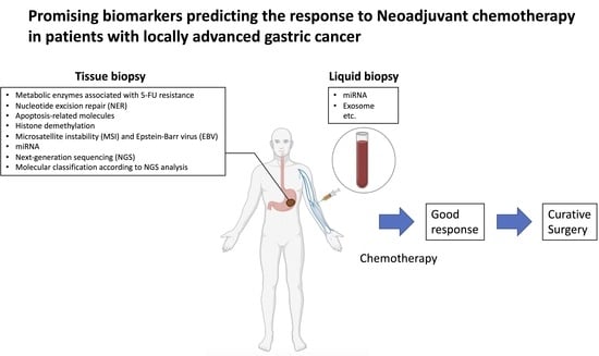
2. Biomarkers Involved in NAC

To date, 5-FU/CDDP-based combination therapy has been widely used for NAC [15], but due to drug resistance, the single-drug efficacy rates are not more than 20%, and the overall efficacy rate of first-line chemotherapy based on 5-FU or CDDP is less than 50%. Thus, some patients cannot benefit from NAC [16].
Therefore, there is an urgent need to explore the indicators of enzyme profiles related to CDDP and 5-FU or S-1 metabolism as predictors of the response to treatment. The most examined indicators include thymidylate synthase (TS), thymidine phosphorylase (TP), dihydropyrimidine dehydrogenase (DPD), and excision repair cross-complementation group 1 (ERCC1). In addition, apoptosis-associated proteins, histone demethylases, microsatellite instability (MSI), miRNAs, and exosomes have been reported as potential predictors (Table 2).

3. Metabolic Enzymes Associated with 5-FU Resistance

In the body, 5-FU is phosphorylated by TP to form the metabolically active substance fluorodeoxyuridine monophosphate and binds to TS, which is necessary for DNA synthesis, and forms a ternary complex with reduced folate. This ternary complex inhibits DNA synthesis, resulting in the suppression of cell proliferation. However, more than 85% of 5-FU is reduced to inactive metabolites by DPD in the liver and other organs and excreted through the kidneys. Therefore, the activity of DPD plays an important role in the efficiency of 5-FU [26].
In 2002, Terashima et al. [27] reported that the activity of DPD in gastric cancer tissues could predict drug resistance to 5-FU. Later, Napieralski et al. found that patients with a high expression of DPD were not sensitive to 5-FU and had a poor prognosis, whereas the opposite was observed in patients with a low expression of DPD [17]. In addition, Wang et al. detected TS overexpression in a DNA microarray analysis of 5-FU-resistant cancer cell lines [28]. A meta-analysis of 555 gastric cancer patients treated with S-1 showed a significant difference in response rate depending on the expression of DPD. However, there was no significant difference in the overall response rate based on the expression levels of TS and TP [29]. In contrast, Ott et al. identified a TS tandem repeat polymorphism in blood samples as an independent prognostic factor in the NAC group in LAGC patients treated with 5-FU-based preoperative chemotherapy [18]. A significant improvement in survival was also observed in the 2rpt/2rpt and 2rpt/3rpt genotypes [18]. These results suggest that TS and DPD are useful markers among the enzymes related to fluorouracil metabolism, but their clinical significance in NAC has not been fully established and further studies are needed.

4. Nucleotide Excision Repair (NER)

The NER pathway repairs relatively widespread DNA damage—several tens of base pairs—caused mainly by UV light [30,31]. The major NER pathways and other functional protein complexes are responsible for complicated NER reactions [32]. After DNA damage, ERCC1 forms a complex with XPA, XPF, and RPA proteins and binds to the DNA damage site for subsequent cleavage, removal, and repair of the damaged DNA. Platinum drugs, such as CDDP, induce cell death by forming cross-links within and between DNA strands. Since cross-linked adducts are suitable substrates for NER, the relationship between ERCC1 expression and CDDP sensitivity has attracted much attention. Metzger et al. reported that the expression of ERCC1 mRNA correlated with prognosis in 38 patients with gastric cancer treated with preoperative chemotherapy (CDDP+5FU) [19]. Similar results were also reported by Wei et al. in a study using a modified folinic acid, fluorouracil, and oxaliplatin (FOLFOX) regimen [33].
In addition, Fareed et al. reported that CDDP-based preoperative chemotherapy for gastroesophageal adenocarcinoma had significantly better pathologic tumor reduction and survival in ERCC1-negative tumors diagnosed using immunohistochemistry, which can be used as a predictive marker for treatment [20]. Kwon et al. reported that the response and survival rates to chemotherapy were significantly better in patients with ERCC1-negative tumors than in those with ERCC1-positive tumors in a study involving 64 patients treated with 5-FU and oxaliplatin before surgery. Moreover, ERCC1 expression was a prognostic factor in a multivariate analysis [34]. We investigated the relationship between the expression of major NER proteins and treatment responses in patients enrolled in a phase II study of docetaxel, CDDP, and S-1 (DCS) NAC for LAGC and found that damaged DNA binding protein complex subunit 2 (DDB2) and ERCC1 were associated with the treatment response [21,35]. DDB2 is known to be a sensor protein for early damage recognition during NER, and loss of its function increases the susceptibility of cancer cells to DNA damage [36]. To investigate the relationship between the expression of ERCC1 and/or DDB2 and the clinical effect of DCS therapy, we examined the expression of these proteins in tumor tissues before treatment by immunohistochemistry and analyzed the correlation with the anti-tumor effect (pathological response) of DCS therapy. The results showed that the positive predictive rates of ERCC1 and DDB2 expression for predicting resistance to DCS therapy were 72.9% and 78.3%, respectively. The positive predictive rate for predicting resistance to DCS therapy was as high as 82.5% when both were combined, suggesting their potential to be useful as markers of resistance to preoperative DCS therapy.

5. Apoptosis-Related Molecules

The relationship between apoptosis-related molecules and gastric cancer chemotherapy resistance has also been investigated. For example, low expression of BAX has been associated with lower response rates in patients treated with 5-FU in combination with CDDP [37], capecitabine, oxaliplatin plus irinotecan (COI), or FOLFOX [38]. BCL2-homology domain 3 (BH3) proteins, such as BAD, BIM, and BID, activate BAX and inhibit anti-apoptotic factors of the intrinsic apoptotic pathway. Altering the expression of these proteins may promote chemoresistance in gastric cancer. Therefore, we investigated gastric cancer cell lines by using a BH3 profiling method [39,40], which quantitatively evaluates the dependency of apoptosis on BH3 peptides, and found that docetaxel-induced apoptosis correlates with BIM and BAK protein expression, and that BAK knockdown causes docetaxel resistance [22]. We also determined the BAK expression index of 69 gastric cancer specimens before DCS therapy by multiplying the BAK positivity with a number representing the staining intensity. We found that patients with a good histopathological response to chemotherapy had a higher BAK expression index than those with incomplete response, and those with a BAK expression index of three or higher had a better progression-free survival and overall survival, indicating that BAK protein expression can predict the antitumor effect of docetaxel-containing therapy using pretreatment biopsy tissues. Similarly, Wu et al. also reported that decreased expression of BIM was associated with decreased overall survival in docetaxel-treated patients [41].

6. Histone Demethylation

Histone methylation can positively or negatively affect gene transcription, and dysregulation of histone methyltransferases is known to be involved in tumorigenesis [42]. Among them, Jumonji domain-containing protein 2A (JMJD2A), a member of the JMJD2 family, catalyzes the demethylation of H3K36 or H1.4K26. JMJD2A is overexpressed in a variety of cancers and promotes tumor growth [43], and is associated with drug resistance and poor clinical outcomes [44,45]. Using microarray analysis of gene expression in pretreatment biopsies of gastric cancer, Nakagawa et al. identified a functional gene signature consisting of 29 genes that are predictive of response to DCS therapy [46], among which JMJD2A was involved in gastric cancer chemosensitivity. They showed that overexpression of JMJD2A was positively correlated with the response rate in 34 patients treated with DCS [47]. These findings suggest that histone demethylation may be a novel epigenetic factor that regulates sensitivity to chemotherapy for gastric cancer.

7. Microsatellite Instability (MSI) and Epstein-Barr Virus (EBV)

MSI expression has been reported as a predictor of chemotherapy efficacy and response to immune checkpoint inhibitors [48]. However, the benefit of perioperative chemotherapy in MSI-high (MSH-H) gastric cancer remains controversial, due to the limited number of these patients in various clinical studies [49].
For example, in a meta-analysis of postoperative adjuvant chemotherapy for resectable gastric cancer, MSI-high (HIS-H) status was shown to be a negative predictor of prognostic benefit from adjuvant chemotherapy. In a meta-analysis of individual patient data from MAGIC, the Capecitabine and Oxaliplatin Adjuvant Study in Stomach Cancer (CLASSIC), the Adjuvant Chemoradiotherapy in Stomach Tumors (ARTIST), and the Intergroup Trial in Adjuvant Chemotherapy for Adenocarcinoma of the Stomach (ITACA-S) trials, Pietrantonio et al. found that the 5-year overall survival rate in the MSI-H group was significantly prolonged than in the MSI-low and microsatellite stable (MSS) groups (77.5% vs. 59.3%). Furthermore, they reported that additional chemotherapy was effective in the MSI-low/MSS group but not in the MSI-H group (70% vs. 77%) [50]. Hashimoto et al. investigated the expression of MLH1 and PD-L1 in surgical specimens from 110 and 285 patients who were treated with NAC and surgery alone, respectively. The results showed that the response rate to preoperative chemotherapy was significantly lower in MLH1-negative patients than in MLH1-positive patients, but there was no significant difference between patients with high and low PD-L1 expression. Conversely, the relapse-free survival of patients who did not receive preoperative chemotherapy was significantly longer in the MLH1-negative group than in the MLH1-positive group, and there was no significant difference in relapse-free survival between the two groups in patients who received preoperative chemotherapy. In addition, PD-L1 expression was not associated with relapse-free survival in patients with or without chemotherapy [23]. Similarly, Haag et al. reported a poor histological response to NAC in MSI-H tumors [24]. Therefore, it is suggested that MLH1-negative or MSI-H gastric cancers are unlikely to have a histological response to NAC. It is expected that immune checkpoint inhibitors can be used against them in the future.
Interestingly, Biesma et al. reported substantial histopathologic responses after NAC in patients with MSI-high gastric cancer, but only those with a mucinous phenotype from the D1/D2 trial in which patients underwent surgery alone and the ChemoRadiotherapy after Induction chemoTherapy In Cancer of the Stomach (CRITICS) trial in which patients underwent surgery and perioperative treatment [51,52]. These results indicate that the mucinous phenotype may be a relevant parameter in future clinical trials for patients with MSI-H.
Epstein–Barr virus-positive (EBV+) gastric cancer is one of the distinct molecular subtypes in The Cancer Genome Atlas (TCGA) classification [53]. Patients with EBV-negative gastric cancer have better outcomes than patients with EBV-negative and microsatellite stable (EBV−/MSS) gastric cancer [50,53,54,55,56]. However, data on response rates to NAC in EBV+ resectable gastric cancer are limited [57]. In the CRITICS trial, among the molecular subgroups of gastric cancer, EBV+ tumors had the highest histopathologic response rate and better outcomes than EBV-/MSS tumors [49].
In one retrospective series, Kohlruss et al. reviewed 760 NAC cases and found that MSI-H and EBV+ do not predict response to platinum- and 5-FU-based NAC but indicates a good prognosis. Particularly, MSI-H indicates a good prognosis regardless of treatment with NAC. Since MSS predicts a good response to NAC and suggests a poor prognosis for patients treated with surgery alone, MSS may help identify patients who would benefit more from preoperative chemotherapy [54].

8. miRNA and Exosomes

Micro RNAs are single-stranded small RNAs with a length of approximately 18–25 nucleotides, of which more than 1000 have been identified [58]. miRNAs regulate gene expression by binding to the 3′ untranslated region of mRNA and are involved in regulating a variety of biological processes [59].
Recently, miRNAs have been investigated as possible molecular markers and are expected to be used for the diagnosis [60,61] and prognosis of various cancers [62], as well as for predicting the effects of anticancer drugs. For example, let-7i is an miRNA involved in chemoresistance. Liu et al. examined the tissues of 86 patients with LAGC who had undergone preoperative chemotherapy and curative resection. They found that a lower level of let-7i expression in tumor tissues prior to treatment was associated with a lower histological response rate to NAC (FOLFOX regimen), indicating that let-7i expression could be a predictive marker of chemotherapy resistance in patients with LAGC [25]. Tan et al. found that the expression levels of miR-145 and miR-185 in the peripheral blood of 120 patients undergoing NAC with S-1 and oxaliplatin (SOX) tended to be lower in the tumor progression group, indicating that miR-145 and miR-185 may help predict the efficacy of SOX therapy when used as NAC [63].
miRNAs are present in cell-secreted vesicles called exosomes, which protect miRNA from degradation in the bloodstream and allow for the detection of miRNA in the blood [64,65]. Therefore, liquid biopsy, which is a method to detect exosomal miRNAs secreted by tumor cells, has been attracting considerable attention as a promising method for monitoring chemoresistance [66,67,68]. For example, Zhang et al. reported that CDDP and paclitaxel promoted the secretion of miR-522 in exosomes from cancer-associated fibroblasts, suppressed arachidonate lipoxygenase 15 (ALOX15), and decreased the accumulation of lipid-ROS (toxic lipid peroxides) in gastric cancer cells, resulting in reduced sensitivity to anticancer drugs [69]. In addition, Wang et al. reported that exosomal miR-155-5p can directly inhibit GATA binding protein 3 (GATA3) and tumor protein p53-induced nuclear protein 1 (TP53INP1) and can overcome paclitaxel resistance in gastric cancer cells [70].
In the future, it is expected that more sensitive and specific exosomal miRNA markers related to chemotherapy sensitivity will be identified. Additionally, the monitoring of miRNAs by liquid biopsy will enable the development of a method to evaluate the efficacy of chemotherapy more efficiently and select the appropriate timing of surgery on a real-time basis. However, clinical validation and standardization of the procedure are needed before liquid biopsy can be widely used on a routine basis. Early experiments analyzing liquid biopsies appear to be very promising in patients with advanced gastric cancer, but more prospective studies are needed to validate the efficacy of liquid biopsy and understand the molecular mechanisms underlying chemotherapy resistance.

9. Molecular Classification According to NGS Analysis

Over the past decade, next-generation sequencing (NGS) technology has been a powerful tool for studying the complexity of gastric cancers, with important implications for both the molecular characterization of the neoplasm and the therapeutic management of gastric cancer patients [71]. Indeed, the increasingly frequent integration of NGS in the molecular assessment of biological samples has the potential to greatly improve the selection of patients to be included in clinical trials of molecularly targeted drugs.
In fact, several amplicon-based NGS assays have been clinically approved and are currently being used to detect the most frequent and actionable genomic alterations in tumor samples [72]. The main NGS-based approaches (i.e., whole-genome sequencing [WGS], whole-exome sequencing [WES], RNA sequencing [RNA Seq], and targeted sequencing) have been systematically applied to characterize molecular alterations in gastric cancer [71]. This large amount of genomic, transcriptomic, and epigenomic data will significantly improve our understanding of the molecular landscape of gastric cancer, unravel its molecular heterogeneity, and pave the way for a comprehensive molecular classification of this complex disease, which will contribute to the development of new molecularly targeted drugs and the selection of patients to be included in clinical trials [73,74].
While the well-known Lauren classification criteria for gastric cancer were developed six decades ago according to histologic features (intestinal or diffuse), the use of genomic data has recently led to the development of new molecular classification schemes. In 2014, The Cancer Genome Atlas (TCGA) network developed a new molecular classification scheme using somatic cell copy number analysis, WES, DNA methylation profiling, messenger RNAseq, microRNA sequencing, and reverse-phase protein array profiling to characterize 295 localized and untreated gastric cancers [53]. According to TCGA results, gastric cancer can be classified into four molecular subtypes: (i) EBV-positive tumors (9%), (ii) MSI tumors (22%), (iii) genome stable (GS) tumors (20%), and (iv) tumors with chromosomal instability (CIN) (50%).
More importantly, this subclassification system was shown to have the potential to guide targeted therapy for different types of patients with gastric cancer subtypes. However, clinical data obtained by the Prodige group in France and the AIO group in Germany were negative for response discrimination regarding patients with diffuse (GS) versus intestinal (CIN) types [11,75,76]. No other robust predictive markers have been found for the GS and CIN groups.
In 2015, the Asian Cancer Research Group (ACRG) proposed another molecular classification based on the evaluation of 300 gastric cancer samples in Korea by gene expression profiling, genome-wide copy number microarray, and targeted gene sequencing [77]. Four molecular subtypes of gastric cancer with different clinical and genomic features have been identified: (i) MSI tumors (23%); (ii) MSS with epithelial–mesenchymal transition features (MSS/EMT) tumors (15%); (iii) MSS with TP53 active (MSS/TP53þ) tumors (26%); and (iv) MSS with TP53 inactive (MSS/ P53-) tumors (36%). It should be noted that the differences between the TCGA and ACRG classifications are not perfect and uniform as they reflect differences in the approaches and platforms used and the ethnicity of the samples (i.e., global for TCGA and Korean for ACTG). However, it reveals molecular characteristics of gastric cancer that are not available from conventional histology-based classification, and thus is expected to improve our understanding of gastric cancer, improve treatment outcomes for gastric cancer patients, and potentially pave the way for better gastric cancer diagnosis and new drug development. A better understanding of the genomics of gastric cancer will allow optimization of treatment before or during NAC for individual patients. In other words, if this platform is incorporated into clinical research in the future and ultimately applied to routine medical practice, tailored NAC treatment for each patient will be possible.

10. Future Perspectives

Good predictive markers are expected to eliminate unnecessary and potentially detrimental NAC. In other words, more accurate predictive markers can identify patients at higher risk of locoregional recurrence or distant metastasis and increase the chances of curing the disease with lower toxicity by targeting key molecules and pathways. On the other hand, patients with biomarkers that suggest a low risk of local recurrence or distant metastasis, or markers of resistance to NAC, can proceed to surgery without receiving NAC.
Tissue heterogeneity, a hallmark of gastric cancer, has been an obstacle to the development of predictive and prognostic biomarkers. Recently, Murugaesu et al. performed WES on eight patients, including more than 40 tumor regions, before and after neoadjuvant chemotherapy to assess the proportion of subclonal alterations in different tumor sites and to evaluate the degree of intratumor heterogeneity (ITH) in cancer [78]. Interestingly, more than half of all mutations were heterogeneously present in different tumor subclones. Using this approach, they reported that there is a strong correlation between the ITH index (mean of the proportion of heterogeneous mutations relative to the total number of mutations) and response to chemotherapy. This is consistent with the expectation that, from a biological perspective, tumors with high genomic heterogeneity would respond poorly to neoadjuvant therapy. Future larger, well-designed, prospective studies are needed to confirm the utility of ITH as a predictive and prognostic biomarker. From a diagnostic and monitoring perspective, the subclonal heterogeneity of gastric cancer suggests the utility of liquid biopsy, which can better reflect the subclonal mutational status in individual patients [79].
Furthermore, the heterogeneity of gastric cancer has necessitated different types of therapy. With advances in genomic and epigenomic research, further subclassification of gastric cancer into new molecular entities is expected to facilitate therapeutic decision making. In the foreseeable future, the integration of well-established clinicopathologic markers with modern molecular profiling will enable accurate prediction of NAC chemosensitivity in gastric cancer.

11. Conclusions

In this review, we provided an overview of promising biomarkers that could play a vital role in predicting the response to NAC in patients with LAGC. To date, various predictive factors for the therapeutic effect of conventional cytotoxic chemotherapy on gastric cancer have been identified. Some molecular targeted therapies and immune checkpoint inhibitors are now being introduced for gastric cancer, and their molecular markers such as human epidermal growth factor receptor 2 (HER2) and MSI are good predictive markers. However, these predictive factors have not yet been implemented in clinical practice for predicting the response to NAC.
More recently, gene-panel tests using NGS techniques have also been introduced into clinical practice. This new trend of personalized cancer treatment is expected to make the best use of the advantages of NAC by enabling the selection of refractory cases and effective prediction of treatment efficacy. Subsequently, a better understanding of the molecular characterization of gastric cancers will likely help employ targeted and biological therapies in relation to surgery, chemotherapy, and radiotherapy in gastric cancers, to improve outcomes in patient subsets with historically poor prognoses. In addition, further development of predictive markers will help define subgroups of patients who will benefit optimally from adjuvant treatment. Furthermore, the introduction of liquid biopsy methods will enable minimally invasive and reproducible preoperative sampling, which will lead to the appropriate selection and modification of treatment regimens based on prognostic risk and treatment-resistant biomarkers, thereby allowing NAC to become a more valid treatment strategy.

Author Contributions

Writing—original draft, Y.S.; writing—review and editing, Y.S. and T.T.; approval of the final manuscript, Y.S., K.O., T.K., F.N., H.M. and T.T. All authors have read and agreed to the published version of the manuscript.

Funding

This research received no external funding.

Institutional Review Board Statement

Not applicable.

Informed Consent Statement

Not applicable.

Data Availability Statement

Not applicable.

Conflicts of Interest

The authors declare no conflict of interest.

References

  1. Bray, F.; Ferlay, J.; Soerjomataram, I.; Siegel, R.L.; Torre, L.A.; Jemal, A. Global cancer statistics 2018: GLOBOCAN estimates of incidence and mortality worldwide for 36 cancers in 185 countries. CA Cancer J. Clin. 2018, 68, 394–424. [Google Scholar] [CrossRef] [PubMed] [Green Version]
  2. Khan, U.; Shah, M.A. Optimizing Therapies in the Perioperative Management of Gastric Cancer. Curr. Treat. Options Oncol. 2019, 20, 57. [Google Scholar] [CrossRef] [PubMed]
  3. Petrillo, A.; Smyth, E.C. Multimodality treatment for localized gastric cancer: State of the art and new insights. Curr. Opin. Oncol. 2020, 32, 347–355. [Google Scholar] [CrossRef] [PubMed]
  4. D’Ugo, D.; Rausei, S.; Biondi, A.; Persiani, R. Preoperative treatment and surgery in gastric cancer: Friends or foes? Lancet. Oncol. 2009, 10, 191–195. [Google Scholar] [CrossRef]
  5. Moehler, M.; Schimanski, C.C.; Gockel, I.; Junginger, T.; Galle, P.R. (Neo)adjuvant strategies of advanced gastric carcinoma: Time for a change? Dig. Dis. 2004, 22, 345–350. [Google Scholar] [CrossRef]
  6. Fujitani, K. Overview of Adjuvant and Neoadjuvant Therapy for Resectable Gastric Cancer in the East. Dig. Surg. 2013, 30, 119–129. [Google Scholar] [CrossRef]
  7. Terashima, M.; Iwasaki, Y.; Mizusawa, J.; Katayama, H.; Nakamura, K.; Katai, H.; Yoshikawa, T.; Ito, Y.; Kaji, M.; Kimura, Y.; et al. Randomized phase III trial of gastrectomy with or without neoadjuvant S-1 plus cisplatin for type 4 or large type 3 gastric cancer, the short-term safety and surgical results: Japan Clinical Oncology Group Study (JCOG0501). Gastric Cancer 2019, 22, 1044–1052. [Google Scholar] [CrossRef] [Green Version]
  8. Iwasaki, Y.; Terashima, M.; Mizusawa, J.; Katayama, H.; Nakamura, K.; Katai, H.; Yoshikawa, T.; Ito, S.; Kaji, M.; Kimura, Y.; et al. Gastrectomy with or without neoadjuvant S-1 plus cisplatin for type 4 or large type 3 gastric cancer (JCOG0501): An open-label, phase 3, randomized controlled trial. Gastric Cancer 2021, 24, 492–502. [Google Scholar] [CrossRef]
  9. Cunningham, D.; Allum, W.H.; Stenning, S.P.; Thompson, J.N.; Van de Velde, C.J.H.; Nicolson, M.; Scarffe, J.H.; Lofts, F.J.; Falk, S.J.; Iveson, T.J.; et al. Perioperative Chemotherapy versus Surgery Alone for Resectable Gastroesophageal Cancer. N. Engl. J. Med. 2006, 355, 11–20. [Google Scholar] [CrossRef] [Green Version]
  10. Al-Batran, S.-E.; Hofheinz, R.D.; Pauligk, C.; Kopp, H.-G.; Haag, G.M.; Luley, K.B.; Meiler, J.; Homann, N.; Lorenzen, S.; Schmalenberg, H.; et al. Histopathological regression after neoadjuvant docetaxel, oxaliplatin, fluorouracil, and leucovorin versus epirubicin, cisplatin, and fluorouracil or capecitabine in patients with resectable gastric or gastro-oesophageal junction adenocarcinoma (FLOT4-AIO): Results from the phase 2 part of a multicentre, open-label, randomised phase 2/3 trial. Lancet Oncol. 2016, 17, 1697–1708. [Google Scholar] [CrossRef]
  11. Al-Batran, S.-E.; Homann, N.; Pauligk, C.; Goetze, T.O.; Meiler, J.; Kasper, S.; Kopp, H.-G.; Mayer, F.; Haag, G.M.; Luley, K.; et al. Perioperative chemotherapy with fluorouracil plus leucovorin, oxaliplatin, and docetaxel versus fluorouracil or capecitabine plus cisplatin and epirubicin for locally advanced, resectable gastric or gastro-oesophageal junction adenocarcinoma (FLOT4): A randomised, phase 2/3 trial. Lancet 2019, 393, 1948–1957. [Google Scholar] [CrossRef] [PubMed]
  12. Joshi, S.S.; Badgwell, B.D. Current treatment and recent progress in gastric cancer. CA Cancer J. Clin. 2021, 71, 264–279. [Google Scholar] [CrossRef] [PubMed]
  13. Ajani, J.A.; Mansfield, P.F.; Crane, C.H.; Wu, T.T.; Lunagomez, S.; Lynch, P.M.; Janjan, N.; Feig, B.; Faust, J.; Yao, J.C.; et al. Paclitaxel-Based Chemoradiotherapy in Localized Gastric Carcinoma: Degree of Pathologic Response and Not Clinical Parameters Dictated Patient Outcome. J. Clin. Oncol. 2005, 23, 1237–1244. [Google Scholar] [CrossRef]
  14. Gervaso, L.; Pellicori, S.; Cella, C.A.; Bagnardi, V.; Lordick, F.; Fazio, N. Biomarker evaluation in radically resectable locally advanced gastric cancer treated with neoadjuvant chemotherapy: An evidence reappraisal. Ther. Adv. Med. Oncol. 2021, 13, 1–11. [Google Scholar] [CrossRef] [PubMed]
  15. Wang, X.-Z.; Zeng, Z.-Y.; Ye, X.; Sun, J.; Zhang, Z.-M.; Kang, W.-M. Interpretation of the development of neoadjuvant therapy for gastric cancer based on the vicissitudes of the NCCN guidelines. World J. Gastrointest. Oncol. 2020, 12, 37–53. [Google Scholar] [CrossRef]
  16. Wagner, A.D.; Syn, N.L.; Moehler, M.; Grothe, W.; Yong, W.P.; Tai, B.-C.; Ho, J.; Unverzagt, S. Chemotherapy for advanced gastric cancer. Cochrane Database Syst. Rev. 2017, 8, CD004064. [Google Scholar] [CrossRef] [Green Version]
  17. Napieralski, R.; Ott, K.; Kremer, M.; Specht, K.; Vogelsang, H.; Becker, K.; Müller, M.; Lordick, F.; Fink, U.; Rüdiger Siewert, J.; et al. Combined GADD45A and Thymidine Phosphorylase Expression Levels Predict Response and Survival of Neoadjuvant-Treated Gastric Cancer Patients. Clin. Cancer Res. 2005, 11, 3025–3031. [Google Scholar] [CrossRef] [Green Version]
  18. Ott, K.; Vogelsang, H.; Marton, N.; Becker, K.; Lordick, F.; Kobl, M.; Schuhmacher, C.; Novotny, A.; Mueller, J.; Fink, U.; et al. Thethymidylate synthase tandem repeat promoter polymorphism: A predictor for tumor-related survival in neoadjuvant treated locally advanced gastric cancer. Int. J. Cancer 2006, 119, 2885–2894. [Google Scholar] [CrossRef]
  19. Metzger, R.; Leichman, C.G.; Danenberg, K.D.; Danenberg, P.V.; Lenz, H.J.; Hayashi, K.; Groshen, S.; Salonga, D.; Cohen, H.; Laine, L.; et al. ERCC1 mRNA levels complement thymidylate synthase mRNA levels in predicting response and survival for gastric cancer patients receiving combination cisplatin and fluorouracil chemotherapy. J. Clin. Oncol. 1998, 16, 309–316. [Google Scholar] [CrossRef]
  20. Fareed, K.R.; Al-Attar, A.; Soomro, I.N.; Kaye, P.V.; Patel, J.; Lobo, D.N.; Parsons, S.L.; Madhusudan, S. Tumour regression and ERCC1 nuclear protein expression predict clinical outcome in patients with gastro-oesophageal cancer treated with neoadjuvant chemotherapy. Br. J. Cancer 2010, 102, 1600–1607. [Google Scholar] [CrossRef]
  21. Hirakawa, M.; Sato, Y.; Ohnuma, H.; Takayama, T.; Sagawa, T.; Nobuoka, T.; Harada, K.; Miyamoto, H.; Sato, Y.; Takahashi, Y.; et al. A phase II study of neoadjuvant combination chemotherapy with docetaxel, cisplatin, and S-1 for locally advanced resectable gastric cancer: Nucleotide excision repair (NER) as potential chemoresistance marker. Cancer Chemother. Pharmacol. 2013, 71, 789–797. [Google Scholar] [CrossRef] [PubMed]
  22. Kubo, T.; Kawano, Y.; Himuro, N.; Sugita, S.; Sato, Y.; Ishikawa, K.; Takada, K.; Murase, K.; Miyanishi, K.; Sato, T.; et al. BAK is a predictive and prognostic biomarker for the therapeutic effect of docetaxel treatment in patients with advanced gastric cancer. Gastric Cancer 2016, 19, 827–838. [Google Scholar] [CrossRef] [PubMed] [Green Version]
  23. Hashimoto, T.; Kurokawa, Y.; Takahashi, T.; Miyazaki, Y.; Tanaka, K.; Makino, T.; Yamasaki, M.; Nakajima, K.; Ikeda, J.; Mori, M.; et al. Predictive value of MLH1 and PD-L1 expression for prognosis and response to preoperative chemotherapy in gastric cancer. Gastric Cancer 2019, 22, 785–792. [Google Scholar] [CrossRef] [PubMed] [Green Version]
  24. Haag, G.M.; Czink, E.; Ahadova, A.; Schmidt, T.; Sisic, L.; Blank, S.; Heger, U.; Apostolidis, L.; Berger, A.K.; Springfeld, C.; et al. Prognostic significance of microsatellite-instability in gastric and gastroesophageal junction cancer patients undergoing neoadjuvant chemotherapy. Int. J. Cancer 2019, 144, 1697–1703. [Google Scholar] [CrossRef]
  25. Liu, K.; Qian, T.; Tang, L.; Wang, J.; Yang, H.; Ren, J. Decreased expression of microRNA let-7i and its association with chemotherapeutic response in human gastric cancer. World J. Surg. Oncol. 2012, 10, 3–8. [Google Scholar] [CrossRef] [Green Version]
  26. Diasio, R.B. Oral DPD-inhibitory fluoropyrimidine drugs. Oncology 2000, 14, 19–23. [Google Scholar]
  27. Terashima, M.; Irinoda, T.; Fujiwara, H.; Nakaya, T.; Takagane, A.; Abe, K.; Yonezawa, H.; Oyama, K.; Inaba, T.; Saito, K.; et al. Roles of thymidylate synthase and dihydropyrimidine dehydrogenase in tumor progression and sensitivity to 5-fluorouracil in human gastric cancer. Anticancer Res. 2002, 22, 761–768. [Google Scholar]
  28. Wang, W.; Cassidy, J.; O’Brien, V.; Ryan, K.M.; Collie-Duguid, E. Mechanistic and Predictive Profiling of 5-Fluorouracil Resistance in Human Cancer Cells. Cancer Res. 2004, 64, 8167–8176. [Google Scholar] [CrossRef] [Green Version]
  29. Wang, D.; Yu, X.; Wang, X. High/Positive Expression of 5-Fluorouracil Metabolic Enzymes Predicts Better Response to S-1 in Patients with Gastric Cancer: A Meta-Analysis. Int. J. Biol. Mark. 2016, 31, 101–109. [Google Scholar] [CrossRef]
  30. Marteijn, J.A.; Lans, H.; Vermeulen, W.; Hoeijmakers, J.H.J. Understanding nucleotide excision repair and its roles in cancer and ageing. Nat. Rev. Mol. Cell Biol. 2014, 15, 465–481. [Google Scholar] [CrossRef]
  31. Jeggo, P.A.; Pearl, L.H.; Carr, A.M. DNA repair, genome stability and cancer: A historical perspective. Nat. Rev. Cancer 2016, 16, 35–42. [Google Scholar] [CrossRef] [PubMed] [Green Version]
  32. Sugasawa, K. Regulation of damage recognition in mammalian global genomic nucleotide excision repair. Mutat. Res. 2010, 685, 29–37. [Google Scholar] [CrossRef] [PubMed]
  33. Wei, J.; Zou, Z.; Qian, X.; Ding, Y.; Xie, L.; Sanchez, J.J.; Zhao, Y.; Feng, J.; Ling, Y.; Liu, Y.; et al. ERCC1 mRNA levels and survival of advanced gastric cancer patients treated with a modified FOLFOX regimen. Br. J. Cancer 2008, 98, 1398–1402. [Google Scholar] [CrossRef] [Green Version]
  34. Kwon, H.-C.; Roh, M.S.; Oh, S.Y.; Kim, S.-H.; Kim, M.C.; Kim, J.-S.; Kim, H.-J. Prognostic value of expression of ERCC1, thymidylate synthase, and glutathione S-transferase P1 for 5-fluorouracil/oxaliplatin chemotherapy in advanced gastric cancer. Ann. Oncol. 2007, 18, 504–509. [Google Scholar] [CrossRef] [PubMed]
  35. Sato, Y.; Takayama, T.; Sagawa, T.; Takahashi, Y.; Ohnuma, H.; Okubo, S.; Shintani, N.; Tanaka, S.; Kida, M.; Sato, Y.; et al. Phase II study of S-1, docetaxel and cisplatin combination chemotherapy in patients with unresectable metastatic gastric cancer. Cancer Chemother. Pharmacol. 2010, 66, 721–728. [Google Scholar] [CrossRef]
  36. Takimoto, R.; MacLachlan, T.K.; Dicker, D.T.; Niitsu, Y.; Mori, T.; El-Deiry, W.S. BRCA1 Transcriptionally Regulates Damaged DNA Binding Protein (DDB2) In the DNA Repair Response Following UV-Irradiation. Cancer Biol. Ther. 2002, 1, 177–186. [Google Scholar] [CrossRef] [Green Version]
  37. Wang, X.; Lin, Y.; Lan, F.; Yu, Y.; Ouyang, X.; Liu, W.; Xie, F.; Wang, X.; Huang, Q. BAX and CDKN1A polymorphisms correlated with clinical outcomes of gastric cancer patients treated with postoperative chemotherapy. Med. Oncol. 2014, 31, 249. [Google Scholar] [CrossRef]
  38. Jeong, S.H.; Han, J.H.; Kim, J.H.; Ahn, M.S.; Hwang, Y.H.; Lee, H.W.; Kang, S.Y.; Park, J.S.; Choi, J.-H.; Lee, K.J.; et al. Bax Predicts Outcome in Gastric Cancer Patients Treated with 5-fluorouracil, Leucovorin, and Oxaliplatin Palliative Chemotherapy. Dig. Dis. Sci. 2011, 56, 131–138. [Google Scholar] [CrossRef]
  39. Chonghaile, T.N.; Sarosiek, K.A.; Vo, T.-T.; Ryan, J.A.; Tammareddi, A.; Moore, V.D.G.; Deng, J.; Anderson, K.C.; Richardson, P.; Tai, Y.-T.; et al. Pretreatment Mitochondrial Priming Correlates with Clinical Response to Cytotoxic Chemotherapy. Science 2011, 334, 1129–1133. [Google Scholar] [CrossRef] [Green Version]
  40. Vo, T.-T.; Ryan, J.; Carrasco, R.; Neuberg, D.; Rossi, D.J.; Stone, R.M.; DeAngelo, D.J.; Frattini, M.G.; Letai, A. Relative Mitochondrial Priming of Myeloblasts and Normal HSCs Determines Chemotherapeutic Success in AML. Cell 2012, 151, 344–355. [Google Scholar] [CrossRef] [Green Version]
  41. Wu, N.; Huang, Y.; Zou, Z.; Gimenez-Capitan, A.; Yu, L.; Hu, W.; Zhu, L.; Sun, X.; Sanchez, J.J.; Guan, W.; et al. High BIM mRNA levels are associated with longer survival in advanced gastric cancer. Oncol. Lett. 2017, 13, 1826–1834. [Google Scholar] [CrossRef] [PubMed] [Green Version]
  42. Chi, P.; Allis, C.D.; Wang, G.G. Covalent histone modifications—Miswritten, misinterpreted and mis-erased in human cancers. Nat. Rev. Cancer 2010, 10, 457–469. [Google Scholar] [CrossRef] [PubMed] [Green Version]
  43. Kogure, M.; Takawa, M.; Cho, H.-S.; Toyokawa, G.; Hayashi, K.; Tsunoda, T.; Kobayashi, T.; Daigo, Y.; Sugiyama, M.; Atomi, Y.; et al. Deregulation of the histone demethylase JMJD2A is involved in human carcinogenesis through regulation of the G1/S transition. Cancer Lett. 2013, 336, 76–84. [Google Scholar] [CrossRef] [PubMed]
  44. Berry, W.L.; Shin, S.; Lightfoot, S.A.; Janknecht, R. Oncogenic features of the JMJD2A histone demethylase in breast cancer. Int. J. Oncol. 2012, 41, 1701–1706. [Google Scholar] [CrossRef] [Green Version]
  45. Hu, C.-E.; Liu, Y.-C.; Zhang, H.-D.; Huang, G.-J. JMJD2A predicts prognosis and regulates cell growth in human gastric cancer. Biochem. Biophys. Res. Commun. 2014, 449, 1–7. [Google Scholar] [CrossRef]
  46. Kitamura, S.; Tanahashi, T.; Aoyagi, E.; Nakagawa, T.; Okamoto, K.; Kimura, T.; Miyamoto, H.; Mitsui, Y.; Rokutan, K.; Muguruma, N.; et al. Response Predictors of S-1, Cisplatin, and Docetaxel Combination Chemotherapy for Metastatic Gastric Cancer: Microarray Analysis of Whole Human Genes. Oncology 2017, 93, 127–135. [Google Scholar] [CrossRef]
  47. Nakagawa, T.; Sato, Y.; Tanahashi, T.; Mitsui, Y.; Kida, Y.; Fujino, Y.; Hirata, M.; Kitamura, S.; Miyamoto, H.; Okamoto, K.; et al. JMJD2A sensitizes gastric cancer to chemotherapy by cooperating with CCDC8. Gastric Cancer 2020, 23, 426–436. [Google Scholar] [CrossRef]
  48. Baretti, M.; Le, D.T. DNA mismatch repair in cancer. Pharmacol. Ther. 2018, 189, 45–62. [Google Scholar] [CrossRef]
  49. Biesma, H.D.; Soeratram, T.T.D.; Sikorska, K.; Caspers, I.A.; van Essen, H.F.; Egthuijsen, J.M.P.; Mookhoek, A.; van Laarhoven, H.W.M.; van Berge Henegouwen, M.I.; Nordsmark, M.; et al. Response to neoadjuvant chemotherapy and survival in molecular subtypes of resectable gastric cancer: A post hoc analysis of the D1/D2 and CRITICS trials. Gastric Cancer 2022, 25, 640–651. [Google Scholar] [CrossRef]
  50. Pietrantonio, F.; Miceli, R.; Raimondi, A.; Kim, Y.W.; Kang, W.K.; Langley, R.E.; Choi, Y.Y.; Kim, K.-M.; Nankivell, M.G.; Morano, F.; et al. Individual Patient Data Meta-Analysis of the Value of Microsatellite Instability As a Biomarker in Gastric Cancer. J. Clin. Oncol. Off. J. Am. Soc. Clin. Oncol. 2019, 37, 3392–3400. [Google Scholar] [CrossRef]
  51. Cats, A.; Jansen, E.P.M.; van Grieken, N.C.T.; Sikorska, K.; Lind, P.; Nordsmark, M.; Meershoek-Klein Kranenbarg, E.; Boot, H.; Trip, A.K.; Swellengrebel, H.A.M.; et al. Chemotherapy versus chemoradiotherapy after surgery and preoperative chemotherapy for resectable gastric cancer (CRITICS): An international, open-label, randomised phase 3 trial. Lancet Oncol. 2018, 19, 616–628. [Google Scholar] [CrossRef]
  52. Songun, I.; Putter, H.; Kranenbarg, E.M.K.; Sasako, M.; van de Velde, C.J.H. Surgical treatment of gastric cancer: 15-year follow-up results of the randomised nationwide Dutch D1D2 trial. Lancet Oncol. 2010, 11, 439–449. [Google Scholar] [CrossRef]
  53. Bass, A.J.; Thorsson, V.; Shmulevich, I.; Reynolds, S.M.; Miller, M.; Bernard, B.; Hinoue, T.; Laird, P.W.; Curtis, C.; Shen, H.; et al. Comprehensive molecular characterization of gastric adenocarcinoma. Nature 2014, 513, 202–209. [Google Scholar] [CrossRef] [Green Version]
  54. Kohlruss, M.; Grosser, B.; Krenauer, M.; Slotta-Huspenina, J.; Jesinghaus, M.; Blank, S.; Novotny, A.; Reiche, M.; Schmidt, T.; Ismani, L.; et al. Prognostic implication of molecular subtypes and response to neoadjuvant chemotherapy in 760 gastric carcinomas: Role of Epstein–Barr virus infection and high- and low-microsatellite instability. J. Pathol. Clin. Res. 2019, 5, 227–239. [Google Scholar] [CrossRef] [PubMed] [Green Version]
  55. Marrelli, D.; Polom, K.; Pascale, V.; Vindigni, C.; Piagnerelli, R.; De Franco, L.; Ferrara, F.; Roviello, G.; Garosi, L.; Petrioli, R.; et al. Strong Prognostic Value of Microsatellite Instability in Intestinal Type Non-cardia Gastric Cancer. Ann. Surg. Oncol. 2016, 23, 943–950. [Google Scholar] [CrossRef]
  56. Van Beek, J.; zur Hausen, A.; Kranenbarg, E.K.; van de Velde, C.J.H.; Middeldorp, J.M.; van den Brule, A.J.C.; Meijer, C.J.L.M.; Bloemena, E. EBV-positive gastric adenocarcinomas: A distinct clinicopathologic entity with a low frequency of lymph node involvement. J. Clin. Oncol. 2004, 22, 664–670. [Google Scholar] [CrossRef]
  57. Ignatova, E.; Seriak, D.; Fedyanin, M.; Tryakin, A.; Pokataev, I.; Menshikova, S.; Vakhabova, Y.; Smirnova, K.; Tjulandin, S.; Ajani, J.A. Epstein–Barr virus-associated gastric cancer: Disease that requires special approach. Gastric Cancer 2020, 23, 951–960. [Google Scholar] [CrossRef]
  58. Hussen, B.M.; Hidayat, H.J.; Salihi, A.; Sabir, D.K.; Taheri, M.; Ghafouri-Fard, S. MicroRNA: A signature for cancer progression. Biomed. Pharmacother. 2021, 138, 111528. [Google Scholar] [CrossRef]
  59. O’Brien, J.; Hayder, H.; Zayed, Y.; Peng, C. Overview of MicroRNA Biogenesis, Mechanisms of Actions, and Circulation. Front. Endocrinol. 2018, 9, 00402. [Google Scholar] [CrossRef] [Green Version]
  60. Yao, Y.; Ding, Y.; Bai, Y.; Zhou, Q.; Lee, H.; Li, X.; Teng, L. Identification of Serum Circulating MicroRNAs as Novel Diagnostic Biomarkers of Gastric Cancer. Front. Genet. 2020, 11, 591515. [Google Scholar] [CrossRef]
  61. So, J.B.Y.; Kapoor, R.; Zhu, F.; Koh, C.; Zhou, L.; Zou, R.; Tang, Y.C.; Goo, P.C.K.; Rha, S.Y.; Chung, H.C.; et al. Development and validation of a serum microRNA biomarker panel for detecting gastric cancer in a high-risk population. Gut 2021, 70, 829–837. [Google Scholar] [CrossRef] [PubMed]
  62. Ueda, T.; Volinia, S.; Okumura, H.; Shimizu, M.; Taccioli, C.; Rossi, S.; Alder, H.; Liu, C.; Oue, N.; Yasui, W.; et al. Relation between microRNA expression and progression and prognosis of gastric cancer: A microRNA expression analysis. Lancet Oncol. 2010, 11, 136–146. [Google Scholar] [CrossRef] [Green Version]
  63. Tan, B.; Li, Y.; Di, Y.; Fan, L.; Zhao, Q.; Liu, Q.; Wang, D.; Jia, N. Clinical value of peripheral blood microRNA detection in evaluation of SOX regimen as neoadjuvant chemotherapy for gastric cancer. J. Clin. Lab. Anal. 2018, 32, e22363. [Google Scholar] [CrossRef] [PubMed] [Green Version]
  64. Tang, Z.; Li, D.; Hou, S.; Zhu, X. The cancer exosomes: Clinical implications, applications and challenges. Int. J. Cancer 2020, 146, 2946–2959. [Google Scholar] [CrossRef]
  65. Liu, J.; Ren, L.; Li, S.; Li, W.; Zheng, X.; Yang, Y.; Fu, W.; Yi, J.; Wang, J.; Du, G. The biology, function, and applications of exosomes in cancer. Acta Pharm. Sin. B 2021, 11, 2783–2797. [Google Scholar] [CrossRef]
  66. Uchôa Guimarães, C.T.; Ferreira Martins, N.N.; Cristina da Silva Oliveira, K.; Almeida, C.M.; Pinheiro, T.M.; Gigek, C.O.; Roberto de Araújo Cavallero, S.; Assumpção, P.P.; Cardoso Smith, M.A.; Burbano, R.R.; et al. Liquid biopsy provides new insights into gastric cancer. Oncotarget 2018, 9, 15144–15156. [Google Scholar] [CrossRef] [Green Version]
  67. Lengyel, C.G.; Hussain, S.; Trapani, D.; El Bairi, K.; Altuna, S.C.; Seeber, A.; Odhiambo, A.; Habeeb, B.S.; Seid, F. The emerging role of liquid biopsy in gastric cancer. J. Clin. Med. 2021, 10, 2108. [Google Scholar] [CrossRef]
  68. Huang, T.; Song, C.; Zheng, L.; Xia, L.; Li, Y.; Zhou, Y. The roles of extracellular vesicles in gastric cancer development, microenvironment, anti-cancer drug resistance, and therapy. Mol. Cancer 2019, 18, 62. [Google Scholar] [CrossRef]
  69. Zhang, H.; Deng, T.; Liu, R.; Ning, T.; Yang, H.; Liu, D.; Zhang, Q.; Lin, D.; Ge, S.; Bai, M.; et al. CAF secreted miR-522 suppresses ferroptosis and promotes acquired chemo-resistance in gastric cancer. Mol. Cancer 2020, 19, 43. [Google Scholar] [CrossRef] [Green Version]
  70. Wang, M.; Qiu, R.; Yu, S.; Xu, X.; Li, G.; Gu, R.; Tan, C.; Zhu, W.; Shen, B. Paclitaxel-resistant gastric cancer MGC-803 cells promote epithelial-to-mesenchymal transition and chemoresistance in paclitaxel-sensitive cells via exosomal delivery of miR-155-5p. Int. J. Oncol. 2018, 54, 326–338. [Google Scholar] [CrossRef]
  71. Verma, R.; Sharma, P.C. Next generation sequencing-based emerging trends in molecular biology of gastric cancer. Am. J. Cancer Res. 2018, 8, 207–225. [Google Scholar] [PubMed]
  72. Nemtsova, M.V.; Kalinkin, A.I.; Kuznetsova, E.B.; Bure, I.V.; Alekseeva, E.A.; Bykov, I.I.; Khorobrykh, T.V.; Mikhaylenko, D.S.; Tanas, A.S.; Kutsev, S.I.; et al. Clinical relevance of somatic mutations in main driver genes detected in gastric cancer patients by next-generation DNA sequencing. Sci. Rep. 2020, 10, 504. [Google Scholar] [CrossRef] [PubMed] [Green Version]
  73. Fassan, M.; Simbolo, M.; Bria, E.; Mafficini, A.; Pilotto, S.; Capelli, P.; Bencivenga, M.; Pecori, S.; Luchini, C.; Neves, D.; et al. High-throughput mutation profiling identifies novel molecular dysregulation in high-grade intraepithelial neoplasia and early gastric cancers. Gastric Cancer 2014, 17, 442–449. [Google Scholar] [CrossRef]
  74. Bria, E.; Pilotto, S.; Simbolo, M.; Fassan, M.; de Manzoni, G.; Carbognin, L.; Sperduti, I.; Brunelli, M.; Cataldo, I.; Tomezzoli, A.; et al. Comprehensive molecular portrait using next generation sequencing of resected intestinal-type gastric cancer patients dichotomized according to prognosis. Sci. Rep. 2016, 6, 22982. [Google Scholar] [CrossRef] [PubMed] [Green Version]
  75. Al-Batran, S.-E.; Homann, N.; Pauligk, C.; Illerhaus, G.; Martens, U.M.; Stoehlmacher, J.; Schmalenberg, H.; Luley, K.B.; Prasnikar, N.; Egger, M.; et al. Effect of Neoadjuvant Chemotherapy Followed by Surgical Resection on Survival in Patients With Limited Metastatic Gastric or Gastroesophageal Junction Cancer: The AIO-FLOT3 Trial. JAMA Oncol. 2017, 3, 1237–1244. [Google Scholar] [CrossRef] [PubMed]
  76. Eveno, C.; Adenis, A.; Bouche, O.; Le Malicot, K.; Hautefeuille, V.; Faroux, R.; Thirot Bidault, A.; Egreteau, J.; Meunier, B.; Mabro, M.; et al. Adjuvant chemotherapy versus perioperative chemotherapy (CTx) for resectable gastric signet ring cell (SRC) gastric cancer: A multicenter, randomized phase II study (PRODIGE 19). J. Clin. Oncol. 2019, 37, 4019. [Google Scholar] [CrossRef]
  77. Cristescu, R.; Lee, J.; Nebozhyn, M.; Kim, K.M.; Ting, J.C.; Wong, S.S.; Liu, J.; Yue, Y.G.; Wang, J.; Yu, K.; et al. Molecular analysis of gastric cancer identifies subtypes associated with distinct clinical outcomes. Nat. Med. 2015, 21, 449–456. [Google Scholar] [CrossRef]
  78. Murugaesu, N.; Wilson, G.A.; Birkbak, N.J.; Watkins, T.B.K.; McGranahan, N.; Kumar, S.; Abbassi-Ghadi, N.; Salm, M.; Mitter, R.; Horswell, S.; et al. Tracking the genomic evolution of esophageal adenocarcinoma through neoadjuvant chemotherapy. Cancer Discov. 2015, 5, 821–832. [Google Scholar] [CrossRef] [Green Version]
  79. Paschold, L.; Binder, M. Circulating Tumor DNA in Gastric and Gastroesophageal Junction Cancer. Curr. Oncol. 2022, 29, 1430–1441. [Google Scholar] [CrossRef]
Table 1. List of potential benefits and risks of neoadjuvant chemotherapy.
Possible AdvantagesPossible Disadvantages
Downsizing or downstaging of the primary tumorDelayed definitive surgery
Improvement of the possibility of subsequent R0 resectionWorsening general performance status
Eliminating systemic micrometastasesChemotherapy-related peritumoral fibrotic reaction
Evaluation of a chemosensitivity-guide for adjuvant chemotherapyPerioperative complication
More efficient delivery of chemotherapy due to prior surgical disruption of the vasculatureDisease progression (leads to inoperable disease)
Better tolerability than postoperative chemotherapy
Table 2. Predictors of response to preoperative chemotherapy for advanced gastric cancer.
BiomarkerChemotherapySamplesCasesMethodResultsAuthor
DPD, TP, GADD45A5-FU/cisplatinBiopsy61Real-time PCRHigh DPD levels were found more frequently in non-responding patients and were associated with worse survival.
The combination of GADD45A and TP revealed the strongest predictive effect.
Napieralski et al. [17]
TS, MTHFR5-FUBlood238PCRA significant survival benefit for the patients with NAC was found for the 2rpt/2rpt and 2rpt/3rpt genotypesOtt et al. [18]
ERCC15-FU/cisplatinBiopsy38PCRERCC1 mRNA levels had a statistically significant association with survivalMetzger et al. [19]
ERCC1Platinum-based chemotherapyTissue142ImmunohistochemistryERCC1 expression correlated with lack of histopathological response to NAC and was associated with OSFareed et al. [20]
DDB2/ERCC1Docetaxel, cisplatin, S-1Biopsy43ImmunohistochemistryDDB2- and/or ERCC1-high phenotype was significantly correlated with non-responding patientsHirakawa et al. [21]
BAKDocetaxel, cisplatin, S-1Biopsy69ImmunohistochemistryBAK expression was predictive of chemotherapeutic responses and survival.Kubo et al. [22]
MLH1Fluorouracil-based doublet or triplet chemotherapyTissue285ImmunohistochemistryLoss of MLH1 was associated with chemoresistance and did not prolong survival following neoadjuvant chemotherapy.Hashimoto et al. [23]
MSIPlatinum-based chemotherapyTissue101ImmunohistochemistryMSI-H phenotype was a favorable prognostic marker in patients with gastric cancer receiving NACHaag et al. [24]
MicroRNA
(let-7i)
Folinic acid, fluorouracil, and oxaliplatinTissue68Quantitative RT-PCR.Low let-7i expression was an unfavorable prognostic factor of OS.Liu et al. [25]
Foot note: DPD, dihydropyrimidine dehydrogenase; TP, thymidine phosphorylase; TS, thymidylate synthase; MTHFR, 5,10-methylene-tetrahydrofolate reductase; ERCC1, gene excision repair cross-complementing; DDB2, damage DNA binding protein complex subunit 2; BAK, Bcl-2 homologous antagonist killer; MLH1, MutL homolog 1; MSI, microsatellite instability; 5-FU, 5-fluorouracil; RT-PCR, reverse transcription polymerase chain reaction; OS, overall survival; NAC, neoadjuvant chemotherapy.
Publisher’s Note: MDPI stays neutral with regard to jurisdictional claims in published maps and institutional affiliations.

