
Safety and Effectiveness of Abatacept in a Prospective Cohort of Patients with Rheumatoid Arthritis–Associated Interstitial Lung Disease


 , add
Show full author list
, add
Show full author list
Abstract
1. Introduction
2. Materials and Methods
2.1. Design
2.2. Study Population
2.3. Protocol
2.4. Working Definitions and Variables
2.5. Statistical Analysis
3. Results
3.1. Baseline Clinical Characteristics
3.2. Progression of Lung Disease
3.3. Adverse Events
3.4. Factors Associated with Progression of Lung Disease in Patients with RA-ILD Treated with Abatacept
4. Discussion
5. Conclusions
Supplementary Materials
Author Contributions
Funding
Institutional Review Board Statement
Informed Consent Statement
Data Availability Statement
Acknowledgments
Conflicts of Interest
References
- Smolen, J.S.; Aletaha, D.; McInnes, I.B. Rheumatoid arthritis. Lancet 2016, 388, 2023–2038. [Google Scholar] [CrossRef]
- Hallowell, R.W.; Horton, M.R. Interstitial lung disease in patients with rheumatoid arthritis: Spontaneous and drug induced. Drugs 2014, 74, 443–450. [Google Scholar] [CrossRef] [PubMed]
- Kim, D.; Cho, S.K.; Choi, C.B.; Choe, J.Y.; Chung, W.T.; Hong, S.J.; Jun, J.B.; Jung, Y.O.; Kim, T.H.; Kim, T.J.; et al. Impact of interstitial lung disease on mortality of patients with rheumatoid arthritis. Rheumatol. Int. 2017, 37, 1735–1745. [Google Scholar] [CrossRef] [PubMed]
- Bongartz, T.; Nannini, C.; Medina-Velasquez, Y.F.; Achenbach, S.J.; Crowson, C.S.; Ryu, J.H.; Vassallo, R.; Gabriel, S.E.; Matteson, E.L. Incidence and mortality of interstitial lung disease in rheumatoid arthritis: A population-based study. Arthritis Rheum. 2010, 62, 1583–1591. [Google Scholar] [CrossRef]
- Bilgici, A.; Ulusoy, H.; Kuru, O.; Celenk, C.; Unsal, M.; Danaci, M. Pulmonary involvement in rheumatoid arthritis. Rheumatol. Int. 2005, 25, 429–435. [Google Scholar] [CrossRef]
- Fu, Q.; Wang, L.; Li, L.; Li, Y.; Liu, R.; Zheng, Y. Risk factors for progression and prognosis of rheumatoid arthritis-associated interstitial lung disease: Single center study with a large sample of Chinese population. Clin. Rheumatol. 2019, 38, 1109–1116. [Google Scholar] [CrossRef]
- Hozumi, H.; Nakamura, Y.; Johkoh, T.; Sumikawa, H.; Colby, T.V.; Kono, M.; Hashimoto, D.; Enomoto, N.; Fujisawa, T.; Inui, N.; et al. Acute exacerbation in rheumatoid arthritis-associated interstitial lung disease: A retrospective case control study. BMJ Open 2013, 3, e003132. [Google Scholar] [CrossRef]
- Jacob, J.; Song, J.W.; Yoon, H.-Y.; Cross, G.; Barnett, J.; Woo, W.L.; Adams, F.; Kokosi, M.; Devaraj, A.; Renzoni, E.; et al. Prevalence and Effects of Emphysema in Never-Smokers with Rheumatoid Arthritis Interstitial Lung Disease. EBioMedicine 2018, 28, 303–310. [Google Scholar] [CrossRef]
- Lee, H.-K.; Kim, D.S.; Yoo, B.; Seo, J.B.; Rho, J.-Y.; Colby, T.V.; Kitaichi, M. Histopathologic pattern and clinical features of rheumatoid arthritis-associated interstitial lung disease. Chest 2005, 127, 2019–2027. [Google Scholar] [CrossRef]
- Nurmi, H.M.; Purokivi, M.K.; Kärkkäinen, M.S.; Kettunen, H.-P.; Selander, T.A.; Kaarteenaho, R.L. Variable course of disease of rheumatoid arthritis-associated usual interstitial pneumonia compared to other subtypes. BMC Pulm. Med. 2016, 16, 107. [Google Scholar] [CrossRef]
- Rojas-Serrano, J.; Herrera-Bringas, D.; Pérez-Román, D.I.; Pérez-Dorame, R.; Mateos-Toledo, H.; Mejía, M. Rheumatoid arthritis-related interstitial lung disease (RA-ILD): Methotrexate and the severity of lung disease are associated to prognosis. Clin. Rheumatol. 2017, 36, 1493–1500. [Google Scholar] [CrossRef] [PubMed]
- Solomon, J.J.; Chung, J.H.; Cosgrove, G.P.; Demoruelle, M.K.; Fernandez-Perez, E.R.; Fischer, A.; Frankel, S.K.; Hobbs, S.B.; Huie, T.J.; Ketzer, J.; et al. Predictors of mortality in rheumatoid arthritis-associated interstitial lung disease. Eur. Respir. J. 2016, 47, 588–596. [Google Scholar] [CrossRef] [PubMed]
- Tsuchiya, Y.; Takayanagi, N.; Sugiura, H.; Miyahara, Y.; Tokunaga, D.; Kawabata, Y.; Sugita, Y. Lung diseases directly associated with rheumatoid arthritis and their relationship to outcome. Eur. Respir. J. 2011, 37, 1411–1417. [Google Scholar] [CrossRef] [PubMed]
- Yang, J.A.; Lee, J.S.; Park, J.K.; Lee, E.B.; Song, Y.W.; Lee, E.Y. Clinical characteristics associated with occurrence and poor prognosis of interstitial lung disease in rheumatoid arthritis. Korean J. Intern. Med. 2019, 34, 434–441. [Google Scholar] [CrossRef] [PubMed]
- Zamora-Legoff, J.A.; Krause, M.L.; Crowson, C.S.; Ryu, J.H.; Matteson, E.L. Patterns of interstitial lung disease and mortality in rheumatoid arthritis. Rheumatology 2017, 56, 344–350. [Google Scholar] [CrossRef]
- Saketkoo, L.A.; Espinoza, L.R. Rheumatoid arthritis interstitial lung disease: Mycophenolate mofetil as an antifibrotic and disease-modifying antirheumatic drug. Arch. Intern. Med. 2008, 168, 1718–1719. [Google Scholar]
- Oldham, J.M.; Lee, C.; Valenzi, E.; Witt, L.J.; Adegunsoye, A.; Hsu, S.; Chen, L.; Montner, S.; Chung, J.H.; Noth, I.; et al. Azathioprine response in patients with fibrotic connective tissue disease-associated interstitial lung disease. Respir. Med. 2016, 121, 117–122. [Google Scholar] [CrossRef]
- Barnes, H.; Holland, A.E.; Westall, G.P.; Goh, N.S.; Glaspole, I.N. Cyclophosphamide for connective tissue disease-associated interstitial lung disease. Cochrane Database Syst. Rev. 2018, 1, Cd010908. [Google Scholar] [CrossRef]
- Flaherty, K.R.; Wells, A.U.; Cottin, V.; Devaraj, A.; Walsh, S.L.F.; Inoue, Y.; Richeldi, L.; Kolb, M.; Tetzlaff, K.; Stowasser, S.; et al. Nintedanib in Progressive Fibrosing Interstitial Lung Diseases. N. Engl. J. Med. 2019, 381, 1718–1727. [Google Scholar] [CrossRef]
- Narváez, J.; Robles-Pérez, A.; Molina-Molina, M.; Vicens-Zygmunt, V.; Luburich, P.; Yañez, M.A.; Alegre, J.J.; Nolla, J.M. Real-world clinical effectiveness of rituximab rescue therapy in patients with progressive rheumatoid arthritis-related interstitial lung disease. Semin. Arthritis Rheum. 2020, 50, 902–910. [Google Scholar] [CrossRef]
- Mena-Vázquez, N.; Godoy-Navarrete, F.J.; Manrique-Arija, S.; Aguilar-Hurtado, M.C.; Romero-Barco, C.M.; Ureña-Garnica, I.; Espildora, F.; Añón-Oñate, I.; Pérez-Albaladejo, L.; Gomez-Cano, C.; et al. Non-anti-TNF biologic agents are associated with slower worsening of interstitial lung disease secondary to rheumatoid arthritis. Clin. Rheumatol. 2020, 40, 133–142. [Google Scholar] [CrossRef] [PubMed]
- Cubero, C.C.; Carmona, E.C.; Casasempere, P.V. Systematic Review of the Impact of Drugs on Diffuse Interstitial Lung Disease Associated with Rheumatoid Arthritis. Reumatol. Clin. 2020, 17, 504–513. [Google Scholar]
- Ibfelt, E.H.; Jacobsen, R.K.; Kopp, T.I.; Cordtz, R.L.; Jakobsen, A.S.; Seersholm, N.; Shaker, S.B.; Dreyer, L. Methotrexate and risk of interstitial lung disease and respiratory failure in rheumatoid arthritis: A nationwide population-based study. Rheumatology 2020, 60, 346–352. [Google Scholar] [CrossRef] [PubMed]
- Kiely, P.; Busby, A.D.; Nikiphorou, E.; Sullivan, K.; Walsh, D.A.; Creamer, P.; Dixey, J.; Young, A. Is incident rheumatoid arthritis interstitial lung disease associated with methotrexate treatment? Results from a multivariate analysis in the ERAS and ERAN inception cohorts. BMJ Open 2019, 9, e028466. [Google Scholar] [CrossRef] [PubMed]
- Md Yusof, M.Y.; Kabia, A.; Darby, M.; Lettieri, G.; Beirne, P.; Vital, E.M.; Dass, S.; Emery, P. Effect of rituximab on the progression of rheumatoid arthritis-related interstitial lung disease: 10 years’ experience at a single centre. Rheumatology 2017, 56, 1348–1357. [Google Scholar] [CrossRef]
- Fernandez-Diaz, C.; Loricera, J.; Castaneda, S.; Lopez-Mejias, R.; Ojeda-Garcia, C.; Olive, A.; Rodríguez-Muguruza, S.; Carreira, P.E.; Pérez-Sandoval, T.; Retuerto, M.; et al. Abatacept in patients with rheumatoid arthritis and interstitial lung disease: A national multicenter study of 63 patients. Semin. Arthritis Rheum. 2018, 48, 22–27. [Google Scholar] [CrossRef]
- Fernández-Díaz, C.; Castañeda, S.; Melero-González, R.B.; Ortiz-Sanjuán, F.; Juan-Mas, A.; Carrasco-Cubero, C.; Casafont-Solé, I.; Olivé, A.; Rodríguez-Muguruza, S.; Almodóvar-González, R.; et al. Abatacept in interstitial lung disease associated with rheumatoid arthritis: National multicenter study of 263 patients. Rheumatology 2020, 59, 3906–3916. [Google Scholar] [CrossRef]
- Mena-Vázquez, N.; Rojas-Gimenez, M.; Romero-Barco, C.M.; Manrique-Arija, S.; Francisco, E.; Aguilar-Hurtado, M.C.; Añón-Oñate, I.; Pérez-Albaladejo, L.; Ortega-Castro, R.; Godoy-Navarrete, F.J.; et al. Predictors of Progression and Mortality in Patients with Prevalent Rheumatoid Arthritis and Interstitial Lung Disease: A Prospective Cohort Study. J. Clin. Med. 2021, 10, 874. [Google Scholar] [CrossRef]
- Genovese, M.C.; Becker, J.C.; Schiff, M.; Luggen, M.; Sherrer, Y.; Kremer, J.; Birbara, C.; Box, J.; Natarajan, K.; Nuamah, I.; et al. Abatacept for rheumatoid arthritis refractory to tumor necrosis factor alpha inhibition. N. Engl. J. Med. 2005, 353, 1114–1123. [Google Scholar] [CrossRef]
- Aletaha, D.; Neogi, T.; Silman, A.J.; Funovits, J.; Felson, D.T.; Bingham, C.O., 3rd; Birnbaum, N.S.; Burmester, G.R.; Bykerk, V.P.; Cohen, M.D.; et al. 2010 Rheumatoid arthritis classification criteria: An American College of Rheumatology/European League Against Rheumatism collaborative initiative. Arthritis Rheum. 2010, 62, 2569–2581. [Google Scholar] [CrossRef]
- Ito, Y.; Arita, M.; Kumagai, S.; Takei, R.; Noyama, M.; Tokioka, F.; Nishimura, K.; Koyama, T.; Tachibana, H.; Ishida, T. Radiological fibrosis score is strongly associated with worse survival in rheumatoid arthritis-related interstitial lung disease. Mod. Rheumatol. 2019, 29, 98–104. [Google Scholar] [CrossRef] [PubMed]
- Kelly, C.A.; Saravanan, V.; Nisar, M.; Arthanari, S.; Woodhead, F.A.; Price-Forbes, A.N.; Dawson, J.; Sathi, N.; Ahmad, Y.; Koduri, G.; et al. Rheumatoid arthritis-related interstitial lung disease: Associations, prognostic factors and physiological and radiological characteristics—A large multicentre UK study. Rheumatology 2014, 53, 1676–1682. [Google Scholar] [CrossRef] [PubMed]
- Goh, N.S.; Desai, S.R.; Veeraraghavan, S.; Hansell, D.M.; Copley, S.J.; Maher, T.M.; Corte, T.J.; Sander, C.R.; Ratoff, J.; Devaraj, A.; et al. Interstitial lung disease in systemic sclerosis: A simple staging system. Am. J. Respir. Crit. Care Med. 2008, 177, 1248–1254. [Google Scholar] [CrossRef] [PubMed]
- Hyldgaard, C.; Ellingsen, T.; Hilberg, O.; Bendstrup, E. Rheumatoid Arthritis-Associated Interstitial Lung Disease: Clinical Characteristics and Predictors of Mortality. Respiration 2019, 98, 455–460. [Google Scholar] [CrossRef] [PubMed]
- Song, J.W.; Lee, H.K.; Lee, C.K.; Chae, E.J.; Jang, S.J.; Colby, T.V.; Kim, D.S. Clinical course and outcome of rheumatoid arthritis-related usual interstitial pneumonia. Sarcoidosis Vasc. Diffus. Lung Dis. 2013, 30, 103–112. [Google Scholar]
- Jacob, J.; Hirani, N.; van Moorsel, C.H.M.; Rajagopalan, S.; Murchison, J.T.; van Es, H.W.; Bartholmai, B.J.; van Beek, F.T.; Struik, M.H.L.; Stewart, G.A.; et al. Predicting outcomes in rheumatoid arthritis related interstitial lung disease. Eur. Respir. J. 2019, 53, 1800869. [Google Scholar] [CrossRef]
- Travis, W.D.; Costabel, U.; Hansell, D.M.; King, T.E., Jr.; Lynch, D.A.; Nicholson, A.G.; Ryerson, C.J.; Ryu, J.H.; Selman, M.; Wells, A.U.; et al. An official American Thoracic Society/European Respiratory Society statement: Update of the international multidisciplinary classification of the idiopathic interstitial pneumonias. Am. J. Respir. Crit. Care Med. 2013, 188, 733–748. [Google Scholar] [CrossRef] [PubMed]
- Lee, Y.S.; Kim, H.C.; Lee, B.Y.; Lee, C.K.; Kim, M.Y.; Jang, S.J.; Lee, H.S.; Moon, J.; Colby, T.V.; Kim, D.S. The Value of Biomarkers as Predictors of Outcome in Patients with Rheumatoid Arthritis-Associated Usual Interstitial Pneumonia. Sarcoidosis Vasc. Diffus. Lung Dis. 2016, 33, 216–223. [Google Scholar]
- Iwamoto, N.; Kawakami, A.; Fujikawa, K.; Aramaki, T.; Kawashiri, S.Y.; Tamai, M.; Arima, K.; Ichinose, K.; Kamachi, M.; Yamasaki, S.; et al. Prediction of DAS28-ESR remission at 6 months by baseline variables in patients with rheumatoid arthritis treated with etanercept in Japanese population. Mod. Rheumatol. 2009, 19, 488–492. [Google Scholar] [CrossRef][Green Version]
- Maska, L.; Anderson, J.; Michaud, K. Measures of functional status and quality of life in rheumatoid arthritis: Health Assessment Questionnaire Disability Index (HAQ), Modified Health Assessment Questionnaire (MHAQ), Multidimensional Health Assessment Questionnaire (MDHAQ), Health Assessment Questionnaire II (HAQ-II), Improved Health Assessment Questionnaire (Improved HAQ), and Rheumatoid Arthritis Quality of Life (RAQoL). Arthritis Care Res. 2011, 63 (Suppl. 11), S4–S13. [Google Scholar]
- Agencia Española de Medicamentos y Productos Sanitarios. Información para las Notificaciones de Sospechas de Reacciones Adversas a Medicamentos por Parte de Profesionales Sanitarios. Available online: https://www.aemps.gob.es/vigilancia/medicamentosUsoHumano/SEFVH/NRA-SEFV-H/notificaSospechas-RAM-profSanitarios.htm#NSRAPS (accessed on 8 February 2022).
- Mera-Varela, A.; Perez-Pampin, E. Abatacept therapy in rheumatoid arthritis with interstitial lung disease. J. Clin. Rheumatol. 2014, 20, 445–446. [Google Scholar] [CrossRef] [PubMed]
- Curtis, J.R.; Sarsour, K.; Napalkov, P.; Costa, L.A.; Schulman, K.L. Incidence and complications of interstitial lung disease in users of tocilizumab, rituximab, abatacept and anti-tumor necrosis factor alpha agents, a retrospective cohort study. Arthritis Res. Ther. 2015, 17, 319. [Google Scholar] [CrossRef] [PubMed]
- Nakashita, T.; Ando, K.; Takahashi, K.; Motojima, S. Possible effect of abatacept on the progression of interstitial lung disease in rheumatoid arthritis patients. Respir. Investig. 2016, 54, 376–379. [Google Scholar] [CrossRef] [PubMed]
- Castellví, I.; Elhai, M.; Bruni, C.; Airò, P.; Jordan, S.; Beretta, L.; Codullo, V.; Montecucco, C.M.; Bokarewa, M.; Iannonne, F.; et al. Safety and effectiveness of abatacept in systemic sclerosis: The EUSTAR experience. Semin. Arthritis Rheum. 2020, 50, 1489–1493. [Google Scholar] [CrossRef]
- Tardella, M.; Di Carlo, M.; Carotti, M.; Giovagnoni, A.; Salaffi, F. Abatacept in rheumatoid arthritis-associated interstitial lung disease: Short-term outcomes and predictors of progression. Clin. Rheumatol. 2021, 40, 4861–4867. [Google Scholar] [CrossRef]
- Cassone, G.; Manfredi, A.; Atzeni, F.; Venerito, V.; Vacchi, C.; Picerno, V.; Furini, F.; Erre, G.L.; Tomietto, P.; Fedele, A.L.; et al. Safety of Abatacept in Italian Patients with Rheumatoid Arthritis and Interstitial Lung Disease: A Multicenter Retrospective Study. J. Clin. Med. 2020, 9, 277. [Google Scholar] [CrossRef]
- Kurata, I.; Tsuboi, H.; Terasaki, M.; Shimizu, M.; Toko, H.; Honda, F.; Ohyama, A.; Yagishita, M.; Osada, A.; Ebe, H.; et al. Effect of Biological Disease-modifying Anti-rheumatic Drugs on Airway and Interstitial Lung Disease in Patients with Rheumatoid Arthritis. Intern. Med. 2019, 58, 1703–1712. [Google Scholar] [CrossRef]
- Restrepo, J.F.; del Rincon, I.; Battafarano, D.F.; Haas, R.W.; Doria, M.; Escalante, A. Clinical and laboratory factors associated with interstitial lung disease in rheumatoid arthritis. Clin. Rheumatol. 2015, 34, 1529–1536. [Google Scholar] [CrossRef]
- Sparks, J.A.; He, X.; Huang, J.; Fletcher, E.A.; Zaccardelli, A.; Friedlander, H.M.; Gill, R.R.; Hatabu, H.; Nishino, M.; Murphy, D.J.; et al. Rheumatoid Arthritis Disease Activity Predicting Incident Clinically Apparent Rheumatoid Arthritis-Associated Interstitial Lung Disease: A Prospective Cohort Study. Arthritis Rheumatol. 2019, 71, 1472–1482. [Google Scholar] [CrossRef]
- Jimenez-Alvarez, L.; Arreola, J.L.; Ramirez-Martinez, G.; Ortiz-Quintero, B.; Gaxiola, M.; Reynoso-Robles, R.; Avila-Moreno, F.; Urrea, F.; Pardo, A.; Selman, M.; et al. The effect of CTLA-4Ig, a CD28/B7 antagonist, on the lung inflammation and T cell subset profile during murine hypersensitivity pneumonitis. Exp. Mol. Pathol. 2011, 91, 718–722. [Google Scholar] [CrossRef]
- Juge, P.A.; Lee, J.S.; Lau, J.; Kawano-Dourado, L.; Rojas Serrano, J.; Sebastiani, M.; Koduri, G.; Matteson, E.; Bonfiglioli, K.; Sawamura, M.; et al. Methotrexate and rheumatoid arthritis associated interstitial lung disease. Eur. Respir. J. 2021, 57. [Google Scholar] [CrossRef] [PubMed]
- Mena-Vázquez, N.; Rojas-Gimenez, M.; Romero-Barco, C.M.; Manrique-Arija, S.; Hidalgo Conde, A.; Arnedo Díez de Los Ríos, R.; Cabrera César, E.; Ortega-Castro, R.; Espildora, F.; Aguilar-Hurtado, M.C.; et al. Characteristics and Predictors of Progression Interstitial Lung Disease in Rheumatoid Arthritis Compared with Other Autoimmune Disease: A Retrospective Cohort Study. Diagnostics 2021, 11, 1794–1806. [Google Scholar] [CrossRef] [PubMed]
- Qiu, M.; Jiang, J.; Nian, X.; Wang, Y.; Yu, P.; Song, J.; Zou, S. Factors associated with mortality in rheumatoid arthritis-associated interstitial lung disease: A systematic review and meta-analysis. Respir. Res. 2021, 22, 264. [Google Scholar] [CrossRef] [PubMed]
- Vicente-Rabaneda, E.F.; Atienza-Mateo, B.; Blanco, R.; Cavagna, L.; Ancochea, J.; Castañeda, S.; González-Gay, M.Á. Efficacy and safety of abatacept in interstitial lung disease of rheumatoid arthritis: A systematic literature review. Autoimmun. Rev. 2021, 20, 102830. [Google Scholar] [CrossRef] [PubMed]
- Singh, J.A.; Wells, G.A.; Christensen, R.; Ghogomu, E.T.; Maxwell, L.; Macdonald, J.K.; Filippini, G.; Skoetz, N.; Francis, D.; Lopes, L.C.; et al. Adverse effects of biologics: A network meta-analysis and Cochrane overview. Cochrane Database Syst. Rev. 2011, 2, Cd008794. [Google Scholar]


| Variable | Sample = 57 |
|---|---|
| Epidemiologic characteristics | |
| Female sex, n (%) | 32 (56.1) |
| Caucasian race, n (%) | 56 (98.2) |
| Age in years, mean (SD) | 67.7 (22.0) |
| Clinical and laboratory characteristics | |
| Smoking status | |
| Nonsmoker, n (%) | 39 (68.4) |
| Smoker, n (%) | 9 (15.8) |
| Ex-smoker, n (%) | 9 (15.8) |
| Body mass index, mean (SD) | 27.9 (5.1) |
| Time since diagnosis of RA, months, median (IQR) | 120.6 (66.0–220.9) |
| Diagnostic delay, months, median (IQR) | 6.3 (3.7–16.0) |
| Time since diagnosis of ILD, months, median (IQR) | 48.0 (25.8–93.9) |
| Positive rheumatoid factor (>10 U/mL), n (%) | 54 (94.7) |
| Positive antipeptide citrullinated antibody (>20 U/mL), n (%) | 48 (84.2) |
| Erosive disease, n (%) | 36 (63.2) |
| Inflammatory activity | |
| 28-joint Disease Activity Score, mean (SD) | 3.8 (1.5) |
| Health Assessment Questionnaire, median (IQR) | 1.1 (0.5–1.5) |
| C-reactive protein (mg/dl), median (IQR) | 7.0 (2.5–20.7) |
| Erythrocyte sedimentation rate (mm/h), median (IQR) | 33.0 (15.0–47.0) |
| Conventional synthetic DMARDs | 47 (82.5) |
| Methotrexate, n (%) | 22 (38.5) |
| Leflunomide, n (%) | 17 (29.8) |
| Sulfasalazine, n (%) | 2 (3.6) |
| Hydroxychloroquine, n (%) | 6 (10.6) |
| Immunosuppressant | 5 (8.8) |
| Mycophenolate, n (%) | 3 (5.3) |
| Azathioprine, n (%) | 2 (3.5) |
| Antifibrotic agents, nintedanib n (%) | 1 (1.8) |
| Baseline corticosteroids, n (%) | 39 (68.4) |
| Baseline dose of corticosteroids (grams), median (IQR) | 5.0 (2.5–5.0) |
| Variable | Baseline | 12 Months | End of Follow-Up | p-Value Baseline vs. 12 Months | p-Value Baseline vs. End of Follow-Up |
|---|---|---|---|---|---|
| Follow-up in months, median (RIC) | - | 12.6 (11.9–12.8) | 27.3 (12.2–42.8) | - | - |
| Lung function | |||||
| Oxygen saturation, mean (SD) | 95.9 (2.4) | 95.3 (3.6) | 95.1 (3.9) | 0.340 | 0.171 |
| Pulmonary function tests | |||||
| FVC predicted (%), mean (SD) | 76.5 (18.2) | 76.3 (16.1) | 75.7 (14.4) | 0.990 | 0.883 |
| FVC < 80%, n (%) | 24 (42.1) | 22 (43.1) | 27 (47.3) | 0.690 | 0.202 |
| FVC ≥ 80%, n (%) | 33 (57.9) | 29 (56.8) | 30 (52.6) | 0.690 | 0.202 |
| FEV1 predicted (%), mean (SD) | 85.9 (10.4) | 85.1 (19.1) | 84.7 (13.9) | 0.560 | 0.364 |
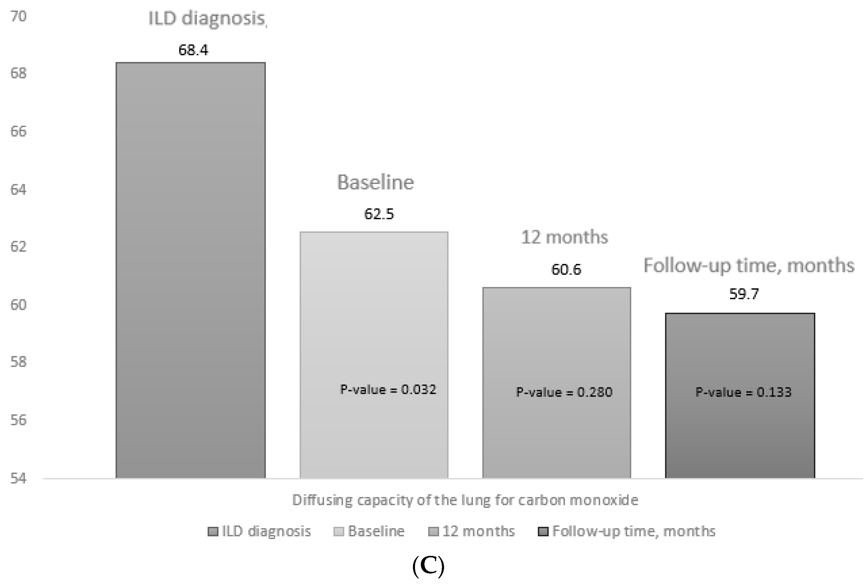
| DLCO-SB predicted (%), mean (SD) | 62.5 (16.5) | 60.6 (16.0) | 59.7 (15.7) | 0.340 | 0.133 |
| Clinical course | |||||
| Progression, n (%) | 25 (43.8) * | 10 (19.6) | 13 (22.8) | 0.014 | 0.036 |
| Stabilization, n (%) | 29 (50.8) * | 34 (66.6) | 38 (66.7) | ||
| Improvement, n (%) | 3 (5.3) * | 7 (13.7) | 6 (10.5) | ||
| HRCT | |||||
| Radiological type | 0.540 | 0.297 | |||
| UIP, n (%) | 34 (59.6) | 31 (60.7) | 36 (63.2) | ||
| NSIP, n (%) | 18 (31.6) | 16 (31.3) | 16 (28.1) | ||
| Fibrotic NSIP, n (%) | 5 (8.8) | 4 (7.8) | 5 (8.8) | ||
| Clinical course | 0.019 | 0.028 | |||
| Progression, n (%) | 20 (35.0) * | 10 (19.6) | 13 (22.8) | ||
| Stabilization, n (%) | 34 (59.6) * | 35 (68.6) | 39 (68.4) | ||
| Improvement, n (%) | 3 (5.3) * | 6 (11.7) | 5 (8.8) | ||
| Overall progress of lung disease ** | 0.010 | 0.019 | |||
| Improvement, n (%) | 3 (5.3) * | 7 (13.7) | 6 (10.5) | ||
| Stabilization, n (%) | 28 (48.4) * | 34 (66.6) | 35 (61.4) | ||
| Worsening, n (%) | 26 (45.6) * | 10 (17.5) | 13 (22.8) | ||
| Death, n (%) | - | - | 3 (5.3) | ||
| Inflammatory activity | |||||
| DAS28, mean (SD) | 3.8 (1.5) | 3.0 (1.6) | 3.1 (1.2) | 0.019 | 0.024 |
| C-reactive protein (mg/dl), median (IQR) | 7.0 (5.0–17.7) | 3.6 (2.4–10.1) | 3.3 (2.6–7.9) | 0.041 | 0.018 |
| ESR (mm/h), median (IQR) | 33.0 (15.0–47.0) | 19.0 (6.0–32.0) | 20.0 (8.0–29.0) | 0.045 | 0.046 |
| HAQ, median (IQR) | 1.0 (0.2–1.8) | 1.0 (0.3–1.8) | 1.1 (0.6–1.9) | 0.629 | 0.424 |
| Corticosteroids, n (%) | 39 (68.4) | 30 (58.8) | 28 (48.4) | 0.360 | 0.040 |
| Variable | Sample = 57 |
|---|---|
| Infections, n (%) | 25 (43.9) |
| Respiratory infection, n (%) | 20 (35.0) |
| Other infections, n (%) | 6 (10.5) |
| Cutaneous disease, n (%) | 2 (3.5) |
| Urinary infection, n (%) | 4 (7.0) |
| Transient neutropenia, n (%) | 1 (3.5) |
| Hospitalization, n (%) | 6 (10.5) |
| Reasons for hospitalization | |
| Progression of ILD, n (%) | 3 (5.2) |
| Respiratory infection, n (%) | 2 (3.5) |
| Pyelonephritis, n (%) | 1 (3.5) |
| Mortality, n (%) | 3 (5.2) |
| Variable | Univariate HR (95%CI) | Multivariate HR (IC 95%) | p-Value |
|---|---|---|---|
| Age, years | 1.000 (0.974–1.028) | ||
| Male sex | 2.975 (0.618–7.109) | ||
| Current or previous smoking history | 1.292 (0.386–4.321) | ||
| ACPA (>20 U/mL) | 0.417 (0.096–1.809) | ||
| UIP radiological pattern | 4.480 (0.896–12.390) | ||
| DAS28-ESR | 2.001 (1.194–3.354) | 2.520 (1.038–3.120) | 0.041 |
| FVC predicted (%) | 0.879 (0.819–0.944) | 0.826 (0.704–0.969) | 0.019 |
| DLCO-SB predicted (%) | 0.889 (0.835–0.946) | 0.837 (0.723–0.969) | 0.018 |
| Combination with methotrexate | 0.267 (0.066–0.996) | ||
| Corticosteroids (grams) | 1.272 (1.047–1.545) |
Publisher’s Note: MDPI stays neutral with regard to jurisdictional claims in published maps and institutional affiliations. |
© 2022 by the authors. Licensee MDPI, Basel, Switzerland. This article is an open access article distributed under the terms and conditions of the Creative Commons Attribution (CC BY) license (https://creativecommons.org/licenses/by/4.0/).
Share and Cite
Mena-Vázquez, N.; Rojas-Gimenez, M.; Fuego-Varela, C.; García-Studer, A.; Perez-Gómez, N.; Romero-Barco, C.M.; Godoy-Navarrete, F.J.; Manrique-Arija, S.; Gandía-Martínez, M.; Calvo-Gutiérrez, J.; et al. Safety and Effectiveness of Abatacept in a Prospective Cohort of Patients with Rheumatoid Arthritis–Associated Interstitial Lung Disease. Biomedicines 2022, 10, 1480. https://doi.org/10.3390/biomedicines10071480
Mena-Vázquez N, Rojas-Gimenez M, Fuego-Varela C, García-Studer A, Perez-Gómez N, Romero-Barco CM, Godoy-Navarrete FJ, Manrique-Arija S, Gandía-Martínez M, Calvo-Gutiérrez J, et al. Safety and Effectiveness of Abatacept in a Prospective Cohort of Patients with Rheumatoid Arthritis–Associated Interstitial Lung Disease. Biomedicines. 2022; 10(7):1480. https://doi.org/10.3390/biomedicines10071480
Chicago/Turabian StyleMena-Vázquez, Natalia, Marta Rojas-Gimenez, Clara Fuego-Varela, Aimara García-Studer, Nair Perez-Gómez, Carmen María Romero-Barco, Francisco Javier Godoy-Navarrete, Sara Manrique-Arija, Myriam Gandía-Martínez, Jerusalem Calvo-Gutiérrez, and et al. 2022. "Safety and Effectiveness of Abatacept in a Prospective Cohort of Patients with Rheumatoid Arthritis–Associated Interstitial Lung Disease" Biomedicines 10, no. 7: 1480. https://doi.org/10.3390/biomedicines10071480
APA StyleMena-Vázquez, N., Rojas-Gimenez, M., Fuego-Varela, C., García-Studer, A., Perez-Gómez, N., Romero-Barco, C. M., Godoy-Navarrete, F. J., Manrique-Arija, S., Gandía-Martínez, M., Calvo-Gutiérrez, J., Morales-Garrido, P., Mouriño-Rodriguez, C., Castro-Pérez, P., Añón-Oñate, I., Espildora, F., Aguilar-Hurtado, M. C., Hidalgo Conde, A., Arnedo Díez de los Ríos, R., Cabrera César, E., ... Fernández-Nebro, A. (2022). Safety and Effectiveness of Abatacept in a Prospective Cohort of Patients with Rheumatoid Arthritis–Associated Interstitial Lung Disease. Biomedicines, 10(7), 1480. https://doi.org/10.3390/biomedicines10071480