Article

Different Effects of Valproic Acid on SLC12A2, SLC12A5 and SLC5A8 Gene Expression in Pediatric Glioblastoma Cells as an Approach to Personalised Therapy

by
Eligija Damanskienė
1,
Ingrida Balnytė
1,
Angelija Valančiūtė
1,
Marta Marija Alonso
2 and
Donatas Stakišaitis
1,3,*
1
Department of Histology and Embryology, Medical Academy, Lithuanian University of Health Sciences, 44307 Kaunas, Lithuania
2
Department of Pediatrics, Clínica Universidad de Navarra, University of Navarra, 31008 Pamplona, Spain
3
Laboratory of Molecular Oncology, National Cancer Institute, 08660 Vilnius, Lithuania
*
Author to whom correspondence should be addressed.
Biomedicines 2022, 10(5), 968; https://doi.org/10.3390/biomedicines10050968
Submission received: 12 March 2022 / Revised: 19 April 2022 / Accepted: 20 April 2022 / Published: 22 April 2022
(This article belongs to the Special Issue Gender Medicine and Pharmacology)

Abstract

:
Valproic acid (VPA) is a histone deacetylase inhibitor with sex-specific immunomodulatory and anticancer effects. This study aimed to investigate the effect of 0.5 and 0.75 mM VPA on NKCC1 (SLC12A2), KCC2 (SLC12A5) and SLC5A8 (SLC5A8) co-transporter gene expressions in pediatric PBT24 (boy’s) and SF8628 (girl’s) glioblastoma cells. The SLC12A2, SLC12A5 and SLC5A8 RNA expressions were determined by the RT-PCR method. The SLC12A2 and SLC5A8 expressions did not differ between the PBT24 and SF8628 controls. The SLC12A5 expression in the PBT24 control was significantly higher than in the SF8628 control. VPA treatment significantly increased the expression of SLC12A2 in PBT24 but did not affect SF8628 cells. VPA increased the SLC12A5 expression in PBT24 and SF8628 cells. The SLC12A5 expression of the PBT24-treated cells was significantly higher than in corresponding SF8628 groups. Both VPA doses increased the SLC5A8 expression in PBT24 and SF8628 cells, but the expression was significantly higher in the PBT24-treated, compared to the respective SF8628 groups. The SLC5A8 expression in PBT24-treated cells was 10-fold higher than in SF8628 cells. The distinct effects of VPA on the expression of SLC12A2, SLC12A5 and SLC5A8 in PBT24 and SF8628 glioblastoma cells suggest differences in tumor cell biology that may be gender-related.

1. Introduction

Pediatric high-grade glioblastoma multiforme (phGBM) is a highly malignant brain tumor that is the leading cause of death in the pediatric brain cancer population [1,2]. Due to the difference in biological factors from adult glioblastoma and the uniqueness of this pediatric heterogeneous group of the tumor, reasonable therapeutic strategies for adult glioblastoma have not improved the outcome of phGBM treatment [3,4]. To effectively treat phGBM, it is essential to determine the individual sensitivity of the drug to carcinogenesis. The development of safe and effective anticancer medicines aims to improve the bioavailability of medicines and reduce the development of resistance to chemotherapy [5]. Equally important is the identification of the factors that make treatment effective. Such determinants may be gender-specific, which could be key to developing individualised treatments [6].
Valproic acid (2-n-propyl-pentanoic acid; VPA) is an investigational preparation for glioma treatment [7]. VPA is a well-known histone deacetylase inhibitor (HDACi) [8,9]. In combination with radiotherapy or chemotherapy, VPA delays the development of glioblastoma resistance to chemotherapy or radiotherapy [10,11,12,13,14]; in combination with radiotherapy, it increases the sensitivity of primary glioma cells to radiotherapy and γ-radiation-induced cytotoxicity [14,15]. Anti-tumor effects of VPA in combination with chemotherapy and radiotherapy are of interest for phGBM [16,17].
The metabolism of VPA is sex-specific. VPA is transported more via the G protein (MRP) in females than in males of experimental animals and humans [18,19]. Testosterone reduces drug transport [20]. Hepatic metabolism of VPA is more potent in females [21]; in males, due to lower expression of efflux pumps, a saturation of hepatobiliary transport results in longer retention of VPA in the hepatocyte, resulting in slower clearance of VPA [22].
One of the main anticancer effects of drugs is to inhibit tumor cell proliferation and induce apoptosis. VPA inhibits glioma cell proliferation and induces apoptosis [23]. The chloride anion (Cl) is involved in cell volume regulation and cell apoptosis [24]. An early sign of apoptosis is a decrease in cell volume due to loss of intracellular K+ and chloride ([Cl]i) [25,26]. High-grade glioblastoma cells are associated with increased [Cl]i [27], which is associated with increased Na-K-2Cl co-transporter (NKCC1) and decreased K-Cl co-transporter (KCC2) activity [28,29]. The sodium and Cl ion-dependent SLC5A8 co-transporter triggers cell apoptosis via pyruvate-dependent HDAC inhibition [30]. In elucidating the pathogenesis of glioblastoma, it is essential to investigate the mechanisms involved in the regulation of [Cl]i concerning the efficacy of therapy. The mechanisms by which VPA is involved in the regulation of [Cl]i are illustrated in Figure 1.
Increased expression of the NKCC1 in glioblastoma cells is linked to cell proliferation [31]. Increased NKCC1 protein expression in human glioblastoma is directly related to tumor grade and cell migration; inhibition of NKCC1 attenuates glioblastoma cell migration and tumor invasion [27,32,33]. VPA suppresses NKCC1 gene expression in male, but not female, rat thymocytes [34]. Reduction of KCC2 neuropil staining was reported in adult patients with glioblastoma and epilepsy [29]. VPA produces both kaliuretic and chloriduretic effects in males but not in female rats [35]. Whether the VPA initiates KCC2 activity in pediatric glioblastoma cells, leading to a parallel loss of K+ and Cl ions, needs to be investigated.
SLC5A8 is a sodium (Na+) and Cl dependent and sodium-linked monocarboxylate co-transporter that transports lactate, pyruvate, acetate, propionate, valerate, butyrate, and monocarboxylate preparations (dichloroacetate, nicotinate, salicylate) into cells [36,37]. SLC5A8 triggers cell apoptosis through pyruvate-dependent HDAC inhibition [30]. SLC5A8 is a tumor growth-inhibitory gene in primary human and experimental animal glioma and is suppressed by epigenetic mechanisms [35]. The epigenetic reprogramming of cancer cells induced by VPA is dependent on estrogen receptors [38]. Suppression of the SLC5A8 gene is linked to DNA methylation, and treatment of cancer cells with DNA demethylating agents increases SLC5A8 expression [37]. VPA may also activate genes regulated by DNA methylation, and the VPA treatment may be triggered by active DNA demethylation in cancer cells [39,40]. SLC5A8 activation in male rat duodenal enterocytes is correlated with NKCC1 activity [41]. In fibroblasts treated with the NKCC1 inhibitor bumetanide, an enhancement of maximal respiration indicates an increase in substrate availability and mitochondrial oxidation [42], indicating that altered NKCC1 activity may result in alterations in mitochondrial function.
The study aimed to investigate if there are differences in VPA’s effect on the expression of NKCC1, KCC2 and SLC5A8 co-transporter genes between high-grade SF8628 cells and high-grade PBT24 cells.

2. Materials and Methods

2.1. Cell Lines and Cell Culture

A 13-year-old boy’s high-grade glioblastoma PBT24 cell line cells were donated by Prof. M. M. Alonso (University of Navarra, Spain) [43] for the study. A 3-year-old girl’s diffuse intrinsic pontine glioblastoma (DIPG) SF8628 cell line cells—harboring the histone H3.3 Lys 27-to-methionine (Sigma Aldrich, St. Louis, MO, USA)—were also studied [44,45]. The PBT24 cells were cultivated in Roswell Park Memorial Institute 1640 (RPMI), medium (Sigma Aldrich, St. Louis, MO, USA) supplemented with 10% fetal bovine serum (FBS; Sigma Aldrich, St. Louis, MO, USA) containing 100 IU/mL of penicillin and 100 µg/mL of streptomycin (P/S; Sigma Aldrich, St. Louis, MO, USA). The SF8628 cells were cultivated in Dulbecco’s Modified Eagle Medium (DMEM)–High Glucose Media (Sigma Aldrich, St. Louis, MO, USA), supplemented with 10% fetal bovine serum (FBS; Sigma Aldrich, St. Louis, MO, USA) containing 100 IU/mL of penicillin and 100 µg/mL of streptomycin (P/S; Sigma Aldrich, St. Louis, MO, USA) and 2 mM L-Glutamine (Sigma Aldrich, St. Louis, MO, USA). Cells were incubated at 37 °C in a humidified 5% CO2 atmosphere.

2.2. Extraction of RNA from PBT24 and SF8628 Cell Line Cells

PBT24 and SF8628 cell line cells were treated with 0.5 mM VPA and 0.75 mM VPA for 24 h. The concentration of 0.5 mM VPA and 0.75 mM VPA was chosen because it corresponds to the mean plasma concentration of the drug in VPA-treated patients [46,47]. Control groups were cultured in a cell culture medium depending on the cell line. According to the manufacturer’s instructions, the total RNA was extracted using the TRIzol Plus RNA Purification Kit (Life Technologies, New York, NY, USA). The RNA quality and concentration were assessed using a NanoDrop2000 spectrophotometer (Thermo Scientific, Branchburg, NJ, USA). The total RNA integrity was analyzed using the Agilent 2100 Bioanalyzer system (Agilent Technologies, Santa Clara, CA, USA) with an Agilent RNA 6000 Nano Kit (Agilent Technologies, Santa Clara, CA, USA). The samples of RNA were stored at −80 °C until further analysis.

2.3. Determination of the SLC12A2, SLC12A5 and SLC5A8 Expression in PBT24 and SF8628 Cell Line Cells

RNA expression assays were performed for SLC12A2 (Hs00169032_m1), SLC12A5 (Hs00221168_m1), SLC5A8 (Hs00377618_m1) and GAPDH (Hs02786624_g1) genes. According to the manufacturer’s instructions, reverse transcription was performed with the High-Capacity cDNA Reverse Transcription Kit with RNase Inhibitor (Applied Biosystems, Waltham, MA, USA) in a 20 µL reaction volume containing 50 ng RNA using the Biometra TAdvanced thermal cycler (Analytik Jena AG, Jena, Germany). The synthesized copy DNA (cDNA) was stored at 4 °C until use, or at −80 °C for a longer time. Real-time polymerase chain reaction (PCR) was performed using an Applied Biosystems 7900 Fast Real-Time PCR System (Applied Bio-systems, Waltham, MA, USA). The reactions were run in triplicate with 4 µL of cDNA template in a 20 µL reaction volume, 10 µL of TaqMan Universal Master Mix II, no UNG (Applied Biosystems, Waltham, MA, USA), 1 µL of TaqMan Gene Expression Assay 20× (Applied Biosystems, Waltham, MA, USA), 5 µL of nuclease-free water (Invitrogen, Carlsbad, CA, USA), with the program running at 95 °C for 10 min, followed by 45 cycles of 95 °C for 15 s, 50 °C for 2 min and 60 °C for 1 min.
The control and 24 h VPA-treated groups (n = 6 per group) were tested for SLC12A2, SLC12A5, SLC5A8 and GAPDH expression.

2.4. Statistical Analysis

To investigate the SLC12A2, SLC12A5, and SLC5A8 RNA gene expression in the VPA-treated and control groups, the threshold cycle (CT) value was normalized with the control GAPDH, and the ΔCT value was obtained. The Livak method (∆∆CT) was used for calculating the relative fold change in expression levels [48]. The Spearman’s rank correlation coefficient (r) was used to assess relationships between the SLC12A2 and SLC12A5 (ΔCT values were used). Differences at the value of p < 0.05 were considered significant. The figures were created using GraphPad Prism 7 and IBM SPSS Statistics 23.0 software.

3. Results

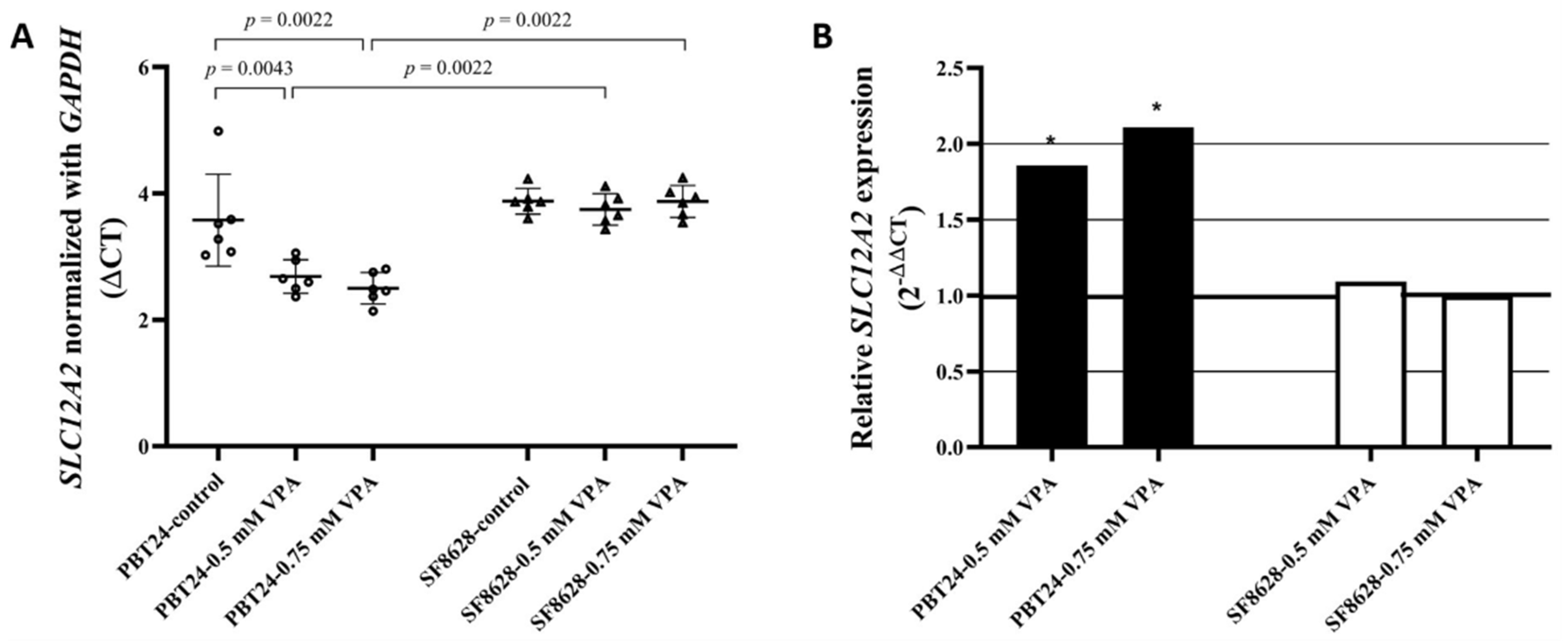
3.1. The Expression of SLC12A2 in PBT24 and SF8628 Cell Study Groups

The SLC12A2 expression data for the PBT24 and SF8628 cell groups tested are shown in Table 1 and Figure 2.
A comparison of the SLC12A2 expression of PBT24 and SF8628 cells between the control groups showed no difference. Treatment with the tested doses of VPA significantly increased the SLC12A2 expression of PBT24 cells. No significant VPA effect on SF8628 cells was observed. After 24 h of treatment with the tested doses of VPA, the SLC12A2 expression of SF8628 cells was significantly lower than that of PBT24 cells treated with 0.5 and 0.75 mM of VPA.
Compared to the PBT24 control, the 0.5 mM VPA dose increased SLC12A2 expression in PBT24 cells 1.9-fold (2−ΔΔCT = 1.858), while the 0.75 mM VPA dose increased SLC12A2 expression in PBT24 cells 2.1-fold without significant difference between the two treated groups (Figure 2B).

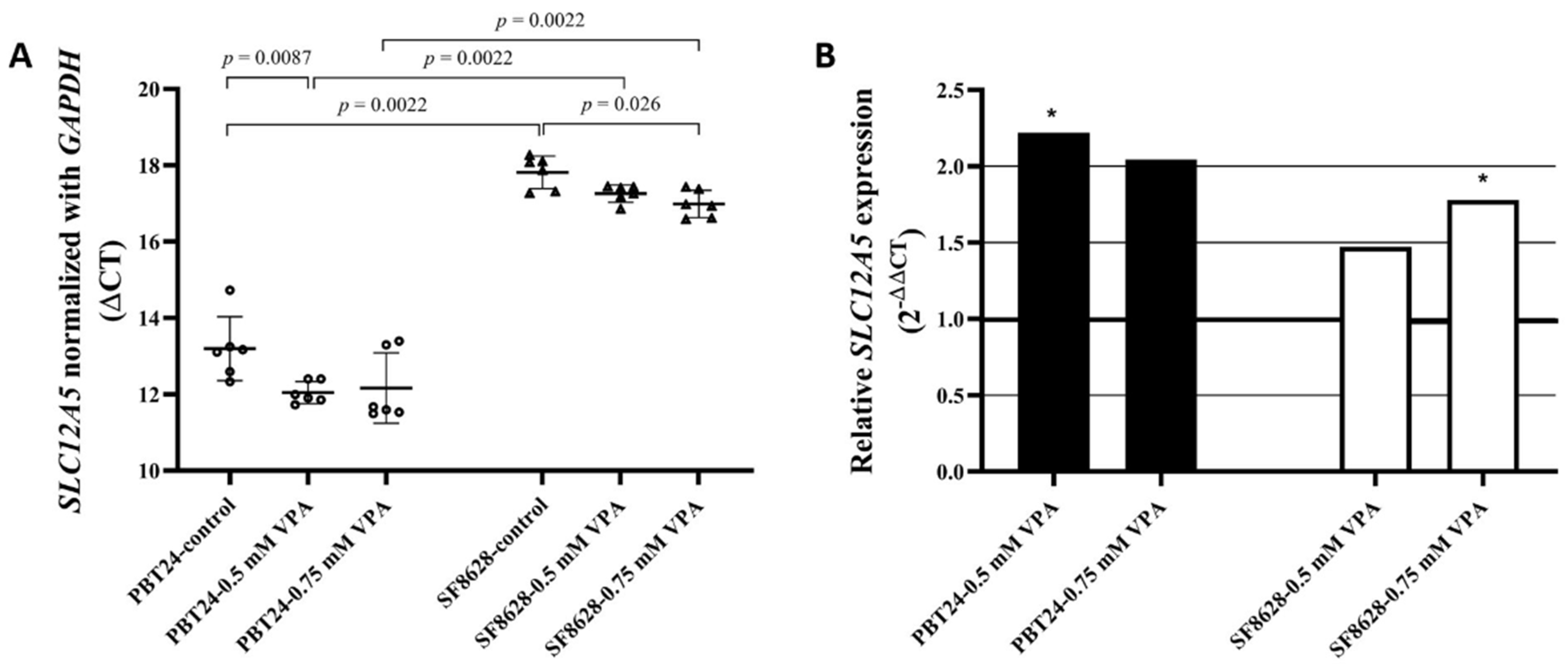
3.2. The Expression of SLC12A5 in PBT24 and SF8628 Cell Study Groups

The expression data of SLC12A5 and GAPDH in PBT24 and SF8628 cell controls and in the groups treated with two doses of VPA for 24 h are shown in Table 2.
The expression of SLC12A5 in PBT24 control cells is significantly higher than that of SF8628 control cells. Comparison of PBT24 control and 0.5 mM VPA-treated cells showed substantially higher SLC12A5 expression in treated cells. Comparing the expression of SLC12A5 in PBT24 control and 0.75 mM VPA-treated cells, there is a clear trend towards an increase in expression in treated cells, but no statistical difference was found. No difference in the SLC12A5 expression was determined when comparing 0.5 mM and 0.75 mM PBT24 treated cells. The SLC12A5 expression in PBT24 cells treated with the tested doses of VPA was significantly higher than in the corresponding SF8628 cell groups.
When comparing the expression of SLC12A5 in SF8628 control with those treated with a 0.75 mM dose of VPA, the expression was significantly higher in treated cells. Compared to control, no statistically significant change in SLC12A5 expression was observed in dose cells treated with 0.5 mM VPA (Table 2; Figure 3A). There was no significant difference in SLC12A5 expression between SF8628 cells treated with different VPA doses (p > 0.05).
Compared to the respective control, treatment with 0.5 mM VPA dose increased SLC12A5 expression 2.2-fold (2−ΔΔCT = 2.220) in PBT24 cells and 1.5-fold in SF8628 cells; treatment with 0.75 mM VPA increased the expression of SLC12A5 in PBT24 cells 2-fold and that of SF8628 cells 1.8-fold (Figure 3B).
Comparing the gene expression of the tested cell controls, PBT24 cells’ SLC12A5 was expressed 24.6-fold (2−ΔΔCT = 24.625) over SF8628 cells. The SLC12A5 expression of PBT24 cells treated with 0.5 mM VPA was 37-fold (2−ΔΔCT) higher over SF8626-0.5 mM VPA cells; those treated with 0.75 mM VPA were 28-fold higher in PBT24 cells than in SF8628 cells.
The correlation (r) between ΔCT SLC12A2 and ΔCT SLC12A5 values of the study groups was the following: 0.71 for the PBT24 cell control (p < 0.05), 0.14 for the SF8628 cell control (p > 0.05), 0.03 for the PBT24 cells treated with 0.5 mM VPA (p > 0.05) and 0.37 for the SF8628 cells treated with 0.5 mM VPA (p > 0.05; Figure 4); 0.37 for the PBT24 cells treated with 0.75 mM VPA, and −0.08 for the SF8628 cells treated with 0.75 mM VPA (p > 0.05 in groups). VPA treatment significantly altered the correlation between SLC12A2 and SLC12A5 in the PBT24 cell. These changes suggest that VPA may reduce the [Cl]i of the PBT24 cell through activation of SLC12A5 and down-regulation of SLC12A2. In contrast, we did not find a trend towards such an effect in the SF8628 cells.

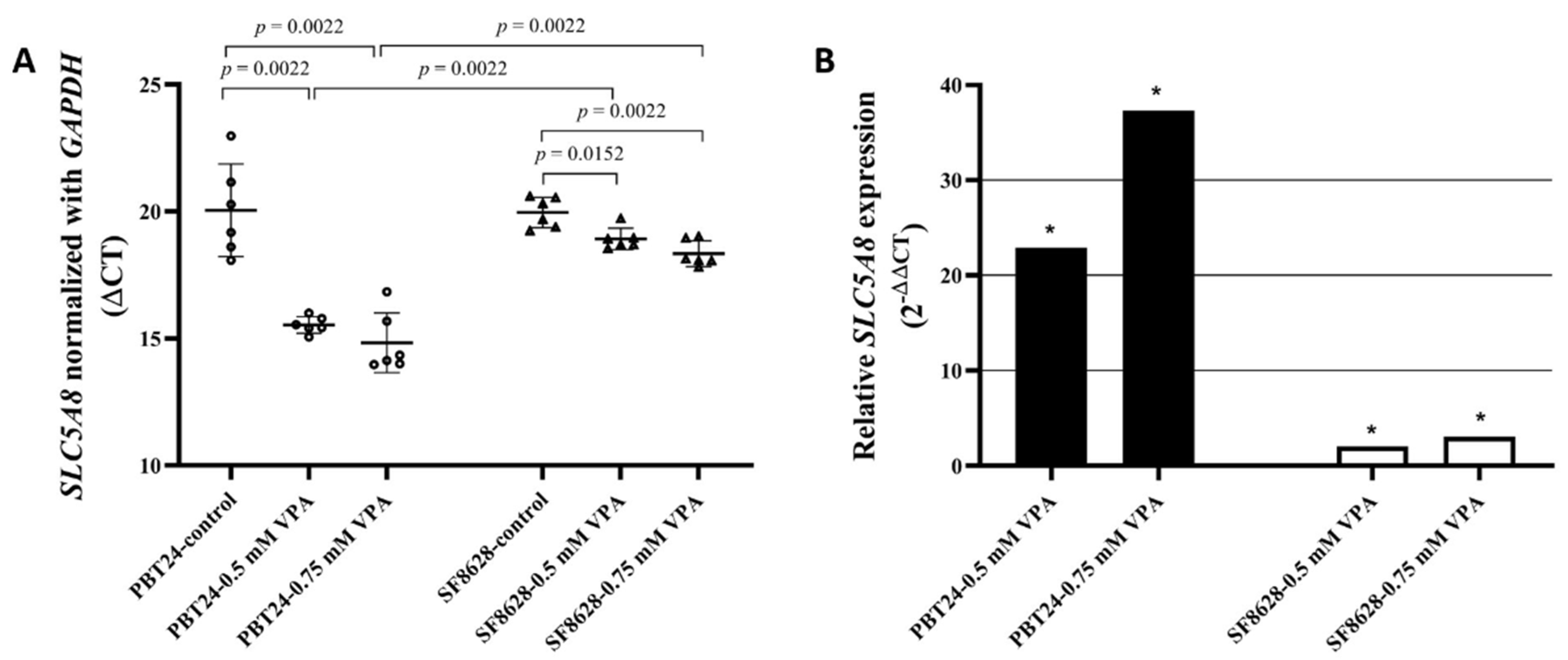
3.3. The Expression of SLC5A8 in PBT24 and SF8628 Cell Study Groups

The expression data for SLC5A8 and GAPDH in PBT24 and SF8628 cells in the control and 0.5 and 0.75 mM VPA-treated groups for 24 h are shown in Table 3.
The expression of SLC5A8 in PBT24 control cells was not different from that in SF8628 control. SLC5A8 expression was significantly higher in the PBT24 cell groups treated with both VPA doses than in the corresponding groups of SF8628 cells. Compared with the respective control, treatment with VPA doses significantly increased SLC5A8 expression in PBT24 and SF8628 cells (Table 3; Figure 5A).
Compared to the respective control, treatment with 0.5 mM VPA dose increased the SLC5A8 expression 22.9-fold (2−ΔΔCT = 22.907) in PBT24 cells and 2-fold in SF8628 cells; treatment with 0.75 mM VPA increased the SLC5A8 expression in PBT24 cells by 37.3-fold and by 3-fold in the SF8628 cells (Figure 5B).
The SLC5A8 expression in PBT24 cells treated with 0.5 mM VPA was 10.5-fold (2−ΔΔCT = 10.475) higher than that in SF8628 cells, while SLC5A8 expression in PBT24 cells treated with 0.75 mM VPA was 11.4-fold more heightened than in SF8628 cells.

4. Discussion

The efficacy of treatment of glioblastoma in the presence of high tumor polymorphism suggests that a single strategy to achieve an effective treatment is unwise [49]. The goal should be the personalised treatment of pediatric glioblastoma, tailored to the individual patient’s sex-specific molecular and genetic characteristics of tumor carcinogenesis [4].
Recently reported experimental in vivo data have shown the different effects of temozolomide (TMZ) on PBT24 and SF6828 xenografts and the molecular mechanisms involved. TMZ reduced PBT24 tumor growth, and treatment efficacy was related to significantly increased SLC12A5 expression in PBT24 cells, but there were no such effects on SLC12A5 expression of SF8628 cells [50].
The [Cl]i content of high-grade glioblastoma cells is significantly higher than the average in grade II glioma and normal cortical cells [27]. Increased [Cl]i level in glioblastoma cells are associated with increased NKCC1 and decreased K-Cl co-transporter activity [28,29]. Na-K-2Cl co-transporter activity in glioblastoma is associated with cell proliferation [31]; the increased expression of NKCC1 protein in human glioblastoma is associated with tumor grade; inhibition of NKCC1 activity impairs tumor invasion [27,32]. Strong NKCC1 immune reactivity is characteristic of the aberrant neuronal component of the glioblastoma, while the increase in neuronal NKCC1 expression is not characteristic of the peri-lesional zone of tumor samples. The high levels of neuronal NKCC1 in phGBM support the hypothesis of abnormal and immature neuronal cells [29]. The study showed no difference in SLC12A2 expression between PBT24 and SF8628 cell controls, but VPA treatment significantly increased the expression of SLC12A2 in PBT24 cells, while no such effect was determined in SF8628 cells. The mechanism of this VPA action is unknown. TMZ treatment increased NKCC1 expression in PBT24 and SF8626 cells [50]. TMZ-induced NKCC1 activation is associated with increased WNK kinases protein phosphorylation [51,52,53]. The suppressive effect on glioma cell [Cl]i may be due to inhibition of NKCC1 activity and other Cl transport mechanisms involved in Cl efflux from the cell [33,50].
Our study shows that the PBT24 control cell SLC12A5 expression is significantly higher than the SF8628 controls. When comparing the SLC12A5 expression between the PBT24 and the SF8628 controls and cells treated with VPA, the expression was significantly higher in both lines of treated cells, while the expression of SLC12A5 was explicitly higher in VPA-treated PBT24 cells than in the corresponding SF8628 groups. It cannot be ruled out that such differences between study cells are also related to gender.
A key feature of cancer cell apoptosis is a reduction in cell volume due to a decrease in [K+]i and [Cl]i [25,26]. Apoptosis requires a steady decrease in cell volume due to the loss of [K+]i and [Cl]i, which occurs before other detectable signs of apoptosis [24,54,55]. The decrease in [K+]i and [Cl]i is associated with caspase activation and caspase cascade-related apoptotic mechanisms [26]. Furthermore, KCC2 is a neuron-specific K+ and Cl extruder that uses a K+ gradient to maintain low [Cl]i levels and ensure the proper functioning of postsynaptic GABAA receptors. The paradoxical excitatory effects of GABAA depend on the relatively high [Cl]i content [56]. Increased activity of NKCC1 can lead to astrocyte swelling [57,58] and induce a GABAA receptor-mediated excitatory response that promotes seizures [56,58,59]. Reduced KCC2 neuropil staining has been found in glioblastoma of patients with marked epilepsy [29]. Reduced KCC2 expression and function is a hallmark of evolving cerebral epileptic disorders [60]. The effect of drugs that activate KCC2 function in glioblastoma cells is of interest as a potential new anticancer therapeutic target. Future research on the co-localization of Cl co-transporters with the GABAA receptor may shed light on the importance of the functional interaction of Cl transporters in glioblastoma cells.
The VPA-associated anti-tumor activity has been observed in children with refractory and those with phGBM who received VPA monotherapy for progressive phGBM after radiotherapy and chemotherapy [16,61,62,63]. However, a meta-analysis of 1869 patients enrolled in five phase III trials for newly diagnosed glioblastoma found no improvement with the addition of VPA to standard chemotherapy [64]. Such conflicting data on VPA efficacy suggest that future studies could differentiate treatment efficacy by individual characteristics. An example of a potential non-effective treatment or induced tumor progression could be the combination of VPA with temozolomide—a case where both preparations would activate NKCC1 function synergistically.
Another important cancer marker in determining the effectiveness of VPA could be the SLC5A8 co-transporter, which acts as an electrogenic sodium- and chloride-dependent sodium-coupled co-transporter [65,66]. The SLC5A8 acts as a tumor growth suppressor gene, often silenced by epigenetic mechanisms in primary gliomas [35]. Silencing the SLC5A8 is associated with DNA methylation, and treatment of cancer cells with DNA demethylating agents increases the SLC5A8 expression [37]. VPA inhibits HDAC activity [67], inducing DNA demethylation and increasing the SLC5A8 expression [37,39,40]. SLC5A8 induces cell apoptosis via pyruvate-dependent inhibition of HDAC [30]. Our data show that the expression of SLC5A8 in control PBT24 cells did not differ from that of SF8628 controls. Compared to the respective control, treatment with both doses of VPA significantly increased SLC5A8 expression in PBT24 and SF8628 cells, but SLC5A8 expression was explicitly higher in VPA-treated PBT24 cells. These data show that VPA has a stronger tumor-suppressive effect on PBT24 cells (boy’s) than SF8626 (girl’s) cells. The SLC5A8 activity in the male rat duodenum enterocytes depends on Na+-K+-2Cl cotransporter (NKCC1) activity [41]. Changes in the NKCC1 activity might lead to mitochondrial SLC5A8-related function changes [42]. The differences in VPA effects found between PBT24 and SF8628 pediatric glioblastoma may indicate medicine differential effects on carcinogenesis linked to [Cl]i regulation, as PBT24 cells showed a higher expression of the KCC2 gene as well as a more pronounced activation of the co-transporter gene under VPA exposure. The effect of VPA on KCC2 is inseparable from the activity of the GABAA receptor. The role of VPA treatment on NKCC1, KCC2, and GABAA in the regulation of [Cl]i is an important point for further investigation. VPA also differently activated the expression of the SLC5A8 gene in the study cells. Furthermore, it cannot be excluded that the found differences between PBT24 and SF8628 are related to sex. Thus, assessing the gender of the cell is key to determining the efficacy of anticancer drugs, as it allows the molecular subtype of glioblastoma to be identified and allows for more effective personalised therapies to be developed based on molecular mechanisms that are also linked to sex [68].
The detected differences in VPA treatment between the compared glioblastoma cells may be therapeutically significant. The several-fold increase in the expression of SLC12A5 and SLC5A8 after VPA treatment directly reflects correlation with the RNA gene methylation. The mRNA assay is used to quantify gene expression and is a more accurate identifier of gene activity than the DNA methylation test [69]. According to the scientific literature, the correlation between differentially expressed mRNA and mRNA/protein of the same gene is debatable. Genome-wide correlations between mRNA and protein are low [70,71,72]. Thus, the limitation of the protein expression investigation of the studied genes can also be justified by the doubtful likelihood that short-term treatment with VPA would be associated with changes in protein expression. Therefore, evidence of apparent differences in gene expression obtained after VPA treatment is significant. A limitation of our study is that other mechanisms underlying protein activity were not excluded. The study data include NKCC1, which determines Cl influx, and KCC2, which resolves anion efflux in a cell. Therefore, it would be further meaningful to investigate the VPA effect on [Cl]i, which reflects Cl- transport mechanisms and is a signaling pathway. Such follow-up research would also allow assessment of the effect of VPA on the functioning of the Cl anion-dependent SLC5A8 co-transporter.

5. Conclusions

The different effects of VPA on PBT24 and SF8628 glioblastoma cells are associated with a higher impact of VPA on SLC12A5, which may be related to the more effective reduction of [Cl]i level in PBT24 cells. VPA more strongly promotes SLC5A8 activation in PBT24 cells. The different effects of VPA on the expression of SLC12A2, SLC12A5, and SLC5A8 in PBT24 and SF8628 glioblastoma cells suggest differences in tumor cell biology. The study shows the importance of investigating the mechanisms of chloride anion transport across the cell membrane and their possible relationship with VPA treatment. Further research is needed on the association of VPA and gender with the treatment effectiveness on glioma.

Author Contributions

Conceptualization, D.S., I.B., A.V. and E.D.; Methodology, E.D.; Validation, D.S., I.B. and A.V.; Formal analysis, E.D., D.S., I.B. and A.V.; Investigation, E.D.; Resources, D.S., I.B. and M.M.A.; Data curation, E.D.; Writing—Original Draft Preparation, E.D. and D.S.; Writing—Review and Editing, E.D., D.S., A.V., I.B, and M.M.A.; Visualization, E.D.; Supervision, D.S.; Project Administration, D.S. and I.B.; Funding Acquisition, D.S. and I.B. All authors have read and agreed to the published version of the manuscript.

Funding

This research was funded by the Research Council of Lithuania, grant number P-MIP-20-36.

Institutional Review Board Statement

Not applicable.

Informed Consent Statement

Not applicable.

Data Availability Statement

The data presented in this study are available on request from the corresponding author.

Conflicts of Interest

The authors declare no conflict of interest.

References

  1. Njonkou, R.; Jackson, C.M.; Woodworth, G.F.; Hersh, D.S. Pediatric Glioblastoma: Mechanisms of Immune Evasion and Potential Therapeutic Opportunities. Cancer Immunol. Immunother. 2022. [Google Scholar] [CrossRef] [PubMed]
  2. Coleman, C.; Stoller, S.; Grotzer, M.; Stucklin, A.G.; Nazarian, J.; Mueller, S. Pediatric Hemispheric High-Grade Glioma: Targeting the Future. Cancer Metastasis Rev. 2020, 39, 245–260. [Google Scholar] [CrossRef]
  3. Jones, C.; Karajannis, M.A.; Jones, D.T.W.; Kieran, M.W.; Monje, M.; Baker, S.J.; Becher, O.J.; Cho, Y.-J.; Gupta, N.; Hawkins, C.; et al. Pediatric High-Grade Glioma: Biologically and Clinically in Need of New Thinking. Neuro Oncol. 2017, 19, 153–161. [Google Scholar] [CrossRef] [Green Version]
  4. Ene, C.I.; Holland, E.C. Personalized Medicine for Gliomas. Surg. Neurol. Int. 2015, 6, S89–S95. [Google Scholar] [CrossRef]
  5. Puiu, R.A.; Balaure, P.C.; Constantinescu, E.; Grumezescu, A.M.; Andronescu, E.; Oprea, O.-C.; Vasile, B.S.; Grumezescu, V.; Negut, I.; Nica, I.C.; et al. Anti-Cancer Nanopowders and MAPLE-Fabricated Thin Films Based on SPIONs Surface Modified with Paclitaxel Loaded β-Cyclodextrin. Pharmaceutics 2021, 13, 1356. [Google Scholar] [CrossRef] [PubMed]
  6. Fisher, J.L.; Jones, E.F.; Flanary, V.L.; Williams, A.S.; Ramsey, E.J.; Lasseigne, B.N. Considerations and Challenges for Sex-Aware Drug Repurposing. Biol. Sex Differ. 2022, 13, 13. [Google Scholar] [CrossRef] [PubMed]
  7. Li, C.; Chen, H.; Tan, Q.; Xie, C.; Zhan, W.; Sharma, A.; Sharma, H.S.; Zhang, Z. The Therapeutic and Neuroprotective Effects of an Antiepileptic Drug Valproic Acid in Glioma Patients. Prog. Brain Res. 2020, 258, 369–379. [Google Scholar] [CrossRef] [PubMed]
  8. Daśko, M.; de Pascual-Teresa, B.; Ortín, I.; Ramos, A. HDAC Inhibitors: Innovative Strategies for Their Design and Applications. Molecules 2022, 27, 715. [Google Scholar] [CrossRef]
  9. Wawruszak, A.; Halasa, M.; Okon, E.; Kukula-Koch, W.; Stepulak, A. Valproic Acid and Breast Cancer: State of the Art in 2021. Cancers 2021, 13, 3409. [Google Scholar] [CrossRef]
  10. Monga, V.; Swami, U.; Tanas, M.; Bossler, A.; Mott, S.L.; Smith, B.J.; Milhem, M. A Phase I/II Study Targeting Angiogenesis Using Bevacizumab Combined with Chemotherapy and a Histone Deacetylase Inhibitor (Valproic Acid) in Advanced Sarcomas. Cancers 2018, 10, 53. [Google Scholar] [CrossRef] [Green Version]
  11. Han, W.; Yu, F.; Wang, R.; Guan, W.; Zhi, F. Valproic Acid Sensitizes Glioma Cells to Luteolin through Induction of Apoptosis and Autophagy via Akt Signaling. Cell. Mol. Neurobiol. 2021, 41, 1625–1634. [Google Scholar] [CrossRef] [PubMed]
  12. Shao, C.; Wu, M.; Chen, F.; Li, C.; Xia, Y.; Chen, Z. Histone Deacetylase Inhibitor, 2-Propylpentanoic Acid, Increases the Chemosensitivity and Radiosensitivity of Human Glioma Cell Lines in Vitro. Chin. Med. J. 2012, 125, 4338–4343. [Google Scholar] [PubMed]
  13. Chinnaiyan, P.; Cerna, D.; Burgan, W.E.; Beam, K.; Williams, E.S.; Camphausen, K.; Tofilon, P.J. Postradiation Sensitization of the Histone Deacetylase Inhibitor Valproic Acid. Clin. Cancer Res. 2008, 14, 5410–5415. [Google Scholar] [CrossRef] [PubMed] [Green Version]
  14. Van Nifterik, K.A.; Van den Berg, J.; Slotman, B.J.; Lafleur, M.V.M.; Sminia, P.; Stalpers, L.J.A. Valproic Acid Sensitizes Human Glioma Cells for Temozolomide and γ-Radiation. J. Neuro-Oncol. 2012, 107, 61–67. [Google Scholar] [CrossRef]
  15. Pont, L.M.E.B.; Naipal, K.; Kloezeman, J.J.; Venkatesan, S.; van den Bent, M.; van Gent, D.C.; Dirven, C.M.F.; Kanaar, R.; Lamfers, M.L.M.; Leenstra, S. DNA Damage Response and Anti-Apoptotic Proteins Predict Radiosensitization Efficacy of HDAC Inhibitors SAHA and LBH589 in Patient-Derived Glioblastoma Cells. Cancer Lett. 2015, 356, 525–535. [Google Scholar] [CrossRef] [PubMed]
  16. Su, J.M.-F.; Murray, J.C.; McNall-Knapp, R.Y.; Bowers, D.C.; Shah, S.; Adesina, A.M.; Paulino, A.C.; Jo, E.; Mo, Q.; Baxter, P.A.; et al. A Phase 2 Study of Valproic Acid and Radiation, Followed by Maintenance Valproic Acid and Bevacizumab in Children with Newly Diagnosed Diffuse Intrinsic Pontine Glioma or High-Grade Glioma. Pediatr. Blood Cancer 2020, 67, e28283. [Google Scholar] [CrossRef]
  17. Berendsen, S.; Broekman, M.; Seute, T.; Snijders, T.; van Es, C.; de Vos, F.; Regli, L.; Robe, P. Valproic Acid for the Treatment of Malignant Gliomas: Review of the Preclinical Rationale and Published Clinical Results. Expert Opin. Investig. Drugs 2012, 21, 1391–1415. [Google Scholar] [CrossRef]
  18. Rost, D.; Kopplow, K.; Gehrke, S.; Mueller, S.; Friess, H.; Ittrich, C.; Mayer, D.; Stiehl, A. Gender-Specific Expression of Liver Organic Anion Transporters in Rat. Eur. J. Clin. Investig. 2005, 35, 635–643. [Google Scholar] [CrossRef]
  19. Smirnova, O.V. Sex differences in drug action: The role of multidrug-resistance proteins (MRPs). Fiziol Cheloveka 2012, 38, 124–136. [Google Scholar]
  20. Suzuki, T.; Zhao, Y.L.; Nadai, M.; Naruhashi, K.; Shimizu, A.; Takagi, K.; Takagi, K.; Hasegawa, T. Gender-Related Differences in Expression and Function of Hepatic P-Glycoprotein and Multidrug Resistance-Associated Protein (Mrp2) in Rats. Life Sci. 2006, 79, 455–461. [Google Scholar] [CrossRef]
  21. Ibarra, M.; Vázquez, M.; Fagiolino, P.; Derendorf, H. Sex Related Differences on Valproic Acid Pharmacokinetics after Oral Single Dose. J. Pharmacokinet. Pharmacodyn. 2013, 40, 479–486. [Google Scholar] [CrossRef] [PubMed]
  22. Wong, H.; Xia, B.; Tong, V.; Kumar, S.; Kenny, J. Atypical Kinetics of Valproic Acid Glucuronidation In vitro and In vivo in Humans. Biol. Med. 2012, S11, 6. [Google Scholar] [CrossRef] [Green Version]
  23. Riva, G.; Cilibrasi, C.; Bazzoni, R.; Cadamuro, M.; Negroni, C.; Butta, V.; Strazzabosco, M.; Dalprà, L.; Lavitrano, M.; Bentivegna, A. Valproic Acid Inhibits Proliferation and Reduces Invasiveness in Glioma Stem Cells Through Wnt/β Catenin Signalling Activation. Genes 2018, 9, 522. [Google Scholar] [CrossRef] [PubMed] [Green Version]
  24. Okada, Y.; Maeno, E. Apoptosis, Cell Volume Regulation and Volume-Regulatory Chloride Channels. Comp. Biochem. Physiol. A Mol. Integr. Physiol. 2001, 130, 377–383. [Google Scholar] [CrossRef]
  25. Maeno, E.; Ishizaki, Y.; Kanaseki, T.; Hazama, A.; Okada, Y. Normotonic Cell Shrinkage Because of Disordered Volume Regulation is an Early Prerequisite to Apoptosis. Proc. Natl. Acad. Sci. USA 2000, 97, 9487–9492. [Google Scholar] [CrossRef] [PubMed] [Green Version]
  26. Bortner, C.D.; Cidlowski, J.A. Cell Shrinkage and Monovalent Cation Fluxes: Role in Apoptosis. Arch. Biochem. Biophys. 2007, 462, 176–188. [Google Scholar] [CrossRef] [Green Version]
  27. Garzon-Muvdi, T.; Schiapparelli, P.; ap Rhys, C.; Guerrero-Cazares, H.; Smith, C.; Kim, D.-H.; Kone, L.; Farber, H.; Lee, D.Y.; An, S.S.; et al. Regulation of Brain Tumor Dispersal by NKCC1 through a Novel Role in Focal Adhesion Regulation. PLoS Biol. 2012, 10, e1001320. [Google Scholar] [CrossRef] [Green Version]
  28. Pallud, J.; Le Van Quyen, M.; Bielle, F.; Pellegrino, C.; Varlet, P.; Cresto, N.; Baulac, M.; Duyckaerts, C.; Kourdougli, N.; Chazal, G.; et al. Cortical GABAergic Excitation Contributes to Epileptic Activities around Human Glioma. Sci. Transl. Med. 2014, 6, 244ra89. [Google Scholar] [CrossRef] [Green Version]
  29. Aronica, E.; Boer, K.; Redeker, S.; Spliet, W.G.M.; van Rijen, P.C.; Troost, D.; Gorter, J.A. Differential Expression Patterns of Chloride Transporters, Na+-K+-2Cl--Cotransporter and K+-Cl--Cotransporter, in Epilepsy-Associated Malformations of Cortical Development. Neuroscience 2007, 145, 185–196. [Google Scholar] [CrossRef]
  30. Thangaraju, M.; Carswell, K.N.; Prasad, P.D.; Ganapathy, V. Colon Cancer Cells Maintain Low Levels of Pyruvate to Avoid Cell Death Caused by Inhibition of HDAC1/HDAC3. Biochem. J. 2009, 417, 379–389. [Google Scholar] [CrossRef]
  31. Cong, D.; Zhu, W.; Kuo, J.S.; Hu, S.; Sun, D. Ion Transporters in Brain Tumors. Curr. Med. Chem. 2015, 22, 1171–1181. [Google Scholar] [CrossRef] [PubMed] [Green Version]
  32. Haas, B.R.; Cuddapah, V.A.; Watkins, S.; Rohn, K.J.; Dy, T.E.; Sontheimer, H. With-No-Lysine Kinase 3 (WNK3) Stimulates Glioma Invasion by Regulating Cell Volume. Am. J. Physiol. Cell Physiol. 2011, 301, C1150–C1160. [Google Scholar] [CrossRef] [PubMed] [Green Version]
  33. Haas, B.R.; Sontheimer, H. Inhibition of the Sodium-Potassium-Chloride Cotransporter Isoform-1 Reduces Glioma Invasion. Cancer Res. 2010, 70, 5597–5606. [Google Scholar] [CrossRef] [PubMed] [Green Version]
  34. Juknevičienė, M.; Balnytė, I.; Valančiūtė, A.; Lesauskaitė, V.; Stanevičiūtė, J.; Curkūnavičiūtė, R.; Stakišaitis, D. Valproic Acid Inhibits NA-K-2CL Cotransporter RNA Expression in Male But Not in Female Rat Thymocytes. Dose Response 2019, 17, 1559325819852444. [Google Scholar] [CrossRef] [PubMed]
  35. Hong, C.; Maunakea, A.; Jun, P.; Bollen, A.W.; Hodgson, J.G.; Goldenberg, D.D.; Weiss, W.A.; Costello, J.F. Shared Epigenetic Mechanisms in Human and Mouse Gliomas Inactivate Expression of the Growth Suppressor SLC5A8. Cancer Res. 2005, 65, 3617–3623. [Google Scholar] [CrossRef] [Green Version]
  36. Gopal, E.; Fei, Y.-J.; Miyauchi, S.; Zhuang, L.; Prasad, P.D.; Ganapathy, V. Sodium-Coupled and Electrogenic Transport of B-Complex Vitamin Nicotinic Acid by Slc5a8, a Member of the Na/Glucose Co-Transporter Gene Family. Biochem. J. 2005, 388, 309–316. [Google Scholar] [CrossRef] [Green Version]
  37. Babu, E.; Ramachandran, S.; CoothanKandaswamy, V.; Elangovan, S.; Prasad, P.D.; Ganapathy, V.; Thangaraju, M. Role of SLC5A8, a Plasma Membrane Transporter and a Tumor Suppressor, in the Antitumor Activity of Dichloroacetate. Oncogene 2011, 30, 4026–4037. [Google Scholar] [CrossRef] [Green Version]
  38. Travaglini, L.; Vian, L.; Billi, M.; Grignani, F.; Nervi, C. Epigenetic Reprogramming of Breast Cancer Cells by Valproic Acid Occurs Regardless of Estrogen Receptor Status. Int. J. Biochem. Cell Biol. 2009, 41, 225–234. [Google Scholar] [CrossRef]
  39. Milutinovic, S.; D’Alessio, A.C.; Detich, N.; Szyf, M. Valproate Induces Widespread Epigenetic Reprogramming Which Involves Demethylation of Specific Genes. Carcinogenesis 2007, 28, 560–571. [Google Scholar] [CrossRef] [Green Version]
  40. Veronezi, G.M.B.; Felisbino, M.B.; Gatti, M.S.V.; Mello, M.L.S.; de Vidal, B.C. DNA Methylation Changes in Valproic Acid-Treated HeLa Cells as Assessed by Image Analysis, Immunofluorescence and Vibrational Microspectroscopy. PLoS ONE 2017, 12, e0170740. [Google Scholar] [CrossRef]
  41. Kaji, I.; Iwanaga, T.; Watanabe, M.; Guth, P.H.; Engel, E.; Kaunitz, J.D.; Akiba, Y. SCFA Transport in Rat Duodenum. Am. J. Physiol. Gastrointest Liver Physiol. 2015, 308, G188–G197. [Google Scholar] [CrossRef] [PubMed] [Green Version]
  42. Omer, S.; Koumangoye, R.; Delpire, E. A Mutation in the Na-K-2Cl Cotransporter-1 Leads to Changes in Cellular Metabolism. J. Cell. Physiol. 2020, 235, 7239–7250. [Google Scholar] [CrossRef] [PubMed]
  43. Martinez-Velez, N.; Marigil, M.; García-Moure, M.; Gonzalez-Huarriz, M.; Aristu, J.J.; Ramos-García, L.-I.; Tejada, S.; Díez-Valle, R.; Patiño-García, A.; Becher, O.J.; et al. Delta-24-RGD Combined with Radiotherapy Exerts a Potent Antitumor Effect in Diffuse Intrinsic Pontine Glioma and Pediatric High Grade Glioma Models. Acta Neuropathol. Commun. 2019, 7, 64. [Google Scholar] [CrossRef] [Green Version]
  44. Sigma-Aldrich. SF8628 Human DIPG H3.3-K27M Cell Line SF8628 Pediatric Diffuse Intrinsic Pontine Glioma (DIPG) Cell Line Harbors the Histone H3.3 Lys 27-to-Methionine (K27M) Mutation and Can Support Research and Drug Development Efforts Targeting DIPG. Available online: http://www.sigmaaldrich.com/ (accessed on 8 December 2021).
  45. Mueller, S.; Hashizume, R.; Yang, X.; Kolkowitz, I.; Olow, A.K.; Phillips, J.; Smirnov, I.; Tom, M.W.; Prados, M.D.; James, C.D.; et al. Targeting Wee1 for the Treatment of Pediatric High-Grade Gliomas. Neuro Oncol. 2014, 16, 352–360. [Google Scholar] [CrossRef] [PubMed]
  46. Coulter, D.W.; Walko, C.; Patel, J.; Moats-Staats, B.M.; McFadden, A.; Smith, S.V.; Khan, W.A.; Bridges, A.S.; Deal, A.M.; Oesterheld, J.; et al. Valproic Acid Reduces the Tolerability of Temsirolimus in Children with Solid Tumors. Anticancer Drugs 2013, 24, 415–421. [Google Scholar] [CrossRef] [Green Version]
  47. Mei, S.; Feng, W.; Zhu, L.; Yu, Y.; Yang, W.; Gao, B.; Wu, X.; Zhao, Z.; Fang, F. Genetic Polymorphisms and Valproic Acid Plasma Concentration in Children with Epilepsy on Valproic Acid Monotherapy. Seizure-Eur. J. Epilepsy 2017, 51, 22–26. [Google Scholar] [CrossRef] [Green Version]
  48. Livak, K.J.; Schmittgen, T.D. Analysis of Relative Gene Expression Data Using Real-Time Quantitative PCR and the 2(-Delta Delta C(T)) Method. Methods 2001, 25, 402–408. [Google Scholar] [CrossRef]
  49. Torrisi, F.; Alberghina, C.; D’Aprile, S.; Pavone, A.M.; Longhitano, L.; Giallongo, S.; Tibullo, D.; Di Rosa, M.; Zappalà, A.; Cammarata, F.P.; et al. The Hallmarks of Glioblastoma: Heterogeneity, Intercellular Crosstalk and Molecular Signature of Invasiveness and Progression. Biomedicines 2022, 10, 806. [Google Scholar] [CrossRef]
  50. Damanskienė, E.; Balnytė, I.; Valančiūtė, A.; Alonso, M.M.; Preikšaitis, A.; Stakišaitis, D. The Different Temozolomide Effects on Tumorigenesis Mechanisms of Pediatric Glioblastoma PBT24 and SF8628 Cell Tumor in CAM Model and on Cells In Vitro. Int. J. Mol. Sci. 2022, 23, 2001. [Google Scholar] [CrossRef]
  51. Algharabil, J.; Kintner, D.B.; Wang, Q.; Begum, G.; Clark, P.A.; Yang, S.-S.; Lin, S.-H.; Kahle, K.T.; Kuo, J.S.; Sun, D. Inhibition of Na(+)-K(+)-2Cl(-) Cotransporter Isoform 1 Accelerates Temozolomide-Mediated Apoptosis in Glioblastoma Cancer Cells. Cell. Physiol. Biochem. 2012, 30, 33–48. [Google Scholar] [CrossRef]
  52. Gagnon, K.B.E.; England, R.; Delpire, E. Characterization of SPAK and OSR1, Regulatory Kinases of the Na-K-2Cl Cotransporter. Mol. Cell. Biol. 2006, 26, 689–698. [Google Scholar] [CrossRef] [PubMed] [Green Version]
  53. Kahle, K.T.; Rinehart, J.; Lifton, R.P. Phosphoregulation of the Na-K-2Cl and K-Cl Cotransporters by the WNK Kinases. Biochim. Biophys. Acta 2010, 1802, 1150–1158. [Google Scholar] [CrossRef] [PubMed] [Green Version]
  54. Bortner, C.D.; Sifre, M.I.; Cidlowski, J.A. Cationic Gradient Reversal and Cytoskeleton-Independent Volume Regulatory Pathways Define an Early Stage of Apoptosis. J. Biol. Chem. 2008, 283, 7219–7229. [Google Scholar] [CrossRef] [PubMed] [Green Version]
  55. Bortner, C.D.; Hughes, F.M.; Cidlowski, J.A. A Primary Role for K+ and Na+ Efflux in the Activation of Apoptosis. J. Biol. Chem. 1997, 272, 32436–32442. [Google Scholar] [CrossRef] [PubMed] [Green Version]
  56. Yamada, J.; Okabe, A.; Toyoda, H.; Kilb, W.; Luhmann, H.J.; Fukuda, A. Cl- Uptake Promoting Depolarizing GABA Actions in Immature Rat Neocortical Neurones Is Mediated by NKCC1. J. Physiol. 2004, 557, 829–841. [Google Scholar] [CrossRef]
  57. Chen, H.; Sun, D. The Role of Na-K-Cl Co-Transporter in Cerebral Ischemia. Neurol. Res. 2005, 27, 280–286. [Google Scholar] [CrossRef]
  58. Ben-Ari, Y. Excitatory Actions of Gaba during Development: The Nature of the Nurture. Nat. Rev. Neurosci. 2002, 3, 728–739. [Google Scholar] [CrossRef]
  59. Owens, D.F.; Kriegstein, A.R. Is There More to GABA than Synaptic Inhibition? Nat. Rev. Neurosci. 2002, 3, 715–727. [Google Scholar] [CrossRef]
  60. Di Cristo, G.; Awad, P.N.; Hamidi, S.; Avoli, M. KCC2, Epileptiform Synchronization, and Epileptic Disorders. Prog. Neurobiol. 2018, 162, 1–16. [Google Scholar] [CrossRef]
  61. Su, J.M.; Li, X.-N.; Thompson, P.; Ou, C.-N.; Ingle, A.M.; Russell, H.; Lau, C.C.; Adamson, P.C.; Blaney, S.M. Phase 1 Study of Valproic Acid in Pediatric Patients with Refractory Solid or CNS Tumors: A Children’s Oncology Group Report. Clin. Cancer Res. 2011, 17, 589–597. [Google Scholar] [CrossRef] [Green Version]
  62. Wolff, J.E.A.; Kramm, C.; Kortmann, R.-D.; Pietsch, T.; Rutkowski, S.; Jorch, N.; Gnekow, A.; Driever, P.H. Valproic Acid Was Well Tolerated in Heavily Pretreated Pediatric Patients with High-Grade Glioma. J. Neuro-Oncol. 2008, 90, 309–314. [Google Scholar] [CrossRef] [PubMed]
  63. Witt, O.; Schweigerer, L.; Driever, P.H.; Wolff, J.; Pekrun, A. Valproic Acid Treatment of Glioblastoma Multiforme in a Child. Pediatr. Blood Cancer 2004, 43, 181. [Google Scholar] [CrossRef] [PubMed]
  64. Happold, C.; Gorlia, T.; Chinot, O.; Gilbert, M.; Nabors, L.B.; Wick, W.; Pugh, S.L.; Hegi, M.; Cloughesy, T.; Roth, P.; et al. 26LBA Does Valproic Acid Improve Survival in Glioblastoma? A Meta-Analysis of Randomized Trials in Newly Diagnosed Glioblastoma. Eur. J. Cancer 2015, 51, S723. [Google Scholar] [CrossRef]
  65. Thangaraju, M.; Ananth, S.; Martin, P.M.; Roon, P.; Smith, S.B.; Sterneck, E.; Prasad, P.D.; Ganapathy, V. C/Ebpdelta Null Mouse as a Model for the Double Knock-out of Slc5a8 and Slc5a12 in Kidney. J. Biol. Chem. 2006, 281, 26769–26773. [Google Scholar] [CrossRef] [PubMed] [Green Version]
  66. Frank, H.; Gröger, N.; Diener, M.; Becker, C.; Braun, T.; Boettger, T. Lactaturia and Loss of Sodium-Dependent Lactate Uptake in the Colon of SLC5A8-Deficient Mice. J. Biol. Chem. 2008, 283, 24729–24737. [Google Scholar] [CrossRef] [PubMed] [Green Version]
  67. Mihaylova, M.M.; Vasquez, D.S.; Ravnskjaer, K.; Denechaud, P.-D.; Yu, R.T.; Alvarez, J.G.; Downes, M.; Evans, R.M.; Montminy, M.; Shaw, R.J. Class IIa Histone Deacetylases Are Hormone-Activated Regulators of FOXO and Mammalian Glucose Homeostasis. Cell 2011, 145, 607–621. [Google Scholar] [CrossRef] [PubMed] [Green Version]
  68. Yang, W.; Warrington, N.M.; Taylor, S.J.; Whitmire, P.; Carrasco, E.; Singleton, K.W.; Wu, N.; Lathia, J.D.; Berens, M.E.; Kim, A.H.; et al. Sex Differences in GBM Revealed by Analysis of Patient Imaging, Transcriptome, and Survival Data. Sci. Transl. Med. 2019, 11, eaao5253. [Google Scholar] [CrossRef] [Green Version]
  69. Learn Science at Scitable. The Role of Methylation in Gene Expression. Available online: https://www.nature.com/scitable/topicpage/the-role-of-methylation-in-gene-expression-1070/ (accessed on 19 April 2022).
  70. de Sousa Abreu, R.; Penalva, L.O.; Marcotte, E.M.; Vogel, C. Global Signatures of Protein and MRNA Expression Levels. Mol. Biosyst. 2009, 5, 1512–1526. [Google Scholar] [CrossRef] [Green Version]
  71. Vogel, C.; Marcotte, E.M. Insights into the Regulation of Protein Abundance from Proteomic and Transcriptomic Analyses. Nat. Rev. Genet. 2012, 13, 227–232. [Google Scholar] [CrossRef]
  72. Koussounadis, A.; Langdon, S.P.; Um, I.H.; Harrison, D.J.; Smith, V.A. Relationship between Differentially Expressed MRNA and MRNA-Protein Correlations in a Xenograft Model System. Sci. Rep. 2015, 5, 10775. [Google Scholar] [CrossRef] [Green Version]
Figure 1. Mechanisms of Cl transport in glioblastoma cell and possible effects of VPA. The Na+-K+-2Cl co-transporter activity (Cl influx) is elevated in the glioblastoma cell, and the drug should inhibit this transport. The activity of the K+-Cl co-transporter (Cl efflux) is blunted (the target effect of the treatment is to activate the carrier). The activity of the latter carrier stimulates the activity of the GABA A receptor, which is a Cl channel transporting Cl out of the cell. As the concentration of [Cl]i in the glioblastoma cell is significantly increased, the resulting Cl concentration gradient makes the Cl/HCO3 exchanger inactive. It is expected that the VPA can perform the necessary regulation of Cl transport across cell membrane according to the markings in figure traffic light colors.
Biomedicines 10 00968 g001
Figure 2. (A) SLC12A2 expression in PBT24 and SF8628 control groups and VPA-treated groups. Data are after normalization with GAPDH. Delta threshold cycle (ΔCT) values were used for the graph (the horizontal bars represent the mean; the short horizontal lines show standard deviation (SD) values). (B) SLC12A2 relative expression in PBT24 and SF8628 VPA-treated groups compared with respective control. The 1.0 line shows the starting point of gene expression; * p < 0.05.
Biomedicines 10 00968 g002
Figure 3. (A) SLC12A5 expression in PBT24 and SF8628 control groups and VPA-treated groups. Data are after normalization with GAPDH. Delta threshold cycle (ΔCT) values were used for the graph (the horizontal bars represent the mean; the short horizontal lines show SD values). (B) The SLC12A5 relative expression in PBT24 and SF8628 VPA-treated groups compared with respective control. The 1.0 line shows the starting point of gene expression; * p < 0.05.
Biomedicines 10 00968 g003
Figure 4. Correlation plots of the SLC12A2 and SLC12A5 in the PBT24 (A) and SF8628 (B) cell study groups. Blue color represents control group, black—0.5 mM VPA-treated group.
Biomedicines 10 00968 g004
Figure 5. (A) SLC5A8 expression in PBT24 and SF8628 control groups and VPA-treated groups. Data are after normalization with GAPDH. Delta threshold cycle (ΔCT) values were used for the graph (the horizontal bars represent the mean; the short horizontal lines show SD values). (B) SLC5A8 relative expression in PBT24 and SF8628 VPA-treated groups compared with respective control. The 1.0 line shows the starting point of gene expression; * p < 0.05.
Biomedicines 10 00968 g005
Table 1. RNA expression of SLC12A2 in PBT24 and SF8628 cell study groups.
Study Group nCT MeanΔCT Mean ± SDΔΔCT
SLC12A2GAPDH
PBT24-control622.95119.3723.579 ± 0.73
0.5-mM VPA621.93419.2492.685 ± 0.27 a−0.894
0.75-mM VPA621.84719.3462.501 ± 0.25 b−1.077
SF8628-control622.89419.0173.876 ± 0.21
0.5-mM VPA623.34019.5923.748 ± 0.25 c−0.128
0.75-mM VPA623.58419.7093.875 ± 0.25 d−0.002
a p = 0.0043, compared with PBT24-control; b p = 0.0022, compared with PBT24-control; c p = 0.0022, compared with PBT24-0.5 mM VPA; d p = 0.0022, compared with PBT24-0.75 mM VPA.
Table 2. RNA expression of SLC12A5 in PBT24 and SF8628 cell study groups.
Study Group nCT MeanΔCT Mean ± SDΔΔCT
SLC12A5GAPDH
PBT24-control632.56419.37213.191 ± 0.83
0.5-mM VPA631.29019.24912.041 ± 0.29 a−1.150
0.75-mM VPA631.50519.34612.159 ± 0.92−1.032
SF8628-control636.83119.01717.814 ± 0.43 b
0.5-mM VPA636.84819.59217.256 ± 0.23 c−0.558
0.75-mM VPA636.69119.70916.982 ± 0.36 d,e−0.832
ap = 0.0087, compared with PBT24-control; b p = 0.0022, compared with PBT24-control; c p = 0.0022, compared with PBT24-0.5 mM VPA; d p = 0.0260, compared with SF8628-control; e p = 0.0022, compared with PBT24-0.75 mM VPA.
Table 3. RNA expression of SLC5A8 in PBT24 and SF8628 cell study groups.
Study Group nCT MeanΔCT Mean ± SDΔΔCT
SLC5A8GAPDH
PBT24-control639.41619.37220.044 ± 1.82
0.5-mM VPA634.77519.24915.526 ± 0.33 a−4.518
0.75-mM VPA634.17019.34614.824 ± 1.17 b−5.220
SF8628-control638.97219.01719.955 ± 0.59
0.5-mM VPA638.50719.59218.915 ± 0.42 c,d−1.040
0.75-mM VPA638.04419.70918.335 ± 0.51 e,f−1.620
ap = 0.0022, compared with PBT24-control; b p = 0.0022, compared with PBT24-control; c p = 0.0152, compared with SF8628-control; d p = 0.0022, compared with PBT24-0.5 mM VPA; e p = 0.0022, compared with SF8628-control; f p = 0.0022, compared with PBT24-0.75 mM VPA.
Publisher’s Note: MDPI stays neutral with regard to jurisdictional claims in published maps and institutional affiliations.

