Review

Hypersensitivity Reactions to Iodinated Contrast Media

1
Department of Dermatology, Changhua Christian Hospital, Changhua City 50073, Taiwan
2
Institute of Medicine, Chung Shan Medical University, Taichung 40201, Taiwan
3
Department of Medical Imaging and Intervention, Chang Gung Memorial Hospital, Linkou Branch, Taoyuan 33305, Taiwan
*
Author to whom correspondence should be addressed.
Biomedicines 2022, 10(5), 1036; https://doi.org/10.3390/biomedicines10051036
Submission received: 30 March 2022 / Revised: 26 April 2022 / Accepted: 28 April 2022 / Published: 30 April 2022

Abstract

:
At present, iodinated contrast media (ICM) are mostly non-ionic, have low osmolality, and are safe. Even if adverse drug reactions (ADRs) occur, most are chemo-toxic symptoms and require only observation or H1 antihistamines. However, rare, unpredictable, and even life-threatening hypersensitivity can still occur. The aim of this review is to summarize the issues that all relevant staff need to know about and be able to respond to. The most significant risk factor for ICM hypersensitivity is a history of ICM hypersensitivity. For high-risk populations, we must cautiously weigh the advantages and disadvantages of premedication and be aware that breakthrough reactions may still occur. The best policy for patients with a history of severe ICM hypersensitivity is to avoid the same ICM. If ICM are inevitable, skin tests, in vitro tests, and drug provocation tests may help to find a feasible alternative that is safer. The severity of the hypersensitivity is correlated with the positivity rate of these tests, so there is no need for further investigations for patients with only mild reactions. We should also keep in mind that even excipients in ICM may induce hypersensitivity. Detailed, standardized documentation is essential for correct diagnosis and the prevention of future occurrence.

1. Introduction

In medical imaging, contrast media can help to distinguish between different tissues in the body and improve the accuracy of disease diagnosis [1]. Different contrast media are available for various medical imaging technologies, such as sonography, magnetic resonance imaging, and X-ray-based imaging (e.g., angiography, computed tomography (CT), and upper-gastrointestinal series studies) [2]. However, the administration of contrast media is associated with adverse drug reactions (ADRs). The incidence of ADRs is higher for contrast media used in X-ray-based imaging than that for sonography and magnetic resonance imaging [2].
X-ray-based contrast media include barium sulfate, iodinated contrast media (ICM), and carbon dioxide (CO2). Barium sulfate is only used in fluoroscopic examinations for the gastrointestinal tract, with seldom ADRs [3,4]. For patients with impaired renal function or a history of hypersensitivity to ICM, CO2 can be used as a contrast medium in angiography or venography [5].
ICM can be divided into oil-soluble and water-soluble ICM. Oil-soluble ethiodized oil (lipiodol) is limited to use in hysterosalpingography, lymphangiography, and transarterial chemoembolization [6], and has an extremely low incidence of hypersensitivity. However, water-soluble iodinated contrast media also play an important role in the diagnosis of disease and evaluation of the therapeutic response, especially in CT imaging of vessels and tumors. Thus, this review focuses on the hypersensitivity of this type of ICM.

2. Classification of ICM

The first generation of ICM was ionic ICM, which has one benzene ring monomer containing three iodine atoms and a side chain with a carboxylic acid (–COOH) group [7]. The osmolality of ionic ICM is 5–7 times that of normal serum. Ionic ICM has been classified as a hypertonic and high-osmolar ICM. The second generation of ICM was non-ionic ICM, which also has one benzene ring monomer with various side chains containing polar alcohol (–OH) groups, but no –COOH groups. Due to its non-ionic characteristics, the osmolality is decreased to 2–3 times that of normal serum, but its radiopacity remains similar. Compared to the osmolality of ionic ICM, monomeric non-ionic ICM are classified as hypotonic or low-osmolar ICM.
Another new type of non-ionic ICM is dimeric ICM, which contains two covalently bound tri-iodinated benzene rings. The osmolality of this non-ionic dimeric ICM is similar to that of normal serum, and non-ionic dimeric ICM are classified as isotonic or iso-osmolar ICM [8].
The radiopacity of ICM is dependent on the iodine concentration (mg/mL), with higher iodine concentration corresponding with higher osmolality. In general, ionic monomeric ICM has higher osmolality than non-ionic monomeric ICM. The different types of ICM and their characteristics are summarized in Table 1.
The ICM solution contains not only contrast media, but also excipients. Sodium calcium edetate is an excipient of both ionic and non-ionic ICM. Tromethamine (trometamol, tris) can be found in the different monomeric and dimeric non-ionic ICM. These excipients may play a role in ICM hypersensitivity [9,10,11]. The most common method for administration of ICM into the body is intravascular injection. The injection speed varies depending on the purpose of imaging and ranges from slow manual injection to injection at 20 mL/s using a power injector.

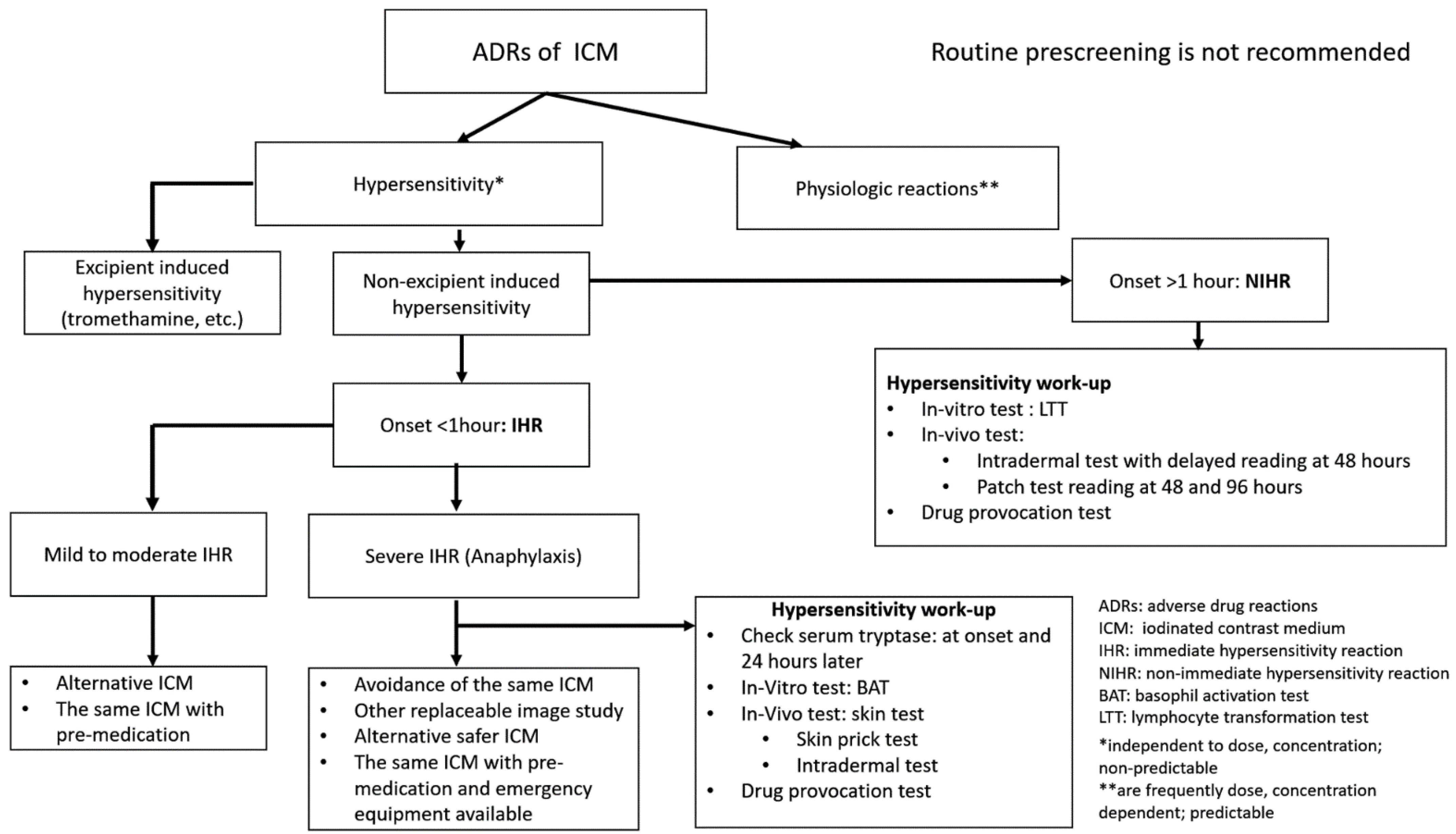
3. Classification of ADRs of ICM

The ADRs related to the intravascular administration of ICM can be divided into two types: hypersensitivity (allergic-like) events and chemo-toxic events [2,4,12]. According to the ACR Manual on Contrast Media [2], ADRs can also be divided into three grades: “mild” (signs and symptoms are self-limited without evidence of progression), “moderate” (signs and symptoms are more pronounced and commonly require medical management), and “severe” (signs and symptoms are often life-threatening and can result in permanent morbidity or death if not managed appropriately). Some mild and moderate reactions have the potential to become severe if not treated.
The detailed symptoms and signs of different grades are summarized in Table 2. Most ADRs of ICM are immediate (acute) with onset within 1 h and commonly within 30 min after intravenous administration of ICM [2,13]. About 70% of them occur within 5 min [14]. Non-immediate (delayed) cutaneous reaction usually has a latency of hours to one week after receiving the ICM [2]. Nearly all life-threatening ADRs occur within 20 min after intravascular administration of ICM. Chemo-toxic reactions to ICM are frequently dependent on the dose and concentration, while hypersensitivity is likely independent of them [2]. Nausea, vomiting, or flushing are occasionally difficult to classify as hypersensitivity or chemo-toxic events.

4. Epidemiology

Several studies have revealed a higher incidence of ADRs with ionic ICM (4.17–12.66%) than non-ionic ICM (0.69–3.13%) [14,15,16]. Therefore, non-ionic ICM have mostly been used in X-ray-related studies in recent years, especially in CT and angiography. Standardized classification systems for ADRs of ICM have been established to differentiate the hypersensitivity and chemo-toxic events [2,4], and the incidence of hypersensitivity to different non-ionic ICM has been reported as being 0.37–0.99% [17]. The potential risks of ADRs are allergy, asthma, cardiac status, anxiety, age, and gender [2,18,19]. No external warming of ICM [20], injection dose [21,22,23], injection speed [21,22,23], and family history of hypersensitivity to ICM [17] are also risk factors.

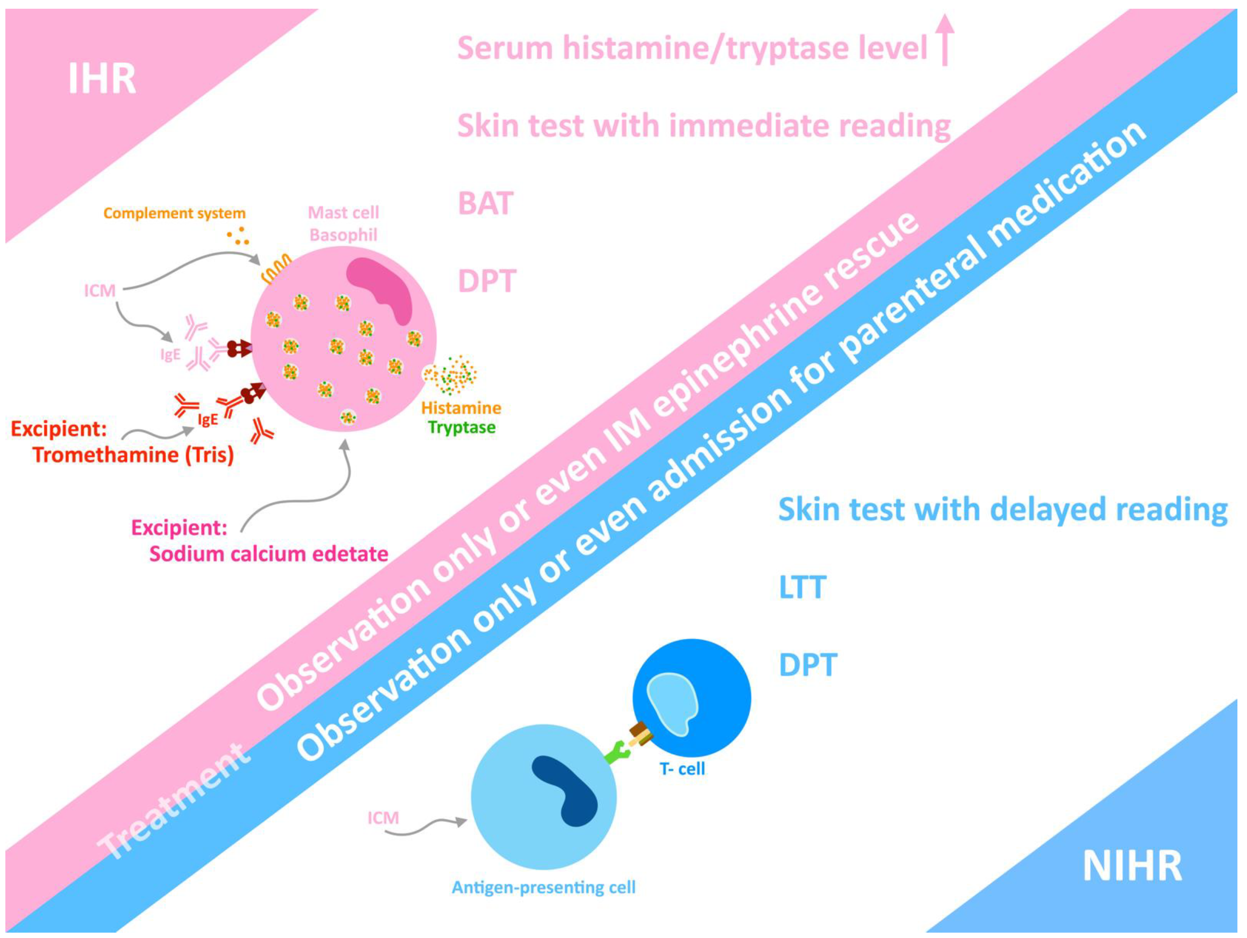
5. Phenotype of Hypersensitivity

Hypersensitivity to ICM is divided into immediate hypersensitivity reaction (IHR), in which reactions occur within 1 h of ICM administration, and non-immediate hypersensitivity reaction (NIHR), which occurs more than 1 h later [24,25,26,27]. The latency of most NIHRs is 3 h to 2 days, and they usually resolve in one week. In patients receiving non-ionic ICM, the incidence of IHR and NIHR is about 0.5–3% [25,28]. The manifestations of IHR include urticaria, angioedema, vomiting, abdominal pain, diarrhea, dyspnea, bronchospasm, and drop in blood pressure [24,25,26,27]. The most severe form of IHR is life-threatening anaphylactic shock with hypotension and loss of consciousness, which has a prevalence of 0.04–0.28% [29].
The pathogenesis of most ICM-induced hypersensitivity is unclear. Multiple possible pathomechanisms result in the activation of effector cells and the release of mediators. Histamine release occurs when patients develop urticaria. In the case of IHR, the pathophysiological explanations include the activation of mast cells and basophils (Figure 1), in addition to the release of histamine, tryptase, and other mediators [30]. The activation and release of mediators can occur through the IgE-mediated immune pathway and non-specific pathways, such as activation of the complement systems, activation of the XII clotting system (leading to the production of bradykinin and conversion of L-arginine into nitric oxide) [31], and formation of “pseudoantigens” [6]. A possible underlying specific IgE-mediated mechanism may be responsible for some IHRs of ICM because some individuals have positive skin test results [32]. A recent mouse study showed that iodixanol mechanistically increases the degranulation of mast cells, and more inflammatory factors are synthesized through activation of the PLC-γ and PI3K-related pathways [33].
The most common type of NIHR is maculopapular exanthema (MPE), which accounts for more than 50% of affected individuals [34]. Non-ionic dimeric ICM induces more NIHR than non-ionic monomeric ICM [35]. Other types of NIHR are rare but more severe and even potentially fatal, such as Stevens–Johnson syndrome, toxic epidermal necrolysis, drug reaction with eosinophilia and systemic symptoms, acute generalized exanthematous pustulosis, symmetrical drug-related intertriginous and flexural exanthema, and fixed drug eruption [24,25,27,36,37,38,39,40,41]. Delayed urticaria and angioedema may sometimes appear. NIHR is T cell-mediated, and a skin biopsy may reveal CD4+ and CD8+ T cell infiltration of the skin lesions, whereas a delayed-reading skin test (such as an intradermal test (IDT) or patch test) may show a positive reaction.
The latency of T cell-mediated delayed-type hypersensitivity to ICM ranges from hours to days after administration of the ICM. Usually, drug reactions with eosinophilia and systemic symptoms develop after exposure to the culprit drug for 2 to 4 weeks, but if induced by ICM, the latency is shorter (usually within the week following ICM administration) [39]. A positive lymphocyte transformation test (LTT) and ICM-specific T cell clones have been demonstrated in some patients [42]. The classification and recommendations for ADRs of ICM are shown in Figure 2.

6. Diagnosis

When dealing with a patient who has suspected hypersensitivity to a drug, food, or ICM, it is most important to obtain a detailed clinical history. To confirm the severe form of IHR, anaphylaxis, it is suggested to check serum levels of the mediators histamine and tryptase, which are released from mast cells and basophils after activation. The checks should be performed immediately after the event (within 4 h) and 24 h later (baseline). The elimination half-lives of histamine and tryptase are 15 to 20 min and 90 min to 2 h, respectively. Checking the serum level of tryptase is more practical because histamine degrades quickly and is more complicated to measure [30]. Serum levels of tryptase are positively correlated with the severity of IHR, and it is a valuable biomarker that supports the diagnosis of anaphylaxis. A clinically significant rise is defined as a serum tryptase level greater than one of the following: ([1.2 × baseline tryptase] + 2) μg/L, ([baseline tryptase] + 3) μg/L, 1.35 × baseline tryptase, or more than 11.4 μg/L. Among these options, the first has the greatest Youden’s index, which means it has the most significant clinical practicability [44].
The most important criterion for testing ICM hypersensitivity is a history of severe hypersensitivity, either immediate or non-immediate. Although the sensitivity of in vivo and in vitro tests of ICM allergy correlates with the severity of the hypersensitivity [45], a skin test may be positive in cases of mild reactions if performed within a maximum of 6 months from the reaction. Hence, a skin test can be performed in all patients with a history of hypersensitivity reactions if feasible. However, the significance of both tests is limited due to unsatisfactory sensitivity and specificity, as shown in Table 3. In vitro tests are preferred for severe and even life-threatening hypersensitivity reactions such as anaphylaxis.
Once the IHR event is resolved, a skin test can be performed to identify whether the IHR is IgE-mediated. The sensitivity range of skin tests in IHR is 4.2 to 73% and correlates with the severity of the phenotype of IHR [46,47,48,49,50,51,52,53]. In patients with IHR to ICM, pooled per-patient positivity rates of skin tests were 17% (95% CI, 10–26%) in a meta-analysis from 2015, but the positivity rates were increased to 52% (95% CI, 31–72%) in patients with severe IHR. The pooled per-patient positivity rates of skin tests in NIHR patients were 26% (95% CI, 15–41%). Among the skin tests, the rate for the skin prick test (SPT) is 7% (95% CI, 1–30%), the rate for IDT is 22% (95% CI, 13–34%), and the rate for the patch test is 16% (95% CI, 15–41%). If we combine IDT with the patch test, the positivity rate is increased [50].
The cross-reactivity of ICM in skin tests in the IHR phenotype population is 68% (95% CI, 48–83%), and that in the NIHR phenotype population is 39% (95% CI, 29–50%). The specificity of SPT for ICM-induced IHR is about 94.6%, and that of IDT is 91.4–96.3% [51,52]. The negative predictive value of skin tests is 93% (95% CI, 86–96%) [50]. The sensitivity and specificity of skin tests in ICM-induced NIHR are 72% and 96%, respectively, but the skin test is usually performed long after the hypersensitivity event, which decreases the positivity rate [12,48,54,55].
According to these findings, even with negative skin-test results for possible alternative ICM, physicians should pay attention to the patient because recurrence of hypersensitivity reaction may occur. In another study, there was no recurrence of severe hypersensitivity reactions in patients receiving alternative ICM with a negative skin test [56]. Hence, skin tests are invaluable for the investigation of possible alternative ICM, with negative predictive values of 94.2% (95% CI, 89.6% to 97.2%) and 86.1% (95% CI, 72.1–94.7%) for IHR and NIHR, respectively [57]. The skin test should be performed using a panel of many kinds of ICM, including the suspected culprit ICM.
SPT is performed with undiluted ICM (300–320 mg/mL), and then IDT is performed with ICM at dilutions of 1:1000 to 1:10 if the SPT result is negative [27,52]. It is notable that lower concentration of the tested ICM corresponds with lower positivity rates of reactions [53]. IDT is read at 20 min for IHR. Delayed reading of IDT after 48 and 72 h is necessary for delayed-type hypersensitivity, and a positive reaction should present wheals and possibly erythema instead of wheals only, which may be due to osmolality [42]. In severe hypersensitivity reactions, the investigations should be performed very cautiously. For NIHR, a safe stepwise approach may be started with a patch test, followed by delayed-reading IDT with a 1:10 dilution of ICM, and finally, delayed-reading IDT with undiluted ICM (1:1) [50,57,58,59].
Patch tests are performed in delayed-type hypersensitivity and read after 48 h and 96–120 h. However, these tests are more difficult and expensive [52,60]. In fixed drug eruption, PD should be tested in situ on previous lesion sites to avoid false-negative results [34]. The reason for this is that drug-specific resident-memory T cells are present in previously fixed drug eruption lesions [61]. The allergological work-up should be performed with a latency of 1 and 6 months from the latest ICM reaction, or else the sensitivity can be low [46]. The reason for the limited duration of skin reactivity is considered to be IgE clearance [62]. The time interval between the skin test and the immediate hypersensitivity event is 3 months and 48 months in groups with positive and negative skin-test results, respectively [46].
Once the hypersensitivity has resolved, in vitro tests for identifying the culprit ICM are indicated for patients with high risk or severe hypersensitivity phenotype, in addition to cases when a skin test is not available [49]. The basophil activation test (BAT) has been considered to be a useful tool in identifying culprit drugs or ICM in cases of IHR, and its correlation with the skin test and drug provocation test is good [48]. BAT is especially useful for severe forms of IHR such as anaphylactic shock, for which skin tests and drug provocation test are contraindicated. One should also be aware that many factors influence the performance of BAT, such as the selection of stimulants, designed protocol, gating strategies, and cutoff value [48,63]. The sensitivity and specificity of BAT are 46–63% and 89–100%, respectively, depending on the threshold chosen [63,64,65].
The lymphocyte transformation test (LTT) detects and measures the proliferation of circulating lymphocytes specific to the antigen of the culprit drug or ICM upon stimulation by the antigen. The sensitivity and specificity of LTT differ for different antigens. LTT is useful for detecting culprit ICM in patients with NIHR, but a negative result may not rule out the cause [27,66,67]. The sensitivity of LTT ranges from 13 to 75% [65]. It is suggested that LTT be performed at 4–6 weeks after resolution of the event and the withdrawal of corticosteroids and other immunosuppressants, but no more than 2 to 3 years after the reaction [68,69]. Corticosteroid (more than 0.2 mg/kg of prednisolone) and equivalent immunosuppressants may interfere with the performance of LTT.
There is controversy regarding the need for a drug provocation test (DPT), which is considered to be the last step of the diagnostic algorithm. Even if there is a negative result in pretesting, recurrence of the hypersensitivity reaction following re-exposure to ICM may occur [70]. Administration of ICM is potentially harmful, so many contraindications should be noted before performing DPT, such as impaired renal function, current medications with renal toxicity, pregnancy, lactation, thyrotoxic crisis, lactic acidosis, and patients with severe IHR or NIHR [46].
Currently, no standard protocol for drug provocation has been established, such as a graded challenge or full-dose challenge test. The dose of ICM used in DPT ranges from 49 to 100 mL. The negative predictive value of DPT at a Spanish center was 97.3 and 80% when using 10 mL and 50 mL of ICM, respectively [71]. Two protocols are currently used: one involving 5, 15, 30, and 35 mL at 45-min intervals; and another involving 0.05, 0.5, 1, 5, 7.5, 10, and 25 mL at 30-min intervals [72].
A consensus on protocols should be established and validated. Drug provocation tests must be performed by well-trained experts in well-equipped centers for possible emergency treatment. DPT should only be performed for patients with negative skin test, basophil activation test, and lymphocyte transformation test results, or when there is no alternative diagnostic tool and the diagnosis is uncertain [73]. The recommended in vivo and in vitro tests for IHR and NIHR are illustrated in Figure 2.
The excipient tromethamine is a buffering agent and is contained in many cosmetics, drugs, and COVID-19 vaccines. It may cause contact dermatitis, allergy, and even anaphylaxis [9,74]. Another excipient in ICM is sodium calcium edetate. There is one report of a patient with allergy to sodium calcium edetate, who was identified through investigation of the reactions to ICM [10]. Investigations to determine the culprit ICM should include the excipients. Hypersensitivity reactions to excipients are immediate; hence, BAT is performed to identify if excipient or ICM per se is the real culprit. If hypersensitivity to tromethamine or sodium calcium edetate is proven, the patient should be informed to avoid drugs or cosmetics containing the ingredient in the future. Furthermore, pharmaceutical companies should put every effort into developing ICM that contain promising stabilizing agents with reduced immunogenicity.

7. Treatment

The reactions after injection of ICM range from mild chemo-toxic reactions to severe life-threatening situations. Most of the adverse reactions to low-osmolality ICM are mild and only require observation or oral H1-antihistamine. Although life-threatening anaphylaxis is rare and unpredictable, remaining vigilant is very important. Staff such as radiologists, technologists, and nurses should be familiar with standard operating procedures for all kinds of reactions. When adverse reactions occur, personnel should notify the supervising radiologist, monitor the vital signs of the patient, administer certain medications, and call for assistance (such as from the emergency department). All emergency equipment and medication should be prepared.
H1 antihistamines are sufficient for mild hypersensitivity symptoms such as itching, but for life-threatening anaphylaxis, intramuscular epinephrine is necessary, and many radiologists are unaware of this [75]. The first-line treatment is 0.01 mg per kilogram of body weight to a maximum of 0.5 mg of epinephrine at a concentration of 1:1000, which should be injected intramuscularly in the lateral aspect of the thigh [76]. When patients manifest airway, breathing, and circulation symptoms, staff should assess and manage them immediately and adequate oxygen supply is critical.
The most common type of NIHR is maculopapular exanthema [34]. NIHR usually has mild to moderate severity and is usually self-limiting, with most cases requiring little or no therapy. However, severe NIHR may occur, such as acute generalized exanthematous pustulosis, drug reaction with eosinophilia and systemic symptoms, Stevens–Johnson syndrome, and toxic epidermal necrolysis. Such cases usually need to be referred to a specialist for treatment. In these circumstances, systemic corticosteroids and even admission are usually needed.

8. Prescreening, Prevention and Rapid Drug Desensitization

A questionnaire before the procedure may help to determine whether patients have higher risk of a hypersensitivity reaction. Apart from this, no in vitro or in vivo test has shown good evidence for prescreening populations with high risk. A recent prospective study was performed for the validation of prescreening IDT to predict ICM hypersensitivity, but the results revealed extremely low sensitivity and a low positive predictive value [77]. In patients with a history of hypersensitivity reaction, the intradermal skin test has good negative predictive value [78]. In conclusion, prescreening with skin tests is not recommended due to the extremely low sensitivity and low positive predictive value.
In patients with a hypersensitivity history to their first-choice treatment, rapid drug desensitization is a therapeutic approach which temporarily builds tolerance to the drug in a short period of time. The principle of desensitization is to gradually eliminate the IgE activation of mast cells through small doses of drugs. This is done by starting with a small dose of the drug and slowly increasing the dose until achieving the therapeutic dose needed or the maximum tolerated dose. Rapid drug desensitization has been practiced for many years in taxanes for cancer patients and biologics such as rituximab, and there is a great clinical need. With rapid drug desensitization, the chance of breakthrough reaction is usually less than 10%, and this is usually a mild reaction. Premedication is also included in some desensitization protocols. Despite the long-existing therapeutic approach, there is no evidence to support such drug desensitization in radiocontrast agent hypersensitivity [78,79].

9. Recommendations

For patients with a history of hypersensitivity to ICM, the recommendations range from strict avoidance of ICM to imaging alternatives that do not use ICM, premedication, alternative ICM, and the drug provocation test [12,25,80]. Avoidance of imaging studies with ICM is recommended if alternative imaging techniques such as magnetic resonance imaging are applicable [25,27]. Although systemic corticosteroids and antihistamines have been used widely for a long time as premedication, there is no gold-standard regimen of premedication for preventing hypersensitivity reactions to ICM. Breakthrough reactions develop despite premedication.
The American College of Radiology recommends changing an alternative ICM in patients with a prior allergic-like reaction [2]. In patients using alternative ICM with a negative skin test for the ICM, the recurrence rate of IHR is 7% (95% CI, 4–14%), and that of NIHR is 35% (95% CI, 19–55%) [50]. In patients with a history of mild hypersensitivity reaction, the recurrence rate of similar mild reactions is 31.1% with the same ICM, 12% with another ICM, and 7.6% with antihistamine premedication [81,82,83]. In patients with mild ICM-related IHR limited to the skin, the rate of severe IHR after re-administration of the ICM, such as anaphylaxis, is below 1% [84].
The American College of Radiology recommends premedication for patients with prior allergy-like reactions to ICM or for populations at risk [2]. The purpose of corticosteroids is to mitigate the likelihood of hypersensitivity in high-risk individuals or in patients with a history of hypersensitivity to ICM. In the era of high-osmolality ICM administration, premedication may be beneficial for reducing the likelihood of IHR [85]; however, now that high-osmolality ICM are no longer used, the efficacy of premedication remains questionable.
Premedication with systemic corticosteroids and antihistamines may prevent the occurrence of mild to moderate IHR to ICM in circumstances where ICM administration is needed, but the breakthrough reaction rate is 3–4 times that of the population without a history of hypersensitivity [27,81,84]. In patients with a history of severe hypersensitivity reactions, such as anaphylaxis, use of the same ICM should not be attempted, even with premedication. Imaging without ICM is suggested. If the patient must undergo imaging with ICM, prescreening to find a safer alternative is suggested.
In general, avoiding the known culprit ICM is more helpful than premedication with corticosteroid and antihistamines in reducing the recurrence of IHR to ICM [81,83,86]. Premedication is not effective in patients with a history of NIHR to ICM [39,87]. In practice, the most common protocol of oral corticosteroids involves several time points (e.g., 50 mg of prednisone orally at 13, 7, and 1 h before administration of ICM) and H1 antihistamine (e.g., 50 mg of diphenhydramine 1 h before administration of ICM) [88]. Another study suggests performing prophylaxis using 5 h of intravenous corticosteroid (200 mg of hydrocortisone administered at 5 and 1 h, with 50 mg of diphenhydramine administered 1 h before the imaging study). The results suggest that this approach is not inferior to an oral regimen [89]. Nevertheless, premedication is not a panacea, and the radiology team must be prepared for treating breakthrough reactions.
There are many drawbacks of premedication with corticosteroids and antihistamines, such as transient leukocytosis, asymptomatic hyperglycemia, increased risk of infection, lengthened hospitalization, and drowsiness. Furthermore, it may cause more indirect suffering than the direct distress that it is meant to prevent [90]. The use of a substitute for the known ICM is more helpful in reducing the possibility of breakthrough reactions. In summary, no known precautional measures can guarantee against repeat reaction after re-administration of the culprit ICM, and avoiding the known ICM is more helpful than premedication in reducing the recurrence of hypersensitivity to ICM. The recommendations for IHR and NIHR of ICM are demonstrated in Figure 2. A history of chemo-toxic reactions (such as nausea, vomiting, or flushing) does not increase the risk of future ICM-related hypersensitivity, and premedication is not recommended in such cases.

10. Standardization of the Documentation of ICM-Induced Hypersensitivity

It is critical to standardize documentation with a detailed description of ICM-induced hypersensitivity for correct diagnosis and further investigations. Staff should avoid using inaccurate terms such as “iodine allergy,” which may interfere with judgment. The suggested standardized documentation should include the following [91]:
  • The precise name of the injected culprit ICM and its dose (volume, concentration);
  • The manifestations of the reaction (e.g., itching, generalized urticaria, dizziness, drop of blood pressure, tachycardia, etc.) to establish the severity of the reaction;
  • The chronology of the adverse reaction;
  • Specific treatment for the ICM-induced hypersensitivity.
Correct medical records lead to logical thinking about the adverse event and may help to prevent recurrence of the same reactions.

11. Conclusions

All ICM used in practice are relatively safe but have a possible risk of hypersensitivity, even when used correctly as prescribed. In general, however, the benefits of performing the procedure with ICM when needed outweigh the risks. In patients with a history of hypersensitivity, using an alternative ICM is preferred to using the culprit ICM with premedication because breakthrough reactions may occur. Skin tests (SPT, IDT, PT) and in vitro tests (BAT and LTT) may help to confirm the causality of ICM and hypersensitivity reactions, and help to find a safer alternative ICM. Standardized documentation of the hypersensitivity event leads to better clarification and may help with future research on ICM hypersensitivity.

Author Contributions

Conceptualization and writing (original draft preparation, review, and editing): T.-M.C., S.-Y.C. All authors have read and agreed to the published version of the manuscript.

Funding

This research received no external funding.

Institutional Review Board Statement

Not applicable.

Informed Consent Statement

Not applicable.

Data Availability Statement

Not applicable.

Conflicts of Interest

The authors declare no conflict of interest.

Abbreviations

ADRAdverse drug reaction
BATBasophil activation test
DPTDrug provocation test
ICMIodinated contrast media
IDTIntradermal test
IHRImmediate hypersensitivity reaction
LTTLymphocyte transformation test
MPEMaculopapular exanthema
NIHRNon-immediate hypersensitivity reaction
SPTSkin prick test

References

  1. Caschera, L.; Lazzara, A.; Piergallini, L.; Ricci, D.; Tuscano, B.; Vanzulli, A. Contrast agents in diagnostic imaging: Present and future. Pharmacol. Res. 2016, 110, 65–75. [Google Scholar] [CrossRef] [PubMed]
  2. Radiology, A.C.O. ACR Manual on Contrast Media; American College of Radiology: Reston, VA, USA, 2021; ISBN 978-1-55903-012-0. [Google Scholar]
  3. O’Connor, S.D.; Summers, R.M. Summers, Revisiting oral barium sulfate contrast agents. Acad. Radiol. 2007, 14, 72–80. [Google Scholar] [CrossRef] [PubMed]
  4. European Society of Urogenital Radiology. ESUR Guidelines on Contrast Agents 10.0. 2019. Available online: https://www.esur.org/fileadmin/content/2019/ESUR_Guidelines_10.0_Final_Version.pdf (accessed on 15 December 2021).
  5. Sharafuddin, M.J.; Marjan, A.E. Current status of carbon dioxide angiography. J. Vasc. Surg. 2017, 66, 618–637. [Google Scholar] [CrossRef] [PubMed]
  6. Lasser, E.C. The multipotential pseudoantigenicity of X-ray contrast media. Pseudoantigen excess may downregulate the release of hypotensive mediators.pdf. Int. Arch. Allergy Immunol. 2000, 123, 282–290. [Google Scholar] [PubMed]
  7. Arun Kuar Gupta, A.G.; Sandhu, M.S. Iodinated contrast media: An update. In Diagnostic Radiology: Advances in Imaging Technique; Jaypee Brothers Medical Publishers: New Delhi, India, 2019; Chapter 18; p. 374. [Google Scholar]
  8. Gueant-Rodriguez, R.M.; Romano, A.; Barbaud, A.; Brockow, K.; Gueant, J.-L. Hypersensitivity reactions to iodinated contrast media. Curr. Pharm. Des. 2006, 12, 3359–3372. [Google Scholar] [CrossRef]
  9. Lukawska, J.; Mandaliya, D.; Chan, A.E.; Foggitt, A.; Bidder, T.; Harvey, J.; Stycharczuk, L.; Bisdas, S. Anaphylaxis to trometamol excipient in gadolinium-based contrast agents for clinical imaging. J. Allergy Clin. Immunol. Pract. 2019, 7, 1086–1087. [Google Scholar] [CrossRef]
  10. R usso, P.A.; Banovic, T.; Wiese, M.D.; Whyte, A.F.; Smith, W.B. Systemic allergy to EDTA in local anesthetic and radiocontrast media. J. Allergy Clin. Immunol. Pract. 2014, 2, 225–229. [Google Scholar] [CrossRef]
  11. Ali, S.B.; Perkins, G.; Ryoo, D.; Lee, M.; Tunbridge, M.; Yuson, C.; Smith, W.; Hissaria, P.; Le, T.T. AstraZeneca ChAdOx1-S COVID-19 vaccine can be safely administered in patients with EDTA allergy. Allergy Asthma Clin. Immunol. 2022, 18, 22. [Google Scholar] [CrossRef]
  12. Rosado Ingelmo, A.; Dona Diaz, I.; Cabanas Moreno, R.; Moya Quesada, M.C.; Garcia-Aviles, C.; Garcia Nunez, I.; Martinez Tadeo, J.I.; Mielgo Ballesteros, R.; Ortega-Rodriguez, N.; Padial Vilchez, M.A.; et al. Clinical Practice Guidelines for Diagnosis and Management of Hypersensitivity Reactions to Contrast Media. J. Investig. Allergol. Clin. Immunol. 2016, 26, 144–155. [Google Scholar] [CrossRef] [Green Version]
  13. Li, X.; Chen, J.; Zhang, L.; Liu, H.; Wang, S.; Chen, X.; Fang, J.; Wang, S.; Zhang, W. Clinical observation of the adverse drug reactions caused by non-ionic iodinated contrast media: Results from 109,255 cases who underwent enhanced CT examination in Chongqing, China. Br. J. Radiol. 2015, 88, 20140491. [Google Scholar] [CrossRef] [Green Version]
  14. Katayama, H.; Yamaguchi, K.; Kozuka, T.; Takashima, T.; Seez, P.; Matsuura, K. Adverse reactions to ionic and nonionic contrast media. A report from the Japanese Committee on the Safety of Contrast Media. Radiology 1990, 175, 621–628. [Google Scholar] [CrossRef] [PubMed]
  15. Wolf, G.L.; Arenson, R.L.; Cross, A.P. A prospective trial of ionic vs nonionic contrast agents in routine clinical practice: Comparison of adverse effects. Am. J. Roentgenol. 1989, 152, 939–944. [Google Scholar] [CrossRef] [PubMed]
  16. Cochran, S.T.; Bomyea, K. Trends in adverse events from iodinated contrast media. Acad. Radiol. 2002, 9, S65–S68. [Google Scholar] [CrossRef]
  17. Cha, M.J.; Kang, D.Y.; Lee, W.; Yoon, S.H.; Choi, Y.H.; Byun, J.S.; Lee, J.; Kim, Y.H.; Choo, K.S.; Cho, B.S.; et al. Hypersensitivity Reactions to Iodinated Contrast Media: A Multicenter Study of 196,081 Patients. Radiology 2019, 293, 117–124. [Google Scholar] [CrossRef] [PubMed]
  18. Voltolini, S.; Cofini, V.; Murzilli, F.; Bignardi, D.; Borro, M.; Calamari, M.; Caruso, C.; Cittadini, G.; Contatore, M.; Cortellini, G.; et al. Hypersensitivity reactions to iodinate contrast media in Italy: A retrospective study. Characteristics of patients and risk factors. Eur. Ann. Allergy Clin. Immunol. 2021, 54, 60. [Google Scholar] [CrossRef] [PubMed]
  19. Klostranec, J.M.; Rohringer, T.; Gerber, R.; Murphy, K.J. The Role of Biologic Sex in Anaphylactoid Contrast Reactions: An Important Consideration for Women of Reproductive Age and Undergoing Hormone Replacement Therapy. Radiology 2021, 299, 272–275. [Google Scholar] [CrossRef]
  20. Zhang, B.; Liu, J.; Dong, Y.; Guo, B.; Lian, Z.; Yu, H.; Luo, X.; Mo, X.; Zhang, L.; Huang, W.; et al. Extrinsic warming of low-osmolality iodinated contrast media to 37 degrees C reduced the rate of allergic-like reaction. Allergy Asthma Proc. 2018, 39, e55–e63. [Google Scholar] [CrossRef]
  21. Li, X.; Liu, H.; Zhao, L.; Liu, J.; Cai, L.; Liu, L.; Zhang, W. Clinical observation of adverse drug reactions to non-ionic iodinated contrast media in population with underlying diseases and risk factors. Br. J. Radiol. 2017, 90, 20160729. [Google Scholar] [CrossRef] [Green Version]
  22. Brockow, K. Reduced iodinated contrast media dose and injection speed for CT: How much does this decrease the risk of a hypersensitivity reactions? Quant. Imaging Med. Surg. 2020, 10, 537–540. [Google Scholar] [CrossRef]
  23. Park, H.J.; Son, J.H.; Kim, T.B.; Kang, M.K.; Han, K.; Kim, E.H.; Kim, A.Y.; Park, S.H. Relationship between Lower Dose and Injection Speed of Iodinated Contrast Material for CT and Acute Hypersensitivity Reactions: An Observational Study. Radiology 2019, 293, 565–572. [Google Scholar] [CrossRef]
  24. Böhm, I.; Heverhagen, J.T.; Klose, K.J. Classification of acute and delayed contrast media-induced reactions: Proposal of a three-step system. Contrast Media Mol. Imaging 2012, 7, 537–541. [Google Scholar] [CrossRef] [PubMed]
  25. Brockow, K.; Christiansen, C.; Kanny, G.; Clément, O.; Barbaud, A.; Bircher, A.; Dewachter, P.; Guéant, J.L.; Rodriguez Guéant, R.M.; Mouton-Faivre, C.; et al. Management of hypersensitivity reactions to iodinated contrast media. Allergy 2005, 60, 150–158. [Google Scholar] [CrossRef] [PubMed]
  26. Tanno, L.K.; Torres, M.J.; Castells, M.; Demoly, P. What can we learn in drug allergy management from World Health Organization’s international classifications? Allergy 2018, 73, 987–992. [Google Scholar] [CrossRef] [PubMed] [Green Version]
  27. Brockow, K. Medical algorithm: Diagnosis and treatment of radiocontrast media hypersensitivity. Allergy 2020, 75, 1278–1280. [Google Scholar] [CrossRef]
  28. Christiansen, C.; Pichler, W.J.; Skotland, T. Delayed allergy-like reactions to X-ray contrast media: Mechanistic considerations. Eur. Radiol. 2000, 10, 1965–1975. [Google Scholar] [CrossRef]
  29. Lee, S.Y.; Lim, K.W.; Chang, Y.S. Radiocontrast media hypersensitivity in the Asia Pacific region. Asia Pac. Allergy 2014, 4, 119–125. [Google Scholar] [CrossRef] [Green Version]
  30. Laroche, D. Immediate reactions to contrast media: Mediator release and value of diagnostic testing. Toxicology 2005, 209, 193–194. [Google Scholar] [CrossRef]
  31. Morcos, S.K. Review article: Acute serious and fatal reactions to contrast media: Our current understanding. Br. J. Radiol. 2005, 78, 686–693. [Google Scholar] [CrossRef]
  32. Morales-Cabeza, C.; Roa-Medellin, D.; Torrado, I.; De Barrio, M.; Fernandez-Alvarez, C.; Montes-Acenero, J.F.; De La Riva, I.; Prieto-Garcia, A. Immediate reactions to iodinated contrast media. Ann. Allergy Asthma Immunol. 2017, 119, 553–557. [Google Scholar] [CrossRef]
  33. Zou, W.; Yang, S.; Chen, L.; Hu, S.; Hao, G.; Hu, C. Iodixanol activation of mast cells: Implications in the pathogenesis of iodixanol-induced delayed cutaneous adverse reactions. Toxicology 2022, 465, 153034. [Google Scholar] [CrossRef]
  34. Böhm, I.; Schild, H.H. Immediate and non-immediate reaction after non-ionic X-ray contrast medium injection: Case report and review of the literature. Eur. J. Radiol. Extra 2007, 61, 129–133. [Google Scholar] [CrossRef]
  35. Schild, H.H.; Kuhl, C.K.; Hübner-Steiner, U.; Böhm, I.; Speck, U. Adverse events after unenhanced and monomeric and dimeric contrast-enhanced CT: A prospective randomized controlled trial. Radiology 2006, 240, 56–64. [Google Scholar] [CrossRef] [PubMed]
  36. Brockow, K.; Ardern-Jones, M.R.; Mockenhaupt, M.; Aberer, W.; Barbaud, A.; Caubet, J.C.; Spiewak, R.; Torres, M.J.; Mortz, C.G. EAACI position paper on how to classify cutaneous manifestations of drug hypersensitivity. Allergy 2019, 74, 14–27. [Google Scholar] [CrossRef] [PubMed] [Green Version]
  37. Böhm, I.; Schild, H.H. A practical guide to diagnose lesser-known immediate and delayed contrast media-induced adverse cutaneous reactions. Eur. Radiol. 2006, 16, 1570–1579. [Google Scholar] [CrossRef] [PubMed]
  38. Rosado, A.; Canto, G.; Veleiro, B.; Rodríguez, J. Toxic epidermal necrolysis after repeated injections of iohexol. Am. J. Roentgenol. 2001, 176, 262–263. [Google Scholar] [CrossRef] [PubMed]
  39. Soria, A.; Bernier, C.; Veyrac, G.; Barbaud, A.; Puymirat, E.; Milpied, B. Drug reaction with eosinophilia and systemic symptoms may occur within 2 weeks of drug exposure: A retrospective study. J. Am. Acad. Dermatol. 2020, 82, 606–611. [Google Scholar] [CrossRef]
  40. Soria, A.; Amsler, E.; Bernier, C.; Milpied, B.; Tetart, F.; Morice, C.; Dezoteux, F.; Ferrier-Le Bouedec, M.C.; Barbaud, A.; Staumont-Salle, D.; et al. DRESS and AGEP Reactions to Iodinated Contrast Media: A French Case Series. J. Allergy Clin. Immunol. Pract. 2021, 9, 3041–3050. [Google Scholar] [CrossRef]
  41. Tasker, F.; Fleming, H.; McNeill, G.; Creamer, D.; Walsh, S. Contrast media and cutaneous reactions. Part 2: Delayed hypersensitivity reactions to iodinated contrast media. Clin. Exp. Dermatol. 2019, 44, 844–860. [Google Scholar] [CrossRef] [Green Version]
  42. Torres, M.J.; Gomez, F.; Dona, I.; Rosado, A.; Mayorga, C.; Garcia, I.; Blanca-Lopez, N.; Canto, G.; Blanca, M. Diagnostic evaluation of patients with nonimmediate cutaneous hypersensitivity reactions to iodinated contrast media. Allergy 2012, 67, 929–935. [Google Scholar] [CrossRef]
  43. Costantino, M.T.; Romanini, L.; Gaeta, F.; Stacul, F.; Valluzzi, R.L.; Passamonti, M.; Bonadonna, P.; Cerri, G.; Pucci, S.; Ricci, P.; et al. SIRM-SIAAIC consensus, an Italian document on management of patients at risk of hypersensitivity reactions to contrast media. Clin. Mol. Allergy 2020, 18, 13. [Google Scholar] [CrossRef]
  44. Vitte, J.; Amadei, L.; Gouitaa, M.; Mezouar, S.; Zieleskiewicz, L.; Albanese, J.; Bruder, N.; Lagier, D.; Mertès, P.M.; Mège, J.L.; et al. Paired acute-baseline serum tryptase levels in perioperative anaphylaxis: An observational study. Allergy 2019, 74, 1157–1165. [Google Scholar] [CrossRef] [PubMed]
  45. Worm, M.; Francuzik, W.; Renaudin, J.M.; Bilo, M.B.; Cardona, V.; Scherer Hofmeier, K.; Köhli, A.; Bauer, A.; Christoff, G.; Cichocka-Jarosz, E.; et al. Factors increasing the risk for a severe reaction in anaphylaxis: An analysis of data from The European Anaphylaxis Registry. Allergy 2018, 73, 1322–1330. [Google Scholar] [CrossRef] [PubMed]
  46. Kvedariene, V.; Martins, P.; Rouanet, L.; Demoly, P. Diagnosis of iodinated contrast media hypersensitivity: Results of a 6-year period. Clin. Exp. Allergy. 2006, 36, 1072–1077. [Google Scholar] [CrossRef] [PubMed]
  47. Trcka, J.; Schmidt, C.; Seitz, C.S.; Bröcker, E.B.; Gross, G.E.; Trautmann, A. Anaphylaxis to iodinated contrast material: Nonallergic hypersensitivity or IgE-mediated allergy? Am. J. Roentgenol. 2008, 190, 666–670. [Google Scholar] [CrossRef]
  48. Salas, M.; Gomez, F.; Fernandez, T.D.; Doña, I.; Aranda, A.; Ariza, A.; Blanca-López, N.; Mayorga, C.; Blanca, M.; Torres, M.J. Diagnosis of immediate hypersensitivity reactions to radiocontrast media. Allergy 2013, 68, 1203–1206. [Google Scholar] [CrossRef]
  49. Mayorga, C.; Fernandez, T.D.; Montañez, M.I.; Moreno, E.; Torres, M.J. Recent developments and highlights in drug hypersensitivity. Allergy 2019, 74, 2368–2381. [Google Scholar] [CrossRef] [Green Version]
  50. Yoon, S.H.; Lee, S.Y.; Kang, H.R.; Kim, J.Y.; Hahn, S.; Park, C.M.; Chang, Y.S.; Goo, J.M.; Cho, S.H. Skin tests in patients with hypersensitivity reaction to iodinated contrast media: A meta-analysis. Allergy 2015, 70, 625–637. [Google Scholar] [CrossRef]
  51. Goksel, O.; Aydın, O.; Atasoy, C.; Akyar, S.; Demirel, Y.S.; Misirligil, Z.; Bavbek, S. Hypersensitivity reactions to contrast media: Prevalence, risk factors and the role of skin tests in diagnosis—A cross-sectional survey. Int. Arch. Allergy Immunol. 2011, 155, 297–305. [Google Scholar] [CrossRef]
  52. Brockow, K.; Romano, A.; Aberer, W.; Bircher, A.J.; Barbaud, A.; Bonadonna, P.; Faria, E.; Kanny, G.; Lerch, M.; Pichler, W.J.; et al. Skin testing in patients with hypersensitivity reactions to iodinated contrast media—A European multicenter study. Allergy 2009, 64, 234–241. [Google Scholar] [CrossRef]
  53. Dewachter, P.; Laroche, D.; Mouton-Faivre, C.; Bloch-Morot, E.; Cercueil, J.P.; Metge, L.; Carette, M.F.; Vergnaud, M.C.; Clément, O. Immediate reactions following iodinated contrast media injection: A study of 38 cases. Eur. J. Radiol. 2011, 77, 495–501. [Google Scholar] [CrossRef]
  54. Kvedariene, V.; Orvydaite, M.; Petraityte, P.; Rudyte, J.; Edvardas Tamosiunas, A. Inherent clinical properties of non-immediate hypersensitivity to iodinated contrast media. Int. J. Clin. Pract. 2021, 75, e14766. [Google Scholar] [CrossRef] [PubMed]
  55. Nucera, E.; Parrinello, G.; Gangemi, S.; Buonomo, A.; Aruanno, A.; Lohmeyer, F.M.; Inchingolo, R.; Rizzi, A. Contrast Medium Hypersensitivity: A Large Italian Study with Long-Term Follow-Up. Biomedicines 2022, 10, 759. [Google Scholar] [CrossRef] [PubMed]
  56. Caimmi, S.; Benyahia, B.; Suau, D.; Bousquet-Rouanet, L.; Caimmi, D.; Bousquet, P.J.; Demoly, P. Clinical value of negative skin tests to iodinated contrast media. Clin. Exp. Allergy 2010, 40, 805–810. [Google Scholar] [CrossRef] [PubMed]
  57. Schrijvers, R.; Breynaert, C.; Ahmedali, Y.; Bourrain, J.L.; Demoly, P.; Chiriac, A.M. Skin Testing for Suspected Iodinated Contrast Media Hypersensitivity. J. Allergy Clin. Immunol. Pract. 2018, 6, 1246–1254. [Google Scholar] [CrossRef] [PubMed]
  58. Ahn, Y.H.; Koh, Y.I.; Kim, J.H.; Ban, G.Y.; Lee, Y.K.; Hong, G.N.; Jin, U.R.; Choi, B.J.; Shin, Y.S.; Park, H.S.; et al. The potential utility of iodinated contrast media (ICM) skin testing in patients with ICM hypersensitivity. J. Korean Med. Sci. 2015, 30, 245–251. [Google Scholar] [CrossRef] [Green Version]
  59. Vernassiere, C.; Trechot, P.; Commun, N.; Schmutz, J.L.; Barbaud, A. Low negative predictive value of skin tests in investigating delayed reactions to radio-contrast media. Contact Dermat. 2004, 50, 359–366. [Google Scholar] [CrossRef]
  60. Bhujoo, Z.; Ingen-Housz-Oro, S.; Gener, G.; Gaudin, O.; Fleck, M.; Verlinde-Carvalho, M.; Paul, M.; Chosidow, O.; Wolkenstein, P.; Assier, H. Patch tests in nonimmediate cutaneous adverse drug reactions: The importance of late readings on day 4. Contact Dermat. 2022, 86, 29–33. [Google Scholar] [CrossRef]
  61. Gaide, O.; Emerson, R.O.; Jiang, X.; Gulati, N.; Nizza, S.; Desmarais, C.; Robins, H.; Krueger, J.G.; Clark, R.A.; Kupper, T.S. Common clonal origin of central and resident memory T cells following skin immunization. Nat. Med. 2015, 21, 647–653. [Google Scholar] [CrossRef]
  62. Boehm, I. Limited duration of hypersensitivity reactions to contrast and exact documentation of such adverse events. Reg. Anesth. Pain Med. 2020, 45, 246. [Google Scholar] [CrossRef]
  63. Steiner, M.; Harrer, A.; Himly, M. Basophil Reactivity as Biomarker in Immediate Drug Hypersensitivity Reactions-Potential and Limitations. Front. Pharmacol. 2016, 7, 171. [Google Scholar] [CrossRef] [Green Version]
  64. Pinnobphun, P.; Buranapraditkun, S.; Kampitak, T.; Hirankarn, N.; Klaewsongkram, J. The diagnostic value of basophil activation test in patients with an immediate hypersensitivity reaction to radiocontrast media. Ann. Allergy Asthma Immunol. 2011, 106, 387–393. [Google Scholar] [CrossRef] [PubMed]
  65. Gomez, E.; Ariza, A.; Blanca-Lopez, N.; Torres, M.J. Nonimmediate hypersensitivity reactions to iodinated contrast media. Curr. Opin. Allergy Clin. Immunol. 2013, 13, 345–353. [Google Scholar] [CrossRef]
  66. Kanny, G.; Pichler, W.; Morisset, M.; Franck, P.; Marie, B.; Kohler, C.; Renaudin, J.M.; Beaudouin, E.; Laudy, J.S.; Moneret-Vautrin, D.A. T cell-mediated reactions to iodinated contrast media: Evaluation by skin and lymphocyte activation tests. J. Allergy Clin. Immunol. 2005, 115, 179–185. [Google Scholar] [CrossRef] [PubMed]
  67. Torres, M.J.; Mayorga, C.; Cornejo-Garcia, J.A.; Lopez, S.; Chaves, P.; Rondon, C.; Fernandez, T.; Blanca, M. Monitoring non-immediate allergic reactions to iodine contrast media. Clin. Exp. Immunol. 2008, 152, 233–238. [Google Scholar] [CrossRef] [PubMed]
  68. Pichler, W.J.; Tilch, J. The lymphocyte transformation test in the diagnosis of drug hypersensitivity. Allergy 2004, 59, 809–820. [Google Scholar] [CrossRef]
  69. Hari, Y.; Frutig-Schnyder, K.; Hurni, M.; Yawalkar, N.; Zanni, M.P.; Schnyder, B.; Kappeler, A.; von Greyerz, S.; Braathen, L.R.; Pichler, W.J. T cell involvement in cutaneous drug eruptions. Clin. Exp. Allergy 2001, 31, 1398–1408. [Google Scholar] [CrossRef]
  70. Sese, L.; Gaouar, H.; Autegarden, J.E.; Alari, A.; Amsler, E.; Vial-Dupuy, A.; Pecquet, C.; Frances, C.; Soria, A. Immediate hypersensitivity to iodinated contrast media: Diagnostic accuracy of skin tests and intravenous provocation test with low dose. Clin. Exp. Allergy 2016, 46, 472–478. [Google Scholar] [CrossRef]
  71. Soria, A.; Masson, N.; Vial-Dupuy, A.; Gaouar, H.; Amsler, E.; Chollet-Martin, S.; Nicaise-Roland, P.; Autegarden, J.E.; Barbaud, A. Allergological workup with half-dose challenge in iodinated contrast media hypersensitivity. Allergy 2019, 74, 414–417. [Google Scholar] [CrossRef]
  72. Trautmann, A.; Brockow, K.; Behle, V.; Stoevesandt, J. Radiocontrast Media Hypersensitivity: Skin Testing Differentiates Allergy From Nonallergic Reactions and Identifies a Safe Alternative as Proven by Intravenous Provocation. J. Allergy Clin. Immunol. Pract. 2019, 7, 2218–2224. [Google Scholar] [CrossRef]
  73. Bansie, R.D.; Karim, A.F.; van Maaren, M.S.; Hermans, M.A.; van Daele, P.; Gerth van Wijk, R.; Rombach, S.M. Assessment of immediate and non-immediate hypersensitivity contrast reactions by skin tests and provocation tests: A review. Int. J. Immunopathol. Pharmacol. 2021, 35, 20587384211015061. [Google Scholar] [CrossRef]
  74. Kounis, N.G.; Koniari, I.; de Gregorio, C.; Assimakopoulos, S.F.; Velissaris, D.; Hung, M.Y.; Mplani, V.; Saba, L.; Brinia, A.; Kouni, S.N.; et al. COVID-19 Disease, Women’s Predominant Non-Heparin Vaccine-Induced Thrombotic Thrombocytopenia and Kounis Syndrome: A Passepartout Cytokine Storm Interplay. Biomedicines 2021, 9, 950. [Google Scholar] [CrossRef] [PubMed]
  75. Lightfoot, C.B.; Abraham, R.J.; Mammen, T.; Abdolell, M.; Kapur, S.; Abraham, R.J. Survey of radiologists’ knowledge regarding the management of severe contrast material-induced allergic reactions. Radiology 2009, 251, 691–696. [Google Scholar] [CrossRef] [PubMed] [Green Version]
  76. Lieberman, P.; Kemp, S.F.; Oppenheimer, J.; Lang, D.M.; Bernstein, I.L.; Nicklas, R.A.; Anderson, J.A.; Bernstein, D.I.; Bernstein, J.A.; Fink, J.N.; et al. The diagnosis and management of anaphylaxis: An updated practice parameter. J. Allergy Clin. Immunol. 2005, 115, S483–S523. [Google Scholar] [CrossRef] [PubMed]
  77. Lee, J.H.; Kwon, O.Y.; Park, S.Y.; Seo, B.; Won, H.K.; Kang, Y.; An, J.; Kwon, H.S.; Song, W.J.; Cho, Y.S.; et al. Validation of the Prescreening Intradermal Skin Test for Predicting Hypersensitivity to Iodinated Contrast Media: A Prospective Study with ICM Challenge. J. Allergy Clin. Immunol. Pract. 2020, 8, 267–272. [Google Scholar] [CrossRef]
  78. Thong, B.Y.; Vultaggio, A.; Rerkpattanapipat, T.; Schrijvers, R. Prevention of Drug Hypersensitivity Reactions: Prescreening and Premedication. J. Allergy Clin. Immunol. Pract. 2021, 9, 2958–2966. [Google Scholar] [CrossRef]
  79. Madrigal-Burgaleta, R.; Bernal-Rubio, L.; Berges-Gimeno, M.P.; Carpio-Escalona, L.V.; Gehlhaar, P.; Alvarez-Cuesta, E. A Large Single-Hospital Experience Using Drug Provocation Testing and Rapid Drug Desensitization in Hypersensitivity to Antineoplastic and Biological Agents. J. Allergy Clin. Immunol. Pract. 2019, 7, 618–632. [Google Scholar] [CrossRef]
  80. Morcos, S.K.; Thomsen, H.S.; Webb, J.A. Prevention of generalized reactions to contrast media: A consensus report and guidelines. Eur. Radiol. 2001, 11, 1720–1728. [Google Scholar] [CrossRef]
  81. Lee, S.Y.; Yang, M.S.; Choi, Y.H.; Park, C.M.; Park, H.W.; Cho, S.H.; Kang, H.R. Stratified premedication strategy for the prevention of contrast media hypersensitivity in high-risk patients. Ann. Allergy Asthma Immunol. 2017, 118, 339–344.e1. [Google Scholar] [CrossRef]
  82. Park, S.J.; Kang, D.Y.; Sohn, K.H.; Yoon, S.H.; Lee, W.; Choi, Y.H.; Cho, S.H.; Kang, H.R. Immediate Mild Reactions to CT with Iodinated Contrast Media: Strategy of Contrast Media Readministration without Corticosteroids. Radiology 2018, 288, 710–716. [Google Scholar] [CrossRef] [Green Version]
  83. Abe, S.; Fukuda, H.; Tobe, K.; Ibukuro, K. Protective effect against repeat adverse reactions to iodinated contrast medium: Premedication vs. changing the contrast medium. Eur. Radiol. 2016, 26, 2148–2154. [Google Scholar] [CrossRef]
  84. Mervak, B.M.; Davenport, M.S.; Ellis, J.H.; Cohan, R.H. Rates of Breakthrough Reactions in Inpatients at High Risk Receiving Premedication Before Contrast-Enhanced CT. Am. J. Roentgenol. 2015, 205, 77–84. [Google Scholar] [CrossRef] [PubMed]
  85. Lasser, E.C.; Berry, C.C.; Talner, L.B.; Santini, L.C.; Lang, E.K.; Gerber, F.H.; Stolberg, H.O. Pretreatment with corticosteroids to alleviate reactions to intravenous contrast material. N. Engl. J. Med. 1987, 317, 845–849. [Google Scholar] [CrossRef] [PubMed]
  86. McDonald, J.S.; Larson, N.B.; Kolbe, A.B.; Hunt, C.H.; Schmitz, J.J.; Maddox, D.E.; Hartman, R.P.; Kallmes, D.F.; McDonald, R.J. Prevention of Allergic-like Reactions at Repeat CT: Steroid Pretreatment versus Contrast Material Substitution. Radiology 2021, 301, 133–140. [Google Scholar] [CrossRef]
  87. Romano, A.; Artesani, M.C.; Andriolo, M.; Viola, M.; Pettinato, R.; Vecchioli-Scaldazza, A. Effective prophylactic protocol in delayed hypersensitivity to contrast media: Report of a case involving lymphocyte transformation studies with different compounds. Radiology 2002, 225, 466–470. [Google Scholar] [CrossRef] [PubMed]
  88. Torres, M.J.; Trautmann, A.; Bohm, I.; Scherer, K.; Barbaud, A.; Bavbek, S.; Bonadonna, P.; Cernadas, J.R.; Chiriac, A.M.; Gaeta, F.; et al. Practice parameters for diagnosing and managing iodinated contrast media hypersensitivity. Allergy 2021, 76, 1325–1339. [Google Scholar] [CrossRef]
  89. Mervak, B.M.; Cohan, R.H.; Ellis, J.H.; Khalatbari, S.; Davenport, M.S. Intravenous Corticosteroid Premedication Administered 5 Hours before CT Compared with a Traditional 13-Hour Oral Regimen. Radiology 2017, 285, 425–433. [Google Scholar] [CrossRef] [Green Version]
  90. Davenport, M.S.; Mervak, B.M.; Ellis, J.H.; Dillman, J.R.; Dunnick, N.R.; Cohan, R.H. Indirect Cost and Harm Attributable to Oral 13-Hour Inpatient Corticosteroid Prophylaxis before Contrast-enhanced CT. Radiology 2016, 279, 492–501. [Google Scholar] [CrossRef]
  91. Boehm, I. Three Important Points on the Documentation of Contrast Hypersensitivity Reactions to Improve Contrast Medium Safety. J. Am. Coll. Radiol. 2020, 17, 207. [Google Scholar] [CrossRef]
Figure 1. Summary of iodinated contrast media (ICM)-induced hypersensitivity. ICM or excipients may activate the basophil or mast cell to release histamine and other mediators, and then induce immediate hypersensitivity reaction (IHR) via IgE-mediated or non-IgE pathways, such as the complement system pathway. Non-immediate hypersensitivity reaction (NIHR) induced by ICM can be evoked by a T-cell mediated pathway. Skin test, basophil activation test (BAT), drug provocation test (DPT), and lymphocyte transformation test (LTT) can achieve the diagnosis of ICM hypersensitivity. In patients with anaphylaxis or severe, prolonged symptoms of NIHR, prompt and appropriate intervention is needed.
Biomedicines 10 01036 g001
Figure 2. Algorithm for classification and recommendations for ADRs of ICM. For excipient-induced hypersensitivity, investigations can be performed as in IHR work-up. The algorithm is the authors’ proposal and modified with references [25,43].
Biomedicines 10 01036 g002
Table 1. Types of iodinated contrast media and their characteristics.
Generic NameTrade NameIonic/Non-IonicMonomer/DimerIodine Content (mg/mL)Osmolality (mOsm/kg H2O)OsmolarExcipients with Reported Hypersensitivity
IothalamateConrayIonicMonomer3251843HighSodium calcium edetate
AmidotrizoateUrografin 76%IonicMonomer3702100HighSodium calcium edetate
IoxithalamateTelebrix 30IonicMonomer3001710HighSodium calcium edetate
DiatrizoateHypaque 50IonicMonomer3001550HighSodium calcium edetate
IoxaglateHexabrixIonicDimer320580LowSodium calcium edetate
IopromideUltravist 370Non-ionicMonomer370774LowSodium calcium edetate,
Tromethamine
IohexolOmnipaque 350Non-ionicMonomer350884LowSodium calcium edetate,
Tromethamine
IoversolOptiray 300Non-ionicMonomer300651LowSodium calcium edetate,
Tromethamine
IopamidolIsovue-370Non-ionicMonomer370796LowSodium calcium edetate,
Tromethamine
IobitridolXenetix 350Non-ionicMonomer350915LowSodium calcium edetate,
Tromethamine
IoxilanOxilan 350Non-ionicMonomer350695LowSodium calcium edetate,
Tromethamine
IomeprolIomeron 350Non-ionicMonomer350618LowTromethamine
IopentolImagopaque 300Non-ionicMonomer300640LowSodium calcium edetate,
Tromethamine
IodixanolVisipaque 320Non-ionicDimer320290IsoSodium calcium edetate,
Tromethamine
IotrolanIsovist 300Non-ionicDimer300291IsoSodium calcium edetate
Table 2. Categories of acute adverse reactions adapted from the ACR Manual on Contrast Media and ESUR Guideline.
Allergic-Like/HypersensitivityChemo-Toxic
Mild
Limited urticaria/pruritisLimited nausea/vomiting limited
Cutaneous edemaTransient flushing/warmth/chills
Limited “itchy”/”scratchy” throatHeadache/dizziness/anxiety/altered taste
Nasal congestionMild hypertension
Sneezing/conjunctivitis/rhinorrheaVasovagal reaction that resolves spontaneously
Moderate
Diffuse urticaria/pruritisProtracted nausea/vomiting
Diffuse erythema, stable vital signsHypertensive urgency
Facial edema without dyspneaIsolated chest pain
Throat tightness or hoarseness without dyspneaVasovagal reaction that requires and is responsive to treatment
Wheezing/bronchospasm, mild or no hypoxia
Severe
Diffuse edema, or facial edema with dyspneaVasovagal reaction resistant to treatment
Diffuse erythema with hypotensionArrhythmia
Laryngeal edema with stridor and/or hypoxiaConvulsions, seizures
Wheezing/bronchospasm, significant hypoxiaHypertensive emergency
Anaphylactic shock (hypotension + tachycardia)
Table 3. Positivity rate of various kinds of in vivo and in vitro tests for hypersensitivity of ICM.
CategoryTestPercentage [References]
IHRSensitivity of skin test4.2 to 73% (correlate with the severity of the phenotype) [42,43,44,45,46,47,48,49]
Specificity of SPT94.6% [47,48]
Specificity of IDT91.4–96.3% [47,48]
Pooled per-patient positivity rates of skin tests17% (95% CI, 10–26%) [46]
Severe IHR-pooled per-patient positivity rates of skin tests52% (95% CI, 31–72%) [46]
Negative predictive value of skin test93% (95% CI, 86–96%) [46]
Cross-reactivity in skin test68% (95% CI, 48–83%) [47,48]
Sensitivity of BAT46–63% [40,41,42]
Specificity of BAT89–100% [40,41,42]
NIHRSensitivity of skin test72% [44,50,51]
Specificity of skin test96% [44,50,51]
Pooled per-patient positivity rates of skin tests26% (95% CI, 15–41%) [46]
Pooled per-patient positivity rates of SPT7% (95% CI, 1–30%) [46]
Pooled per-patient positivity rates of IDT22% (95% CT, 13–34%) [46]
Pooled per-patient positivity rates of patch test16% (95% CI, 15–41%) [46]
Cross-reactivity in skin test39% (95% CI, 29–50%) [47,48]
Sensitivity of LTT13 to 75% [61]
Skin test for alternative ICMNegative predictive value for IHR94.2% (95% CI, 89.6% to 97.2%) [53]
Negative predictive value for NIHR86.1% (95% CI, 72.1–94.7%) [53]
Abbreviations: BAT, basophil activation test; ICM, iodinated contrast media; IDT, intradermal test; IHR, immediate hypersensitivity reaction; LTT, lymphocyte transformation test; NIHR, non-immediate hypersensitivity reaction; SPT, skin prick test.
Publisher’s Note: MDPI stays neutral with regard to jurisdictional claims in published maps and institutional affiliations.

