Article

Whole-Exome Sequencing of Germline Variants in Non-BRCA Families with Hereditary Breast Cancer

by
Yaxuan Liu
1,*,
Hafdis T. Helgadottir
2,3,
Pedram Kharaziha
1,
Jungmin Choi
4,5,
Francesc López-Giráldez
4,
Shrikant M. Mane
4,
Veronica Höiom
1,
Carl Christofer Juhlin
1,6,
Catharina Larsson
1 and
Svetlana Bajalica-Lagercrantz
1
1
Department of Oncology-Pathology, Karolinska Institutet, Bioclinicum, Karolinska University Hospital, 17176 Stockholm, Sweden
2
Department of Molecular Medicine and Surgery, Karolinska Institutet, 17176 Stockholm, Sweden
3
Department of Clinical Genetics, Karolinska University Hospital, 17176 Stockholm, Sweden
4
Yale Center for Genome Analysis, Yale University, New Haven, CT 06511, USA
5
Department of Biomedical Sciences, Korea University College of Medicine, Seoul 136701, Korea
6
Department of Pathology and Cancer Diagnostics, Karolinska University Hospital, 17176 Stockholm, Sweden
*
Author to whom correspondence should be addressed.
Biomedicines 2022, 10(5), 1004; https://doi.org/10.3390/biomedicines10051004
Submission received: 23 March 2022 / Revised: 20 April 2022 / Accepted: 24 April 2022 / Published: 26 April 2022
(This article belongs to the Special Issue Breast Cancer: From Biology to Therapeutic Opportunities)

Abstract

:
Breast cancer is the most prevalent malignancy among women worldwide and hereditary breast cancer (HBC) accounts for about 5–10% of the cases. Today, the most recurrent genes known are BRCA1 and BRCA2, accounting for around 25% of familial cases. Although thousands of loss-of-function variants in more than twenty predisposing genes have been found, the majority of familial cases of HBC remain unexplained. The aim of this study was to identify new predisposing genes for HBC in three non-BRCA families with autosomal dominant inheritance pattern using whole-exome sequencing and functional prediction tools. No pathogenic variants in known hereditary cancer-related genes could explain the breast cancer susceptibility in these families. Among 2122 exonic variants with maximum minor allele frequency (MMAF) < 0.1%, between 17–35 variants with combined annotation-dependent depletion (CADD) > 20 segregated with disease in the three analyzed families. Selected candidate genes, i.e., UBASH3A, MYH13, UTP11L, and PAX7, were further evaluated using protein expression analysis but no alterations of cancer-related pathways were observed. In conclusion, identification of new high-risk cancer genes using whole-exome sequencing has been more challenging than initially anticipated, in spite of selected families with pronounced family history of breast cancer. A combination of low- and intermediate-genetic-risk variants may instead contribute the breast cancer susceptibility in these families.

1. Introduction

Breast cancer is the most prevalent malignancy among women worldwide affecting 1.7 million new cases every year [1]. The overall prognosis is relatively good and today’s treatment strategies and possibilities for early detection in screening programs have contributed to a high survival. In general, 10-year survival is close to 90%; however, there are large variations between patients of different disease stages, ranging from close to 100% survival in stage 0 or 1 disease to 20% in a stage 4 disease.
The occurrence of familial forms of breast cancer has long been recognized, and the present notion is that approximately 10% of breast cancer patients have a genetic background predisposing to the disease [2,3]. Typical features of familial breast cancer are early age of onset, bilateral disease, male breast cancer, coincidence of breast cancer in close relatives, and co-occurrence with associated tumors, mainly ovarian cancer. Examples of hereditary tumor syndromes where breast cancer is a component include the hereditary breast and ovarian cancer (HBOC) syndrome [3], Cowden or PTEN hamartoma tumor syndrome [4], Li-Fraumeni or heritable TP53-realted cancer syndrome [5], Peutz–Jegher syndrome, and the hereditary gastric cancer syndrome [6].
The first identified breast-cancer-predisposing genes BRCA1 and BRCA2 are responsible for about 25% of HBOC [7]. In addition to breast and ovarian cancer, BRCA1 carriers also have an increased risk of developing other tumors such as prostate cancer and pancreatic cancer [8,9,10].
The development of sequencing techniques such as whole-exome sequencing (WES) has contributed to the identification of more than 20 hereditary breast cancer (HBC)-associated genes, including, among others, TP53, PALB2, PTEN, ATM, and CHEK2. ATM and CHEK2 are examples of moderate-penetrance genes with lower lifetime risk of developing breast cancer as compared to the high-penetrance genes BRCA1 and BRCA2, and these moderate-penetrance genes account for around 6% of HBC [11]. However, in most families fulfilling the HBC criteria, the specific breast-cancer-predisposing risk gene has not been identified. In these families, the genetic susceptibility to develop breast cancer can instead be due to more commonly occurring low- and intermediate-risk variants.
In Sweden, more than 9000 individuals are diagnosed with breast cancer each year. In an attempt to outline the frequency of breast-cancer-associated risk genes in Sweden, a Swedish BRCA1/2 Extended Analysis (SWEA) study was conducted during 2012–2017 collecting nearly 4000 families with HBC, which were genetically tested using a 64-gene panel including the known breast-cancer-associated genes. In the current study, we selected three families that were SWEA-negative but had a striking pedigree of HBC for WES analysis, in an attempt to identify new predisposing genes for HBC. Four potential breast-cancer-predisposing genes, i.e., UBASH3A, MYH13, UTP11L, and PAX7, were selected for further validation through transfection and ectopic expression of wild-type and mutated constructs followed by mass spectrometry profiling and/or Western blot analyses.

2. Materials and Methods

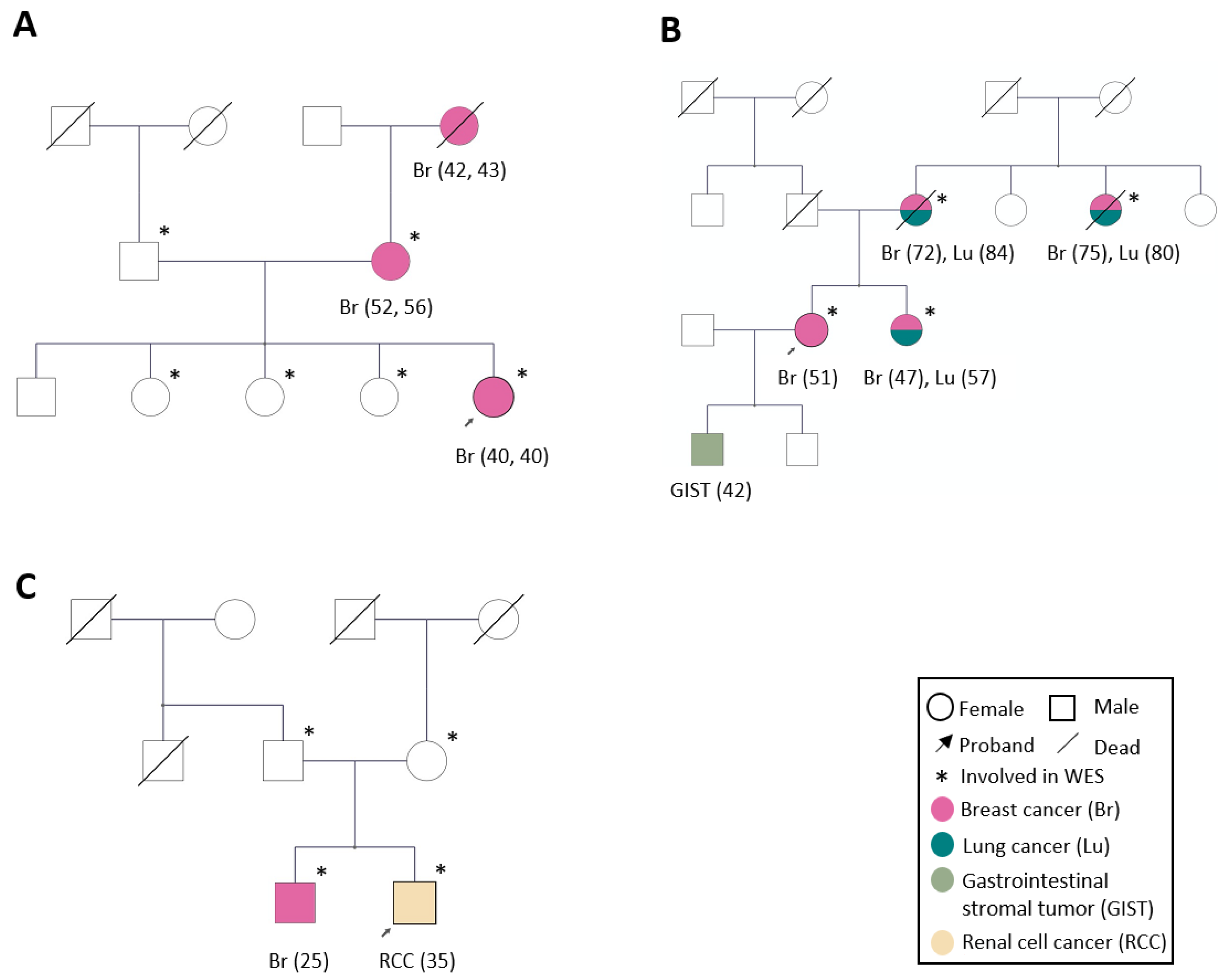
2.1. Family Selection

Initially, 17 SWEA-negative families with a striking pedigree of HBC were selected. The probands were approached at the Hereditary Unit at the Oncology clinic at Karolinska University Hospital for inclusion in the study for WES analysis. These probands had thus been screened negatively using the 64-gene panel including the known genes associated with hereditary breast cancer: BRCA1, BRCA2, CDH1, CHEK2, PTEN, STK11, TP53, ATM, BARD1, BRIP1, CDKN2A, MRE11A, NBN, PALB2, RAD50, RAD51C, RAD51D, ATR, BABAM1, BAP1, BCCIP, BLM, BRAP, BRCC3, BRE, C17orf70, C19orf40, CDK4, FAM175A, FAN1, FANCA, FANCB, FANCC, FANCD2, FANCE, FANCF, FANCG, FANCI, FANCL, FANCM, HUS1, KLLN, MDC1, RAD1, RAD17, RAD51, RAD51B, RAD52, RAD54L, RAD9A, RBBP8, RMI1, RMI2, SDHB, SDHD, SLX4, TOP3A, TOPBP1, TP53BP1, UIMC1, XRCC2, XRCC3, XRCC4, and ZNF350 (personal communication with Prof. Åke Borg, Lund University Cancer Center). In 4 out of these 17 families, we were able to sample blood from both healthy and diseased relatives or from at least three individuals with breast cancer. In one family we were not able to confirm the cancer diagnosis from the paternal side; therefore, this family was excluded. Finally, three families were selected for further analysis.

2.2. Family Characteristics

Family 1 is characterized by three generations of bilateral ductal breast adenocarcinoma with disease onset between 40 and 56 years of age (Figure 1A; Table S1). The index case was diagnosed with bilateral breast cancer at 40 years of age at the first mammogram examination within the national surveillance program. The three healthy sisters have been enrolled in a surveillance program with yearly breast magnetic resonance imaging (MRI) examinations. During the 6 years follow-up, none of them have been diagnosed with breast cancer and their current age is 50, 59, and 61 years, respectively. Blood samples were obtained from the proband, her mother (breast cancer at 52 years of age and contralateral disease at 56 years of age), father, and from the proband’s three healthy sisters. The maternal grandmother who was diagnosed with breast cancer at age 42 and contralaterally at 43 had earlier succumbed to the disease. No tumor material was available despite intensive search at the pathology unit at Karolinska University Hospital.
Family 2 was selected due to four first-degree relatives with breast cancer (age of onset between 47 and 75) of which three were diagnosed with lung cancer (age of onset 57–84), following their breast cancer. All three were non-smokers. The proband was diagnosed exclusively with breast cancer at 51 years of age (no lung cancer) and one of her sons was diagnosed with a gastrointestinal stromal cell tumor (GIST) at the age of 42. Blood samples from all women with breast cancer were collected (Figure 1B; Table S1).
For Family 3, two brothers with early-onset cancer; one with breast cancer at 25 and the other with clear cell renal cell carcinoma at 35. These two brothers and their healthy parents were enrolled in the study (Figure 1C and Table S1).

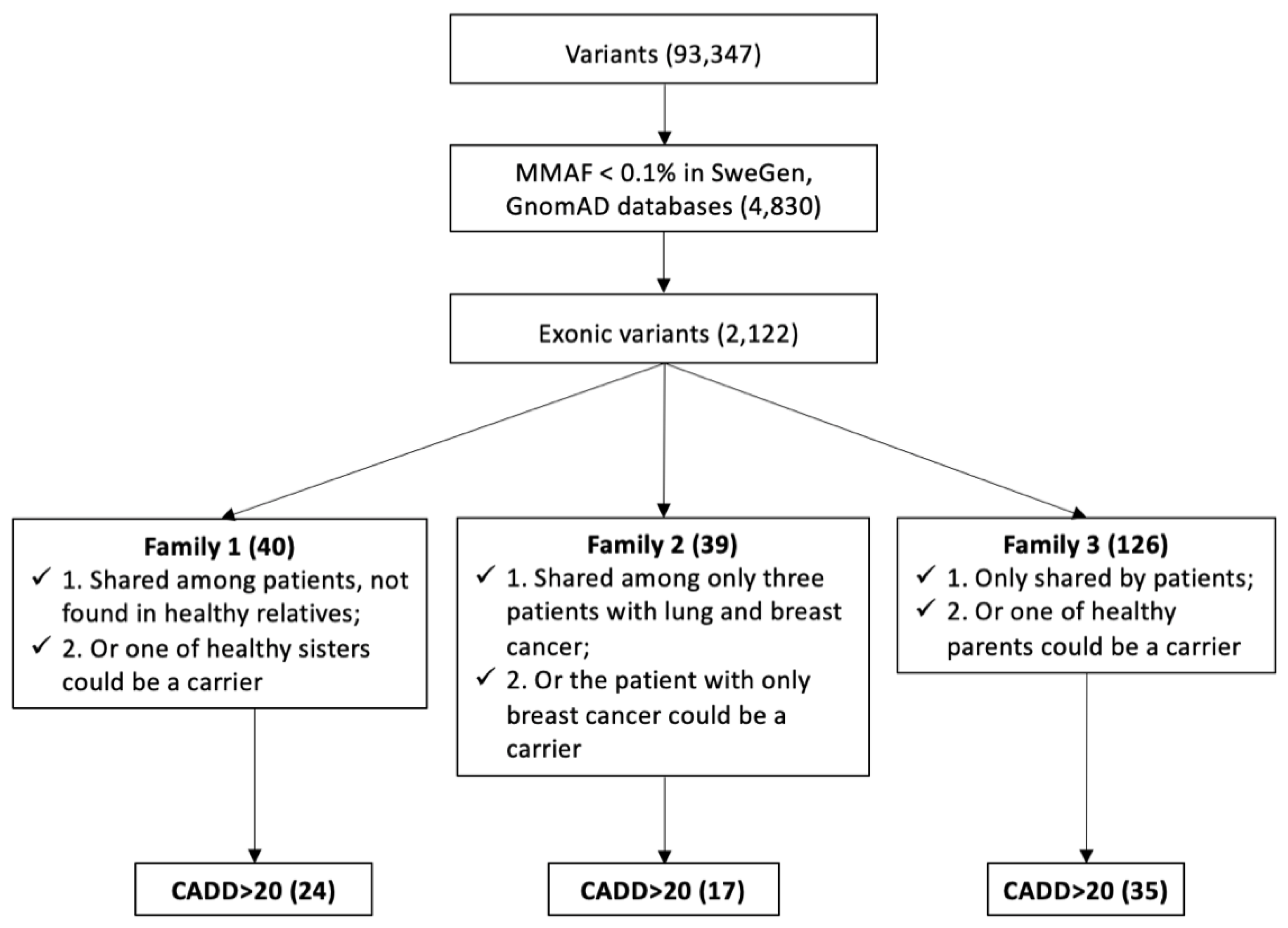
2.3. Whole-Exome Sequencing and Bioinformatics

WES was performed in collaboration with the Yale School of Medicine and the Clinical Genomics, SciLifeLab, Karolinska Institutet. Briefly, genomic DNA was isolated from the blood using Blood & Cell Culture DNA Midi Kit (#13343, Qiagen, Hilden, Germany) and captured using SeqCap EZ version 2 by NimbleGen (Roche NimbleGen, Madison, WI, USA). Captured fragments were sequenced using 76 bp paired-end sequencing reads in an Illumina HiSeq 2000 (Illumina Inc., San Diego, CA, USA) according to Illumina protocols. Sequencing reads were independently aligned to human genome build 37 (GRCh37/hg19) with BWA-MEM [13] and further processed using the GATK 3 Best Practices workflow [14]. Variants were annotated with ANNOVAR [15]. The data was filtered by the max minor allele frequency (MMAF) obtained from the SweGen [16], and GnomAD [17] databases where variants with MMAF ≥ 0.1% were excluded. Furthermore, we selected for exonic variants, as well as for variants segregating with the disease in each family. The functional prediction of the variant was evaluated by combined annotation dependent depletion (CADD) [18], with higher scores (CADD > 20) being more deleterious, and ClinVar [19].

2.4. Cell Culture, Transfection, and Plasmid Construction

The breast cancer cell lines MCF-7 (#HTB-22™, ATCC, Manassas, VA, USA) and MDA-MB-231 (#CRM-HTB-26, ATCC) were cultured in DMEM medium (#31885-023, Gibco, Waltham, MA, USA) with 10% fetal bovine serum, and 3∗105 cells were seeded per well in a 6-well plate 24 h before transfection. After reaching 70–80% cell confluence, the cells were transfected with 2 ug of plasmid per well using lipofectamine 2000 (#11668-019, Invitrogen, Waltham, MA, USA) or lipofectamine 3000 (#L3000-008, Invitrogen) according to the manufacturer’s protocol. Plasmids containing wild-type UBASH3A, MYH13, UTP11L, and PAX7 in the expression vector pcDNA3.1(+) were purchased from Genscipt Biotech (Piscataway, NJ, USA). The Quickchange II XL Site-directed Mutagenesis Kit (#200521, Agilent Technologies, Santa Clara, CA, USA) was used to construct the corresponding mutant plasmid. These plasmids were validated by Sanger sequencing performed at the KI Gene core facility.

2.5. Western Blot

Proteins were isolated from MDA-MB-231/MCF-7 cells transfected with control/wild-type/mutant plasmid and were quantified by BSA assay and 30 ug per sample was prepared with LDS sample buffer and sample reducing agent was heated at 75 °C 10 min and run on a 4–12% Tris-Bis gel with MOPS buffer at 200 V for 1 h. The protein transfer from gel to PVDF membrane with transfer buffer was performed at 40 V for 80 min. Membranes were blocked with 5% no-fat milk in TBST for 1 h and were then incubated overnight with primary antibody at 4 °C. The following day, the membranes were incubated with appropriate secondary antibody for 1 h at 37 °C, followed by developing and analysis on an iBright Western blot imaging system. Primary antibodies used targeted UBASH3A (#CF809272, mouse, Origene, Rockville, MD, USA), MYH13 (#PA5-70713, rabbit, Thermo Fisher, Waltham, MA, USA), UTP11L (#MA5-27022, rabbit, Thermo Fisher), PAX7 (#CF811661, mouse, Origene), EGFR (#4267, rabbit, CST, Danvers, MA, USA), p-EGFR (#3777, rabbit, CST), AKT (#4691, rabbit, CST), p-AKT (#4060, rabbit, CST), ERK (#9102, rabbit, CST), p-ERK (#4376, rabbit, CST), BAX (#14796, rabbit, CST), STAT3 (#12640, rabbit, CST), p-STAT3 (#9145, rabbit, CST), Beta-catenin (#9562, rabbit, CST), and GAPDH (#5174, rabbit, CST). The secondary antibodies used were anti-rabbit (#7074, CST) and anti-mouse (#7076, CST).

2.6. Mass Spectrometry and Pathway Analysis

Proteins were harvested from MCF-7 cells transfected with wild-type plasmid or mutant plasmid of MYH13 and UTP11 genes, respectively, and mass spectrometry was performed at the Clinical proteomics unit, Karolinska Institutet. Differentially expressed proteins, between the cells transfected with wild-type or mutant plasmid, were imported to the WEB-based Gene Set Analysis Toolkit (http://www.webgestalt.org, accessed on 1 January 2021) [20] for the Kyoto Encyclopedia of Genes and Genomes (KEGG) pathway analysis.

3. Results

3.1. Hereditary-Breast-Cancer-Associated Genes and Breast-Cancer-Associated High-Risk SNPs

Even though these families were screened negatively using the 64-gene panel including the known genes associated with hereditary breast cancer, we searched for variants in these 64 genes in the generated WES data. Fifty-seven variants in 29 genes with MMAF  <  0.2 were observed, but none of the variants could explain the breast cancer susceptibility observed in these families. Four variants in three genes (FANCA, FANCM, and TOPBP1) were found in patients of Family 1, and a BRCA2 missense variant (rs80358899) was observed in the proband from Family 2 (Table S2). However, all of them are considered as benign according to ClinVar.
When searching for rare variants contributing to tumorigenesis, we may exclude some more common single-nucleotide polymorphisms (SNPs). In the 24 known breast cancer risk SNPs [21], rs1045485 in CASP8 was only found in Family 2 and was shared by the proband and the proband’s sister (Table S2).

3.2. Identification of Candidate Genes by WES Analysis

In total, 93,347 variants were found in all individuals involved in this WES study, including exonic and exon–intron boundary variants. None of the variants were shared by the healthy or affected individuals when comparing the three families.
After filtering of variants with MMAF ≥ 0.1%, 4830 variants remained and after filtering non-exonic variants, 2122 variants were left. These 2122 exonic variants (27 nonsense, 204 splicing, and 1891 missense, Table S3) were selected for further analysis. In the first two families, maternal inheritance of breast cancer was seen. The flow chart of variant selection is summarized in Figure 2.

3.2.1. Candidate Genes in Family 1

To identify new breast-cancer-predisposing genes in Family 1, two different filtering of the data were applied. First, we applied strict filtering where only the affected individuals were considered as carriers of high-risk predisposing variants, and secondly, less strict filtering was used where one healthy sister could also be a carrier.
Firstly, we postulated that the father and the three healthy sisters do not share the risk variant with their affected sisters and mother. Therefore, variants detected in the father or any of the three healthy sisters were excluded, resulting in seven candidate missense variants (CADD > 20, MMAF < 0.001) that could contribute to the disease in the affected sisters (Table 1). Three of the gene variants, UBASH3A-rs201756769, MYH13-rs767313943, and FBXL4-rs757154231, had CADD  >  25 and were considered as high-risk gene variants. The MMAF of rs369992593 in the STRADB gene, rs141755850 in the CLK1 gene, FBXL4, and MYH13 missense variants were less than 0.0001.
The UBASH3A and MYH13 variants were functionally assessed in MDA-MB-231 and MCF-7 breast cancer cell lines. Concerning UBASH3A, the wild-type UBASH3A protein was highly elevated while the mutated protein showed only a slight increase compared to the basal expression of UBASH3A in both cell lines after transfection (data not shown). The difference between the biological effect of wild-type protein and mutated protein was therefore hard to clarify. After ectopic expression of the MYH13 (Figure S1A), no differences were observed between the mutant and wild-type MYH13 in terms of expressed proteins involved in pathways of proliferation, apoptosis, or adhesion by Western blot (Table S4). We identified 719 differentially expressed proteins from mass spectrometry using MCF-7, and a KEGG pathway analysis revealed that these proteins were enriched for seven different pathways, of which “DNA replication” and “protein processing in endoplasmic reticulum” showed statistical significance (adjusted p value ≤ 0.05; Figure S1B). However, no cancer-associated proteins in these pathways were found for further analysis.
Secondly, the analysis was performed with the assumption that one of the healthy sisters could be a carrier of the risk variant (non-penetrant), so the selection was performed with removal of variants identified in two of the sisters (in different combinations) instead of all three healthy sisters. With this filtering, we identified additional 16 missense variants and 1 splice variant (CADD > 20, MMAF < 0.001) that segregated with breast cancer (Table 1). The SPTLC3-rs372930777 variant had the highest CADD score of 35. The CAPN2-chr1:223947063A/G, COL17A1-rs757388768, and HCLS1-rs757006680 missense variants had the same CADD score of 34. Nine genes were found to have variants with MMAF  <  0.0001 (SPTLC3-rs372930777, CAPN2-chr1:223947063A/G, HCLS1-rs757006680, CNST-rs766380272, APOB-rs778274241, TENM4-rs747100917, DNAH3-rs376279103, NMRK2-chr19:3933697T/G, and DLL3-chr19:39996056G/A; Table 1).

3.2.2. Candidate Genes in Family 2

Two distinct data filtering processes were also applied in Family 2. A rigid filter was used at first, assuming that only patients with lung cancer and breast cancer were considered as carriers of a pathogenic variant in a high-risk susceptibility gene. In the second filtering, we speculated that the proband with breast cancer only also carry the disease-causing variant.
We first identified nine missense variants (CADD > 20, MMAF < 0.001) shared by the three patients with breast and lung cancer, not including the patient with only breast cancer (Table 2). Variants with CADD  >  25 were considered as high-risk variants (AKR1B1-rs201718247, SLC25A25-rs748220703, RYR3-rs760906719, C1orf228-rs575641425, RUFY1-rs754852607, HECTD4-rs779868916, and TIPIN-rs200514985). The MMAFs of RUFY1 and HECTD4 missense variants were less than 0.0001.
We found eight additional variants (CADD > 20, MMAF < 0.001) that were shared among all four affected family members (Table 2), assuming that the patient with breast cancer only could also be a carrier of the risk variant and possibly later develop lung cancer. There were seven missense variants and one splice-site acceptor variant. rs771377582 in the UTP11L gene and rs369607271 in the PAX7 gene had both high CADD scores of 35 and 34, respectively. Variants with MMAF < 0.0001 were found in three genes (PAX7-rs369607271, ZMYM4-chr1:35836075G/A, and WDFY4-rs748753983).
The variants in UTP11L and PAX7 were further studied. After ectopic expression of UTP11L (Figure S2A) and PAX7 (not shown), respectively, no difference in expression of proteins involved in proliferation, apoptosis, or adhesion pathways was observed between cells transfected with wild-type plasmid or mutated plasmid (Table S4). Concerning UTP11, there are limited publications on its involvement in cancer. Therefore, we proceeded with mass spectrometry of MCF-7 and identified 775 differentially expressed proteins that were subjected to KEGG analysis, which did not identify any significantly enriched pathways after false discovery rate adjustment (Figure S2B).

3.2.3. Candidate Genes in Family 3

We applied two strategies to find high-risk susceptibility variants in Family 3 which includes two brothers with early-onset cancer (one with breast cancer and one with renal cell carcinoma) and two healthy parents. Firstly, we assumed that both of the affected individuals shared the same variant, but not their healthy parents. Secondly, we hypothesized that one of parents could be a disease-free carrier (i.e., low penetrance).
Only one variant, chr14:30108088A/C in PRKD1 (CADD > 20, MMAF < 0.001), was shared by the two brothers (Table 3); however, it has been classified as likely benign in the Clinvar database. Considering the chance that one of the parents could carry the same variant, we found 34 additional variants (CADD > 20, MMAF < 0.001) (Table 3). Of these, TNRC6C-rs367710467, STAB1-chr3:52554136G/C, and THOP1-chr19:2799789G/A had the highest CADD score of 33. There were 31 variants with MMAF < 0.0001 (Table 3).

4. Discussion

The aim of this study was to identify genetic variants predisposing for hereditary breast cancer using whole-exome sequencing in families where extended genetic screening within the clinical platform had failed to detect cancer-predisposing genes. We selected three families with a remarkable family history of bilateral breast cancer, breast and lung cancer, and early-onset male breast cancer, respectively, and were able to collect blood samples for genetic analyses from family members covering at least two generations.
The linkage analysis of large pedigrees was the main method in early studies on hereditary cancer to find prevalent high-penetrance cancer risk genes, e.g., BRCA1 and BRCA2 associated with breast and ovarian cancer, and MSH2 and MLH1 associated with Lynch syndrome [22,23,24,25,26]. The other strategy relies on WES analysis to identify candidate genes in patients with clinical features of hereditary cancer (i.e., early tumor onset, family history, multiple tumors, etc.). However, the impact of WES in revealing the genetic cause in hereditary cancer syndromes has been lower than initially anticipated [27], indicating the challenge with this approach. The use of genome-wide association studies (GWAS) in breast cancer families has allowed the identification of genes that are characterized by incomplete penetrance, e.g., CHEK2 and ATM [28,29]. Furthermore, genes known to be altered in sporadic tumors have also been sought as another source of candidate genes for hereditary cancer forms.
After performing WES analysis in our families with striking breast cancer pedigrees, pathogenic variants in known hereditary-breast-cancer-syndrome-predisposing genes were excluded. However, rs1045485 in CASP8 was identified in Family 2 as a potential breast cancer risk variant and it was only shared by the proband and the proband’s sister that developed breast cancer at age 51 and 47, respectively. This SNP could potentially explain the earlier onset of breast cancer compared to their mother and aunt that developed breast cancer at age 72 and 75, respectively. In addition, we also included the splicing variants outside the non-canonical splice sites (−/+ 1, 2 bases in the introns) using VarSEAK (www.varSEAK.bio, accessed on 1 January 2021); however, no variants were predicted as likely pathogenic or pathogenic.
To search for new rare variants, the WES and bioinformatic filtering in our three families with HBC revealed 2122 variants that were selected for further analysis. In Family 1, we performed the first filtering by removing variants that were shared by the proband´s father and her three healthy sisters resulting in seven variants of interest. No functional effects were observed in the protein expression analysis of the UBASH3A and MYH13 missense variants, so other variants could be taken into consideration. The FBXL4 gene is identified as a potential tumor suppressor in prostate cancer, since the loss of FBXL4 has been correlated with advanced tumor stage and poor survival [30]. Meanwhile, the observed FBXL4 missense variant is located in the AMN1 domain, which refers to the regulation of MEN pathway in mitosis. The MMAF of the missense variant in the STRADB gene was less than 0.0001. The STRADB gene plays a role in the cell cycle and an intronic variant in this gene has been associated with ER-positive breast cancer [31]. Among the other interesting candidates, the CLK1 gene is highly expressed in normal breast tissue, according to the human protein atlas, and has also been linked to the regulation of mRNA-splicing processes in gastric cancer [32]. The CLK2 gene has been identified as an oncogene in sporadic breast cancer [33]. CLK1 may have similar function, but no evidence showed its relation to breast cancer. Frameshift variants of the TAF1C gene lead to alterations of RNA polymerase I transcription in gastric and colorectal cancers [34]. The FRZB gene regulates Wnt signaling, and downregulation of the FRZB protein has been observed in breast cancer [35]. Bernascone et al. [36] found that the loss of FRZB predisposed mice to breast cancer. The observed FRZB missense variant is located in the NTR domain (647–976), which may alter the binding to matrix metalloproteinases and possibly affect the downstream Wnt signaling.
It is important to note that this first filtering assumes full disease penetrance, which is not the case in most hereditary cancer syndromes, and we thus risk selecting out disease-causing variants. Considering this possibility of incomplete penetrance, the selection was further performed by removing variants that were identified in at least two healthy sisters instead of all three and then 17 variants remained. One variant with MMAF < 0.0001 and a very high CADD score (34) was located in the CAPN2 gene. The high expression of CAPN2 has previously been reported as associated with adverse prognosis in triple-negative breast cancer [37]. Knockdown of CAPN2 mRNA expression reduced breast cancer cell invasion by regulating invadopodia dynamics [38], that is associated with metastasis. The observed variant is located in the Calpain III domain associated with cytoskeletal remodeling processes and cell differentiation [39]. The other variant with a very high CADD score was seen in the COL17A1 gene, which inhibits cell migration and invasion and is a favorable prognostic marker in breast cancer [40]. The observed variant is located in the Collagen domain involved in the formation of connective tissue structure. These genes could have the potential to increase the risk of breast cancer.
For Family 2, we also performed two different filtering approaches. At first, nine missense variants shared by three patients with breast and lung cancer were selected. Variants with MMAF  <  0.0001 and CADD > 25 were noted in two genes (RUFY1 and HECTD4). RUFY1 has been reported to promote disease progression in gastric cancer and HECTD4 was a favorable prognostic marker in head and neck cancer according to the Human Protein Atlas database. Other interesting gene variants include one in the AKR1B1 gene. The AKR1B1 gene can promote breast cancer development by activating the epithelial-to-mesenchymal transition (EMT) process and the observed missense variant is located in its functional domain [41]. Another variant was found in BCL6 that is reported to contribute to the development of breast cancer. The overexpression of BCL6 in the breast cell line inhibited the expression of β-casein protein and apoptosis [42]. Moreover, the variant discovered in our study is located in a conserved domain. However, the patient with breast cancer has the potential to develop lung cancer in later years, so we also selected eight variants shared among all four patients. No functional impact of UTP11L and PAX7 variants with high CADD scores was found in following protein expression analyses. The FNDC3B gene is an oncogene in hepatocellular cancer and the high expression of FNDC3B induces the EMT process, PI3K/Akt, Rb1, and TGFβ signaling [43]. Except for UTP11L and PAX7, these genes could be candidate genes for breast cancer.
The third family (Family 3) was characterized by two early-onset cancers in two brothers, one with breast cancer and the other with renal cell cancer, while both parents were healthy. Male breast cancer is extremely rare especially at that age [44]. For the first filtering, only one variant with a CADD score above 20 was identified. To find a putative disease-causing gene, we also performed the second filtering as we speculated that the father or mother may carry the variant as well. The SATB1 gene can contribute to tumor growth in a mammary adenocarcinoma mouse model [45], while ZNF507 was reported to affect TGF-β signaling to promote prostate cancer [46]. Pathogenic variants in the DICER1 gene cause the DICER1 syndrome, including benign or malignant tumors of the thyroid, kidney, ovary, brain, and lung [47], but the variant chr14:95570153T/C discovered in our study is not located in a conserved domain; therefore, it may not affect the function of protein. These genes could have the potential to increase the risk of breast cancer. We have searched for a recessive inheritance (i.e., parents with heterozygous variants and brothers with homozygous variants); however, no variants were found. There is of course also a possibility that the brothers developed sporadic tumors with no genetic predisposition.
A limitation of our study is the small cohort of hereditary breast cancer families. The challenge has been to identify families with a striking pedigree of HBC and include only families in whom we were able to draw blood from several generations, and preferably from both healthy and diseased individuals, which leads to a limitation of the cohort size. Another restriction was that we performed WES that focuses on exonic variants, but will miss variants within the intronic areas and may affect splicing. These cannot be identified without a whole-genome-sequencing approach. Moreover, multiple variants in various genes may act in concert and contribute to the disease phenotype, which would also not be picked up by our functional approach testing one variant at the time.
In conclusion, whole-exome sequencing is a useful tool and has led to the identification of many genes associated with rare cancer syndromes. However, it has been more challenging than initially anticipated. In spite of selected families with pronounced family history of breast cancer, we were not able to identify any disease-causing genes with certainty. A wider combination of low- and intermediate-genetic-risk variants may instead contribute the breast cancer susceptibility in these families.

Supplementary Materials

The following supporting information can be downloaded at: https://www.mdpi.com/article/10.3390/biomedicines10051004/s1, Table S1: Clinical characteristics of all patients in the study. Table S2: Five variants in 64 hereditary cancer-related genes with MMAF < 0.2 and one SNP in the study. Table S3: All exonic variants found in the WES with MMAF < 0.001 in all families. Table S4: Western blot analysis of MYH13, UTP11L and PAX7 identified as candidates in Family 1 and Family 2. Figure S1: Ectopic expression of MYH13 identified as a candidate gene in Family 1 and its biological effects analyzed by KEGG pathway analysis. Figure S2: Ectopic expression of UTP11L identified as a candidate gene in Family 2 and its biological effects analyzed by KEGG pathway database.

Author Contributions

Conceptualization, S.B.-L. and Y.L.; methodology, Y.L. and H.T.H.; software, J.C., F.L.-G., S.M.M., H.T.H. and V.H.; validation, Y.L. and P.K.; formal analysis, Y.L. and H.T.H.; investigation, Y.L.; resources, C.C.J. and S.B.-L.; data curation, Y.L.; writing—original draft preparation, Y.L.; writing—review and editing, H.T.H., S.B.-L. and C.L.; visualization, Y.L.; supervision, S.B.-L. and C.L.; project administration, Y.L. All authors have read and agreed to the published version of the manuscript.

Funding

The Swedish Cancer Society (Cancerfonden) (2013/505), The Cancer Research Funds of Radiumhemmet (174012 and 201052), Stiftelsen Längmanska Kulturfonden (PK), Karolinska Institutet, Cancer Research KI, and Stockholm County Council.

Institutional Review Board Statement

The study has been approved by the regional ethical committee in Stockholm (Dnr 2012/2106-31/4).

Informed Consent Statement

All individuals gave their written consent in this study.

Data Availability Statement

Data available in article supplementary files.

Acknowledgments

We are grateful to Kristina Lagerstedt from the Department of Clinical Genetics, Karolinska University Hospital for helpful advice.

Conflicts of Interest

There is no conflict of interest.

References

  1. Ferlay, J.; Soerjomataram, I.; Dikshit, R.; Eser, S.; Mathers, C.; Rebelo, M.; Parkin, D.M.; Forman, D.; Bray, F. Cancer Incidence and Mortality Worldwide: Sources, Methods and Major Patterns in GLOBOCAN 2012. Int. J. Cancer 2015, 136, 359–386. [Google Scholar] [CrossRef] [PubMed]
  2. King-Spohn, K.; Pilarski, R. Beyond BRCA1 and BRCA2. Curr. Probl. Cancer 2014, 38, 235–248. [Google Scholar] [CrossRef] [PubMed]
  3. Terui-Kohbata, H.; Yoshida, M. Current Condition of Genetic Medicine for Hereditary Breast Cancer. Mol. Clin. Oncol. 2017, 7, 98–102. [Google Scholar] [CrossRef] [Green Version]
  4. Pilarski, R. PTEN Hamartoma Tumor Syndrome: A Clinical Overview. Cancers 2019, 11, 844. [Google Scholar] [CrossRef] [PubMed] [Green Version]
  5. Frebourg, T.; Bajalica Lagercrantz, S.; Oliveira, C.; Magenheim, R.; Evans, D.G. Guidelines for the Li–Fraumeni and Heritable TP53-Related Cancer Syndromes. Eur. J. Hum. Genet. 2020, 28, 1379–1386. [Google Scholar] [CrossRef] [PubMed]
  6. Rahman, N. Realizing the Promise of Cancer Predisposition Genes. Nature 2014, 505, 302–308. [Google Scholar] [CrossRef] [PubMed] [Green Version]
  7. Kast, K.; Rhiem, K.; Wappenschmidt, B.; Hahnen, E.; Hauke, J.; Bluemcke, B.; Zarghooni, V.; Herold, N.; Ditsch, N.; Kiechle, M.; et al. Prevalence of BRCA1/2 Germline Mutations in 21 401 Families with Breast and Ovarian Cancer. J. Med. Genet. 2016, 53, 465–471. [Google Scholar] [CrossRef] [Green Version]
  8. Levy-Lahad, E.; Friedman, E. Cancer Risks among BRCA1 and BRCA2 Mutation Carriers. Br. J. Cancer 2007, 96, 11–15. [Google Scholar] [CrossRef]
  9. Kuchenbaecker, K.B.; Hopper, J.L.; Barnes, D.R.; Phillips, K.-A.; Mooij, T.M.; Roos-Blom, M.-J.; Jervis, S.; van Leeuwen, F.E.; Milne, R.L.; Andrieu, N.; et al. Risks of Breast, Ovarian, and Contralateral Breast Cancer for BRCA1 and BRCA2 Mutation Carriers. JAMA 2017, 317, 2402–2416. [Google Scholar] [CrossRef] [Green Version]
  10. McClellan, J.; King, M.-C. Genetic Heterogeneity in Human Disease. Cell 2010, 141, 210–217. [Google Scholar] [CrossRef] [Green Version]
  11. Tung, N.M.; Boughey, J.C.; Pierce, L.J.; Robson, M.E.; Bedrosian, I.; Dietz, J.R.; Dragun, A.; Gelpi, J.B.; Hofstatter, E.W.; Isaacs, C.J.; et al. Management of Hereditary Breast Cancer: American Society of Clinical Oncology, American Society for Radiation Oncology, and Society of Surgical Oncology Guideline. J. Clin. Oncol. Off. J. Am. Soc. Clin. Oncol. 2020, 38, 2080–2106. [Google Scholar] [CrossRef] [PubMed]
  12. Girdea, M.; Dumitriu, S.; Fiume, M.; Bowdin, S.; Boycott, K.M.; Chénier, S.; Chitayat, D.; Faghfoury, H.; Meyn, M.S.; Ray, P.N.; et al. PhenoTips: Patient Phenotyping Software for Clinical and Research Use. Hum. Mutat. 2013, 34, 1057–1065. [Google Scholar] [CrossRef] [PubMed]
  13. Li, H. Aligning sequence reads, clone sequences and assembly contigs with BWA-MEM. arXiv 2013, arXiv:1303.3997. [Google Scholar] [CrossRef]
  14. Van der Auwera, G.A.; Carneiro, M.O.; Hartl, C.; Poplin, R.; Del Angel, G.; Levy-Moonshine, A.; Jordan, T.; Shakir, K.; Roazen, D.; Thibault, J.; et al. From FastQ data to high-confidence variant calls: The genome analysis toolkit best practices pipeline. Curr. Protoc. Bioinform. 2013, 43, 11.10.1–11.10.33. [Google Scholar] [CrossRef] [PubMed]
  15. Wang, K.; Li, M.; Hakonarson, H. ANNOVAR: Functional annotation of genetic variants from high-throughput sequencing data. Nucleic Acids Res. 2010, 38, e164. [Google Scholar] [CrossRef]
  16. Ameur, A.; Dahlberg, J.; Olason, P.; Vezzi, F.; Karlsson, R.; Martin, M.; Viklund, J.; Kähäri, A.K.; Lundin, P.; Che, H.; et al. SweGen: A Whole-Genome Data Resource of Genetic Variability in a Cross-Section of the Swedish Population. Eur. J. Hum. Genet. 2017, 25, 1253–1260. [Google Scholar] [CrossRef] [PubMed] [Green Version]
  17. Karczewski, K.J.; Francioli, L.C.; Tiao, G.; Cummings, B.B.; Alföldi, J.; Wang, Q.; Collins, R.L.; Laricchia, K.M.; Ganna, A.; Birnbaum, D.P.; et al. The Mutational Constraint Spectrum Quantified from Variation in 141,456 Humans. Nature 2020, 581, 434–443. [Google Scholar] [CrossRef] [PubMed]
  18. Kircher, M.; Witten, D.M.; Jain, P.; O’Roak, B.J.; Cooper, G.M.; Shendure, J. A General Framework for Estimating the Relative Pathogenicity of Human Genetic Variants. Nat. Genet. 2014, 46, 310–315. [Google Scholar] [CrossRef] [PubMed] [Green Version]
  19. Landrum, M.J.; Lee, J.M.; Benson, M.; Brown, G.R.; Chao, C.; Chitipiralla, S.; Gu, B.; Hart, J.; Hoffman, D.; Jang, W.; et al. ClinVar: Improving Access to Variant Interpretations and Supporting Evidence. Nucleic Acids Res. 2018, 46, D1062–D1067. [Google Scholar] [CrossRef] [Green Version]
  20. Wang, J.; Vasaikar, S.; Shi, Z.; Greer, M.; Zhang, B. WebGestalt 2017: A More Comprehensive, Powerful, Flexible and Interactive Gene Set Enrichment Analysis Toolkit. Nucleic Acids Res. 2017, 45, W130–W137. [Google Scholar] [CrossRef]
  21. Li, H.; Feng, B.; Miron, A.; Chen, X.; Beesley, J.; Bimeh, E.; Barrowdale, D.; John, E.M.; Daly, M.B.; Andrulis, I.L.; et al. Breast Cancer Risk Prediction Using a Polygenic Risk Score in the Familial Setting: A Prospective Study from the Breast Cancer Family Registry and KConFab. Genet. Med. Off. J. Am. Coll. Med. Genet. 2017, 19, 30–35. [Google Scholar] [CrossRef] [Green Version]
  22. Miki, Y.; Swensen, J.; Shattuck-Eidens, D.; Futreal, P.A.; Harshman, K.; Tavtigian, S.; Liu, Q.; Cochran, C.; Bennett, L.M.; Ding, W. A Strong Candidate for the Breast and Ovarian Cancer Susceptibility Gene BRCA1. Science 1994, 266, 66–71. [Google Scholar] [CrossRef] [Green Version]
  23. Wooster, R.; Neuhausen, S.L.; Mangion, J.; Quirk, Y.; Ford, D.; Collins, N.; Nguyen, K.; Seal, S.; Tran, T.; Averill, D. Localization of a Breast Cancer Susceptibility Gene, BRCA2, to Chromosome 13q12-13. Science 1994, 265, 2088–2090. [Google Scholar] [CrossRef]
  24. Fishel, R.; Lescoe, M.K.; Rao, M.R.; Copeland, N.G.; Jenkins, N.A.; Garber, J.; Kane, M.; Kolodner, R. The Human Mutator Gene Homolog MSH2 and Its Association with Hereditary Nonpolyposis Colon Cancer. Cell 1993, 75, 1027–1038. [Google Scholar] [CrossRef]
  25. Leach, F.S.; Nicolaides, N.C.; Papadopoulos, N.; Liu, B.; Jen, J.; Parsons, R.; Peltomäki, P.; Sistonen, P.; Aaltonen, L.A.; Nyström-Lahti, M. Mutations of a MutS Homolog in Hereditary Nonpolyposis Colorectal Cancer. Cell 1993, 75, 1215–1225. [Google Scholar] [CrossRef]
  26. Papadopoulos, N.; Nicolaides, N.C.; Wei, Y.F.; Ruben, S.M.; Carter, K.C.; Rosen, C.A.; Haseltine, W.A.; Fleischmann, R.D.; Fraser, C.M.; Adams, M.D. Mutation of a MutL Homolog in Hereditary Colon Cancer. Science 1994, 263, 1625–1629. [Google Scholar] [CrossRef] [PubMed]
  27. Stadler, Z.K.; Schrader, K.A.; Vijai, J.; Robson, M.E.; Offit, K. Cancer Genomics and Inherited Risk. J. Clin. Oncol. Off. J. Am. Soc. Clin. Oncol. 2014, 32, 687–698. [Google Scholar] [CrossRef]
  28. Vahteristo, P.; Bartkova, J.; Eerola, H.; Syrjäkoski, K.; Ojala, S.; Kilpivaara, O.; Tamminen, A.; Kononen, J.; Aittomäki, K.; Heikkilä, P.; et al. A CHEK2 Genetic Variant Contributing to a Substantial Fraction of Familial Breast Cancer. Am. J. Hum. Genet. 2002, 71, 432–438. [Google Scholar] [CrossRef] [Green Version]
  29. Bogdanova, N.; Cybulski, C.; Bermisheva, M.; Datsyuk, I.; Yamini, P.; Hillemanns, P.; Antonenkova, N.N.; Khusnutdinova, E.; Lubinski, J.; Dörk, T. A Nonsense Mutation (E1978X) in the ATM Gene Is Associated with Breast Cancer. Breast Cancer Res. Treat. 2009, 118, 207–211. [Google Scholar] [CrossRef] [Green Version]
  30. Stankiewicz, E.; Mao, X.; Mangham, D.C.; Xu, L.; Yeste-Velasco, M.; Fisher, G.; North, B.; Chaplin, T.; Young, B.; Wang, Y.; et al. Identification of FBXL4 as a Metastasis Associated Gene in Prostate Cancer. Sci. Rep. 2017, 7, 5124. [Google Scholar] [CrossRef] [Green Version]
  31. Wang, S.; Huo, D.; Ogundiran, T.O.; Ojengbede, O.; Zheng, W.; Nathanson, K.L.; Nemesure, B.; Ambs, S.; Olopade, O.I.; Zheng, Y. Association of Breast Cancer Risk and the MTOR Pathway in Women of African Ancestry in “The Root” Consortium. Carcinogenesis 2017, 38, 789–796. [Google Scholar] [CrossRef]
  32. Babu, N.; Pinto, S.M.; Biswas, M.; Subbannayya, T.; Rajappa, M.; Mohan, S.V.; Advani, J.; Rajagopalan, P.; Sathe, G.; Syed, N.; et al. Phosphoproteomic Analysis Identifies CLK1 as a Novel Therapeutic Target in Gastric Cancer. Gastric Cancer Off. J. Int. Gastric Cancer Assoc. Jpn. Gastric Cancer Assoc. 2020, 23, 796–810. [Google Scholar] [CrossRef]
  33. Yoshida, T.; Kim, J.H.; Carver, K.; Su, Y.; Weremowicz, S.; Mulvey, L.; Yamamoto, S.; Brennan, C.; Mei, S.; Long, H.; et al. CLK2 Is an Oncogenic Kinase and Splicing Regulator in Breast Cancer. Cancer Res. 2015, 75, 1516–1526. [Google Scholar] [CrossRef] [Green Version]
  34. Oh, H.R.; An, C.H.; Yoo, N.J.; Lee, S.H. Frameshift Mutations of TAF1C Gene, a Core Component for Transcription by RNA Polymerase I, and Its Regional Heterogeneity in Gastric and Colorectal Cancers. Pathology 2015, 47, 101–104. [Google Scholar] [CrossRef] [PubMed]
  35. Wu, Z.-H.; Zhang, Y.-J.; Yue, J.-X.; Zhou, T. Comprehensive Analysis of the Expression and Prognosis for SFRPs in Breast Carcinoma. Cell Transplant. 2020, 29, 963689720962479. [Google Scholar] [CrossRef] [PubMed]
  36. Schlensog, M.; Magnus, L.; Heide, T.; Eschenbruch, J.; Steib, F.; Tator, M.; Kloten, V.; Rose, M.; Noetzel, E.; Gaisa, N.T.; et al. Epigenetic Loss of Putative Tumor Suppressor SFRP3 Correlates with Poor Prognosis of Lung Adenocarcinoma Patients. Epigenetics 2018, 13, 214–227. [Google Scholar] [CrossRef] [Green Version]
  37. Storr, S.J.; Lee, K.W.; Woolston, C.M.; Safuan, S.; Green, A.R.; Macmillan, R.D.; Benhasouna, A.; Parr, T.; Ellis, I.O.; Martin, S.G. Calpain System Protein Expression in Basal-like and Triple-Negative Invasive Breast Cancer. Ann. Oncol. Off. J. Eur. Soc. Med. Oncol. 2012, 23, 2289–2296. [Google Scholar] [CrossRef]
  38. Cortesio, C.L.; Chan, K.T.; Perrin, B.J.; Burton, N.O.; Zhang, S.; Zhang, Z.-Y.; Huttenlocher, A. Calpain 2 and PTP1B Function in a Novel Pathway with Src to Regulate Invadopodia Dynamics and Breast Cancer Cell Invasion. J. Cell Biol. 2008, 180, 957–971. [Google Scholar] [CrossRef] [PubMed] [Green Version]
  39. Reverter, D.; Strobl, S.; Fernandez-Catalan, C.; Sorimachi, H.; Suzuki, K.; Bode, W. Structural Basis for Possible Calcium-Induced Activation Mechanisms of Calpains. Biol. Chem. 2001, 382, 753–766. [Google Scholar] [CrossRef]
  40. Yodsurang, V.; Tanikawa, C.; Miyamoto, T.; Lo, P.H.Y.; Hirata, M.; Matsuda, K. Identification of a Novel P53 Target, COL17A1, That Inhibits Breast Cancer Cell Migration and Invasion. Oncotarget 2017, 8, 55790–55803. [Google Scholar] [CrossRef]
  41. Wu, X.; Li, X.; Fu, Q.; Cao, Q.; Chen, X.; Wang, M.; Yu, J.; Long, J.; Yao, J.; Liu, H.; et al. AKR1B1 Promotes Basal-like Breast Cancer Progression by a Positive Feedback Loop That Activates the EMT Program. J. Exp. Med. 2017, 214, 1065–1079. [Google Scholar] [CrossRef] [Green Version]
  42. Logarajah, S.; Hunter, P.; Kraman, M.; Steele, D.; Lakhani, S.; Bobrow, L.; Venkitaraman, A.; Wagner, S. BCL-6 Is Expressed in Breast Cancer and Prevents Mammary Epithelial Differentiation. Oncogene 2003, 22, 5572–5578. [Google Scholar] [CrossRef] [Green Version]
  43. Cai, C.; Rajaram, M.; Zhou, X.; Liu, Q.; Marchica, J.; Li, J.; Powers, R.S. Activation of Multiple Cancer Pathways and Tumor Maintenance Function of the 3q Amplified Oncogene FNDC3B. Cell Cycle Georget. Tex 2012, 11, 1773–1781. [Google Scholar] [CrossRef] [Green Version]
  44. Gucalp, A.; Traina, T.A.; Eisner, J.R.; Parker, J.S.; Selitsky, S.R.; Park, B.H.; Elias, A.D.; Baskin-Bey, E.S.; Cardoso, F. Male Breast Cancer: A Disease Distinct from Female Breast Cancer. Breast Cancer Res. Treat. 2019, 173, 37–48. [Google Scholar] [CrossRef] [PubMed]
  45. Riabov, V.; Yin, S.; Song, B.; Avdic, A.; Schledzewski, K.; Ovsiy, I.; Gratchev, A.; Verdiell, M.L.; Sticht, C.; Schmuttermaier, C.; et al. Stabilin-1 Is Expressed in Human Breast Cancer and Supports Tumor Growth in Mammary Adenocarcinoma Mouse Model. Oncotarget 2016, 7, 31097–31110. [Google Scholar] [CrossRef] [PubMed] [Green Version]
  46. Kwon, W.; Choi, S.-K.; Kim, D.; Kim, H.-G.; Park, J.-K.; Han, J.E.; Cho, G.-J.; Yun, S.; Yu, W.; Han, S.-H.; et al. ZNF507 Affects TGF-β Signaling via TGFBR1 and MAP3K8 Activation in the Progression of Prostate Cancer to an Aggressive State. J. Exp. Clin. Cancer Res. 2021, 40, 291. [Google Scholar] [CrossRef] [PubMed]
  47. Robertson, J.C.; Jorcyk, C.L.; Oxford, J.T. DICER1 Syndrome: DICER1 Mutations in Rare Cancers. Cancers 2018, 10, 143. [Google Scholar] [CrossRef] [Green Version]
Figure 1. Pedigrees showing the three families in this study. (A). Family 1. Three generations with bilateral breast cancer (Br). (B). Family 2. Two generations with two primary tumors, i.e., breast and lung cancer (Lu). (C). Family 3. Two brothers with early onset cancer, i.e., breast cancer and renal cell carcinoma (RCC), respectively. The proband in each family is indicated with an arrow. Blood samples for WES analysis were collected from relatives marked with a star (*). Pedigree figures were created by the PhenoTips software [12].
Biomedicines 10 01004 g001
Figure 2. The workflow of variant selection in three families. Variants were filtered by max minor allele frequency (MMAF), exonic location, family segregation and combined annotation dependent depletion (CADD) scores. For all families, we applied two filter strategies considering the incomplete penetrance. In total, 24, 17, and 35 candidate variants were identified in Family 1, Family 2, and Family 3, respectively.
Biomedicines 10 01004 g002
Table 1. The twenty-four variants with MMAF < 0.001, CADD > 20 in Family 1.
FilterGenePositionRef/AltSNPTypeChangeMMAFCADD
1FBXL4chr6:993222281A/Crs757154231missenseNM_012160:c.T1739G:p.L580R0.0000132
UBASH3Achr21:43857670C/Trs201756769missenseNM_001001895:c.C1352T:p.T451M0.000230
MYH13chr17:10206712T/Crs767313943missenseNM_003802:c.A5570G:p.Q1857R0.0000227.1
FRZBchr2:183699592C/Trs150679557missenseNM_001463:c.G962A:p.R321Q0.0008324.3
TAF1Cchr16:84213125G/Ars61730960missenseNM_001243158:c.C1036T:p.R346W0.0004124.3
STRADBchr2:202337785C/Grs369992593missenseNM_001206864:c.C301G:p.L101V0.0000823.6
CLK1chr2:201726049C/Ars141755850missenseNM_001162407:c.G428T:p.S143I0.0004123
2SPTLC3chr20:13074186G/Ars372930777missenseNM_018327:c.G788A:p.R263Q0.0000835
CAPN2chr1:223947063A/G.missenseNM_001146068:c.A1175G:p.E392G034
COL17A1chr10:105807514C/Trs757388768missenseNM_000494:c.G2318A:p.G773E0.0004134
HCLS1chr3:121363691C/Trs757006680missenseNM_001292041:c.G373A:p.G125R0.0000134
STARD9chr15:42930971C/Trs369566419missenseNM_020759:c.C520T:p.R174W0.000133
CNSTchr1:246829203C/Trs766380272missenseNM_152609:c.C2174T:p.S725F0.0000128.4
STXBP5Lchr3:120976023C/Trs184420053missenseNM_001308330:c.C1675T:p.L559F0.0008326.4
DNAH3chr16:20975280G/Ars376279103missenseNM_017539:c.C9926T:p.S3309L0.0000826
TENM4chr11:78383268G/Ars747100917missenseNM_001098816:c.C5603T:p.A1868V0.0000125.6
MYOM3chr1:24421405G/Crs200854393missenseNM_152372:c.C866G:p.S289C0.000425.1
ASIC2chr17:31351024C/Trs199589382missenseNM_001094:c.G1051A:p.A351T0.000223.8
NMRK2chr19:3933697T/G.splicing.023.8
APOBchr2:21252574C/Ars778274241missenseNM_000384:c.G1554T:p.K518N0.0000223.4
DERL2chr17:5384651C/Trs202210923missenseNM_001304777:c.G289A:p.V97I0.0008322.9
RIC1chr9:5762545G/Ars771929691missenseNM_001206557:c.G1886A:p.R629H0.0004122.8
DLL3chr19:39996056G/A.missenseNM_016941:c.G1058A:p.R353K022.4
TMEM143chr19:48863405G/Ars544787964missenseNM_001303539:c.C293T:p.A98V0.0008320.2
Filter 1: Includes only affected carriers; Filter 2 (less stringent): Includes one of the healthy sisters as a carrier. Position is based on reference genome hg19; MMAF: max minor allele frequency; CADD: combined annotation dependent depletion.
Table 2. The seventeen variants with MMAF < 0.001, CADD > 20 in Family 2.
FilterGenePositionRef/AltSNPTypeChangeMMAFCADD
1AKR1B1chr7:134136457C/Trs201718247missenseNM_001628:c.G115A:p.G39R0.0008334
SLC25A25chr9:130864666C/Trs748220703missenseNM_001006641:c.C494T:p.T165M0.000434
RYR3chr15:33893707C/Trs760906719missenseNM_001036:c.C1876T:p.R626W0.0004134
RUFY1chr5:178987155A/Trs754852607missenseNM_001040451:c.A116T:p.Q39L0.0000828.6
TIPINchr15:66641436T/Crs200514985missenseNM_001289986:c.A134G:p.D45G0.0004127.9
C1orf228chr17:45166747C/Trs575641425missenseNM_001145636:c.C595T:p.P199S0.0004126.3
HECTD4chr12:112677734T/Crs779868916missenseNM_001109662:c.A4654G:p.I1552V0.0000225.2
BCL6chr3:187444624T/Crs747910667missenseNM_001130845:c.A1603G:p.R535G0.000123.4
CA12chr15:63618533C/Trs149256486missenseNM_001293642:c.G803A:p.G268E0.000223.3
2UTP11Lchr1:38489295C/Trs771377582missenseNM_016037:c.C757T:p.R253C0.000235
PAX7chr1:18961015G/Ars369607271missenseNM_001135254:c.G304A:p.G102S0.0000834
FNDC3Bchr3:171969145C/Trs190147254missenseNM_001135095:c.C604T:p.R202C0.0008331
ZMYM4chr1:35836075G/A.missenseNM_005095:c.G1028A:p.G343D024.6
WDFY4chr10:50013303A/Grs748753983splicing.0.0000523.7
NSD2chr4:1957024C/Grs748922675missenseNM_001042424:c.C2475G:p.H825Q0.0004123.6
MED14chrX:40552004G/Ars763899660missenseNM_004229:c.C1801T:p.R601C0.0002622.7
C17orf53chr17:42225596G/Ars377372267missenseNM_001171251:c.G425A:p.S142N0.000421.8
Filter 1: Includes only carriers affected with breast and lung cancer; Filter 2 (less stringent): All carriers affected with breast cancer (including those with also lung cancer). Position is based on reference genome hg19; MMAF: max minor allele frequency; CADD: combined annotation dependent depletion.
Table 3. The thirty-five variants with MMAF < 0.001, CADD > 20 in Family 3.
FilterGenePositionRef/AltSNPTypeChangeMMAFCADD
1PRKD1chr14:30108088A/C.missenseNM_001330069.2:c.743T > G:p.F248C022.3
2TNRC6Cchr17:76094616C/Trs367710467missenseNM_001142640.1:c.4607C > T:p.S1536L033
STAB1chr3:52554136G/C.missenseNM_015136.3:c.5412G > C:p.E1804D033
THOP1chr19:2799789G/A.missenseNM_003249.5:c.589G > A:p.G197R033
SPDYAchr2:29072798TG/T.splicingNM_001142634.2:c.934del:p.E312KfsTer20032
TMX2-CTNND1chr11:57505465C/Trs375390370missenseNM_001347890.1:c.331C > T:p.R111C032
ZNF793chr19:38023257G/Ars200503532splicingNM_001013659.3:c.16-1G > A032
NOMO3chr16:16363998C/T.missenseNM_001004067.4:c.1915C > T:p.R639C0.0001732
USP17L10chr4:9213003T/A.nonsenseNM_001256852.1:c.621T > A:p.C207Ter032
CNR2chr1:24201377G/Ars201829495missenseNM_001841.3:c.731C > T:p.A244V028.2
ZNF507chr19:32845840T/C.missenseNM_001136156.2:c.2104T > C:p.C702R027.2
USP40chr2:234399901G/GA.splicingNM_001365479.1:c.2923dup:p.S975FfsTer65027
TUBGCP6chr22:50656236G/Ars138609686missenseNM_020461.4:c.5389C > T:p.R1797C0.000064926.6
AGAP3chr7:150839000T/G.missenseNM_001281300.2:c.827T > G:p.F276C025.9
ITPR1chr3:4706906G/A.missenseNM_001099952.3:c.1639G > A:p.A547T025.8
SPDL1chr5:169028384AG/A.splicingNM_001329639.2:c.1426del:p.E476KfsTer19025.7
RUNX1chr21:36164605A/C.missenseNM_001001890.3:c.1189T > G:p.S397A025.5
TCF3chr19:1646413G/C.missenseNM_001136139.4:c.86C > G:p.P29R025.2
NT5C1B-RDH14chr2:18745234C/Trs147855687missenseNM_001002006.3:c.1661G > A:p.R554H0.00045425
ABHD6chr3:58279442G/Ars144907290missenseNM_001320126.2:c.964G > A:p.D322N024.9
EPHA2chr1:16456073C/T.missenseNM_001329090.2:c.2519G > A:p.R840Q024.8
HSPA6chr1:161494581G/C.missenseNM_002155.5:c.133G > C:p.A45P024.4
MYO16chr13:109507831C/T.missenseNM_001198950.3:c.1289C > T:p.T430M023.8
SMC4chr3:160135704T/Crs41272953missenseNM_001002800.3:c.1631T > C:p.I544T0.00011523.5
PRKAR2Achr3:48884770T/G.missenseNM_001321982.2:c.260A > C:p.E87A023.5
USP17L22chr4:9270417G/GA.splicingNM_001256863.1:c.1073_1074insA:p.S358RfsTer20023.3
ZYXchr7:143079991G/Trs150223874missenseNM_001010972.2:c.599G > T:p.W200L023.1
ITFG1chr16:47494745T/Grs376408976splicingNM_001305002.1:c.−132 + 281A > C023.1
NYNRINchr14:24883816T/C.missenseNM_025081.3:c.2861T > C:p.I954T022.6
NIM1Kchr5:43280642C/G.missenseNM_153361.4:c.1122C > G:p.N374K022.3
DICER1chr14:95570153T/C.missenseNM_001271282.3:c.3580A > G:p.R1194G021.8
NXNL1chr19:17571431G/Ars377352923missenseNM_138454.2:c.248C > T:p.T83M021.5
TRPV6chr7:142574988G/Trs139115329missenseNM_018646.6:c.514C > A:p.L172M021.2
ZNF320chr19:53384145G/C.missenseNM_001351773.1:c.1234C > G:p.L412V021
ZNF320chr19:53384768A/Grs144964547missenseNM_001351773.1:c.611T > C:p.L204P0.00090720.7
Filter 1: Includes only affected carriers; Filter 2 (less stringent), Includes that one of the healthy parents could be a carrier. Position is based on reference genome hg19; MMAF: max minor allele frequency; CADD: combined annotation dependent depletion.
Publisher’s Note: MDPI stays neutral with regard to jurisdictional claims in published maps and institutional affiliations.

