Article

Extracorporeal Shock Wave Therapy Combined with Platelet-Rich Plasma during Preventive and Therapeutic Stages of Intrauterine Adhesion in a Rat Model

1
Department of Obstetrics and Gynecology, Kaohsiung Chang Gung Memorial Hospital and Chang Gung University College of Medicine, Kaohsiung 833, Taiwan
2
Graduate Institute of Clinical Medicine, College of Medicine, Kaohsiung Medical University, Kaohsiung 807, Taiwan
3
Department of Obstetrics and Gynecology, Pingtung Christian Hospital, Pingtung 900, Taiwan
4
Center for Shockwave Medicine and Tissue Engineering, Kaohsiung Chang Gung Memorial Hospital and Chang Gung University College of Medicine, Kaohsiung 833, Taiwan
5
Medical Research, Kaohsiung Chang Gung Memorial Hospital and Chang Gung University College of Medicine, Kaohsiung 833, Taiwan
6
Department of Leisure and Sports Management, Cheng Shiu University, Kaohsiung 833, Taiwan
7
Department of Orthopedic Surgery, Kaohsiung Chang Gung Memorial Hospital, Kaohsiung and Chang Gung University College of Medicine, Kaohsiung 833, Taiwan
8
Center for Menopause and Reproductive Medicine Research, Kaohsiung Chang Gung Memorial Hospital and Chang Gung University College of Medicine, Kaohsiung 833, Taiwan
9
Department of Obstetrics and Gynecology, Jen-Ai Hospital, Taichung 412, Taiwan
*
Author to whom correspondence should be addressed.
Biomedicines 2022, 10(2), 476; https://doi.org/10.3390/biomedicines10020476
Submission received: 28 January 2022 / Revised: 15 February 2022 / Accepted: 15 February 2022 / Published: 17 February 2022
(This article belongs to the Special Issue Translational Research in Shock Wave Medicine)

Abstract

:
Intrauterine adhesion (IUA) is caused by artificial endometrial damage during intrauterine cavity surgery. The typical phenotype involves loss of spontaneous endometrium recovery and angiogenesis. Undesirable symptoms include abnormal menstruation and infertility; therefore, prevention and early treatment of IUA remain crucial issues. Extracorporeal shockwave therapy (ESWT) major proposed therapeutic mechanisms include neovascularization, tissue regeneration, and fibrosis. We examined the effects of ESWT and/or platelet-rich plasma (PRP) during preventive and therapeutic stages of IUA by inducing intrauterine mechanical injury in rats. PRP alone, or combined with ESWT, were detected an increased number of endometrial glands, elevated vascular endothelial growth factor protein expression (hematoxylin-eosin staining and immunohistochemistry), and reduced fibrosis rate (Masson trichrome staining). mRNA expression levels of nuclear factor-kappa B, tumor necrosis factor-α, transforming growth factor-β, interleukin (IL)-6, collagen type I alpha 1, and fibronectin were reduced during two stages. However, PRP alone, or ESWT combined with PRP transplantation, not only increased the mRNA levels of vascular endothelial growth factor (VEGF) and progesterone receptor (PR) during the preventive stage but also increased PR, insulin-like growth factor 1 (IGF-1), and IL-4 during the therapeutic stage. These findings revealed that these two treatments inhibited endometrial fibrosis and inflammatory markers, thereby inhibiting the occurrence and development of intrauterine adhesions.

1. Introduction

Intrauterine adhesion (IUA), also known as Asherman syndrome, is a major cause of female secondary infertility [1] and is typically caused by artificial injury to the endometrial basal layer of the uterine cavity during curettage, cesarean section, and hysteromyomectomy [2]. IUA commonly occurs due to trauma and infection, leading to the loss of spontaneous endometrium recovery and angiogenesis, initiating the replacement of the normal endometrium by fibrous connective tissue and the formation of adhesions between uterine walls [3]. Clinical symptoms include chronic pelvic pain, abnormal menstruation (hypomenorrhea or amenorrhea), miscarriage, secondary infertility, and secondary amenorrhea, potentially accompanied by psychological distress [4].
According to epidemiological data, the incidence of IUA in females who have undergone hysteroscopic surgery and abortion ranges between approximately 4–46% [5]. Moreover, there is a high probability of complications and perforation-induced bleeding during reconstructive surgery, while the probability of a successful pregnancy is only 23 to 33% [6,7]. Clinical data also indicate that the prognosis of IUA could be related to failed endometrial regeneration, with a recurrence rate of up to 62.5% [8]. Therefore, the prevention and early treatment of IUA have become particularly important in recent years.
Therapeutic approaches for IUAs include adhesiolysis and preventing adhesion recurrence. To recover the normal shape and volume of the uterine cavity, the objective of most treatment strategies is to alleviate associated symptoms, such as infertility and amenorrhea [9]. The functional layer of the uterus undergoes degeneration, growth, and reorganization throughout the menstrual cycle. Several researchers have attempted to develop different ancillary therapies to accelerate the regeneration of the endometrial layer and prevent the recurrence of adhesions. 17β-Estradiol (E2) and platelet-rich plasma (PRP) have been shown to effectively facilitate endometrial regeneration and the formation of new capillaries after menstruation, as well as promote post-surgical tissue regeneration in patients [10,11]. For IUA therapy, E2 is typically administered as an oral or systemic formulation, which presents restricted clinical application due to its limited half-life in vivo, and is commonly employed combined with other bioactive substances [12,13]. PRP is an autologous concentration of platelets that has been used as a new therapeutic option for different pathologies, such as musculoskeletal diseases [14,15,16], cosmetic medicine [17,18,19], and cardiology [20,21]. PRP releases growth factors and cytokines, such as vascular endothelial growth factor (VEGF) [22], platelet-derived growth factor, epidermal growth factor (EGF) [23], transforming growth factor (TGF) [24], and other cytokines that modulate angiogenesis [25], affect recruitment proliferation [11] and stimulate cell differentiation and growth [26,27,28]. Thus, PRP therapy of the uterus and other tissues is a research hotspot for endometrial regeneration [29,30].
Extracorporeal shockwave therapy (ESWT) is a non-invasive physiotherapy and valid therapeutic modality. It was first used in lithotripsy to destroy kidney stones, and an incidental observation of the osteoblastic response pattern during animal studies began in the mid-1980 [31]. In recent years, studies have shown that ESWT can be utilized for numerous orthopedic disorders, including nonunion of fractures, proximal plantar fasciitis of the heel, tendinitis [32,33,34,35], lateral epicondylitis of the elbow [36,37,38], reproductive functions in female fertility [39,40], and treatment of ischemic diseases such as angina and ischemic heart failure in cardiology [41,42,43]. Although the precise functions of ESWT need to be comprehensively clarified, energy from sonic waves is postulated to increase the release of growth factors, thereby improving endothelial function, angiogenesis, and increasing blood supply [44]. To the best of our knowledge, the role and mechanisms of ESWT on endometrial function have not been evaluated in previous IUA studies.
In the present study, we examined the role of shock wave and/or PRP during the preventive and therapeutic stages of IUA. First, we investigated the role of shock wave and/or PRP for the prevention and early treatment of IUA in a rat model. Next, we determined the impact of ESWT combined with PRP transplantation on IUA development and examined potential underlying mechanisms. These studies are expected to have a significant positive impact by defining the roles of shock waves and/or PRP transplantation, thereby identifying potential therapeutic targets for intrauterine adhesions. The objective of the present study was to identify a new strategy for IUA prevention and treatment.

2. Materials and Methods

2.1. Reagents

17β-Estradiol was obtained from Merck (Oestrogel, 17β-estradiol 0.06% gel; Besins, France), and the PRP disposable kit was obtained from SC Medical (RegenKit® THT tubes, RegenLab, Le Mont-sur-Lausanne, Switzerland). Antibodies against CK-18 and vimentin were obtained from Abcam (Cambridge, UK), and anti-VEGF was obtained from GeneTex (GeneTex, Irvine, CA, USA) unless otherwise indicated.

2.2. Animals

Seven~eight-week-old female Sprague-Dawley rats (weighing 200–250 g) were purchased from BioLasco (Ilan, Taiwan). On arrival, the rats were randomly transferred to plastic cages containing aspen bedding (three rats per cage) and acclimatized for at least one week before experimentation. Animals were housed in a temperature (22 ± 2 °C), humidity (50 ± 20%), and light (12-h light/dark cycle)-controlled environment. Food and water were provided ad libitum. All procedures were reviewed and approved by the Institutional Animal Care and Use Committee of Chang Gung Memorial Hospital (IACUC no. 2019122008, approval date: 20200325) and complied with the National Institutes of Health Guide for Care and Use of Laboratory. Vaginal smears were obtained daily, between 8:00~9:00 am, to assess estrus cycles.

2.3. Establishment of the IUA Model

The mechanical injury IUA model was established as described by Khrouf et al. [45]. Briefly, the animal model was established after achieving regular estrous cyclicity over one week, as determined by vaginal smearing. Rats were anesthetized using tiletamine/zolazepam (Zoletil™ 50, 20–40 mg/kg; Virbac, Carros, France) and xylazine (5–10 mg/kg; Rompun, Bayer AG, Leverkusen, Germany) to induce the intrauterine mechanical injury. After adequate anesthesia was established, the rats were placed in a supine position, and the inferior abdomen was shaved and disinfected with 1% iodine tincture and 70% ethanol. Then, a vertical incision (~2 cm) was placed longitudinally, made sharply, and extended to the peritoneal cavity near the fallopian tube to avoid blood vessels. Subsequently, a 16-G needle was inserted and rotated to scratch the entire inner endometrial surface of both laterals until uterine walls became rough and pale, leaving the uterine serosa intact. Preventive and therapeutic protocols were performed on day 0. For all IUA groups, the uterus was harvested for further study.

2.4. Preparation and Isolation of PRP

Autologous PRP was collected from peripheral venous blood on the day of surgery, using a disposable kit (RegenKit® THT tubes, RegenLab, Le Mont-sur-Lausanne, Switzerland) from redundant male Sprague-Dawley rats; PRP comprises a gel layer of separated blood components. Briefly, 8–10 mL of blood was collected in RegenKit® THT tubes (8 mL per tube), pre-filled with citrate as an anticoagulant. All tubes were centrifuged at 1500× g for 9 min at room temperature using a universal centrifuge Regen Lab PRP-Centri (Regen Lab SA, Le Mont-sur-Lausanne, Switzerland). After centrifugation, the plasma supernatant was used to separate plasma. PRP is the middle layer between plasma and red blood cells, aspirated and collected into a new tube for further study.

2.5. E2 and PRP Treatment during Preventive and Therapeutic Stages of the IUA Model

After two estrous cycles (~9 days), established IUA model rats were randomly assigned to 6 groups to receive different treatments: control (sham) group, untreated; IUA group, entire inner endometrial surface of both laterals was scratched using a 16-G needle; ESWT treatment group, IUA combined with ESWT treatment to the uterus; E2 transplantation group, IUA combined with E2 gel (0.2 mg) injection into the uterus; PRP transplantation group, IUA combined with PRP (0.2 mL) injection to the uterus; ESWT+PRP transplantation group, IUA combined with ESWT and PRP injection (0.2 mL) into the uterus. Total E2 and PRP were transplanted into the uterine subserosa using a 21 G-syringe, and uterine and abdominal wounds were closed. Preventive and therapeutic transplantations were performed on days 0 and 14, respectively. The protocol is shown in Figure 1.

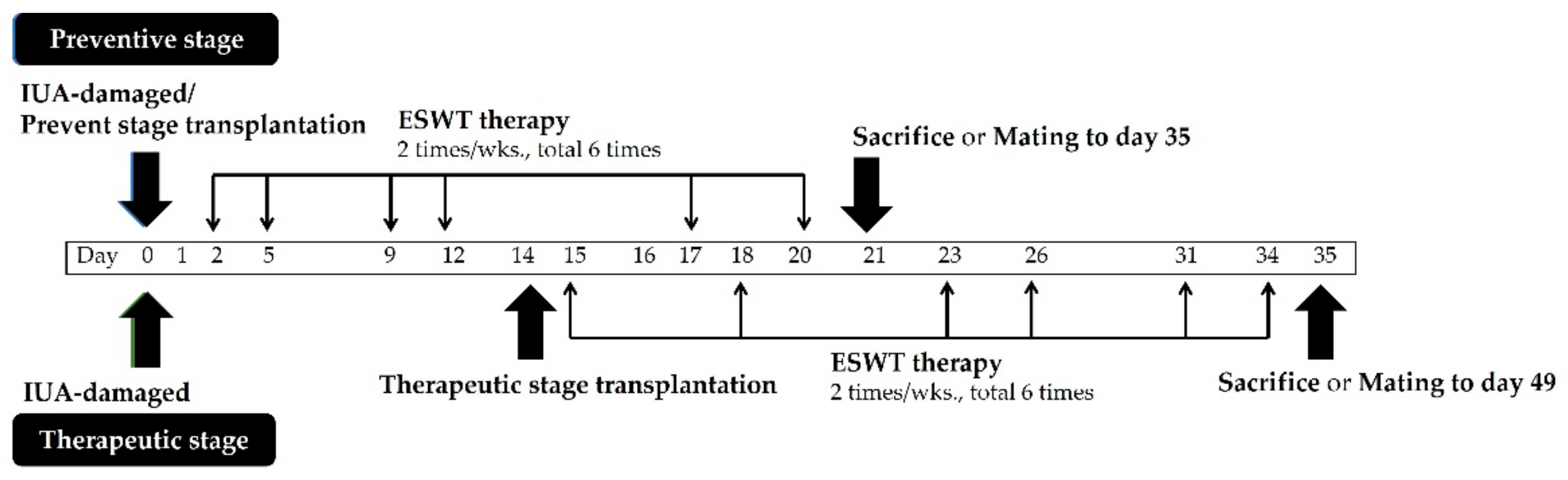
2.6. ESWT for IUA Model

The shock wave applicator (SD-1, Storz, Tägerwilen, Germany) was gently placed over the top, middle, and bottom of the skin surface of the uterine area after applying the ultrasound transmission gel (Toshiba, Tokyo, Japan) and exposed to the shock wave ranging of ESWT (0.1 mJ/mm2, 200 pulses) applicator. The energy dosage of ESWT for treatment was based on a previous study, with some modifications. The detailed protocol followed Zhang et al. [46]. The shock number ranging of ESWT was applied at a frequency of 6 pulses/s, twice weekly for 3 weeks, a total of 1200 pulses initiated 1 day or 14 days after IUA surgery. The current treatment regimen was within the optimal range for LESW. The preventive and therapeutic treatments were performed from day 2 to day 20 and day 15 to day 34, respectively. The protocol is presented in Figure 1. Finally, the rats were sacrificed. In addition, animals were mated at days 21 and 35 after ESWT treatment to observe endometrial morphology and pathology, tissue proliferation, inflammation, pregnancy outcomes, and mechanism of PRP in endometrial regeneration.

2.7. Mating of Rats

The reproductive function of the damaged uterus was evaluated by determining whether the regenerative endometrium could receive a fertilized ovum, provide sufficient nourishment to the developing fetus, and support embryos to the late stage of pregnancy. After days 21 and 35 post-initial injury, 3 female rats from each group were mated with healthy fertile male rats at a ratio of 1:1 and were subjected to two heat cycles. The mating was performed for 3–4 days, after which the male was removed, and the pregnant female rats were routinely housed for 13 days. Pregnant female rats were sacrificed, the uterus was collected for each group, gestational sacs of the implanted embryo were counted, and embryo development was photographed of all groups.

2.8. Histological Analysis

Uterine horns were collected, fixed in 10% neutral buffered formalin, dehydrated, and embedded in paraffin. Serial sections (5-μm thick) were prepared and stained with hematoxylin and eosin (H&E), and Masson trichrome (Sigma-Aldrich; #HT15, Burlington, MA, USA) staining was performed to detect fibrosis according to the manufacturer’s protocol. Undamaged uteri were used as negative controls. All slides were scanned using a Pannoramic SCAN (MIDI II, Sysmex Europe GmbH, Norderstedt, Germany), and a microscope was used to capture images. At least six uterine cross-sections were evaluated for each group, and each section was randomly selected. Epithelial and endometrial thickness were measured using Case Viewer software (3DHISTECH slide converter). The amount of fibrosis (blue) was determined using ImageJ software (National Institutes of Health, Bethesda, MD, USA).

2.9. Immunohistochemical Staining

For immunohistochemistry, uterine sections were dewaxed and rehydrated from graded ethanol, antigen retrieval by autoclave heating in citrate buffer (pH 6.0) (Diagnostic BioSystems; Cat#K035, Malvern, UK). Add enough hydrogen peroxide blocking solution (Abcam; Cat#ab64218, Cambridge, UK) to cover the sections. Incubate for 15 min at room temperature, the slides were incubated with rabbit polyclonal antibodies against vimentin (1:250; Abcam, Cat#ab92547, Cambridge, UK) or anti-cytokeratin 18 (1:500; Abcam, Cat#ab181597, Cambridge, UK) overnight at 4 °C, washed using Tris-buffered saline (TBS) containing 0.01% Tween-20 (TBS-T), and incubated with secondary antibody for 30 min at room temperature. The bound antibody was detected using the DAKO REALTM EnVisionTM Detection System Peroxidase/DAB+, Rabbit/Mouse reagent for 5 min. Subsequently, samples were incubated with anti-VEGF (1:50; GeneTex, Cat#GTX102643, Irvine, CA, USA) overnight at 4 °C, washed with TBS-T, and incubated with the biotinylated universal antibody for 30 min at room temperature, followed by R.T.U. Vectastain Elite ABC Reagent for 30 min at room temperature. The slides were counterstained with hematoxylin for 10 min. All slides were scanned using a Pannoramic SCAN (MIDI II, Sysmex Europe GmbH, Norderstedt, Germany), and a microscope was used to capture images.

2.10. RNA Extraction, Reverse Transcription, and Quantitative Real-Time Polymerase Chain Reaction

Total RNA was isolated using the Quick-RNA™ Miniprep Kit (Zymo Research, Irvine, CA, USA) and reverse-transcribed according to the manufacturer’s instructions. Real-time reverse transcription-PCR (RT-PCR) was performed using Fast SYBR® Green Master Mix (Applied Biosystems, Vilnius, Lithuania) and the ABI 7500 Fast Real-Time PCR System (Applied Biosystems, Singapore). We quantified GAPDH gene transcripts as an endogenous RNA control and normalized each sample with respect to its GAPDH content. Primer sequences used are listed in Table 1.

2.11. Statistical Analysis

Homogeneous data were evaluated by parametric analysis of variance (ANOVA) with Dunnett’s test to assess the statistical differences between ESWT treatment, E2 transplantation, PRP transplantation, ESWT combined with PRP transplantation, and the IUA group by software Prism 5.0 (GraphPad Software Inc., San Diego, CA, USA). All p-values < 0.05 were defined as statistical significance. The mean ± standard error (SE) was presented for individual experiments.

3. Results

3.1. Effects of E2 or PRP Transplantation Combined with ESWT on Body Weight in IUA Female Rats

We first determined the potential effects of PRP and/or ESWT treatment on body weight in IUA-damaged mouse models. We also employed E2 as an ancillary treatment. As shown in Table 2, the baseline body weight values (at day 0) during the preventive stage were 212.1 ± 7.8 g (sham group), 210.3 ± 14.1 g (IUA group), 205.4 ± 10.3 g (ESWT treatment group), 215.8 ± 8.9 g (E2 transplantation group), 201.8 ± 7.0 g (PRP transplantation group), and 201.5 ± 6.0 g (ESWT+PRP transplantation group). ESWT treatment did not significantly impact the body weight, presenting values of 253.5 ± 15.7 g, 244.2 ± 19.0 g, 243.9 ± 23.0 g, 247.8 ± 12.9 g, 243.0 ± 14.9 g, and 235.4 ± 13.9 on day 21, respectively. During the therapeutic stage, the baseline body weight values of each group were 206.1 ± 8.3 g, 212.3 ± 12.2 g, 209.3 ± 6.9 g, 211.6 ± 10.6 g, 221.8 ± 15.1 g, and 224.9 ± 13.9 g, respectively. Following E2 or PRP transplantation and ESWT treatment, the body weight was markedly increased, with values of 262.7 ± 28.3 g, 266.3 ± 20.4 g, 261.6 ± 10.9 g, 265.5 ± 22.6 g, 279.4 ± 16.3 g, and 274.9 ± 20.0 g noted on day 35, respectively. Regardless of E2 gel application, PRP transplantation or ESWT treatment did not affect body weight both pre- or postoperatively. Accordingly, our results revealed that the dose of E2 and PRP, as well as ESWT energy, did not affect body weight in the IUA-induced model.

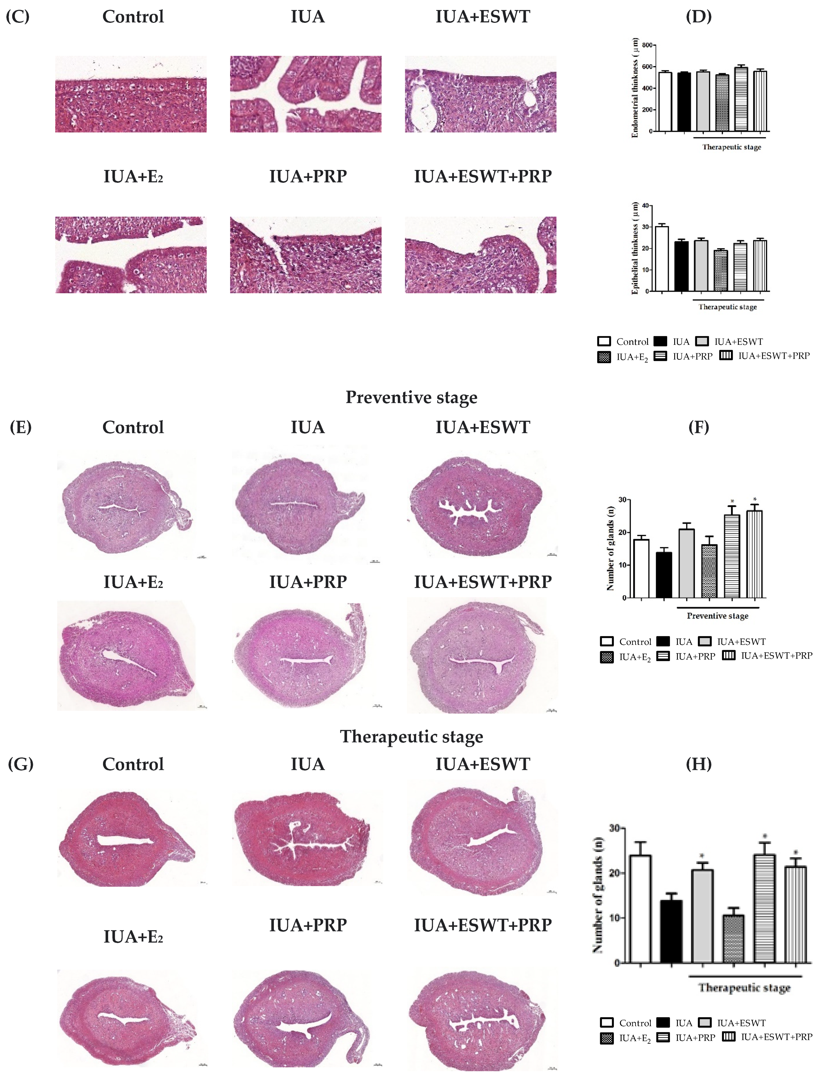
3.2. PRP and ESWT Combined with PRP Transplantation Restored Endometrial Morphology in an IUA Rat Model

To determine the role of E2, PRP, and ESWT on the recovery of damaged endometrium, we established an IUA model in female rats by inducing mechanical injury and examined whether the IUA induction was successfully established. H&E staining was used to analyze endometrial thickness, endometrial glands, and vascularization; the number of endometrial glands and endometrial thickness were quantified and measured. Based on the H&E staining results, uterine tissues in the control (sham) group exhibited simple columnar epithelium, and the submucosa contained abundant glands (Supplementary Data) and numerous blood vessels, regardless of the therapeutic stage. In the IUA group, the uterine cavity presented adhesions and atresia, and the smooth cavity was damaged (Figure 2A,C). Compared with the uterine cavity of the IUA group, uterine tissues of the ESWT, PRP, and ESWT combined with PRP transplantation groups showed improved uterine morphology following uterine cavity damage. In the preventive stage, significantly increased endometrial thickness was observed in the PRP and ESWT combined with PRP transplantation groups (Figure 2B). Additionally, PRP and ESWT combined with PRP transplantation groups showed a significant increase in the number of glands when compared with that in the IUA model group (Figure 2F,H), thus ameliorating the appearance of the submucosa, presenting numerous glands and blood vessels during the preventive and therapeutic stages (Figure 2E,G). However, E2 transplantation did not alter endometrial and epithelial thickness during the preventive and therapeutic stages (Figure 2B,D).

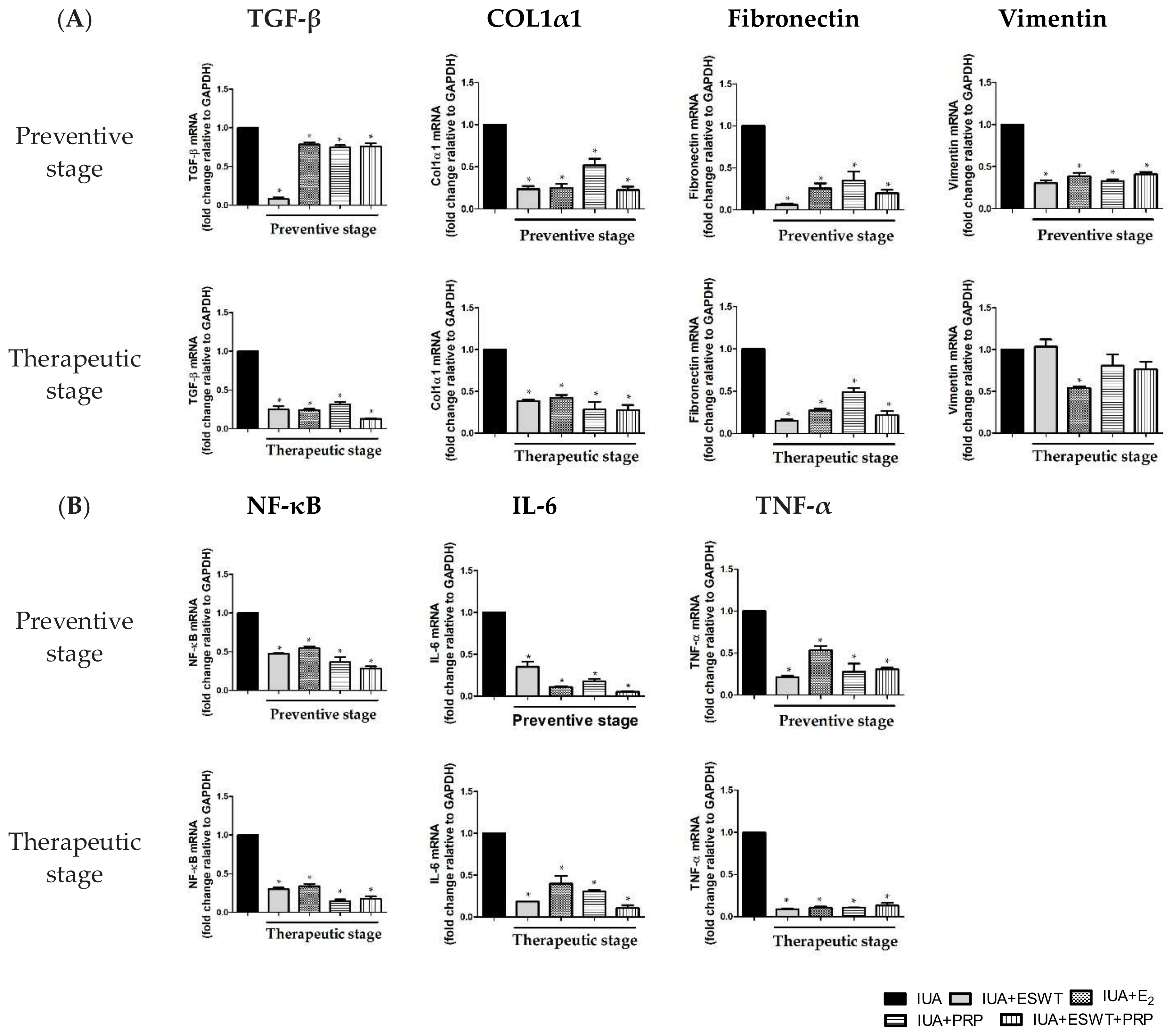
3.3. ESWT, E2, PRP, and ESWT Combined with PRP Transplantation Improved Fibrosis in an IUA Rat Model

Fibrosis is a well-established feature of endometrial adhesions. We performed Masson’s trichrome staining to assess the extent of fibrosis in the IUA-damaged endometrium. In the control group, collagen fibers were stained blue, while cytoplasm, muscle fibers, and red blood cells were stained red, while the nucleus was blue and brown (Figure 3A,C). On examining the morphology of the IUA group, we observed significantly increased fibrosis in the uteri when compared with other groups, and collagen fibers were disordered and unevenly distributed. The ratio of the fibrotic area in the IUA group was also significantly higher (area 61.3% to 64.5%) than that in the normal control group (area 10.6% to 12.22%) during preventive and therapeutic stages (Figure 3B,D). ESWT, PRP, and ESWT combined with PRP significantly decreased the formation of fibrotic scarring, as well as reduced collagen fibrosis formation in the IUA-damaged uterus at both treatment stages. However, E2 transplantation in the IUA-injured uterus afforded no changes when compared with the IUA group in the preventive stage (Figure 3B). The results revealed that IUA induced endometrial damage and led to fibrotic scarring in the uterus compared with corresponding control groups on days 21 and 35.

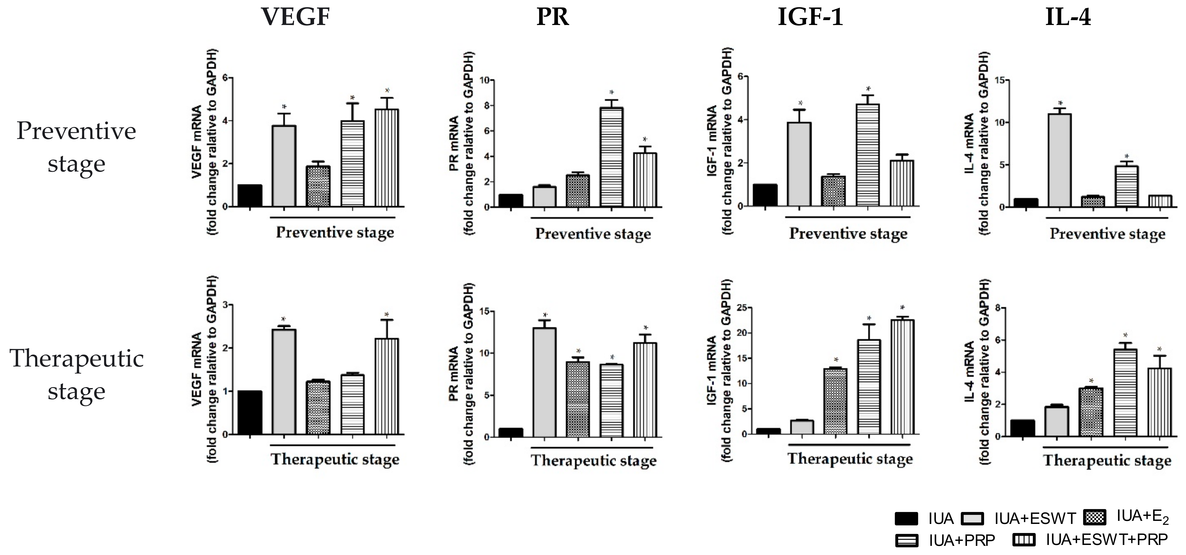
3.4. ESWT Treatment, E2 Transplantation, or ESWT Combined with PRP Regulated Vascular, Fibrosis, Inflammation, and Antiinflammation-Associated Genes in IUA Rats

We next aimed to clarify the possible mechanism underlying ESWT treatment and E2 and PRP transplantation in regulating the biological function of the damaged uterus. Accordingly, we examined the effects of ESWT treatment, E2, and PRP transplantation on vascular, fibrotic, inflammatory, and anti-inflammatory cytokines. As shown in Figure 4A, the mRNA expression levels of TGF-β, collagen-I α 1 (COL1α1), and fibronectin were significantly downregulated by approximately 1.5–2-fold following ESWT treatment, E2 alone, PRP alone, and PRP transplantation when compared with those in the IUA group during the preventive stage; during the therapeutic stage, TGF-β, COL1α1, and fibronectin were attenuated, while vimentin was not significantly altered following ESWT treatment, PRP, and ESWT combined with PRP transplantation. Consistent with the present findings, the mRNA expression levels of nuclear factor-kappa B (NF-κB), IL-6, and tumor necrosis factor (TNF)-α were significantly inhibited following ESWT treatment, E2 transplantation, PRP transplantation, and PRP transplantation combined with ESWT in the two stages (Figure 4B). These findings suggested that ESWT treatment, PRP, and ESWT combined with PRP transplantation regulate fibrosis and inflammation in endometrial stromal cells, particularly during the preventive stage. Conversely, mRNA expression levels of VEGF and progesterone receptor (PR) were dramatically upregulated following PRP alone or ESWT combined with PRP transplantation during the preventive stage when compared with the IUA-damaged group. In the therapeutic stage, the mRNA expression levels of VEGF and PR were significantly upregulated following ESWT treatment, or ESWT treatment combined with PRP transplantation (Figure 5). Moreover, the mRNA expression levels of PR, IGF-1, and IL-4 showed the similarity results, which upregulated following PRP alone or ESWT treatment combined with PRP transplantation (Figure 5). Collectively, ESWT treatment, PRP transplantation, or ESWT combined with PRP significantly suppressed inflammation in the IUA uterus, indicating that these treatment strategies potentially participate in vascularization, endometrial growth, and upregulation of anti-inflammatory-associated genes in the IUA-damaged uterine cavity.

3.5. ESWT Treatment, PRP Transplantation, or ESWT Combined with PRP Mainly Promoted VEGF and Vimentin Expression at the Therapeutic Stage in IUA Rats

Next, we evaluated VEGF, the most potent angiogenic factor, and vimentin, a stromal cell marker, using immunohistochemistry procedures during preventive and therapeutic stages. As shown in Figure 6A, the VEGF-positive area was significantly decreased by approximately 20% in the IUA group and was weakly detected in both stages. The VEGF-positive expression rate was approximately 40% in the endometrial tissue of the control group. Compared with the IUA group, ESWT treatment, PRP transplantation, or ESWT combined with PRP enhanced VEGF expression during the two stages, whereas E2 transplantation in the IUA-affected endometrium only increased VEGF expression during the preventive stage (Figure 6B). Interestingly, ESWT treatment, E2, and PRP transplantation, or ESWT combined with PRP, could facilitate vimentin expression during the therapeutic stage and not the preventive stage (Figure 6F,H).

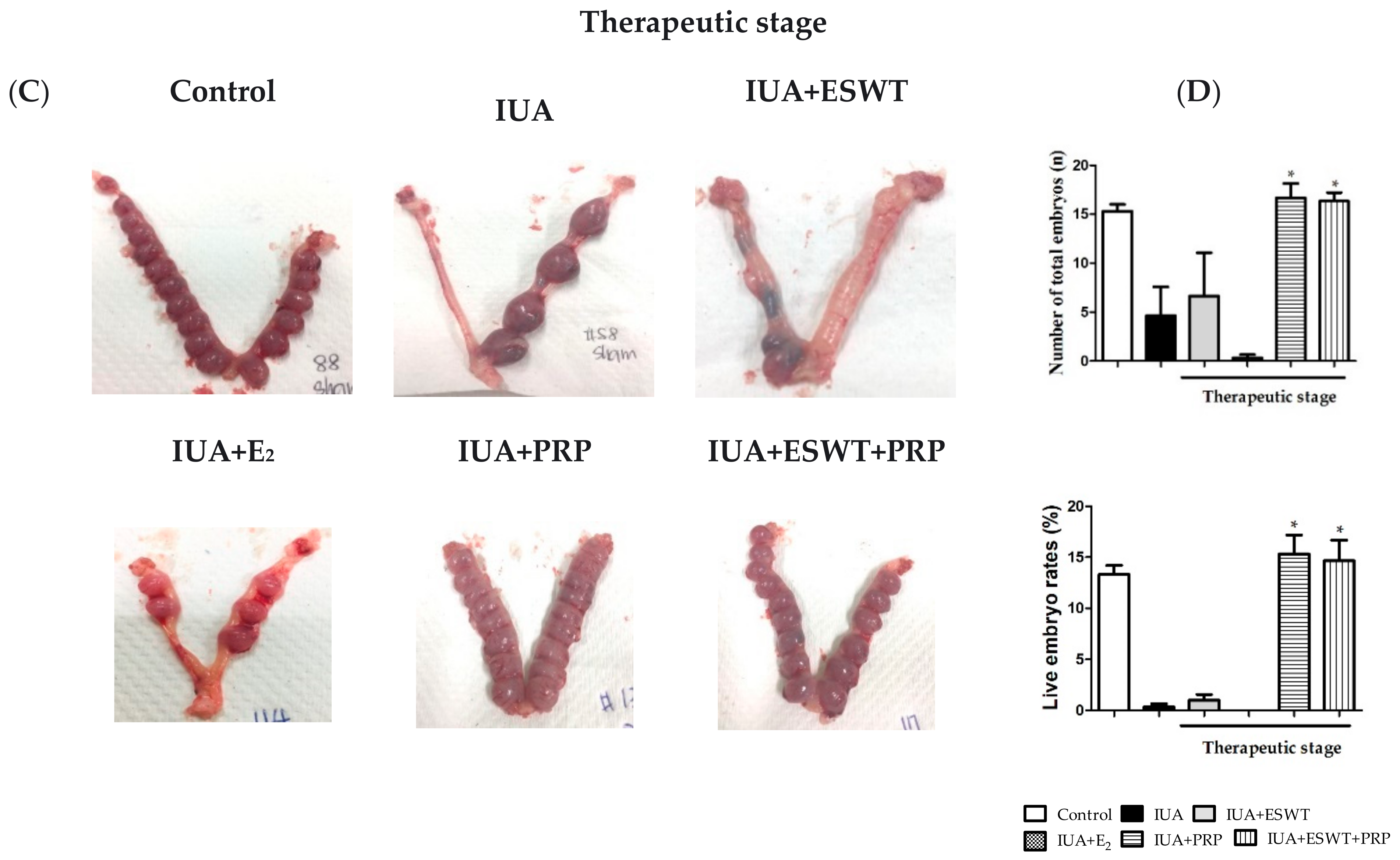
3.6. Pregnancy Outcomes in Rats with IUA-Damaged Endometrium after ESWT Treatment, E2 and PRP Transplantation, or ESWT Combined with PRP Transplantation

We examined functional improvements in the damaged uteri after ESWT treatment, E2 and PRP transplantation, or ESWT combined with PRP transplantation. Accordingly, female rats were mated on days 21 and 35 post-IUA damage induction. Fetuses within the whole uterus were harvested at embryonic day (E) 13.5. Representative images and statistical analyses of total and live embryos are shown in Figure 7. In the preventive stage, transplantation or EWST treatment resulted in an average of 10 to 16 gestational sacs when compared with 11 gestational sacs in the IUA-damaged group; IUA-damaged uteri showed no differences following ESWT treatment, E2 and PRP transplantation, or ESWT combined with PRP transplantation (Figure 7A,B) in terms of the total number of embryos, however, PRP alone significantly increased the live embryo rates (approximately 16 gestational sacs) when compared with the IUA-damaged group (only 2 non-gestational sacs) during the preventive stage (Figure 7B). Furthermore, PRP or ESWT combined with PRP transplantation significantly enhanced total or live embryos (approximately 14 to 16 gestational sacs) when compared with the IUA-damaged group (only 4 non-gestational sacs) during the preventive stage. Regarding improved fertility, PRP transplantation or ESWT combined with PRP transplantation also afforded a better pregnant outcome following treatment of IUA-damaged endometrium (Figure 7C,D).

4. Discussion

In the present study, we evaluated the effects of E2, PRP transplantation, and ESWT combined with PRP using an IUA rat model. The novel approach of ESWT as an ancillary treatment for gynecological diseases has been proposed in the past decade [59,60,61,62]. We selected the medium energy intensity, suitable frequency, and the number of shock waves per treatment session [59]. Our results showed that PRP alone, as well as ESWT combined with PRP in vivo, did not affect body weight but ameliorated endometrial thickness during the preventive stage, but this phenomenon was not observed in the therapeutic stage. In addition, PRP alone, as well as ESWT combined with PRP, increased the number of glands and significantly decreased collagen deposition and fibrosis formation during both stages. In addition, all four treatment strategies regulated fibrosis and inflammation-related cytokines by downregulating mRNA expression levels of TGF-β, Colα1, fibronectin NF-κB, IL-6, and TNF-α; on the other hand, PRP alone, or ESWT combined with PRP transplantation mediated endometrial vascular and the activity of reproductive cytokines by upregulating mRNA expression levels VEGF, and PR during the preventive stage, while these treatments also regulated the endometrial growth and antiinflammation-related cytokines by upregulating mRNA expression levels IGF, and IL-4 during the therapeutic stage. Compared with the IUA group, ESWT, PRP transplantation, or ESWT combined with PRP primarily promoted VEGF and vimentin to regenerate endometrial vascular and stromal cells, as determined by immunochemistry. Interestingly, treatment of damaged uteri with PRP transplantation or ESWT combined with PRP improved embryo implantation and subsequent development. Our findings suggest that PRP administration alone or combined with ESWT could exert proliferative, anti-inflammatory, and anti-fibrotic effects on the damaged endometrium, which may be a potential target for the prevention and early treatment of intrauterine mechanical injury in Sprague-Dawley rats.
IUA is a complex gynecological abnormality that leads to scar tissue formation in the uterus, resulting in infertility and recurrent pregnancy loss. In-depth investigations have been conducted to address this issue. Hysteroscopic adhesiolysis is commonly used to manually remove intrauterine adhesions and reconstruct the uterine cavity [63,64,65], while hormone-associated therapies can help eliminate adhesions and regain the morphology and function of uteri [12,66,67,68]. Hysteroscopy, intrauterine devices, and hydrogels are clinically available for traditional transcervical resection of adhesion and endometrium regeneration [69,70,71]. However, even if adhesiolysis is performed, re-adhesion may occur due to damage to the endometrial epithelial cells and impaired endometrial metabolism and angiogenesis postoperatively [72,73]. Following the classification of IUA types by the American Fertility Society (AFS 1988 version), 209 patients had reduced AFS scores and ameliorated fertility outcomes using hysteroscopic adhesiolysis combined with prolonged oral estrogen after 2–3 months [63]. These strategies afforded limited effects in alleviating endometrial fibrosis and inhibiting adhesion reformation, especially in severe IUA. Therefore, intrauterine injection of hyaluronic acid colloids, amniotic membrane transplantation, and stem cell therapy have been employed [71,74,75,76,77,78,79,80]. Treatment with human umbilical cord mesenchymal stem cell-extracellular vesicles (UCMSC-EVs) alone or combined with estrogen significantly decreased not only inflammatory cytokines, such as TNF-α and IL-6, but also fibrosis markers, such as TGF-β, COL1α1, and VEGF. Xavier et al. have reported a pilot cohort study in which autologous CD133+ bone marrow-derived stem cells offered a safe and efficient therapeutic approach for patients with refractory Asherman’s syndrome, improving endometrial cavity and endometrial thickness [81]. However, the possibility of adhesions recurrence persists, even after adhesiolysis, depending on the disease severity [82]. In addition, certain limitations and poor pregnancy rates need to be addressed [83,84]. Potential complications despite successful pregnancy include abnormal placentation, preterm delivery, intrauterine growth restrictions, and higher rates of cesarean sections [85]. Stem cell therapy has not been widely adopted as it involves invasive and expensive procedures, such as cell storage [86]. In addition, the propensity for tumorigenicity and low retention of stem cell therapy are persistent obstacles [87]. It is crucial to develop a safe and effective therapy to prevent the recurrence of adhesions.
Herein, histological analyses demonstrated the establishment of the IUA model, which was associated with the loss of endometrial epithelial cells and endometrial glands, resulting in the disappearance of the uterine cavity. PRP transplantation and ESWT combined with PRP did not significantly alter the endometrial and epithelial thickness but ameliorated the number of endometrial glands in the preventive and early treatment stages. As endometrial glands transport or secrete bioactive substances that regulate uterine receptivity and impact embryo survival for blastocyst implantation, which are needed in animals for fertility and pregnancy success, the pregnancy rate, and number of live embryos were considerably enhanced following the application of ESWT combined with PRP. At days 21 and 35 post-IUA induction, Masson staining revealed that PRP transplantation and ESWT combined with PRP improved endometrial stromal fibrosis progression when compared with uterine wall adhesion in the IUA group. These results indicate that PRP transplantation or ESWT combined with PRP transplantation could effectively treat refractory endometrial damage.
According to a report by Niu et al., the Tiaoshen Tongluo recipe, a traditional Chinese medicine herbal formula, can upregulate Smad7 and downregulate TGF-β1 to alleviate fibrotic tissue formation in an IUA rat model [88]. Transplantation of decellularized and lyophilized amniotic membranes ameliorated the degree of fibrosis by suppressing TGF-β1 expression [89]. Overexpression of miR-326 potentially inhibited fibrosis formation by downregulating pro-fibrotic genes, such as TGF-β1, COL1α1, and fibronectin in endometrial tissues and endometrial stromal cells derived from patients with IUA [90]. Moreover, high expression of miR-543 downregulated mRNA expression levels of fibronectin and vimentin, which may affect the degree of fibrosis and collagen content in IUA cell lines [91]. Similar results were observed in the present study; expression levels of TGF-β, COL1A1, fibronectin, and vimentin were reduced during the preventive and therapeutic stages following treatment with PRP alone or ESWT combined with PRP. Moreover, the opposite effects of vimentin mRNA levels and protein levels during two stages have been shown, which imply the posttranslational of stromal cell function may involve other pathways, not for the mRNA expression of vimentin.
The transcription factor NF-κB is a pivotal mediator of inflammatory responses triggering the upregulation of various pro-inflammatory gene-related chemokines, cytokines, and adhesion molecules such as monocyte chemoattractant protein-1 (MCP-1), macrophage inflammatory protein 1, IL-6, TNF-α, intercellular adhesion molecule-1 (ICAM-1), and matrix metalloproteinases (MMPs) [92]. Bone marrow mesenchymal stem cells ameliorated IUA in rats by increasing VEGF and decreasing NF-κB expression, as determined by immunohistochemical detection [93]. Our data indicated that the mRNA expression of NF-κB was significantly increased in the IUA-damaged uterus, whereas treatment with PRP alone or ESWT combined with PRP downregulated IL-6 and TNF-α expression when compared with that in the IUA endometrium. Ebrahim et al. revealed that human UCMSCs treated with estrogen exhibited a significant decrease in inflammation and fibrosis, associated with a reduction in TNF-α, TGF-β, IL-6, and collagen-I, in a trichloroacetic acid-induced IUA-damaged uterus when compared with the untreated IUA rats, suggesting that NF-κB and its downstream genes may play crucial roles in promoting the expression of IUA inflammatory factors [78,94].
In contrast, previous in vitro experiments showed that shock wave treatment enhanced endothelial proliferation by upregulating VEGF expression in endothelial cells in a hypoxia-inducible factor 1-independent manner [95]. Li et al. reported that human amniotic epithelial cells induced endometrial thickening, and the number of endometrial glands was increased by upregulation of VEGF expression, indicating that VEGF improves angiogenesis in the IUA rat endometrium [96]. Notably, successful endometrial growth and pregnancy outcomes have been documented following intrauterine PRP infusion in five female patients with infertility presenting a thin endometrium. Reportedly, PRP secretes and recruits VEGF, regulating cell migration, attachment, invasion, proliferation, and differentiation [97]. Ito et al. have reported that a low-energy shock wave could effectively increase VEGF expression in cultured endothelial cells, induce neovascularization, and improve myocardial ischemia in a porcine model of chronic myocardial ischemia in vivo [98]. ESWT also promotes tissue regeneration and improves ischemia-related organ dysfunction, mainly by enhancing angiogenesis, upregulating the expression of stromal cell-derived factor (SDF)-1α, recruiting endothelial progenitor cells, suppressing inflammation, and generating oxidative stress [99,100,101]. In the present study, treatment with PRP alone or combined with ESWT could increase VEGF mRNA expression, as well as upregulate VEGF protein levels, in IUA-injured endometrium during the two treatment stages, suggesting that VEGF improves the endometrial vasculature. Zhang et al. have shown that menstrual blood-derived stromal cells combined with PRP transplantation induced the expression of human-derived secretory proteins, IGF-1, and anti-inflammatory IL-4, as determined by quantitative-PCR and LUMINEX assay. The authors suggested that PRP could promote fertility restoration and more effectively improve uterine proliferation, markedly accelerating endometrial damage repair in IUA rats [30]. Accordingly, PRP provides a highly effective ancillary therapy for IUA treatment [78]. Similar results were observed in the present study.
It is well-known that PR mediates reproductive activity and is expressed in all layers of the uterus. Following the binding of estrogen or progesterone to PR, downstream estrogen receptor and PR signal transductions are activated to initiate gene expression and cell differentiation to maintain uterine function [102,103]. Wang et al. have shown that PR expression was increased after bone marrow-derived mesenchymal stem cell transplantation, which significantly elevated the number of endometrial glands, reduced the fibrotic area, and effectively repaired the damaged endometrium [104].
Herein, we reported the application of PRP and ESWT combined with PRP to treat IUA in an animal model. However, the underlying mechanisms of fertility restoration, endometrial vascularization, and anti-fibrotic and anti-inflammatory effects warrant further investigation. Whether the PRP alone or ESWT combined with PRP transplantation increased the production of extracellular matrix components, and fibrosis are worth further exploration in the endometriosis or adenomyosis due to the anti-inflammatory and anti-oxidant effects in the previous study [59]. In addition, given the differences between humans and rats, the clinical applicability of these findings needs to be confirmed. Furthermore, additional mechanisms would ensure the future application of PRP or ESWT combined with PRP for IUA therapy.

5. Conclusions

In conclusion, the present study’s findings demonstrated the effectiveness of PRP treatment or ESWT combined with PRP using an IUA rat model. Although underlying mechanisms of endometrial restoration are complex, our treatment strategies afforded endometrial vascularization and anti-fibrotic, anti-inflammatory, and fertility restoration during preventive and therapeutic stages. PRP is an innovative therapeutic modality for developing alternative treatment strategies that are safe, affordable, simple, easily performed, and cost-effective. Overall, PRP transplantation or PRP combined with ESWT treatment is a valuable strategy to treat IUA.

Supplementary Materials

The following supporting information can be downloaded at: https://www.mdpi.com/article/10.3390/biomedicines10020476/s1, Supplementary Data: H&E-stained sections of SD rat uterine tissue after curettage.

Author Contributions

Y.-H.C. performed the experiments, analyzed the data, contributed reagents/materials/analysis tools, wrote the paper, prepared figures and/or tables, and reviewed drafts of the paper. N.-C.T., J.-H.C., J.-Y.K. and H.-Y.K. conceptualized and proposed the theory. Y.-J.C. conceived and designed the experiments. P.-L.W. and Y.-C.C. performed the experiments, analyzed the data, and contributed reagents, materials, and analytical tools. K.-C.L. conceived and designed the experiments, performed the experiments, wrote the paper, and reviewed the drafts of the paper. All authors have read and agreed to the published version of the manuscript.

Funding

This work was supported by grant MOST109-2314-B-182A-092-MY3 from the Ministry of Science and Technology of Taiwan (Taipei, Taiwan); Chang Gung Memorial Hospital [Grant number: CMRPG8J1451, CRRPG8K0021-3, and CMRPG8J1131-2] (Taiwan). The funders had no role in the study design, data collection and analysis, decision to publish, nor the manuscript preparation.

Institutional Review Board Statement

All experimental animal procedures were approved by the Institutional Animal Care and Use Committee at Kaohsiung Chang Gung Memorial Hospital (Affidavit of Approval of Animal Use Protocol No. 2019122008) and were performed in accordance with the Guide for the Care and Use of Laboratory Animals, 8th edition (NIH publication No. 85-23, National Academy Press, Washington, DC, USA, revised 2011).

Informed Consent Statement

Not applicable.

Data Availability Statement

The datasets of the present study can be made available from the corresponding author upon request.

Acknowledgments

This work was supported by Grant MOST109-2314-B-182A-092-MY3 from the Ministry of Science and Technology of Taiwan. The authors thank the Department of Medical Research and Development of Kaohsiung Chang Gung Memorial Hospital for providing the service of the ABI 7500 Fast Real-Time PCR System (Applied Biosystems), the Center for Shockwave Medicine and Tissue Engineering, Kaohsiung Chang Gung Memorial Hospital, and Chang Gung University College of Medicine for allowing the use of the SD-1 shock wave applicator (Storz, Tägerwilen, Germany), the Core Lab for Phenomics and Diagnostics of Kaohsiung Chang Gung Memorial Hospital for the use of Pannoramic SCAN (Sysmex Europe GmbH), and Tissue Bank Core Lab (CLRPG8F1702) of Kaohsiung Chang Gung Memorial Hospital for providing technical support.

Conflicts of Interest

The authors declare no conflict of interest. The authors alone are responsible for the content of the manuscript.

References

  1. Warembourg, S.; Huberlant, S.; Garric, X.; Leprince, S.; de Tayrac, R.; Letouzey, V. Prevention and treatment of intra-uterine synechiae: Review of the literature. J. Gynecol. Obstet. Biol. Reprod. 2015, 44, 366–379. [Google Scholar] [CrossRef] [PubMed]
  2. Hooker, A.B.; de Leeuw, R.; van de Ven, P.M.; Bakkum, E.A.; Thurkow, A.L.; Vogel, N.E.A.; van Vliet, H.; Bongers, M.Y.; Emanuel, M.H.; Verdonkschot, A.E.M.; et al. Prevalence of intrauterine adhesions after the application of hyaluronic acid gel after dilatation and curettage in women with at least one previous curettage: Short-term outcomes of a multicenter, prospective randomized controlled trial. Fertil. Steril. 2017, 107, 1223–1231. [Google Scholar] [CrossRef] [Green Version]
  3. Hooker, A.B.; Lemmers, M.; Thurkow, A.L.; Heymans, M.W.; Opmeer, B.C.; Brolmann, H.A.; Mol, B.W.; Huirne, J.A. Systematic review and meta-analysis of intrauterine adhesions after miscarriage: Prevalence, risk factors and long-term reproductive outcome. Hum. Reprod. Update 2014, 20, 262–278. [Google Scholar] [CrossRef] [PubMed] [Green Version]
  4. Dmowski, W.P.; Greenblatt, R.B. Asherman’s syndrome and risk of placenta accreta. Obstet. Gynecol. 1969, 34, 288–299. [Google Scholar] [PubMed]
  5. March, C.M. Management of Asherman’s syndrome. Reprod. Biomed. Online 2011, 23, 63–76. [Google Scholar] [CrossRef] [Green Version]
  6. Capmas, P.; Mihalache, A.; Duminil, L.; Hor, L.S.; Pourcelot, A.G.; Fernandez, H. Intrauterine adhesions: What is the pregnancy rate after hysteroscopic management? J. Gynecol. Obstet. Hum. Reprod. 2020, 49, 101797. [Google Scholar] [CrossRef]
  7. Turocy, J.M.; Rackow, B.W. Uterine factor in recurrent pregnancy loss. Semin. Perinatol. 2019, 43, 74–79. [Google Scholar] [CrossRef]
  8. Gan, L.; Duan, H.; Xu, Q.; Tang, Y.Q.; Li, J.J.; Sun, F.Q.; Wang, S. Human amniotic mesenchymal stromal cell transplantation improves endometrial regeneration in rodent models of intrauterine adhesions. Cytotherapy 2017, 19, 603–616. [Google Scholar] [CrossRef]
  9. Zupi, E.; Centini, G.; Lazzeri, L. Asherman syndrome: An unsolved clinical definition and management. Fertil. Steril. 2015, 104, 1380–1381. [Google Scholar] [CrossRef]
  10. Seyedoshohadaei, F.; Tangestani, L.; Zandvakili, F.; Rashadmanesh, N. Comparison of the Effect of Clomiphene—Estradiol Valerate vs. Letrozole on Endometrial Thickness, Abortion and Pregnancy Rate in Infertile Women with Polycystic Ovarian Syndrome. J. Clin. Diagn. Res. 2016, 10, QC10–QC13. [Google Scholar] [CrossRef]
  11. Etulain, J.; Mena, H.A.; Meiss, R.P.; Frechtel, G.; Gutt, S.; Negrotto, S.; Schattner, M. An optimised protocol for platelet-rich plasma preparation to improve its angiogenic and regenerative properties. Sci. Rep. 2018, 8, 1513. [Google Scholar] [CrossRef] [PubMed]
  12. Zhang, S.S.; Xia, W.T.; Xu, J.; Xu, H.L.; Lu, C.T.; Zhao, Y.Z.; Wu, X.Q. Three-dimensional structure micelles of heparin-poloxamer improve the therapeutic effect of 17beta-estradiol on endometrial regeneration for intrauterine adhesions in a rat model. Int. J. Nanomed. 2017, 12, 5643–5657. [Google Scholar] [CrossRef] [PubMed] [Green Version]
  13. Zhang, S.S.; Xu, X.X.; Xiang, W.W.; Zhang, H.H.; Lin, H.L.; Shen, L.E.; Lin, Q.; Lin, F.; Zhou, Z.Y. Using 17beta-estradiol heparin-poloxamer thermosensitive hydrogel to enhance the endometrial regeneration and functional recovery of intrauterine adhesions in a rat model. FASEB J. 2020, 34, 446–457. [Google Scholar] [CrossRef] [PubMed] [Green Version]
  14. Dhillon, R.S.; Schwarz, E.M.; Maloney, M.D. Platelet-rich plasma therapy—Future or trend? Arthritis Res. Ther. 2012, 14, 219. [Google Scholar] [CrossRef] [Green Version]
  15. Barile, A.; La Marra, A.; Arrigoni, F.; Mariani, S.; Zugaro, L.; Splendiani, A.; Di Cesare, E.; Reginelli, A.; Zappia, M.; Brunese, L.; et al. Anaesthetics, steroids and platelet-rich plasma (PRP) in ultrasound-guided musculoskeletal procedures. Br. J. Radiol. 2016, 89, 20150355. [Google Scholar] [CrossRef]
  16. Bashir, J.; Panero, A.J.; Sherman, A.L. The emerging use of platelet-rich plasma in musculoskeletal medicine. J. Osteopath. Med. 2015, 115, 24–31. [Google Scholar] [CrossRef] [Green Version]
  17. Rinaldi, F.; Marzani, B.; Pinto, D.; Sorbellini, E. Randomized controlled trial on a PRP-like cosmetic, biomimetic peptides based, for the treatment of alopecia areata. J. Dermatol. Treat. 2019, 30, 588–593. [Google Scholar] [CrossRef] [Green Version]
  18. Lin, M.-Y.; Lin, C.-S.; Hu, S.; Chung, W.-H. Progress in the use of platelet-rich plasma in aesthetic and medical dermatology. J. Clin. Aesthetic Dermatol. 2020, 13, 28. [Google Scholar]
  19. Puri, N. Platelet rich plasma in dermatology and aesthetic medicine. Our Dermatol. Online 2015, 6, 207. [Google Scholar] [CrossRef]
  20. Carroll, R.C.; Worthington, R.E.; Craft, R.M.; Snider, C.C.; Dakin, P.A.; Wortham, D.C.; Scott, J.; Jarrett, A. Post interventional cardiology urinary thromboxane correlates with PlateletMapping detected aspirin resistance. Thromb. Res. 2010, 125, e118–e122. [Google Scholar] [CrossRef]
  21. Spartalis, E.; Tomos, P.; Moris, D.; Athanasiou, A.; Markakis, C.; Spartalis, M.D.; Troupis, T.; Dimitroulis, D.; Perrea, D. Role of platelet-rich plasma in ischemic heart disease: An update on the latest evidence. World J. Cardiol. 2015, 7, 665–670. [Google Scholar] [CrossRef] [PubMed]
  22. Biswas, D.; So, K.H.; Hwang, S.U.; Yoon, J.D.; Kim, M.; Kim, D.Y.; Hyun, S.H. Embryotropic effects of vascular endothelial growth factor on porcine embryos produced by in vitro fertilization. Theriogenology 2018, 120, 147–156. [Google Scholar] [CrossRef] [PubMed]
  23. Lee, J.W.; Kwon, O.H.; Kim, T.K.; Cho, Y.K.; Choi, K.Y.; Chung, H.Y.; Cho, B.C.; Yang, J.D.; Shin, J.H. Platelet-rich plasma: Quantitative assessment of growth factor levels and comparative analysis of activated and inactivated groups. Arch. Plast. Surg. 2013, 40, 530–535. [Google Scholar] [CrossRef] [PubMed] [Green Version]
  24. Zadehmodarres, S.; Salehpour, S.; Saharkhiz, N.; Nazari, L. Treatment of thin endometrium with autologous platelet-rich plasma: A pilot study. JBRA Assist. Reprod. 2017, 21, 54–56. [Google Scholar] [CrossRef] [PubMed]
  25. De Pascale, M.R.; Sommese, L.; Casamassimi, A.; Napoli, C. Platelet derivatives in regenerative medicine: An update. Transfus. Med. Rev. 2015, 29, 52–61. [Google Scholar] [CrossRef]
  26. Qian, Y.; Han, Q.; Chen, W.; Song, J.; Zhao, X.; Ouyang, Y.; Yuan, W.; Fan, C. Platelet-Rich Plasma Derived Growth Factors Contribute to Stem Cell Differentiation in Musculoskeletal Regeneration. Front. Chem. 2017, 5, 89. [Google Scholar] [CrossRef] [Green Version]
  27. Jo, C.H.; Kim, J.E.; Yoon, K.S.; Shin, S. Platelet-rich plasma stimulates cell proliferation and enhances matrix gene expression and synthesis in tenocytes from human rotator cuff tendons with degenerative tears. Am. J. Sports Med. 2012, 40, 1035–1045. [Google Scholar] [CrossRef]
  28. Xu, Q.; Li, B.; Yuan, L.; Dong, Z.; Zhang, H.; Wang, H.; Sun, J.; Ge, S.; Jin, Y. Combination of platelet-rich plasma within periodontal ligament stem cell sheets enhances cell differentiation and matrix production. J. Tissue Eng. Regen. Med. 2017, 11, 627–636. [Google Scholar] [CrossRef]
  29. Goncalves, N.J.N.; Frantz, N.; de Oliveira, R.M. Platelet-rich plasma (PRP) therapy: An approach in reproductive medicine based on successful animal models. Anim. Reprod. 2020, 16, 93–98. [Google Scholar] [CrossRef] [Green Version]
  30. Zhang, S.; Li, P.; Yuan, Z.; Tan, J. Platelet-rich plasma improves therapeutic effects of menstrual blood-derived stromal cells in rat model of intrauterine adhesion. Stem Cell Res. Ther. 2019, 10, 61. [Google Scholar] [CrossRef]
  31. Chaussy, C.; Schmiedt, E.; Jocham, D.; Brendel, W.; Forssmann, B.; Walther, V. First clinical experience with extracorporeally induced destruction of kidney stones by shock waves. J. Urol. 1982, 127, 417–420. [Google Scholar] [CrossRef]
  32. Buch, M.; Knorr, U.; Fleming, L.; Theodore, G.; Amendola, A.; Bachmann, C.; Zingas, C.; Siebert, W.E. Extracorporeal shockwave therapy in symptomatic heel spurs. An overview. Orthopade 2002, 31, 637–644. [Google Scholar] [CrossRef] [PubMed]
  33. Wilner, J.M.; Strash, W.W. Extracorporeal shockwave therapy for plantar fasciitis and other musculoskeletal conditions utilizing the Ossatron—An update. Clin. Podiatr. Med. Surg. 2004, 21, 441–447. [Google Scholar] [CrossRef] [PubMed]
  34. Thomson, C.E.; Crawford, F.; Murray, G.D. The effectiveness of extra corporeal shock wave therapy for plantar heel pain: A systematic review and meta-analysis. BMC Musculoskelet. Disord. 2005, 6, 19. [Google Scholar] [CrossRef] [PubMed] [Green Version]
  35. Roehrig, G.J.; Baumhauer, J.; DiGiovanni, B.F.; Flemister, A.S. The role of extracorporeal shock wave on plantar fasciitis. Foot Ankle Clin. 2005, 10, 699–712. [Google Scholar] [CrossRef] [PubMed]
  36. Buchbinder, R.; Green, S.E.; Youd, J.M.; Assendelft, W.J.; Barnsley, L.; Smidt, N. Systematic review of the efficacy and safety of shock wave therapy for lateral elbow pain. J. Rheumatol. 2006, 33, 1351–1363. [Google Scholar]
  37. Stasinopoulos, D.; Johnson, M.I. Effectiveness of extracorporeal shock wave therapy for tennis elbow (lateral epicondylitis). Br. J. Sports Med. 2005, 39, 132–136. [Google Scholar] [CrossRef] [Green Version]
  38. Rompe, J.D.; Theis, C.; Maffulli, N. Shock wave treatment for tennis elbow. Orthopade 2005, 34, 567–570. [Google Scholar] [CrossRef]
  39. Erturk, E.; Ptak, A.M.; Monaghan, J. Fertility measures in women after extracorporeal shockwave lithotripsy of distal ureteral stones. J. Endourol. 1997, 11, 315–317. [Google Scholar] [CrossRef]
  40. Vieweg, J.; Weber, H.M.; Miller, K.; Hautmann, R. Female fertility following extracorporeal shock wave lithotripsy of distal ureteral calculi. J. Urol. 1992, 148, 1007–1010. [Google Scholar] [CrossRef]
  41. Wang, Y.; Guo, T.; Cai, H.Y.; Ma, T.K.; Tao, S.M.; Sun, S.; Chen, M.Q.; Gu, Y.; Pang, J.H.; Xiao, J.M.; et al. Cardiac shock wave therapy reduces angina and improves myocardial function in patients with refractory coronary artery disease. Clin. Cardiol. 2010, 33, 693–699. [Google Scholar] [CrossRef]
  42. Ito, Y.; Ito, K.; Shiroto, T.; Tsuburaya, R.; Yi, G.J.; Takeda, M.; Fukumoto, Y.; Yasuda, S.; Shimokawa, H. Cardiac shock wave therapy ameliorates left ventricular remodeling after myocardial ischemia-reperfusion injury in pigs in vivo. Coron. Artery Dis. 2010, 21, 304–311. [Google Scholar] [CrossRef] [PubMed]
  43. Sung, P.-H.; Lee, M.S.; Chai, H.-T.; Chiang, J.Y.; Li, Y.-C.; Chu, Y.-C.; Huang, C.-R.; Yip, H.-K. Extracorporeal Shock Wave Enhanced Exogenous Mitochondria into Adipose-Derived Mesenchymal Stem Cells and Further Preserved Heart Function in Rat Dilated Cardiomyopathy. Biomedicines 2021, 9, 1362. [Google Scholar] [CrossRef] [PubMed]
  44. Rassweiler, J.J.; Knoll, T.; Kohrmann, K.U.; McAteer, J.A.; Lingeman, J.E.; Cleveland, R.O.; Bailey, M.R.; Chaussy, C. Shock wave technology and application: An update. Eur. Urol. 2011, 59, 784–796. [Google Scholar] [CrossRef] [PubMed] [Green Version]
  45. Khrouf, M.; Morel, O.; Hafiz, A.; Chavatte-Palmer, P. Evaluation of the rabbit as an experimental model for human uterine synechia. J. Hum. Reprod. Sci. 2012, 5, 175. [Google Scholar]
  46. Zhang, X.; Yan, X.; Wang, C.; Tang, T.; Chai, Y. The dose-effect relationship in extracorporeal shock wave therapy: The optimal parameter for extracorporeal shock wave therapy. J. Surg. Res. 2014, 186, 484–492. [Google Scholar] [CrossRef]
  47. Reinehr, S.; Reinhard, J.; Gandej, M.; Gottschalk, I.; Stute, G.; Faissner, A.; Dick, H.B.; Joachim, S.C. S100B immunization triggers NFkappaB and complement activation in an autoimmune glaucoma model. Sci. Rep. 2018, 8, 9821. [Google Scholar] [CrossRef]
  48. Sanchez, R.N.; Chan, C.K.; Garg, S.; Kwong, J.M.; Wong, M.J.; Sadun, A.A.; Lam, T.T. Interleukin-6 in retinal ischemia reperfusion injury in rats. Investig. Ophthalmol. Vis. Sci. 2003, 44, 4006–4011. [Google Scholar] [CrossRef] [Green Version]
  49. Li, M.; O’Sullivan, K.M.; Jones, L.K.; Lo, C.; Semple, T.; Kumanogoh, A.; Kikutani, H.; Holdsworth, S.R.; Kitching, R. Endogenous CD100 promotes glomerular injury and macrophage recruitment in experimental crescentic glomerulonephritis. Immunology 2009, 128, 114–122. [Google Scholar] [CrossRef]
  50. Spittau, B.; Rilka, J.; Steinfath, E.; Zoller, T.; Krieglstein, K. TGFbeta1 increases microglia-mediated engulfment of apoptotic cells via upregulation of the milk fat globule-EGF factor 8. Glia 2014, 63, 142–153. [Google Scholar] [CrossRef]
  51. Du, M.; Chen, B.; Meng, Q.; Liu, S.; Zheng, X.; Zhang, C.; Wang, H.; Li, H.; Wang, N.; Dai, J. 3D bioprinting of BMSC-laden methacrylamide gelatin scaffolds with CBD-BMP2-collagen microfibers. Biofabrication 2015, 7, 044104. [Google Scholar] [CrossRef] [PubMed]
  52. Wu, T.; Chen, J.M.; Xiao, T.G.; Shu, X.B.; Xu, H.C.; Yang, L.L.; Xing, L.J.; Zheng, P.Y.; Ji, G. Qinggan Huoxue Recipe suppresses epithelial-to-mesenchymal transition in alcoholic liver fibrosis through TGF-beta1/Smad signaling pathway. World J. Gastroenterol. 2016, 22, 4695–4706. [Google Scholar] [CrossRef] [PubMed]
  53. Sun, H.; He, X.; Tao, X.; Hou, T.; Chen, M.; He, M.; Liao, H. The CD200/CD200R signaling pathway contributes to spontaneous functional recovery by enhancing synaptic plasticity after stroke. J. Neuroinflamm. 2020, 17, 171. [Google Scholar] [CrossRef] [PubMed]
  54. Imberti, B.; Corna, D.; Rizzo, P.; Xinaris, C.; Abbate, M.; Longaretti, L.; Cassis, P.; Benedetti, V.; Benigni, A.; Zoja, C.; et al. Renal primordia activate kidney regenerative events in a rat model of progressive renal disease. PLoS ONE 2015, 10, e0120235. [Google Scholar] [CrossRef] [Green Version]
  55. Lee, S.W.; Lee, J.E.; Yoo, C.Y.; Ko, M.S.; Park, C.S.; Yang, S.-H. NRP-1 expression is strongly associated with the progression of pituitary adenomas. Oncol. Rep. 2014, 32, 1537–1542. [Google Scholar] [CrossRef] [PubMed]
  56. Yoshino, M.; Mizutani, T.; Yamada, K.; Yazawa, T.; Ogata-Kawata, H.; Sekiguchi, T.; Kajitani, T.; Miyamoto, K. Co-activator p120 is increased by gonadotropins in the rat ovary and enhances progesterone receptor activity. Reprod. Biol. Endocrinol. 2006, 4, 50. [Google Scholar] [CrossRef] [PubMed] [Green Version]
  57. Zhang, X.; Wang, X.; Liu, T.; Mo, M.; Ao, L.; Liu, J.; Cao, J.; Cui, Z. Smad2/3 Upregulates the Expression of Vimentin and Affects Its Distribution in DBP-Exposed Sertoli Cells. PPAR Res. 2015, 2015, 489314. [Google Scholar] [CrossRef]
  58. Xu, L.; Tong, R.; Li, X.; Liang, L.; Huang, H. Effects of CASZI on bronchopulmonary development of neonatal rats. Exp. Ther. Med. 2017, 14, 6243–6246. [Google Scholar] [CrossRef]
  59. Melki, E. Endometriosis treatment with shock waves: A novel approach. Med. Hypotheses 2019, 124, 114–117. [Google Scholar] [CrossRef]
  60. Martinez Portillo, F.J.; Heidenreich, A.; Schwarzer, U.; Michel, M.S.; Alken, P.; Engelmann, U. Microscopic and biochemical fertility characteristics of semen after shockwave lithotripsy of distal ureteral calculi. J. Endourol. 2001, 15, 781–784, discussion 784–785. [Google Scholar] [CrossRef]
  61. Zang, Z.J.; Liu, Q.; Hu, J.; Feng, J.; Zhu, Y.Q.; Ma, G.; Jiang, M.H. The impact of low-intensity extracorporeal shock wave therapy on testicular function in adult rats. Andrology 2018, 6, 936–942. [Google Scholar] [CrossRef] [Green Version]
  62. Wang, H.J.; Tyagi, P.; Lin, T.K.; Huang, C.C.; Lee, W.C.; Chancellor, M.B.; Chuang, Y.C. Low Energy Shock Wave Therapy Attenuates Mitochondrial Dysfunction and Improves Bladder Function in HCl induced Cystitis in Rats. Biomed. J. 2021. [Google Scholar] [CrossRef] [PubMed]
  63. Zhang, L.; Wang, M.; Zhang, Q.; Zhao, W.; Yang, B.; Shang, H.; Shang, X.; Ma, Y.; Wang, B.; Feng, L. Estrogen therapy before hysteroscopic adhesiolysis improves the fertility outcome in patients with intrauterine adhesions. Arch. Gynecol. Obstet. 2019, 300, 933–939. [Google Scholar] [CrossRef] [PubMed]
  64. Zhang, Z.; Li, S.; Deng, J.; Yang, S.; Xiang, Z.; Guo, H.; Xi, H.; Sang, M.; Zhang, W. Aspirin inhibits endometrial fibrosis by suppressing the TGFbeta1Smad2/Smad3 pathway in intrauterine adhesions. Int. J. Mol. Med. 2020, 45, 1351–1360. [Google Scholar] [CrossRef] [PubMed]
  65. Bai, X.; Liu, J.; Yuan, W.; Liu, Y.; Li, W.; Cao, S.; Yu, L.; Wang, L. Therapeutic Effect of Human Amniotic Epithelial Cells in Rat Models of Intrauterine Adhesions. Cell Transplant. 2020, 29, 963689720908495. [Google Scholar] [CrossRef] [Green Version]
  66. Rochwerger, L.; Dho, S.; Parker, L.; Foskett, J.K.; Buchwald, M. Estrogen-dependent expression of the cystic fibrosis transmembrane regulator gene in a novel uterine epithelial cell line. J. Cell Sci. 1994, 107, 2439–2448. [Google Scholar] [CrossRef]
  67. Tonguc, E.A.; Var, T.; Yilmaz, N.; Batioglu, S. Intrauterine device or estrogen treatment after hysteroscopic uterine septum resection. Int. J. Gynaecol. Obstet. 2010, 109, 226–229. [Google Scholar] [CrossRef]
  68. Cai, H.; Li, H.; He, Y. Interceed and Estrogen Reduce Uterine Adhesions and Fibrosis and Improve Endometrial Receptivity in a Rabbit Model of Intrauterine Adhesions. Reprod. Sci. 2016, 23, 1208–1216. [Google Scholar] [CrossRef]
  69. Chen, Y.; Chang, Y.; Yao, S. Role of angiogenesis in endometrial repair of patients with severe intrauterine adhesion. Int. J. Clin. Exp. Pathol. 2013, 6, 1343–1350. [Google Scholar]
  70. Magos, A. Hysteroscopic treatment of Asherman’s syndrome. Reprod. Biomed. Online 2002, 4 (Suppl. 3), 46–51. [Google Scholar] [CrossRef]
  71. Johary, J.; Xue, M.; Zhu, X.; Xu, D.; Velu, P.P. Efficacy of estrogen therapy in patients with intrauterine adhesions: Systematic review. J. Minim. Invasive Gynecol. 2014, 21, 44–54. [Google Scholar] [CrossRef] [PubMed]
  72. Di Spiezio Sardo, A.; Calagna, G.; Scognamiglio, M.; O’Donovan, P.; Campo, R.; De Wilde, R.L. Prevention of intrauterine post-surgical adhesions in hysteroscopy. A systematic review. Eur. J. Obstet. Gynecol. Reprod. Biol. 2016, 203, 182–192. [Google Scholar] [CrossRef] [PubMed]
  73. Deans, R.; Vancaillie, T.; Ledger, W.; Liu, J.; Abbott, J.A. Live birth rate and obstetric complications following the hysteroscopic management of intrauterine adhesions including Asherman syndrome. Hum. Reprod. 2018, 33, 1847–1853. [Google Scholar] [CrossRef] [PubMed]
  74. Salma, U.; Xue, M.; Sayed, A.S.M.; Xu, D. Efficacy of intrauterine device in the treatment of intrauterine adhesions. Biomed. Res. Int. 2014, 2014, 589296. [Google Scholar] [CrossRef] [Green Version]
  75. Tafti, S.Z.G.; Javaheri, A.; Firoozabadi, R.D.; Ashkezar, S.K.; Abarghouei, H.F. Role of hyaluronic acid intrauterine injection in the prevention of Asherman’s syndrome in women undergoing uterine septum resection: An RCT. Int. J. Reprod. Biomed. 2021, 19, 339–346. [Google Scholar] [CrossRef]
  76. Azizi, R.; Aghebati-Maleki, L.; Nouri, M.; Marofi, F.; Negargar, S.; Yousefi, M. Stem cell therapy in Asherman syndrome and thin endometrium: Stem cell-based therapy. Biomed. Pharmacother. 2018, 102, 333–343. [Google Scholar] [CrossRef]
  77. Bai, X.; Liu, J.; Cao, S.; Wang, L. Mechanisms of endometrial fibrosis and the potential application of stem cell therapy. Discov. Med. 2019, 27, 267–279. [Google Scholar]
  78. Ebrahim, N.; Mostafa, O.; El Dosoky, R.E.; Ahmed, I.A.; Saad, A.S.; Mostafa, A.; Sabry, D.; Ibrahim, K.A.; Farid, A.S. Human mesenchymal stem cell-derived extracellular vesicles/estrogen combined therapy safely ameliorates experimentally induced intrauterine adhesions in a female rat model. Stem Cell Res. Ther. 2018, 9, 175. [Google Scholar] [CrossRef]
  79. Kilic, S.; Yuksel, B.; Pinarli, F.; Albayrak, A.; Boztok, B.; Delibasi, T. Effect of stem cell application on Asherman syndrome, an experimental rat model. J. Assist. Reprod. Genet. 2014, 31, 975–982. [Google Scholar] [CrossRef] [Green Version]
  80. Liu, Y.; Cai, J.; Luo, X.; Wen, H.; Luo, Y. Collagen Scaffold with Human Umbilical Cord Mesenchymal Stem Cells Remarkably Improves Intrauterine Adhesions in a Rat Model. Gynecol. Obstet. Investig. 2020, 85, 267–276. [Google Scholar] [CrossRef]
  81. Santamaria, X.; Cabanillas, S.; Cervello, I.; Arbona, C.; Raga, F.; Ferro, J.; Palmero, J.; Remohi, J.; Pellicer, A.; Simon, C. Autologous cell therapy with CD133+ bone marrow-derived stem cells for refractory Asherman’s syndrome and endometrial atrophy: A pilot cohort study. Hum. Reprod. 2016, 31, 1087–1096. [Google Scholar] [CrossRef] [PubMed] [Green Version]
  82. Chen, L.; Zhang, H.; Wang, Q.; Xie, F.; Gao, S.; Song, Y.; Dong, J.; Feng, H.; Xie, K.; Sui, L. Reproductive Outcomes in Patients with Intrauterine Adhesions Following Hysteroscopic Adhesiolysis: Experience from the Largest Women’s Hospital in China. J. Minim. Invasive Gynecol. 2017, 24, 299–304. [Google Scholar] [CrossRef] [PubMed]
  83. Hamerlynck, T.W.; Meyers, D.; Van der Veken, H.; Bosteels, J.; Weyers, S. Fertility outcome after treatment of retained products of conception: A systematic review. Gynecol. Surg. 2018, 15, 12. [Google Scholar] [CrossRef]
  84. Li, H.-J.; He, Y.-L.; Hu, H.-Y.; Li, J.-X.; Xian, W.; Chen, S.-P.; Zhang, D.-M. Effects of Interceed on endometrial receptivity in rabbits with intrauterine adhesion. Med. J. Chin. People. Lib. Army 2015, 40, 344–348. [Google Scholar]
  85. Xue, X.; Chen, Q.; Zhao, G.; Zhao, J.Y.; Duan, Z.; Zheng, P.S. The Overexpression of TGF-beta and CCN2 in Intrauterine Adhesions Involves the NF-kappaB Signaling Pathway. PLoS ONE 2015, 10, e0146159. [Google Scholar] [CrossRef]
  86. Mouhayar, Y.; Sharara, F.I. G-CSF and stem cell therapy for the treatment of refractory thin lining in assisted reproductive technology. J. Assist. Reprod. Genet. 2017, 34, 831–837. [Google Scholar] [CrossRef]
  87. Hu, S.; Ogle, B.M.; Cheng, K. Body builder: From synthetic cells to engineered tissues. Curr. Opin. Cell Biol. 2018, 54, 37–42. [Google Scholar] [CrossRef]
  88. Niu, H.; Miao, X.; Zhan, X.; Zhou, X.; Li, X.; Jiang, L. Tiaoshen Tongluo Attenuates Fibrosis by Modulating the TGF-beta1/Smad Pathway in Endometrial Stromal Cells and a Rat Model of Intrauterine Adhesion. Evid. Based Complement. Altern. Med. 2021, 2021, 6675329. [Google Scholar] [CrossRef]
  89. Chen, X.; Sun, J.; Li, X.; Mao, L.; Zhou, Y.; Cui, L.; Bai, W. Antifibrotic Effects of Decellularized and Lyophilized Human Amniotic Membrane Transplant on the Formation of Intrauterine Adhesion. Exp. Clin. Transplant. 2019, 17, 236–242. [Google Scholar] [CrossRef]
  90. Ning, J.; Zhang, H.; Yang, H. MicroRNA326 inhibits endometrial fibrosis by regulating TGFbeta1/Smad3 pathway in intrauterine adhesions. Mol. Med. Rep. 2018, 18, 2286–2292. [Google Scholar] [CrossRef] [Green Version]
  91. Liu, X.; Xu, Q.; Chen, C.; Duan, H. miR-543 Inhibits the Occurrence and Development of Intrauterine Adhesion by Inhibiting the Proliferation, Migration, and Invasion of Endometrial Cells. BioMed Res. Int. 2021, 2021, 5559102. [Google Scholar] [CrossRef]
  92. Liu, T.; Zhang, L.; Joo, D.; Sun, S.C. NF-kappaB signaling in inflammation. Signal Transduct. Target. Ther. 2017, 2, 17023. [Google Scholar] [CrossRef] [Green Version]
  93. Salama, N.M.; Zaghlol, S.S.; Mohamed, H.H.; Kamar, S.S. Suppression of the inflammation and fibrosis in Asherman syndrome rat model by mesenchymal stem cells: Histological and immunohistochemical studies. Folia Histochem. Cytobiol. 2020, 58, 208–218. [Google Scholar] [CrossRef] [PubMed]
  94. Wang, X.; Ma, N.; Sun, Q.; Huang, C.; Liu, Y.; Luo, X. Elevated NF-kappaB signaling in Asherman syndrome patients and animal models. Oncotarget 2017, 8, 15399–15406. [Google Scholar] [CrossRef] [Green Version]
  95. Peng, Y.Z.; Zheng, K.; Yang, P.; Wang, Y.; Li, R.J.; Li, L.; Pan, J.H.; Guo, T. Shock wave treatment enhances endothelial proliferation via autocrine vascular endothelial growth factor. Genet. Mol. Res. 2015, 14, 19203–19210. [Google Scholar] [CrossRef] [PubMed]
  96. Li, B.; Zhang, Q.; Sun, J.; Lai, D. Human amniotic epithelial cells improve fertility in an intrauterine adhesion mouse model. Stem Cell Res. Ther. 2019, 10, 257. [Google Scholar] [CrossRef] [PubMed] [Green Version]
  97. Chang, Y.; Li, J.; Chen, Y.; Wei, L.; Yang, X.; Shi, Y.; Liang, X. Autologous platelet-rich plasma promotes endometrial growth and improves pregnancy outcome during in vitro fertilization. Int. J. Clin. Exp. Med. 2015, 8, 1286–1290. [Google Scholar] [PubMed]
  98. Ito, K.; Fukumoto, Y.; Shimokawa, H. Extracorporeal shock wave therapy for ischemic cardiovascular disorders. Am. J. Cardiovasc. Drugs 2011, 11, 295–302. [Google Scholar] [CrossRef] [PubMed]
  99. Yeh, K.H.; Sheu, J.J.; Lin, Y.C.; Sun, C.K.; Chang, L.T.; Kao, Y.H.; Yen, C.H.; Shao, P.L.; Tsai, T.H.; Chen, Y.L.; et al. Benefit of combined extracorporeal shock wave and bone marrow-derived endothelial progenitor cells in protection against critical limb ischemia in rats. Crit. Care Med. 2012, 40, 169–177. [Google Scholar] [CrossRef] [Green Version]
  100. Sheu, J.J.; Lee, F.Y.; Yuen, C.M.; Chen, Y.L.; Huang, T.H.; Chua, S.; Chen, Y.L.; Chen, C.H.; Chai, H.T.; Sung, P.H.; et al. Combined therapy with shock wave and autologous bone marrow-derived mesenchymal stem cells alleviates left ventricular dysfunction and remodeling through inhibiting inflammatory stimuli, oxidative stress & enhancing angiogenesis in a swine myocardial infarction model. Int. J. Cardiol. 2015, 193, 69–83. [Google Scholar] [CrossRef]
  101. Huang, T.-H.; Sun, C.-K.; Chen, Y.-L.; Wang, C.-J.; Yin, T.-C.; Lee, M.S.; Yip, H.-K. Shock wave therapy enhances angiogenesis through VEGFR2 activation and recycling. Mol. Med. 2016, 22, 850–862. [Google Scholar] [CrossRef] [Green Version]
  102. Andersson, A.; Bernardi, A.I.; Nurkkala-Karlsson, M.; Stubelius, A.; Grahnemo, L.; Ohlsson, C.; Carlsten, H.; Islander, U. Suppression of Experimental Arthritis and Associated Bone Loss by a Tissue-Selective Estrogen Complex. Endocrinology 2016, 157, 1013–1020. [Google Scholar] [CrossRef] [PubMed] [Green Version]
  103. Su, R.W.; Strug, M.R.; Jeong, J.W.; Miele, L.; Fazleabas, A.T. Aberrant activation of canonical Notch1 signaling in the mouse uterus decreases progesterone receptor by hypermethylation and leads to infertility. Proc. Natl. Acad. Sci. USA 2016, 113, 2300–2305. [Google Scholar] [CrossRef] [PubMed] [Green Version]
  104. Wang, J.; Ju, B.; Pan, C.; Gu, Y.; Zhang, Y.; Sun, L.; Zhang, B.; Zhang, Y. Application of Bone Marrow-Derived Mesenchymal Stem Cells in the Treatment of Intrauterine Adhesions in Rats. Cell. Physiol. Biochem. 2016, 39, 1553–1560. [Google Scholar] [CrossRef] [PubMed]
Figure 1. Protocol for preventive and therapeutic stages of E2, PRP transplantation, and/or ESWT therapy in the IUA model. Female Sprague-Dawley rats were randomly divided into the following groups: control (sham), IUA induction, IUA combined with ESWT therapy, IUA combined with E2 transplantation, IUA combined with PRP transplantation, and IUA combined with ESWT and PRP therapy. The protocol for E2 and PRP transplantation and ESWT are described in the materials and methods. ESWT, Extracorporeal shockwave therapy; IUA, intrauterine adhesion; PRP, platelet-rich plasma.
Biomedicines 10 00476 g001
Figure 2. Establishment of an IUA rat model and the endometrial morphology by H&E staining. (A,C) The uterus from control, IUA, IUA+ESWT, IUA+E2, IUA+PRP, and IUA+ESWT+PRP rats (left to right) at days 21 and 35 post-IUA damage induction during preventive and therapeutic stages. Scale bar = 20 μm (magnification, 50×). (B,D) Analysis of uterine histological data of endometrium and epithelial thickness during preventive and therapeutic stages. (E,G) Effects of E2 and PRP transplantation combined with ESWT treatment on endometrial glands during preventive and therapeutic stages (magnification, 4×). (F,H) The number of glands was calculated in each group during the preventive and therapeutic stages. Data values are expressed as the mean ± SE of triplicate experiments. * p < 0.05 for one-way ANOVA. (n ≥ 7) was significant compared with the IUA group. ESWT, Extracorporeal shockwave therapy; H&E, hematoxylin and eosin; IUA, intrauterine adhesion; PRP, platelet-rich plasma.
Biomedicines 10 00476 g002aBiomedicines 10 00476 g002b
Figure 3. Representative Masson’s trichrome-stained images indicate the degree of endometrial fibrosis. (A,C) The uterus from control, IUA, IUA+ESWT, IUA+E2, IUA+PRP, and IUA+ESWT+PRP rats (left to right) at days 21 and 35 post-IUA damage induction during preventive and therapeutic stages (magnification, 4×). (B,D) The fibrotic area was measured in the preventive and therapeutic stages. Data are expressed as the mean ± SE of triplicate experiments. * p < 0.05 for one-way ANOVA. (n ≥ 7) was significant compared with the IUA group. ESWT, Extracorporeal shockwave therapy; IUA, intrauterine adhesion; PRP, platelet-rich plasma.
Biomedicines 10 00476 g003
Figure 4. Analysis of endometrial inflammatory markers in the uterus of IUA+ESWT, IUA+E2, IUA+PRP, and IUA+ESWT+PRP groups. (A) Real-time PCR analysis of TGF-β, COL1α1, fibronectin, and vimentin mRNA levels in the uterus of all groups at days 21 and 35. (B) Real-time PCR analysis of NF-κB, IL-6, and TNF-α mRNA levels in the uterus in all groups at days 21 and 35. * p < 0.05 for one-way ANOVA. (n ≥ 3) was significant compared with the IUA group. COL1α1, collagen-I α 1; ESWT, Extracorporeal shockwave therapy; IL-6, interleukin-6; IUA, intrauterine adhesion; NF-κB, nuclear factor-kappa B; TGF-β, transforming growth factor-β; TNF-α, tumor necrosis factor-α; PRP, platelet-rich plasma.
Biomedicines 10 00476 g004
Figure 5. Analysis of endometrial inflammatory markers in the uterus of IUA+ESWT, IUA+E2, IUA+PRP, and IUA+ESWT+PRP groups. Real-time PCR analysis of VEGF, PR, IGF-1, and IL-4 mRNA levels in the uterus of all groups at days 21 and 35. * p < 0.05 for one-way ANOVA. (n ≥ 3) was significant compared with the IUA group. ESWT, Extracorporeal shockwave therapy; IGF-1, insulin-like growth factor 1; IL-4, interleukin-4; IUA, intrauterine adhesion; PR, progesterone receptor; PRP, platelet-rich plasma; VEGF, vascular endothelial growth factor.
Biomedicines 10 00476 g005
Figure 6. Immunohistochemical staining of VEGF and vimentin. (A,C) VEGF expression in the immunohistochemically stained endometrium at days 21 and 35 post-IUA damage induction during preventive and therapeutic stages (magnification, 4×). (B,D) VEGF expression was semi-quantified, and the number of positive cells per field was calculated during preventive and therapeutic stages. (E,G) Vimentin expression in the immunohistochemically stained endometrium at days 21 and 35 post-IUA damage induction during preventive and therapeutic stages (magnification, 4×). (F,H) Analysis of vimentin-positive area based on immunohistochemistry results. Data are expressed as the mean ± SE of triplicate experiments. * p < 0.05 for one-way ANOVA. (n ≥ 6) was significant compared with the IUA group. IUA, intrauterine adhesion; VEGF, vascular endothelial growth factor.
Biomedicines 10 00476 g006aBiomedicines 10 00476 g006b
Figure 7. PRP transplantation or PRP combined with ESWT treatment ameliorates pregnancy outcomes in the IUA rat model. (A,C) Pregnancy outcomes in control, IUA, IUA+ESWT, IUA+E2, IUA+PRP, and IUA+ESWT+PRP rats (left to right) at days 21 and 35 post-IUA damage induction in the preventive and therapeutic stages. (B,D) Statistical analysis of total and live embryos in the preventive and therapeutic stages (n ≥ 3). ESWT, Extracorporeal shockwave therapy; IUA, intrauterine adhesion; PRP, platelet-rich plasma, * p < 0.05 for one-way ANOVA.
Biomedicines 10 00476 g007aBiomedicines 10 00476 g007b
Table 1. Primers of specific genes used in quantitative real-time PCR in rat specimens.
GeneForward Primer (5′–3′)Reverse Primer (5′–3′)
NF-κB [47]CTGGCAGCTCTTCTCAAAGCCCAGGTCATAGAGAGGCTCAA
IL-6 [48]TCAACTCCATCTGCCCTTCAGAAGGCAACTGGCTGGAAGTCT
TNF-α [49]GCCTCTTCTCATTCCTGCTTCACTTGGTGGTTTGCTACGA
TGF-β [50]TAATGGTGGACCGCAACAACGGGCACTGCTTCCCGAATGTCT
COL1α1 [51]CATGTTCAGCTTTGTGGACCTGCAGCTGACTTCAGGGATGT
Fibronectin [52]GACTCGCTTTGACTTCACCACGCTGAGACCCAGGAGACCAC
IL-4 [53]CAGGGTGCTTCGCAAATTTTACACCGAGAACCCCAGACTTGTT
IGF-1 [54]GCTTTTACTTCAACAAGCCCACATCAGCGGAGCACAGTACATC
VEGF [55]TATCTTCAAGCCGTCCTGTGGATCCGCATGATCTGCATAG
PR [56]TCAAGGCAATTGGCTTAAGACAGAGCTGTTTCACAAGATCATGCA
Vimentin [57]GCACCCTGCAGTCATTCAGAGCAAGGATTCCACTTTACGTTCA
GAPDH [58]TGGTGAAGGTCGGTGTGAACGACTGTGCCGTTGAACTTGC
Table 2. No effect of IUA-induced combines estrogen and/or ESWT treatment on the body weight in SD rats.
ControlIUAIUA+ESWTIUA+E2IUA+PRPIUA+ESWT+PRP
Bodyweight (g) of preventive stage
Day 0212.1 ± 7.8210.3 ± 14.1205.4 ± 10.3215.8 ± 8.9201.8 ± 7.0201.5 ± 6.0
Day 21253.5 ± 15.7244.2 ± 19.0243.9 ± 23.0247.8 ± 12.9243.0 ± 14.9235.4 ± 13.9
Bodyweight (g) of therapeutic stage
Day 0206.1 ± 8.3212.3 ± 12.2209.3 ± 6.9211.6 ± 10.6221.8 ± 15.1224.9 ± 13.9
Day 35262.7 ± 28.3266.3 ± 20.4261.6 ± 10.9265.5 ± 22.6279.4 ± 16.3274.9 ± 20.0
Publisher’s Note: MDPI stays neutral with regard to jurisdictional claims in published maps and institutional affiliations.

