Review

Targeting Systemic Sclerosis from Pathogenic Mechanisms to Clinical Manifestations: Why IL-6?

by
Anca Cardoneanu
1,2,*,
Alexandra Maria Burlui
1,2,
Luana Andreea Macovei
1,2,
Ioana Bratoiu
1,2,
Patricia Richter
1,2 and
Elena Rezus
1,2
1
Department of Rheumatology, University of Medicine and Pharmacy “Grigore T Popa”, 700115 Iasi, Romania
2
Rehabilitation Hospital, 700661 Iasi, Romania
*
Author to whom correspondence should be addressed.
Biomedicines 2022, 10(2), 318; https://doi.org/10.3390/biomedicines10020318
Submission received: 29 December 2021 / Revised: 25 January 2022 / Accepted: 26 January 2022 / Published: 29 January 2022

Abstract

:
Systemic sclerosis (SS) is a chronic autoimmune disorder, which has both cutaneous and systemic clinical manifestations. The disease pathogenesis includes a triad of manifestations, such as vasculopathy, autoimmunity, and fibrosis. Interleukin-6 (IL-6) has a special role in SS development, both in vascular damage and in the development of fibrosis. In the early stages, IL-6 participates in vascular endothelial activation and apoptosis, leading to the release of damage-associated molecular patterns (DAMPs), which maintain inflammation and autoimmunity. Moreover, IL-6 plays an important role in the development of fibrotic changes by mediating the transformation of fibroblasts into myofibroblasts. All of these are associated with disabling clinical manifestations, such as skin thickening, pulmonary fibrosis, pulmonary arterial hypertension (PAH), heart failure, and dysphagia. Tocilizumab is a humanized monoclonal antibody that inhibits IL-6 by binding to the specific receptor, thus preventing its proinflammatory and fibrotic actions. Anti-IL-6 therapy with Tocilizumab is a new hope for SS patients, with data from clinical trials supporting the favorable effect, especially on skin and lung damage.

1. Introduction

Systemic sclerosis (SS) is a chronic autoimmune disease that has both cutaneous and systemic clinical manifestations, with the latter being associated with high disease morbidity [1]. The pathogenic mechanisms of this condition are not yet fully understood. However, it is known that the pathogenesis of the disease is composed of a triad: vasculopathy, autoimmunity, and fibrosis [2]. All these mechanisms are interconnected, with each having a particular role in the occurrence of clinical manifestations and disease progression. Immunological mechanisms include both innate and adaptative immunity and involve many cells, such as: B and T lymphocytes, mast cells, macrophages, and dendritic cells [3]. Vasculopathy is characterized by damage and endothelial activation and participates in the initiation and perpetuation of autoimmunity [4]. Activation of fibroblasts and excess deposition of extracellular matrix causes the appearance of fibrosis, which is also related to autoimmunity and vasculopathy [3].
In the early stage of the disease, an inflammatory profile was highlighted, characterized by an increased secretion of proinflammatory cytokines, with the predominant immune response being Th1 and Th17 cells [5]. Moreover, innate immunity, by activating NK/NKT-like cells, plays a decisive role in this initial stage of the disease [6]. The late stage of SS has a predominant Th2-type immune response and is defined by the presence of fibrotic changes [5]. Knowing these important data involved in the pathogenesis of the disease, numerous studies have correlated the clinical manifestations with the excessive secretion of some molecules, with the latter being considered potential biomarkers of the disease [7] (Figure 1).
Given the complex pathogenic mechanisms and the multitude of cells and cytokines, we will continue to focus on interleukin 6 (IL-6), which is considered to be one of the major fibrogenic cytokines along with interleukin 4 (IL-4) and transforming growth factor (TGF)-β [3].

2. IL-6: General Data

IL-6 is part of a large family of cytokines, having important roles in regulating immunity, hematopoiesis, inflammation, and oncogenesis. In the past, depending on its biological effect, IL-6 had different names: B lymphocyte-stimulating factor 2 (BSF-2), hepatocyte-stimulating factor (HSF), or interferon-β2 [8]. In 1989, it was found that all these molecules are identical, so it was renamed IL-6, a name used until today [9].
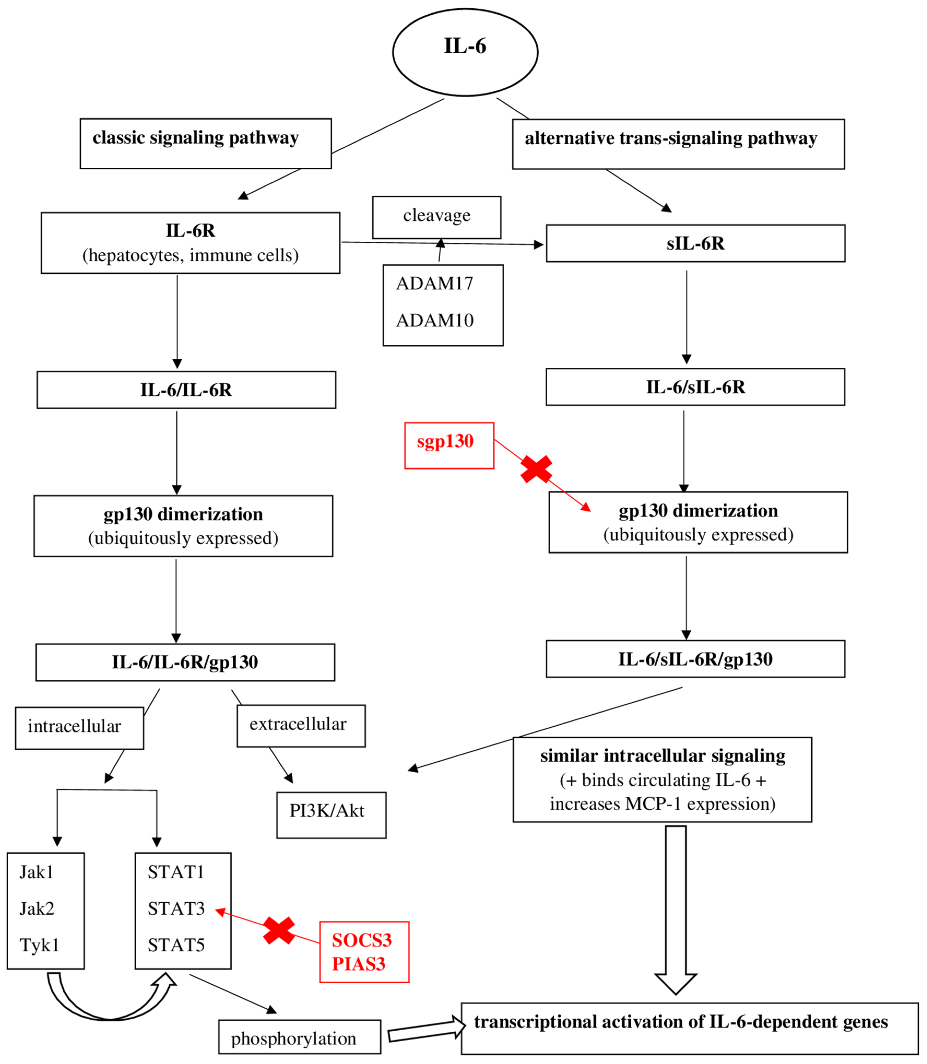
The biological effect of IL-6 can be achieved by two signaling pathways: the classical pathway, which uses a membrane receptor, and the trans-signaling pathway, which involves the presence of a soluble IL-6 receptor. The classical signaling pathway involves the binding of IL-6 to the membrane receptor-IL-6R [10]. Normally, the expression of this receptor is decreased, being found in several cells, especially hepatocytes and some immune cells [11,12]. IL-6R has no intrinsic signaling capacity. The IL-6/IL-6R interaction induces the dimerization of an IL-6R signaling-related cytokine called gp130 [13,14,15]. Unlike IL-6R, this gp130 molecule is ubiquitously expressed and has an intrinsic signaling capacity. The IL-6/IL-6R complex bound to gp130 initiates cellular signaling via several intracellular or extracellular pathways [10,12,16]. Gp130-mediated activation of Janus kinase (Jak) family members and subsequent phosphorylation of specific tyrosine residues cause phosphorylation and dimerization of STAT (signal transducer and activator of transcription proteins) [10,16]. The latter determines the transcriptional activation of IL-6-dependent genes [16].
The alternative IL-6 trans-signaling pathway involves the presence of a soluble receptor (sIL-6R) [16,17,18,19]. This receptor is formed after cleavage of IL-6R by activated ADAM17 (Disintegrin Metalloproteases 17) and ADAM10 (Disintegrin Metalloproteases 10) [20,21]. Binding of IL-6 to sIL-6R and subsequently to gp130 IL-6/sIL-6R determines intracellular signaling, which is very similar to classical IL-6R signaling. In addition, sIL-6R has the advantage of binding circulating IL-6, thereby prolonging the half-life of IL-6 [22]. Moreover, sIL-6R participates in the interaction between leukocytes and vascular endothelium, causing the endothelial production of MCP-1 (monocyte chemoattractant protein-1), a key chemokine that regulates monocyte/macrophage infiltration of the vascular wall [23].
To limit the proinflammatory effects of IL-6, IL-6 signaling is regulated by several inhibitory molecules and processes. Inhibition of IL-6 signaling is important in order to limit inflammation and its side effects, such as extensive tissue damage. Thus, STAT3-dependent transcription is rapidly inactivated by the cytokine 3 signaling suppressor (SOCS3) [24,25,26,27]. IL-6-induced SOCS3 expression acts as a major negative feedback loop for IL-6 signaling. Another inhibitory mechanism, a soluble form of gp130 (sgp130), has been identified in patients’ plasma [28,29]. sgp130 serves as a functional antagonist of IL-6 trans-signaling by binding to the IL-6/sIL-6R complex and preventing IL-6/IL-6R complex binding to the gp130 membrane molecule [30]. A third mechanism of inhibition of IL-6 signaling involves a STAT-3 inhibitor (PIAS3-Protein Inhibitor of Activated STAT 3) that blocks the interaction between phosphorylated STAT3 and cellular DNA and finally genetic transcription [31].
Figure 2 schematically shows these two intracellular signaling pathways of IL-6, and the main negative feedback loops that block the production of its biological effects (Figure 2).
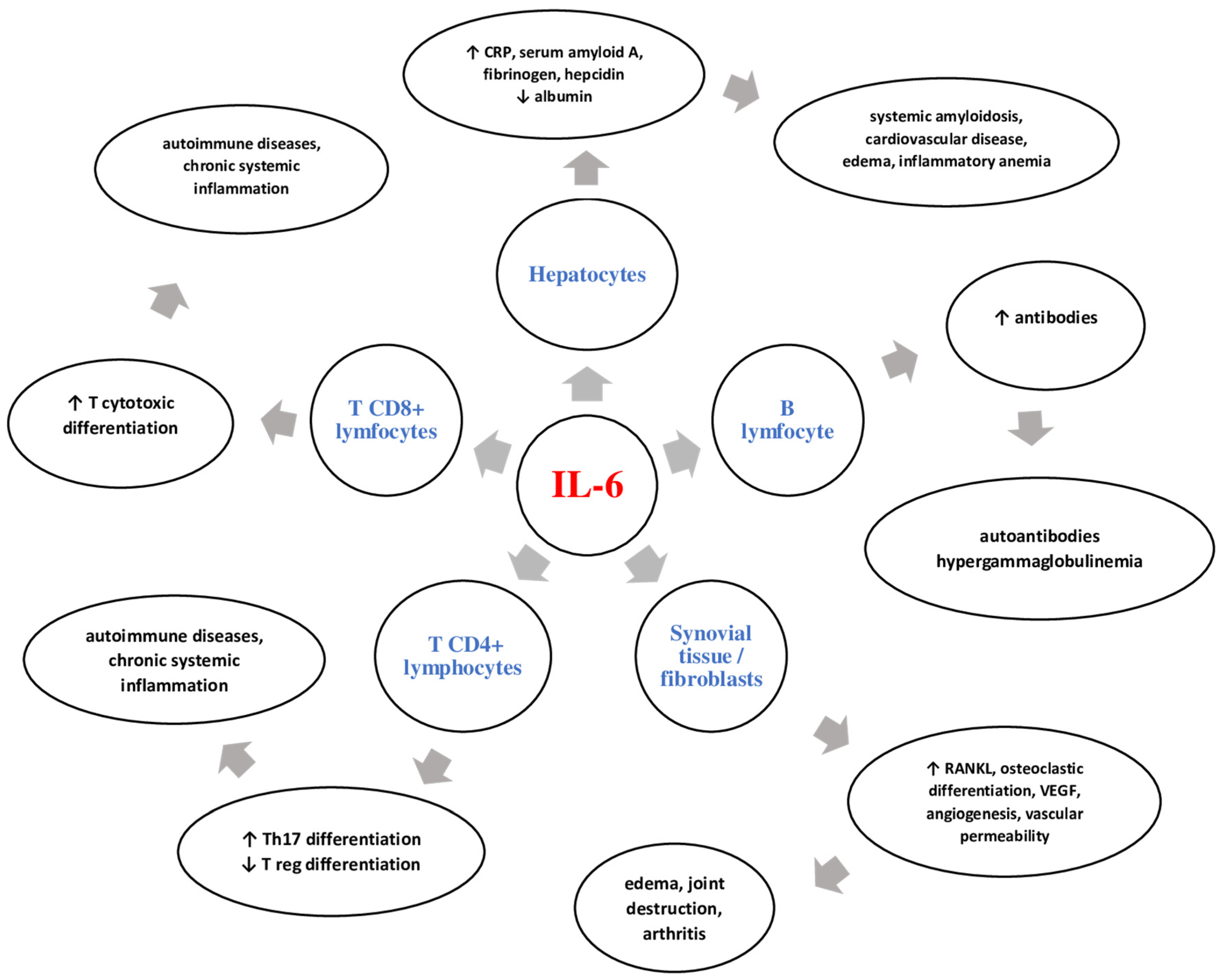
IL-6 is a key cytokine in inflammation, having a systemic polymorphic pathway. Thereby, after the initial stage of inflammation and local production, IL-6 is transported through the blood vessels in the liver. Here, it determines the formation and increased secretion of acute phase proteins, such as C-reactive protein (CRP), serum amyloid A, fibrinogen, and hepcidin. On the other hand, IL-6 has an inhibitory effect on the secretion of fibronectin, albumin, and transferrin [32,33]. Due to the stimulatory effect of hepcidin formation, the iron transporter is blocked in the gut (ferroportin 1) [34]. Thus, the IL-6-hepcidin axis is responsible for hyposideremia and anemia associated with chronic inflammation. In addition, IL-6 has the ability to promote zinc deposition in hepatocytes by increasing the expression of ZIP14-specific transporter. The regulation of ZIP14 by IL-6 contributes to hepatic accumulation of zinc and to the decrease in the serum zinc concentration highlighted in inflammation [32,35].
IL-6 plays an important role in the innate or adaptative immune response by stimulating CD4+ T cell differentiation [32]. Data from the literature support the indispensable role of the IL-6-TGF-β axis in differentiating Th17 cells from naive T CD4+ cells [36]. Moreover, inhibition of regulatory T cell differentiation (Treg) occurs [37]. This Th17/Treg imbalance is responsible for disrupting immune tolerance, leading to chronic inflammatory autoimmune disorders [38]. IL-6 has the ability to stimulate follicular T helper cell differentiation and the production of IL-21, which is responsible for the synthesis of immunoglobulins (Ig), especially IgG4 [39]. IL-6 can induce CD8+ cell differentiation into cytotoxic T cells [40]. IL-6 also acts on B lymphocytes, activating and transforming them into plasma cells, thus promoting the production of autoantibodies and hypergammaglobulinemia [32].
IL-6 can have a direct effect on osteoblast precursors, blocking their differentiation and maturation [41]. IL-6 induces the secretion of matrix metalloproteinases (MMP-1, MMP-3, MMP-13) from chondrocytes and synovial cells. MMP-1 and MMP-13 are able to break type II collagen, and MMP-3 exerts its action on the extracellular matrix, fibronectin, and laminin [42]. In addition, elevated levels of IL-6 in bone marrow stromal cells stimulate the activator receptor of nuclear factor kappa-B ligand (RANKL), leading to osteoclast differentiation and activation [43].
The action of IL-6 is systemic, being a key element in the development and perpetuation of inflammation. IL-6 participates in the formation of a cytokine storm by activating immune cells and stimulating the secretion of new inflammatory mediators. Figure 3 shows the main cells on which IL-6 acts and the systemic effects it can have.

3. IL-6 in Systemic Sclerosis

IL-6 has a particular role in the pathogenesis of SS, both in vascular damage and in the development of fibrosis. The increase in collagen production is achieved through different pathways, such as differentiation of myofibroblasts, inhibition of the secretion of matrix metalloproteinases with collagenolytic effect, and activation of fibroblasts [44]. The trans-signaling pathway is mandatory for collagen production and for the appearance of fibrotic changes due to the presence of sIL-6R [45]. This is supported by data from murine studies. The occurrence of an SS-like condition with pulmonary fibrosis and skin thickening was determined by IL-6 signaling [46]. On the other hand, a decrease in pulmonary fibrosis and inflammation was observed in IL-6-deficient mice [47].
Genetic susceptibility plays an important role in the pathogenesis of SS; therefore, the polymorphism of the IL-6 gene has been studied in various clinical trials. The most studied gene is rs1800795 (also known as −174 C/G), with G alleles being associated with increased IL-6 levels [48,49]. An analysis of −174 C/G IL-6 polymorphism in a cohort of 102 SS patients revealed increased IL-6 gene expression in G-allele carriers. This genetic polymorphism of IL-6 was correlated only with the presence of gastrointestinal manifestations, not with skin and lung damage [50]. Other data obtained after studying 20 cases with SS support the link between IL-6 polymorphism, especially the homozygous GG form, and an active and disabling disease [51].

3.1. IL-6 and SS Features

Elevated serum and skin levels of IL-6 have been highlighted in both early and late stages of SS [52,53]. In the early stages, IL-6 participates in vascular endothelial activation and apoptosis [54], leading to the release of damage-associated molecular patterns (DAMPs), which maintain inflammation and autoimmunity [55].
Numerous data support this increase in IL-6 levels, both in the tissues and in the serum of patients with SS. Feghali’s study showed elevated levels of IL-6 in the skin tissue supernatant cultures of SS patients, even 30 times higher than the control arm [56]. By culturing peripheral blood mononuclear cells isolated from cases of SS with type I collagen, Gurram highlighted an increased level of IL-6 in the supernatant, which shows that IL-6 can be highly secreted by other unaffected tissues [57]. Various methods for measuring serum IL-6 levels have been used over time, such as bioassays and ELISA (enzyme-linked immunosorbent assay). Needleman, using a bioassay method, found detectable concentrations of IL-6 in the serum of SS patients [58]. The ELISA method provided important data on elevated IL-6 levels in these patients, levels that correlated with the degree of skin damage quantified by the cutaneous score [59,60].
It seems that the concentration of IL-6 depends on the duration and form of SS. Thus, a study that included 55 SS patients analyzed the levels of IL-6, sIL-6R, and sgp130 according to the form of the disease. Patients were divided into 4 arms: 12 having an lcSS (limited cutaneous SS) and an early form of disease (less than 3 years from disease onset), 22 with lcSS and a late form (over 3 years from disease onset), 9 cases of an early dcSS form (diffuse cutaneous SS), and 12 cases of a late dcSS form [61]. Following the statistical analysis, the authors found significantly higher levels of IL-6 in the early SS form compared to the control arm, with high levels found especially in patients with pulmonary fibrosis. IL-6 was inversely correlated with vital lung capacity (VPC). IL-6 levels have also been associated with acute phase reactants and A and G immunoglobulin levels [61]. Regarding sIL-6R, elevated levels were observed in a limited SS form compared to the control. For sgp130, no statistically significant data were highlighted [61].
The fact that there is a different secretion of cytokines depending on the stage of the disease has been demonstrated by clinical trials. Thus, Matsushita showed a significant increase in IL-6 and IL-10 in the early stages of the disease, and a significant numerical decrease after 6 years of evolution. Opposite data have been described for IL-12, whose values were decreased in the early stages and more than 15 times increased in the disease with a 6-year evolution [62]. The level of IL-6 has been shown to be inversely proportional to that of IL-13, with early-stage patients having a high IL-6 and low IL-13 amount [63].

3.2. IL-6 and Skin Manifestations

IL-6 plays an important role in the development of fibrotic changes by mediating the transformation of fibroblasts into myofibroblasts, with the latter producing an excessive amount of collagen that infiltrates various organs and tissues, including the heart [64]. Myofibroblasts were found in the dermis in the early and progressive SS forms, with histological analysis indicating their disappearance in late (atrophic) forms of the disease [65]. Furthermore, a study that included a model of bleomycin SS model mice showed a numerical decrease in myofibroblasts after administration of anti-IL-6R antibodies [66]. The same data were highlighted by Kawaguchi, who performed cultures of cutaneous fibroblasts isolated from SS patients and found a high level of procollagen type I in the supernatant. After incubating these cultures with anti-IL-6R antibodies, a numerical decrease in collagen fibers was observed [67].

3.3. IL-6 and Lung Manifestations

The involvement of IL-6 in fibrotic lesions has been proven by clinical trials, especially in lung damage. Pulmonary fibrosis in SS has a multifactorial etiopathogenesis that includes both infiltration with inflammatory cells at the alveolar, peribronchiolar, and interstitium levels, and an intense and aberrant proliferation of fibroblasts that cause the deposition of fibronectin, type I and III collagen fibers, and tenascin [68,69].
Regarding IL-6, Crestani’s data included the analysis of cultures of alveolar macrophages obtained after bronchoalveolar lavage in SS patients. In the supernatant, the researchers found an increased level of IL-6 compared to the control group [70]. Other recently published data point to the same increased level of IL-6 in cases of SS with advanced pulmonary fibrosis due to the perpetuation of chronic inflammation, inhibition of the secretion of metalloproteinases, and increased collagen fiber synthesis [71]. On the other hand, another important pathogenic mechanism involved in the development of pulmonary fibrosis is the enhancement of IL-6 trans-signaling via ADAM-17 by activated macrophages, which increases the extracellular matrix deposits and the proliferation of fibroblasts [72]. In this situation, it seems that inhibition of the IL-6 trans-signaling pathway may be a useful therapeutic approach [71,72].
IL-6 levels, especially in early SS with mild forms of pulmonary fibrosis, appear to have a prognostic value for impaired lung function and increased mortality. This is supported by the analysis of 74 cases of SS associated with pulmonary fibrosis. An IL-6 value of more than 7.67 pg/mL can be considered a predictor of decreased DLCO (diffusing capacity of lung for carbon monoxide) and FVC (forced vital capacity) in the first year. It also correlates with an increase in mortality in the first 30 months of the disease [73]. Other data obtained after analyzing a cohort of 68 SS patients support the prognostic value of IL-6, with increased values being correlated with the extension of skin involvement at the 3-year follow-up, the development of pulmonary fibrosis, and worse long-term survival. Therefore, IL-6 can be considered a predictive marker for disease progression [60,74]. Furthermore, IL-6 levels may be useful in stratifying patients regarding disease activity and survival outcome [75,76,77].

3.4. IL-6 and Cardiovascular Manifestations

IL-6 plays an important role in the cardiovascular involvement of SS. In this regard, an analysis of 20 cases with SS showed positive correlations between the level of IL-6 and the disease duration; EUSTAR (European Scleroderma Study Group) activity score; musculoskeletal, vascular, and respiratory status; and pressure peak in the pulmonary artery [78]. Other positive associations were noted with the pulmonary fibrosis score quantified by HRCT (high-resolution computer tomograph). Negative correlations were scored between IL-6, DLCO, the 6-min walk distance, and right ventricle systolic function parameters [78].
Furthermore, IL-6 may be considered a marker for pulmonary arterial hypertension (PAH) [79]. PAH mainly occurs in the limited form of the disease [80] and is closely related to the proliferation of endothelial cells and the formation of extracellular matrix deposits that cause intimate thickening of the capillaries and pulmonary arterioles [81]. Systemic inflammation, represented by IL-6, plays an important role in the development of PAH due to perivascular inflammatory cell infiltrates [81]. The risk of death is higher compared to idiopathic PAH [82,83], being linked to an accentuated intimal hyperplasia, fewer plexiform lesions, and the involvement of pulmonary veins [81,84].

3.5. IL-6 and Gastrointestinal Manifestations

In addition to lung damage, 90% of SS patients also have gastrointestinal involvement (GI) [85], which is the 3rd leading cause of death [86]. GI impairment is associated with a marked disability and a decreased quality of life in SS patients, leading to reduced survival [86,87]. IL-6 gene polymorphism increases disease susceptibility, being associated with different clinical manifestations [49]. Zekovic’s study, which included 102 SS cases, analyzed the correlations between IL-6 expression and GI manifestations and the presence of a specific disease genotype /phenotype [50]. The results showed that the presence of the C-allele correlates with increased IL-6 gene expression and with GI manifestations, with the most common being abdominal distension and worsening of the GI score (UCLA GIT 2.0) [50].
The pathogenic mechanisms involved in the occurrence of GI manifestations are complex and include concentric intimate thickening and deposition of collagen and mucoid molecules [88], adventitia fibrosis [89], autonomic dysfunction affecting neuromuscular junction, smooth muscle atrophy [90,91], presence of antimyenteric neuronal antibodies, anti-U3 RNP and anti-muscarinic-3 receptor antibodies [92,93,94,95], and small intestinal bacterial overgrowth serologically expressed by an increased fecal calprotectin [96]. The clinical manifestations are polymorphic and may include the entire GI tract, with patients complaining of dysphagia, intestinal transit disorders, fecal incontinence, pseudo-obstruction, or having clinical signs of malabsorption [97].

3.6. IL-6 and Kidney Manifestations

SS kidney disease has an incidence of up to 10% [98] and is associated with increased long-term mortality [99], with scleroderma renal crisis (SRC) being a medical emergency. Genetic predisposition, autoimmune mechanisms, inflammatory cascade, and vasculopathy are the main mechanisms involved in the pathogenesis of SRC. An increased expression of two proteins: GPATCH2L and CTNND2, was highlighted in a recent study that analyzed kidney biopsy pieces in SS patients, suggesting the involvement of genetic susceptibility in the development of the disease [100]. Among autoantibodies, anti-RNA polymerase III antibodies are associated with renal impairment and have a prognostic value [101]. The main inflammatory markers involved in kidney damage are interleukins (IL-2r, IL-6, IL-10, IL-18) and chemokines, such as MCP-1 (monocyte chemoattractant protein-1) [102,103]. Vasculopathy refers to intimal proliferation and thickening of the renal arcuate and interlobular arteries, which causes activation of the renin-angiotensin system [104,105].
Clinically, patients present with rapidly progressive acute renal failure, malignant hypertension that may be complicated by hypertensive encephalopathy and retinopathy, pulmonary edema, or seizures. Microangiopathic hemolytic anemia, cardiac arrhythmias, myocarditis, and fever may also occur [106,107].
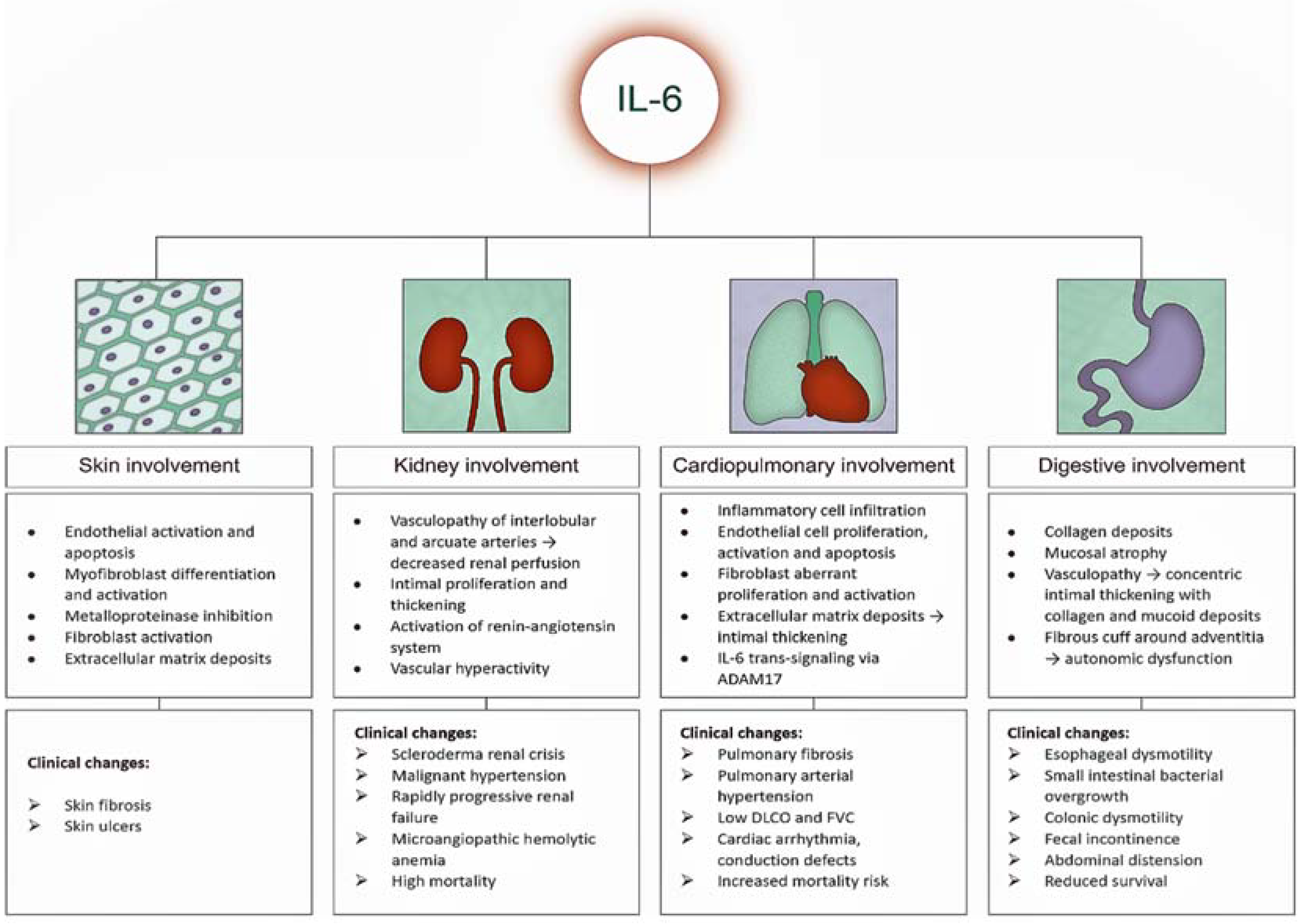
All of these systemic harmful effects of IL-6 are shown schematically in Figure 4.

4. Targeting IL-6 in Systemic Sclerosis

Given the above data, we can sustain that IL-6 can be considered a key cytokine in the development and evolution of SS. For these reasons, anti-IL-6 therapy with Tocilizumab is a new hope for SS patients, with data from clinical trials supporting both cutaneous and pulmonary improvement. Tocilizumab is a humanized monoclonal antibody that inhibits IL-6 binding to mIL-6R (membrane IL-6R) and sIL-6R and prevents its pro-inflammatory and fibrotic effects [108]. In addition to SS, this beneficial effect has been shown in a variety of conditions, such as rheumatoid arthritis, juvenile idiopathic arthritis, Takayasu’s arteritis, Still’s adult disease, giant cell arteritis, and Castleman’s disease [109,110,111,112,113].
The benefits of Tocilizumab in SS have been highlighted in phase 2 and 3 studies since 2016. The FaSScinated study is a randomized, double-blind, placebo-controlled phase 2 trial that included 87 SS patients from 35 sites [114]. Randomization was 1: 1, with 43 cases in the SS group for whom the onset of the disease was less than 5 years and who received subcutaneous Tocilizumab 162 mg weekly versus a placebo arm, which included 44 patients. No patient had been on background immunosuppressive therapy. The primary endpoint was an improvement in the modified RODNAN skin score (mRSS) at 24 weeks, which was not achieved, but the data showed a cutaneous improvement after Tocilizumab administration (mRSS decrease with 3.92 for Tocilizumab vs. 1.22 for placebo, p = 0.0915). A more significant skin improvement was seen after 48 weeks with Tocilizumab, but this still did not reach statistical significance (mRSS decrease with 6.33 for Tocilizumab vs. 2.77 for placebo, p = 0.0579). Data regarding lung damage were promising, showing a significantly smaller decrease in FVC for Tocilizumab at week 48 compared with the placebo (p = 0.0373). Regarding fatigue, itching, clinician global disease severity, and disability, there were no differences between the two groups. The safety of Tocilizumab was comparable to the placebo (42 vs. 40), but more severe infections were reported in the active arm (7 vs. 2), with 1 death. The authors concluded that, although the skin primary endpoint was not achieved, the decrease in lung involvement was significant, thus opening new research perspectives.
Two years later, Khanna published the results of a phase 3 study (focuSSced study) that focused on Tocilizumab’s effectiveness on skin and lung fibrosis, also including data regarding disability, treatment failure, and safety [115]. The randomized, double-blind, placebo-controlled trial included 210 patients with an early dcSS (<5 years) without background immunosuppressive treatment. Randomization was 1: 1, with 104 cases receiving subcutaneous Tocilizumab 162 mg weekly and 106 on placebo. The primary endpoint was an improvement in the mRRS skin score at week 48, which was not achieved (p = 0.10). Secondary objectives included the change in the predicted percentage of FVC, patient- and physician-reported outcomes, and time to treatment failure at week 48. Significant data were obtained for the predicted percentage of FVC, with Tocilizumab being effective in preserving lung functionality (p = 0.002). The results on disability and the overall physician and patient assessment of the disease were not significant. The Tocilizumab safety profile was good, with the most common side effects being infections.
Other recently published data support Tocilizumab’s beneficial effect on skin fibrosis. A study on the effect of IL-6 blockade on the molecular, genomic, and functional characteristics of cultured fibroblasts from skin biopsies of SS patients was performed [116]. The analysis included 12 patients treated with Tocilizumab or placebo for 24 weeks. An activated molecular and functional phenotype was maintained after 24 weeks with placebo. In contrast, fibroblasts in Tocilizumab patient cultures underwent beneficial changes, such as decreased migration, proliferation and contraction, and low collagen fiber production. Furthermore, by analyzing the genetic profile dominated by genes that promote fibrosis (TGFβ-regulated genes, COL1A1), it was found that Tocilizumab can induce normalization of the genetic phenotype, thereby improving skin thickening [116]. The findings of this analysis were promising and highlighted the dual role of IL-6 blockade in inflammation and fibrosis.
Other data on the improvement of cutaneous fibrosis with Tocilizumab come from smaller studies, even from case reports and case series, considering the low incidence of the disease. Shima followed 2 patients with SS refractory to conventional therapies who received Tocilizumab 8 mg/kg/month for 6 months. Skin damage was assessed by both the mRRS score and with a Vesmeter device, which measures viscosity, elasticity, and skin thickening [117]. Tocilizumab therapy resulted in skin softening quantified by both scores. Another study by Shima included seven SS cases; its main purpose was to investigate the cutaneous efficacy of Tocilizumab quantified by the mRRS score and to analyze the factors that contribute to treatment response [63]. Skin improvement was evident after Tocilizumab therapy, but the data were not statistically consistent. Moreover, the decrease in the mRSS score was higher in cases with a significant inflammatory syndrome (quantified by CRP) and shorter disease duration. Negative correlations were found between the mRRS score and chemokine CCL-5 and IL-13 levels.
In addition to improving skin fibrosis, Tocilizumab appears to be effective in osteoarticular changes. Zacay et al. administered Tocilizumab in 16 cases of SS without clinical response to other immunosuppressants. All patients had musculoskeletal involvement. Twelve patients with arthritis/ arthralgia showed a significant improvement after treatment. One case of myalgia improved, and three out of four with myositis showed normalization of muscle enzymes after Tocilizumab [118]. In addition, the authors found a significant improvement in the mRSS skin score, which decreased by 11 points, and an improvement in lung capacity in 46% of cases. All these favorable responses were mainly recorded in patients with an early disease. The same positive effect on musculoskeletal manifestations is supported by EUSTAR (The European Scleroderma Trials and Research group), which analyzed the response to Tocilizumab and abatacept in a cohort of 27 SS cases. All patients with polyarthritis significantly improved after 5 months of therapy. However, the mRSS score did not show a statistically significant decrease [119]. In the two cases presented by Shima [117], one of the patients had a significantly limited range of joint motion, which improved after Tocilizumab therapy, thus strengthening the efficacy of the IL-6 inhibitor on joint involvement and supporting its inclusion in the SS treatment algorithm [120].
Regarding lung damage, one of the most important complications of the disease and the leading cause of death, Tocilizumab therapy is a recently approved solution linked to decreased lung worsening and maintenance of pulmonary function. The results of the extension of the phase 3 focuSSced study support this pulmonary improvement after switching from placebo to Tocilizumab [121]. Thus, 78 patients in the placebo arm switched to Tocilizumab. After 2 years of treatment, 50% of them maintained a constant level of FVC. Only 3% (2 cases) developed an increase in pulmonary fibrosis quantified by a decrease in FVC of over 10%. All of these patients showed not only maintenance of lung capacity, but even an improvement in FVC of 0.6% after 2 years. The results were similar to those in patients treated with Tocilizumab from the beginning. Moreover, the 48-week extension of the faSScinate study strengthened the effect of Tocilizumab therapy in stabilizing lung function. In addition, there was a further improvement in the mRSS score at week 96 [122].
Recently, in 2021, a post hoc analysis of the focuSSced trial was published, which aimed to analyze the preservation of lung function in 136 cases of SS and intestinal lung disease. As a novelty, stratification of patients was conducted according to the degree of pulmonary fibrosis: mild (5–10%), moderate (10–20%), and severe (>20%) [123]. The included patients had an early SS with progressive skin disease and pulmonary fibrosis, so they were in the immunoinflammatory phase of the disease. Moderate and severe fibrotic lung impairment had an increased incidence of 77%. The authors identified inverse correlations between the degree of pulmonary fibrosis and the predicted FVC percentage. FVC stabilization was similar in all study arms regardless of fibrosis and was significantly more important for Tocilizumab compared to the placebo (−0.1% Tocilizumab vs. −6.3% placebo, p < 0.0001). Because Tocilizumab’s favorable pulmonary effects were observed in the early fibrotic phase of SS, the authors hypothesized that a window of therapeutic opportunity exists during this stage.
Tocilizumab may be considered as a rescue therapy in non-responsive cases. Narvaez included in an analysis nine cases of progressive SS with interstitial lung disease refractory to corticosteroid therapy (low and medium doses), rituximab, and other immunosuppressants (cyclophosphamide, azathioprine, mycophenolate mofetil). Patients received Tocilizumab (both intravenously and subcutaneously) in combination with mycophenolate mofetil for 6 months; prednisone below 5 mg/daily was used in 7 cases. The results regarding lung damage were promising, with 44% of patients experiencing both stabilization and an improvement in lung capacity quantified by FVC, DLCO, and the 6-min walking test [124].
Another recent analysis, reinforced by real-world data, indicated again the beneficial effect of Tocilizumab on skin and lung damage, its long-term safety, and an improvement in patient-reported outcomes. Thus, Khanna’s study of 82 SS cases showed an improvement in mRSS and FVC and a good safety profile after 96 weeks of Tocilizumab administration [125]. Panopoulos’ analysis of 21 SS patients receiving subcutaneous Tocilizumab for 1 year showed the same efficacy data on the mRSS score and polyarticular impairment, also providing stabilization of lung damage and an improvement in patient-reported outcomes [126].
The benefit of Tocilizumab therapy in SS heart disease has been less studied so far. Due to repeated lesions of ischemia and reperfusion, histological changes in band-like necrosis, and diffuse macular fibrosis, SS can be considered an independent risk factor for cardiovascular disease [127,128]. This year, the case of a young patient diagnosed with lcSS in 2015 who developed ventricular extrasystoles and changes in cardiac reperfusion highlighted on an interventricular septum biopsy test was published. Mycophenolate mofetil 2 g daily was administered for 3 months, but the heart condition worsened. Intravenous treatment with Tocilizumab 8 mg/kg/monthly was initiated with favorable results regarding clinical symptoms and reperfusion defects [129].
Finally, there are data indicating the favorable effect of Tocilizumab on SS juvenile forms (JSS). The first published study of the efficacy of Tocilizumab in JSS was published last year and included nine JSS cases on anti-IL-6R therapy in combination with methotrexate, mycophenolate mofetil, or corticosteroids. Patients mainly had pulmonary and gastrointestinal manifestations [130]. After a mean follow-up of 24 months, statistically significant data were recorded for skin damage (decreased mRSS), lung function (increased DLCO), global patient assessment (PGA), and Juvenile Systemic Sclerosis Severity (J4S), which improved.
All these data presented above regarding the clinical efficacy of Tocilizumab therapy in SS patients are summarized in Table 1.

5. Conclusions

Although we have a rich therapeutic arsenal, SS remains a serious condition that involves a multidisciplinary approach and targeted therapies. The pathogenesis of the disease is complex and multifactorial, and is still incompletely known. IL-6 has an important role in both vasculopathy and fibrosis and is associated with various clinical manifestations. Blocking IL-6R with Tocilizumab results in many clinical and biological benefits due to its anti-inflammatory and anti-fibrotic effects. Recent studies have opened the way for Tocilizumab in SS, supporting its efficacy and safety.

Author Contributions

Conceptualization, A.C. and E.R.; methodology, A.C. and E.R.; software, A.M.B.; validation, E.R.; formal analysis, L.A.M. and E.R.; Investigation, I.B., P.R. and A.M.B.; resources, A.C., E.R., L.A.M. and A.M.B.; data curation, A.M.B. and E.R.; writing – original draft preparation, A.C.; writing – review and editing, A.C. and A.M.B.; visualization, E.R. and L.A.M.; supervision, E.R.; project administration, E.R.; funding acquisition, A.C. All authors conceived the article, co-wrote the final manuscript, and reviewed the literature. All authors have read and agreed to the published version of the manuscript.

Funding

This research received no external funding.

Institutional Review Board Statement

Not applicable.

Informed Consent Statement

Not applicable.

Data Availability Statement

Not applicable.

Conflicts of Interest

The authors declare no conflict of interests.

References

  1. Allanore, Y.; Simms, R.; Distler, O.; Trojanowska, M.; Pope, J.; Denton, C.P.; Varga, J. Systemic sclerosis. Nat. Rev. Dis. Prim. 2015, 1, 15002. [Google Scholar] [CrossRef] [PubMed]
  2. Denton, C.P.; Khanna, D. Systemic sclerosis. Lancet 2017, 390, 1685–1699. [Google Scholar] [CrossRef]
  3. Brown, M.; O’Reilly, S. The immunopathogenesis of fibrosis in systemic sclerosis. Clin. Exp. Immunol. 2019, 195, 310–321. [Google Scholar] [CrossRef] [PubMed] [Green Version]
  4. Asano, Y.; Sato, S. Vasculopathy in scleroderma. Semin. Immunopathol. 2015, 37, 489–500. [Google Scholar] [CrossRef] [PubMed]
  5. Brembilla, N.C.; Chizzolini, C. T cell abnormalities in systemic sclerosis with a focus on Th17 cells. Eur. Cytokine Netw. 2012, 23, 128–139. [Google Scholar] [CrossRef] [PubMed]
  6. Cossu, M.; van Bon, L.; Nierkens, S.; Bellocchi, C.; Santaniello, A.; Dolstra, H.; Beretta, L.; Radstake, T.R. The magnitude of cytokine production by stimulated CD56+ cells is associated with early stages of systemic sclerosis. Clin. Immunol. 2016, 173, 76–80. [Google Scholar] [CrossRef] [Green Version]
  7. Utsunomiya, A.; Oyama, N.; Hasegawa, M. Potential Biomarkers in Systemic Sclerosis: A Literature Review and Update. J. Clin. Med. 2020, 9, 3388. [Google Scholar] [CrossRef]
  8. Kishimoto, T. Factors Affecting B-Cell Growth and Differentiation. Annu. Rev. Immunol. 1985, 3, 133–157. [Google Scholar] [CrossRef]
  9. Kishimoto, T. The biology of interleukin-6. Blood 1989, 74, 1–10. [Google Scholar] [CrossRef] [Green Version]
  10. Hunter, C.A.; Jones, S.A. IL-6 as a keystone cytokine in health and disease. Nat. Immunol. 2015, 16, 448–457. [Google Scholar] [CrossRef]
  11. Chalaris, A.; Garbers, C.; Rabe, B.; Rose-John, S.; Scheller, J. The soluble Interleukin 6 receptor: Generation and role in inflammation and cancer. Eur. J. Cell Biol. 2011, 90, 484–494. [Google Scholar] [CrossRef] [PubMed]
  12. Mihara, M.; Hashizume, M.; Yoshida, H.; Suzuki, M.; Shiina, M. IL-6/IL-6 receptor system and its role in physiological and pathological conditions. Clin. Sci. 2012, 122, 143–159. [Google Scholar] [CrossRef] [PubMed] [Green Version]
  13. Saito, M.; Yoshida, K.; Hibi, M.; Taga, T.; Kishimoto, T. Molecular cloning of a murine IL-6 receptor-associated signal trans-ducer, gp130, and its regulated expression in vivo. J. Immunol. 1992, 148, 4066–4071. [Google Scholar]
  14. Demyanets, S.; Huber, K.; Wojta, J. Vascular effects of glycoprotein130 ligands—Part II: Biomarkers and therapeutic targets. Vasc. Pharmacol. 2012, 57, 29–40. [Google Scholar] [CrossRef]
  15. Klouche, M.; Bhakdi, S.; Hemmes, M.; Rose-John, S. Novel path to activation of vascular smooth muscle cells: Up-regulation of gp130 creates an autocrine activation loop by IL-6 and its soluble receptor. J. Immunol. 1999, 163, 4583–4589. [Google Scholar]
  16. Ernst, M.; Jenkins, B.J. Acquiring signalling specificity from the cytokine receptor gp130. Trends Genet. 2004, 20, 23–32. [Google Scholar] [CrossRef]
  17. Schöbitz, B.; Pezeshki, G.; Pohl, T.; Hemmann, U.; Heinrich, P.C.; Holsboer, F.; Reul, J.M. Soluble interleukin-6 (IL-6) receptor augments central effects of IL-6 in vivo. FASEB J. 1995, 9, 659–664. [Google Scholar] [CrossRef]
  18. Garbers, C.; Aparicio-Siegmund, S.; Rose-John, S. The IL-6/gp130/STAT3 signaling axis: Recent advances towards specific inhibition. Curr. Opin. Immunol. 2015, 34, 75–82. [Google Scholar] [CrossRef]
  19. Lacroix, M.; Rousseau, F.; Guilhot, F.; Malinge, P.; Magistrelli, G.; Herren, S.; Jones, S.A.; Jones, G.W.; Scheller, J.; Lissilaa, R.; et al. Novel Insights into Interleukin 6 (IL-6) Cis- and Trans-signaling Pathways by Differentially Manipulating the Assembly of the IL-6 Signaling Complex. J. Biol. Chem. 2015, 290, 26943–26953. [Google Scholar] [CrossRef] [Green Version]
  20. Garbers, C.; Jänner, N.; Chalaris, A.; Moss, M.L.; Floss, D.M.; Meyer, D.; Koch-Nolte, F.; Rose-John, S.; Scheller, J. Species specificity of ADAM10 and ADAM17 proteins in interleukin-6 (IL-6) trans-signaling and novel role of ADAM10 in inducible IL-6 receptor shedding. J. Biol. Chem. 2011, 286, 14804–14811. [Google Scholar] [CrossRef] [Green Version]
  21. Schumacher, N.; Meyer, D.; Mauermann, A.; Von Der Heyde, J.; Wolf, J.; Schwarz, J.; Knittler, K.; Murphy, G.; Michalek, M.; Garbers, C.; et al. Shedding of Endogenous Interleukin-6 Receptor (IL-6R) Is Governed by A Disintegrin and Metalloproteinase (ADAM) Proteases while a Full-length IL-6R Isoform Localizes to Circulating Microvesicles. J. Biol. Chem. 2015, 290, 26059–26071. [Google Scholar] [CrossRef] [Green Version]
  22. Peters, M.; Jacobs, S.; Ehlers, M.; Vollmer, P.; Müllberg, J.; Wolf, E.; Brem, G.; Büschenfelde, K.H.M.Z.; Rose-John, S. The function of the soluble interleukin 6 (IL-6) receptor in vivo: Sensitization of human soluble IL-6 receptor transgenic mice towards IL-6 and prolongation of the plasma half-life of IL-6. J. Exp. Med. 1996, 183, 1399–1406. [Google Scholar] [CrossRef] [Green Version]
  23. Marin, V.; Montero-Julian, F.A.; Grès, S.; Boulay, V.; Bongrand, P.; Farnarier, C.; Kaplanski, G. The IL-6-Soluble IL-6Rα Autocrine Loop of Endothelial Activation as an Intermediate Between Acute and Chronic Inflammation: An Experimental Model Involving Thrombin. J. Immunol. 2001, 167, 3435–3442. [Google Scholar] [CrossRef] [Green Version]
  24. Croker, B.A.; Krebs, D.L.; Zhang, J.-G.; Wormald, S.; Willson, T.A.; Stanley, E.G.; Robb, L.; Greenhalgh, C.J.; Förster, I.; Clausen, B.E.; et al. SOCS3 negatively regulates IL-6 signaling in vivo. Nat. Immunol. 2003, 4, 540–545. [Google Scholar] [CrossRef]
  25. Babon, J.J.; Varghese, L.N.; Nicola, N.A. Inhibition of IL-6 family cytokines by SOCS3. Semin. Immunol. 2014, 26, 13–19. [Google Scholar] [CrossRef] [Green Version]
  26. Schmitz, J.; Weissenbach, M.; Haan, S.; Heinrich, P.C.; Schaper, F. SOCS3 Exerts Its Inhibitory Function on Interleukin-6 Signal Transduction through the SHP2 Recruitment Site of gp130. J. Biol. Chem. 2000, 275, 12848–12856. [Google Scholar] [CrossRef] [Green Version]
  27. Zhang, L.; Badgwell, D.B.; Bevers, J.J., III; Schlessinger, K.; Murray, P.J.; Levy, D.E.; Watowich, S.S. IL-6 signaling via the STAT3/SOCS3 pathway: Functional Analysis of the Conserved STAT3 N-domain. Mol. Cell. Biochem. 2006, 288, 179–189. [Google Scholar] [CrossRef] [Green Version]
  28. Narazaki, M.; Yasukawa, K.; Saito, T.; Ohsugi, Y.; Fukui, H.; Koishihara, Y.; Yancopoulos, G.D.; Taga, T.; Kishimoto, T. Soluble forms of the interleukin-6 signal-transducing receptor component gp130 in human serum possessing a potential to inhibit signals through membrane-anchored gp130. Blood 1993, 82, 1120–1126. [Google Scholar] [CrossRef] [Green Version]
  29. Yasukawa, K.; Futatsugi, K.; Saito, T.; Yawata, H.; Narazaki, M.; Suzuki, H.; Taga, T.; Kishimoto, T. Association of recombinant soluble IL-6-signal transducer, gp130, with a complex of IL 6 and soluble IL-6 receptor, and establishment of an ELISA for soluble gp130. Immunol. Lett. 1992, 31, 123–130. [Google Scholar] [CrossRef]
  30. Wolf, J.; Waetzig, G.H.; Chalaris, A.; Reinheimer, T.M.; Wege, H.; Rose-John, S.; Garbers, C. Different Soluble Forms of the Interleukin-6 Family Signal Transducer gp130 Fine-tune the Blockade of Interleukin-6 Trans-signaling. J. Biol. Chem. 2016, 291, 16186–16196. [Google Scholar] [CrossRef] [Green Version]
  31. Heinrich, P.C.; Behrmann, I.; Haan, S.; Hermanns, H.M.; Müller-Newen, G.; Schaper, F. Principles of interleukin (IL)-6-type cytokine signalling and its regulation. Biochem. J. 2003, 374, 1–20. [Google Scholar] [CrossRef] [Green Version]
  32. Tanaka, T.; Narazaki, M.; Kishimoto, T. IL-6 in inflammation, immunity, and disease. Cold Spring Harb. Perspect. Biol. 2014, 6, a016295. [Google Scholar] [CrossRef]
  33. Heinrich, P.C.; Castell, J.V.; Andus, T. Interleukin-6 and the acute phase response. Biochem. J. 1990, 265, 621–636. [Google Scholar] [CrossRef]
  34. Nemeth, E.; Rivera, S.; Gabayan, V.; Keller, C.; Taudorf, S.; Pedersen, B.K.; Ganz, T. IL-6 mediates hypoferremia of in-flammation by inducing the synthesis of the iron regulatory hormone hepcidin. J. Clin. Investig. 2004, 113, 1271–1276. [Google Scholar] [CrossRef] [Green Version]
  35. Liuzzi, J.P.; Lichten, L.A.; Rivera, S.; Blanchard, R.K.; Aydemir, T.B.; Knutson, M.D.; Ganz, T.; Cousins, R.J. Interleukin-6 regulates the zinc transporter Zip14 in liver and contributes to the hypozincemia of the acute-phase response. Proc. Natl. Acad. Sci. USA 2005, 102, 6843–6848. [Google Scholar] [CrossRef] [Green Version]
  36. Korn, T.; Bettelli, E.; Oukka, M.; Kuchroo, V.K. IL-17 and Th17 Cells. Annu. Rev. Immunol. 2009, 27, 485–517. [Google Scholar] [CrossRef]
  37. Bettelli, E.; Carrier, Y.; Gao, W.; Korn, T.; Strom, T.B.; Oukka, M.; Weiner, H.L.; Kuchroo, V.K. Reciprocal developmental pathways for the generation of pathogenic effector TH17 and regulatory T cells. Nature 2006, 441, 235–238. [Google Scholar] [CrossRef]
  38. Kimura, A.; Kishimoto, T. IL-6: Regulator of Treg/Th17 balance. Eur. J. Immunol. 2010, 40, 1830–1835. [Google Scholar] [CrossRef]
  39. Ma, C.S.; Deenick, E.K.; Batten, M.; Tangye, S.G. The origins, function, and regulation of T follicular helper cells. J. Exp. Med. 2012, 209, 1241–1253. [Google Scholar] [CrossRef] [Green Version]
  40. Okada, M.; Kitahara, M.; Kishimoto, S.; Matsuda, T.; Hirano, T.; Kishimoto, T. IL-6/BSF-2 functions as a killer helper factor in the in vitro induction of cytotoxic T cells. J. Immunol. 1988, 141, 1543–1549. [Google Scholar]
  41. Kaneko, Y.; Takeuchi, T. An update on the pathogenic role of IL-6 in rheumatic diseases. Cytokine 2021, 146, 155645. [Google Scholar] [CrossRef] [PubMed]
  42. Bondeson, J.; Wainwright, S.D.; Lauder, S.; Amos, N.; Hughes, C.E. The role of synovial macrophages and macrophage-produced cytokines in driving aggrecanases, matrix metalloproteinases, and other destructive and inflammatory responses in osteoarthritis. Arthritis Res. Ther. 2006, 8, R187. [Google Scholar] [CrossRef] [PubMed] [Green Version]
  43. Yoshitake, F.; Itoh, S.; Narita, H.; Ishihara, K.; Ebisu, S. Interleukin-6 Directly Inhibits Osteoclast Differentiation by Suppressing Receptor Activator of NF-κB Signaling Pathways. J. Biol. Chem. 2008, 283, 11535–11540. [Google Scholar] [CrossRef] [PubMed] [Green Version]
  44. Muangchan, C.; Pope, J.E. Interleukin 6 in Systemic Sclerosis and Potential Implications for Targeted Therapy. J. Rheumatol. 2012, 39, 1120–1124. [Google Scholar] [CrossRef] [PubMed]
  45. Fuschiotti, P.; Medsger, T.A., Jr.; Morel, P.A. Effector CD8+ T cells in systemic sclerosis patients produce abnormally high levels of interleukin-13 associated with increased skin fibrosis. Arthritis Care Res. 2009, 60, 1119–1128. [Google Scholar] [CrossRef]
  46. Yoshizaki, K. Pathogenic Role of IL-6 Combined with TNF-α or IL-1 in the Induction of Acute Phase Proteins SAA and CRP in Chronic Inflammatory Diseases. Adv. Exp. Med. Biol. 2010, 691, 141–150. [Google Scholar] [CrossRef]
  47. Saito, F.; Tasaka, S.; Inoue, K.-I.; Miyamoto, K.; Nakano, Y.; Ogawa, Y.; Yamada, W.; Shiraishi, Y.; Hasegawa, N.; Fujishima, S.; et al. Role of Interleukin-6 in Bleomycin-Induced Lung Inflammatory Changes in Mice. Am. J. Respir. Cell Mol. Biol. 2008, 38, 566–571. [Google Scholar] [CrossRef]
  48. Fishman, D.; Faulds, G.; Jeffery, R.; Mohamed-Ali, V.; Yudkin, J.S.; Humphries, S.; Woo, P. The effect of novel polymorphisms in the interleukin-6 (IL-6) gene on IL-6 transcription and plasma IL-6 levels, and an association with systemic-onset juvenile chronic arthritis. J. Clin. Investig. 1998, 102, 1369–1376. [Google Scholar] [CrossRef] [Green Version]
  49. Steen, V.D.; Medsger, T.A., Jr. Severe organ involvement in systemic sclerosis with diffuse scleroderma. Arthritis Rheum. 2000, 43, 2437–2444. [Google Scholar] [CrossRef]
  50. Khanna, D.; Hays, R.D.; Maranian, P.; Seibold, J.R.; Impens, A.; Mayes, M.D.; Clements, P.J.; Getzug, T.; Fathi, N.; Bechtel, A.; et al. Reliability and validity of the university of california, los angeles scleroderma clinical trial consortium gastrointestinal tract instrument. Arthritis Care Res. 2009, 61, 1257–1263. [Google Scholar] [CrossRef] [Green Version]
  51. Sfrent-Cornateanu, R.; Mihai, C.; Balan, S.; Ionescu, R.; Moldoveanu, E. The IL—6 promoter polymorphism is associated with disease activity and disability in systemic sclerosis. J. Cell. Mol. Med. 2006, 10, 890–895. [Google Scholar] [CrossRef] [Green Version]
  52. Needleman, B.W.; Wigley, F.M.; Stair, R.W. Interleukin-1, Interleukin-2, Interleukin-4, Interleukin-6, Tumor Necrosis Factor α, and Interferon-γ Levels in Sera from Patients with Scleroderma. Arthritis Care Res. 1992, 35, 67–72. [Google Scholar] [CrossRef]
  53. Koch, A.E.; Kronfeld-Harrington, L.B.; Szekanecz, Z.; Cho, M.M.; Haines, K.; Harlow, L.A.; Strieter, R.M.; Kunkel, S.L.; Massa, M.C.; Barr, W.G.; et al. In situ Expression of Cytokines and Cellular Adhesion Molecules in the Skin of Patients with Systemic Sclerosis. Their role in early and late disease. Pathobiology 1993, 61, 239–246. [Google Scholar] [CrossRef]
  54. Barnes, T.C.; Spiller, D.G.; Anderson, M.E.; Edwards, S.W.; Moots, R.J. Endothelial activation and apoptosis mediated by neutrophil-dependent interleukin 6 trans-signalling: A novel target for systemic sclerosis? Ann. Rheum. Dis. 2011, 70, 366–372. [Google Scholar] [CrossRef] [Green Version]
  55. Henderson, J.; Bhattacharyya, S.; Varga, J.; O’Reilly, S. Targeting TLRs and the inflammasome in systemic sclerosis. Pharmacol. Ther. 2018, 192, 163–169. [Google Scholar] [CrossRef]
  56. Feghali, C.A.; Bost, K.L.; Boulware, D.W.; Levy, L.S. Mechanisms of pathogenesis in scleroderma. I. Overproduction of in-terleukin 6 by fibroblasts cultured from affected skin sites of patients with scleroderma. J. Rheumatol. 1992, 19, 1207–1211. [Google Scholar]
  57. Gurram, M.; Pahwa, S.; Frieri, M. Augmented interleukin-6 secretion in collagen-stimulated peripheral blood mononuclear cells from patients with systemic sclerosis. Ann. Allergy 1994, 73, 493–496. [Google Scholar]
  58. Hasegawa, M.; Fujimoto, M.; Matsushita, T.; Hamaguchi, Y.; Takehara, K.; Sato, S. Serum chemokine and cytokine levels as indicators of disease activity in patients with systemic sclerosis. Clin. Rheumatol. 2011, 30, 231–237. [Google Scholar] [CrossRef]
  59. Giacomelli, R.; Cipriani, P.; Danese, C.; Pizzuto, F.; Lattanzio, R.; Parzanese, I.; Passacantando, A.; Perego, M.A.; Tonietti, G. Peripheral blood mononuclear cells of patients with systemic sclerosis produce increased amounts of interleukin 6, but not transforming growth factor beta 1. J. Rheumatol. 1996, 23, 291–296. [Google Scholar]
  60. Sato, S.; Hasegawa, M.; Takehara, K. Serum levels of interleukin-6 and interleukin-10 correlate with total skin thickness score in patients with systemic sclerosis. J. Dermatol. Sci. 2001, 27, 140–146. [Google Scholar] [CrossRef]
  61. Hasegawa, M.; Sato, S.; Fujimoto, M.; Ihn, H.; Kikuchi, K.; Takehara, K. Serum levels of interleukin 6 (IL-6), oncostatin M, soluble IL-6 receptor, and soluble gp130 in patients with systemic sclerosis. J. Rheumatol. 1998, 25, 308–313. [Google Scholar] [PubMed]
  62. Matsushita, T.; Hasegawa, M.; Hamaguchi, Y.; Takehara, K.; Sato, S. Longitudinal analysis of serum cytokine concentrations in systemic sclerosis: Association of interleukin 12 elevation with spontaneous regression of skin sclerosis. J. Rheumatol. 2006, 33, 275–284. [Google Scholar] [PubMed]
  63. Shima, Y.; Kawaguchi, Y.; Kuwana, M. Add-on tocilizumab versus conventional treatment for systemic sclerosis, and cytokine analysis to identify an endotype to tocilizumab therapy. Mod. Rheumatol. 2019, 29, 134–139. [Google Scholar] [CrossRef] [PubMed] [Green Version]
  64. Yamauchi-Takihara, K.; Kishimoto, T. Cytokines and their receptors in cardiovascular diseases—Role of gp130 signalling pathway in cardiac myocyte growth and maintenance. Int. J. Exp. Pathol. 2000, 81, 1–16. [Google Scholar] [CrossRef]
  65. Rajkumar, V.S.; Howell, K.; Csiszar, K.; Denton, C.P.; Black, C.M.; Abraham, D.J. Shared expression of phenotypic markers in systemic sclerosis indicates a convergence of pericytes and fibroblasts to a myofibroblast lineage in fibrosis. Arthritis Res. Ther. 2005, 7, R1113–R1123. [Google Scholar] [CrossRef] [Green Version]
  66. Kitaba, S.; Murota, H.; Terao, M.; Azukizawa, H.; Terabe, F.; Shima, Y.; Fujimoto, M.; Tanaka, T.; Naka, T.; Kishimoto, T.; et al. Blockade of Interleukin-6 Receptor Alleviates Disease in Mouse Model of Scleroderma. Am. J. Pathol. 2012, 180, 165–176. [Google Scholar] [CrossRef] [PubMed]
  67. Kawaguchi, Y.; Hara, M.; Wright, T.M. Endogenous IL-1α from systemic sclerosis fibroblasts induces IL-6 and PDGF-A. J. Clin. Investig. 1999, 103, 1253–1260. [Google Scholar] [CrossRef] [Green Version]
  68. Beon, M.; Harley, R.A.; Wessels, A.; Silver, R.M.; Ludwicka-Bradley, A. Myofibroblast induction and microvascular alteration in scleroderma lung fibrosis. Clin. Exp. Rheumatol. 2004, 22, 733–742. [Google Scholar]
  69. Brissett, M.; Veraldi, K.L.; Pilewski, J.M.; Medsger, T.A., Jr.; Feghali-Bostwick, C.A. Localized expression of tenascin in systemic sclerosis-associated pulmonary fibrosis and its regulation by insulin-like growth factor binding protein 3. Arthritis Care Res. 2012, 64, 272–280. [Google Scholar] [CrossRef] [Green Version]
  70. Crestani, B.; Seta, N.; De Bandt, M.; Soler, P.; Rolland, C.; Dehoux, M.; Boutten, A.; Dombret, M.C.; Palazzo, E.; Kahn, M.F. Interleukin 6 secretion by monocytes and alveolar macrophages in systemic sclerosis with lung involvement. Am. J. Respir. Crit. Care Med. 1994, 149, 1260–1265. [Google Scholar] [CrossRef] [PubMed]
  71. Renaud, L.; da Silveira, W.A.; Takamura, N.; Hardiman, G.; Feghali-Bostwick, C. Prominence of IL6, IGF, TLR, and Bioenergetics Pathway Perturbation in Lung Tissues of Scleroderma Patients with Pulmonary Fibrosis. Front. Immunol. 2020, 11, 383. [Google Scholar] [CrossRef]
  72. Le, T.-T.T.; Karmouty-Quintana, H.; Melicoff, E.; Le, T.-T.T.; Weng, T.; Chen, N.-Y.; Pedroza, M.; Zhou, Y.; Davies, J.; Philip, K.; et al. Blockade of IL-6 Trans Signaling Attenuates Pulmonary Fibrosis. J. Immunol. 2014, 193, 3755–3768. [Google Scholar] [CrossRef] [Green Version]
  73. De Lauretis, A.; Sestini, P.; Pantelidis, P.; Hoyles, R.; Hansell, D.M.; Goh, N.S.; Zappala, C.J.; Visca, D.; Maher, T.M.; Denton, C.P.; et al. Serum Interleukin 6 Is Predictive of Early Functional Decline and Mortality in Interstitial Lung Disease Associated with Systemic Sclerosis. J. Rheumatol. 2013, 40, 435–446. [Google Scholar] [CrossRef] [Green Version]
  74. Khan, K.; Xu, S.; Nihtyanova, S.; Derrett-Smith, E.; Abraham, D.; Denton, C.P.; Ong, V.H. Clinical and pathological significance of interleukin 6 overexpression in systemic sclerosis. Ann. Rheum. Dis. 2012, 71, 1235–1242. [Google Scholar] [CrossRef]
  75. Giuggioli, D.; Lumetti, F.; Colaci, M.; Fallahi, P.; Antonelli, A.; Ferri, C. Rituximab in the treatment of patients with systemic sclerosis. Our experience and review of the literature. Autoimmun. Rev. 2015, 14, 1072–1078. [Google Scholar] [CrossRef]
  76. Bosello, S.; De Santis, M.; Lama, G.; Spanò, C.; Angelucci, C.; Tolusso, B.; Sica, G.; Ferraccioli, G. B cell depletion in diffuse progressive systemic sclerosis: Safety, skin score modification and IL-6 modulation in an up to thirty-six months follow-up open-label trial. Arthritis Res. Ther. 2010, 12, R54. [Google Scholar] [CrossRef] [Green Version]
  77. Abignano, G.; Del Galdo, F. Biomarkers as an opportunity to stratify for outcome in systemic sclerosis. Eur. J. Rheumatol. 2020, 7, 193–202. [Google Scholar] [CrossRef]
  78. Abdel-Magied, R.A.; Kamel, S.; Said, A.F.; Ali, H.M.; Gawad, E.A.A.; Moussa, M.M. WITHDRAWN: Serum interleukin-6 in systemic sclerosis and its correlation with disease parameters and cardiopulmonary involvement. Egypt. J. Chest Dis. Tuberc. 2016, 33, 32. [Google Scholar] [CrossRef] [Green Version]
  79. Pendergrass, S.A.; Hayes, E.; Farina, G.; Lemaire, R.; Farber, H.W.; Whitfield, M.L.; Lafyatis, R. Limited Systemic Sclerosis Patients with Pulmonary Arterial Hypertension Show Biomarkers of Inflammation and Vascular Injury. PLoS ONE 2010, 5, e12106. [Google Scholar] [CrossRef]
  80. Steen, V.; Medsger, T.A., Jr. Predictors of isolated pulmonary hypertension in patients with systemic sclerosis and limited cutaneous involvement. Arthritis Care Res. 2003, 48, 516–522. [Google Scholar] [CrossRef]
  81. Dorfmüller, P.; Humbert, M.; Perros, F.; Sanchez, O.; Simonneau, G.; Müller, K.-M.; Capron, F. Fibrous remodeling of the pulmonary venous system in pulmonary arterial hypertension associated with connective tissue diseases. Hum. Pathol. 2007, 38, 893–902. [Google Scholar] [CrossRef]
  82. Kawut, S.M.; Taichman, D.B.; Archer-Chicko, C.L.; Palevsky, H.I.; Kimmel, S.E. Hemodynamics and Survival in Patients with Pulmonary Arterial Hypertension Related to Systemic Sclerosis. Chest 2003, 123, 344–350. [Google Scholar] [CrossRef] [Green Version]
  83. Fisher, M.R.; Mathai, S.C.; Champion, H.C.; Girgis, R.E.; Housten-Harris, T.; Hummers, L.; Krishnan, J.A.; Wigley, F.; Hassoun, P.M. Clinical differences between idiopathic and scleroderma-related pulmonary hypertension. Arthritis Care Res. 2006, 54, 3043–3050. [Google Scholar] [CrossRef]
  84. Cool, C.D.; Kennedy, D.; Voelkel, N.F.; Tuder, R.M. Pathogenesis and evolution of plexiform lesions in pulmonary hypertension associated with scleroderma and human immunodeficiency virus infection. Hum. Pathol. 1997, 28, 434–442. [Google Scholar] [CrossRef]
  85. Sjogren, R.W. Gastrointestinal motility disorders in scleroderma. Arthritis Care Res. 1994, 37, 1265–1282. [Google Scholar] [CrossRef]
  86. Cénit, M.C.; Simeón, C.P.; Vonk, M.C.; Callejas-Rubio, J.L.; Espinosa, G.; Carreira, P.; Blanco, F.J.; Narvaez, J.; Tolosa, C.; Román-Ivorra, J.A.; et al. Influence of the IL6 Gene in Susceptibility to Systemic Sclerosis. J. Rheumatol. 2012, 39, 2294–2302. [Google Scholar] [CrossRef] [Green Version]
  87. Zekovic, A.; Vreća, M.; Spasovski, V.; Andjelković, M.; Pavlovic, S.; Damjanov, N. Association between the -174 C/G polymorphism in the interleukin-6 (IL-6) gene and gastrointestinal involvement in patients with systemic sclerosis. Clin. Rheumatol. 2018, 37, 2447–2454. [Google Scholar] [CrossRef]
  88. Campbell, P.M.; LeRoy, E.C. Pathogenesis of systemic sclerosis: A vascular hypothesis. Semin. Arthritis Rheum. 1975, 4, 351–368. [Google Scholar] [CrossRef]
  89. Steen, V.D.; Medsger, T.A., Jr. The value of the health assessment questionnaire and special patient-generated scales to demonstrate change in systemic sclerosis patients over time. Arthritis Care Res. 1997, 40, 1984–1991. [Google Scholar] [CrossRef]
  90. Taroni, J.N.; Martyanov, V.; Huang, C.-C.; Mahoney, J.M.; Hirano, I.; Shetuni, B.; Yang, G.-Y.; Brenner, D.; Jung, B.; Wood, T.A.; et al. Molecular characterization of systemic sclerosis esophageal pathology identifies inflammatory and proliferative signatures. Arthritis Res. Ther. 2015, 17, 194. [Google Scholar] [CrossRef] [Green Version]
  91. Thoua, N.M.; Abdel-Halim, M.; Forbes, A.; Denton, C.P.; Emmanuel, A.V. Fecal Incontinence in Systemic Sclerosis Is Secondary to Neuropathy. Am. J. Gastroenterol. 2012, 107, 597–603. [Google Scholar] [CrossRef]
  92. Singh, J.; Mehendiratta, V.; Del Galdo, F.; Jimenez, S.; Cohen, S.; Dimarino, A.J.; Rattan, S. Immunoglobulins from scleroderma patients inhibit the muscarinic receptor activation in internal anal sphincter smooth muscle cells. Am. J. Physiol. Liver Physiol. 2009, 297, G1206–G1213. [Google Scholar] [CrossRef] [Green Version]
  93. Hong, B.Y.; Giang, R.; Mbuagbaw, L.; Larche, M.; Thabane, L. Factors associated with development of gastrointestinal problems in patients with scleroderma: A systematic review. Syst. Rev. 2015, 4, 188. [Google Scholar] [CrossRef]
  94. Fertig, N.; Domsic, R.T.; Rodriguez-Reyna, T.; Kuwana, M.; Lucas, M.; Medsger, T.A., Jr.; Feghali-Bostwick, C.A. Anti-U11/U12 RNP antibodies in systemic sclerosis: A new serologic marker associated with pulmonary fibrosis. Arthritis Care Res. 2009, 61, 958–965. [Google Scholar] [CrossRef] [Green Version]
  95. Kawaguchi, Y.; Nakamura, Y.; Matsumoto, I.; Nishimagi, E.; Satoh, T.; Kuwana, M.; Sumida, T.; Hara, M. Muscarinic-3 acetylcholine receptor autoantibody in patients with systemic sclerosis: Contribution to severe gastrointestinal tract dysmotility. Ann. Rheum. Dis. 2009, 68, 710–714. [Google Scholar] [CrossRef]
  96. Marie, I.; Leroi, A.-M.; Menard, J.-F.; Levesque, H.; Quillard, M.; Ducrotte, P. Fecal calprotectin in systemic sclerosis and review of the literature. Autoimmun. Rev. 2015, 14, 547–554. [Google Scholar] [CrossRef]
  97. Miller, J.B.; Gandhi, N.; Clarke, J.; Mcmahan, Z. Gastrointestinal Involvement in Systemic Sclerosis. JCR J. Clin. Rheumatol. 2018, 24, 328–337. [Google Scholar] [CrossRef]
  98. Denton, C.P.; Black, C.M. Scleroderma—Clinical and pathological advances. Best Pr. Res. Clin. Rheumatol. 2004, 18, 271–290. [Google Scholar] [CrossRef]
  99. Penn, H.; Howie, A.J.; Kingdon, E.J.; Bunn, C.; Stratton, R.; Black, C.; Burns, A.; Denton, C. Scleroderma renal crisis: Patient characteristics and long-term outcomes. QJM Int. J. Med. 2007, 100, 485–494. [Google Scholar] [CrossRef]
  100. Stern, E.P.; Guerra, S.G.; Chinque, H.; Acquaah, V.; González-Serna, D.; Ponticos, M.; Martin, J.; Ong, V.H.; Khan, K.; Nihtyanova, S.I.; et al. Analysis of Anti-RNA Polymerase III Antibody-positive Systemic Sclerosis and Altered GPATCH2L and CTNND2 Expression in Scleroderma Renal Crisis. J. Rheumatol. 2020, 47, 1668–1677. [Google Scholar] [CrossRef]
  101. Liu, C.; Hou, Y.; Xu, D.; Li, L.; Zhang, Y.; Cheng, L.; Yan, S.; Zhang, F.; Li, Y. Analysis of anti-RNA polymerase III antibodies in Chinese Han systemic sclerosis patients. Clin. Rheumatol. 2020, 39, 1191–1197. [Google Scholar] [CrossRef] [Green Version]
  102. Scala, E.; Pallotta, S.; Frezzolini, A.; Abeni, D.; Barbieri, C.; Sampogna, F.; DE Pità, O.; Puddu, P.; Paganelli, R.; Russo, G. Cytokine and chemokine levels in systemic sclerosis: Relationship with cutaneous and internal organ involvement. Clin. Exp. Immunol. 2004, 138, 540–546. [Google Scholar] [CrossRef]
  103. Doran, J.P.; Veale, D.J. Biomarkers in systemic sclerosis. Rheumatology 2008, 47, v36–v38. [Google Scholar] [CrossRef] [Green Version]
  104. Lee, S.; Lee, S.; Sharma, K. The pathogenesis of fibrosis and renal disease in scleroderma: Recent insights from glomerulosclerosis. Curr. Rheumatol. Rep. 2004, 6, 141–148. [Google Scholar] [CrossRef]
  105. Charles, C.; Clements, P.; Furst, D.F. Systemic sclerosis: Hypothesis-driven treatment strategies. Lancet 2006, 367, 1683–1691. [Google Scholar] [CrossRef]
  106. Steen, V.D. Scleroderma renal crisis. Rheum. Dis. Clin. North Am. 2003, 29, 315–333. [Google Scholar] [CrossRef]
  107. Steen, V.D.; Medsger, T.A., Jr. Long-Term Outcomes of Scleroderma Renal Crisis. Ann. Intern. Med. 2000, 133, 600–603. [Google Scholar] [CrossRef]
  108. Mihara, M.; Kasutani, K.; Okazaki, M.; Nakamura, A.; Kawai, S.; Sugimoto, M.; Matsumoto, Y.; Ohsugi, Y. Tocilizumab inhibits signal transduction mediated by both mIL-6R and sIL-6R, but not by the receptors of other members of IL-6 cytokine family. Int. Immunopharmacol. 2005, 5, 1731–1740. [Google Scholar] [CrossRef]
  109. Burmester, G.R.; Feist, E.; Kellner, H.; Braun, J.; Iking-Konert, C.; Rubbert-Roth, A. Effectiveness and safety of the interleukin 6-receptor antagonist tocilizumab after 4 and 24 weeks in patients with active rheumatoid arthritis: The first phase IIIb real-life study (TAMARA). Ann. Rheum. Dis. 2011, 70, 755–759. [Google Scholar] [CrossRef]
  110. Ohsugi, Y. The immunobiology of humanized Anti-IL6 receptor antibody: From basic research to breakthrough medicine. J. Transl. Autoimmun. 2020, 3, 100030. [Google Scholar] [CrossRef]
  111. Castañeda, S.; Martínez-Quintanilla, D.; Martín-Varillas, J.L.; García-Castañeda, N.; Atienza-Mateo, B.; González-Gay, M.A. Tocilizumab for the treatment of adult-onset Still’s disease. Expert Opin. Biol. Ther. 2019, 19, 273–286. [Google Scholar] [CrossRef]
  112. Stone, J.H.; Tuckwell, K.; Dimonaco, S.; Klearman, M.; Aringer, M.; Blockmans, D.E.; Brouwer, E.; Cid, M.C.; Dasgupta, B.; Rech, J.; et al. Trial of Tocilizumab in Giant-Cell Arteritis. N. Engl. J. Med. 2017, 377, 317–328. [Google Scholar] [CrossRef]
  113. Abid, M.B.; Peck, R.; Abid, M.A.; Al-Sakkaf, W.; Zhang, Y.; Dunnill, G.S.; Staines, K.; Sequeiros, I.-M.; Lowry, L. Is tocilizumab a potential therapeutic option for refractory unicentric Castleman disease? Hematol. Oncol. 2017, 36, 320–323. [Google Scholar] [CrossRef] [Green Version]
  114. Khanna, D.; Denton, C.P.; Jahreis, A.; van Laar, J.M.; Frech, T.M.; Anderson, M.E.; Baron, M.; Chung, L.; Fierlbeck, G.; Lakshminarayanan, S.; et al. Safety and efficacy of subcutaneous tocilizumab in adults with systemic sclerosis (faSScinate): A phase 2, randomised, controlled trial. Lancet 2016, 387, 2630–2640. [Google Scholar] [CrossRef]
  115. Khanna, D.; Lin, C.J.F.; Kuwana, M.; Allanore, Y.; Batalov, A.; Butrimiene, I.; Carreira, P.; Matucci Cerinic, M.; Distler, O.; Kaliterna, D.M.; et al. Efficacy and Safety of Tocilizumab for the Treatment of Systemic Sclerosis: Results from a Phase 3 Randomized Controlled Trial [abstract]. Arthritis Rheumatol. 2018, 70, 1000–1002. [Google Scholar]
  116. Denton, C.P.; Ong, V.H.; Xu, S.; Chen-Harris, H.; Modrusan, Z.; Lafyatis, R.; Khanna, D.; Jahreis, A.; Siegel, J.; Sornasse, T. Therapeutic interleukin-6 blockade reverses transforming growth factor-beta pathway activation in dermal fibroblasts: Insights from the faSScinate clinical trial in systemic sclerosis. Ann. Rheum. Dis. 2018, 77, 1362–1371. [Google Scholar] [CrossRef]
  117. Shima, Y.; Kuwahara, Y.; Murota, H.; Kitaba, S.; Kawai, M.; Hirano, T.; Arimitsu, J.; Narazaki, M.; Hagihara, K.; Ogata, A.; et al. The skin of patients with systemic sclerosis softened during the treatment with anti-IL-6 receptor antibody tocilizumab. Rheumatology 2010, 49, 2408–2412. [Google Scholar] [CrossRef] [Green Version]
  118. Zacay, G.; Levy, Y. Outcomes of patients with systemic sclerosis treated with tocilizumab: Case series and review of the literature. Best Pr. Res. Clin. Rheumatol. 2018, 32, 563–571. [Google Scholar] [CrossRef]
  119. Elhai, M.; Meunier, M.; Matucci-Cerinic, M.; Maurer, B.; Riemekasten, G.; Leturcq, T.; Pellerito, R.; Von Mühlen, C.A.; Vacca, A.; Airo, P.; et al. Outcomes of patients with systemic sclerosis-associated polyarthritis and myopathy treated with tocilizumab or abatacept: A EUSTAR observational study. Ann. Rheum. Dis. 2013, 72, 1217–1220. [Google Scholar] [CrossRef]
  120. Fernández-Codina, A.; Walker, K.M.; Pope, J.E. Treatment Algorithms for Systemic Sclerosis According to Experts. Arthritis Rheumatol. 2018, 70, 1820–1828. [Google Scholar] [CrossRef] [Green Version]
  121. Khanna, D.; Lin, C.J.F.; Furst, D.E.; Goldin, J.; Kim, G.; Kuwana, M.; Allanore, Y.; Matucci-Cerinic, M.; Distler, O.; Shima, Y.; et al. Tocilizumab in systemic sclerosis: A randomised, double-blind, placebo-controlled, phase 3 trial. Lancet Respir. Med. 2020, 8, 963–974. [Google Scholar] [CrossRef]
  122. Khanna, D.; Jahreis, A.; Furst, D.E. Tocilizumab Treatment of Patients with Systemic Sclerosis: Clinical Data. J. Scleroderma Relat. Disord. 2017, 2, S29–S35. [Google Scholar] [CrossRef] [Green Version]
  123. Roofeh, D.; Lin, C.J.F.; Goldin, J.; Kim, G.H.; Furst, D.E.; Denton, C.P.; Huang, S.; Khanna, D. Tocilizumab Prevents Progression of Early Systemic Sclerosis–Associated Interstitial Lung Disease. Arthritis Rheumatol. 2021, 73, 1301–1310. [Google Scholar] [CrossRef]
  124. Narváez, J.; Lluch, J.; Alegre Sancho, J.J.A.; Molina-Molina, M.; Nolla, J.M.; Castellvi, I. Effectiveness and safety of tocilizumab for the treatment of refractory systemic sclerosis associated interstitial lung disease: A case series. Ann. Rheum. Dis. 2019, 78, e123. [Google Scholar] [CrossRef] [Green Version]
  125. Khanna, D.; Lin, C.J.F.; Furst, D.E.; Wagner, B.; Zucchetto, M.; Raghu, G.; Martinez, F.J.; Goldin, J.; Siegel, J.; Denton, C.P.; et al. Long-Term Safety and Efficacy of Tocilizumab in Early Systemic Sclerosis–Interstitial Lung Disease: Open Label Extension of a Phase 3 Randomized Controlled Trial. Am. J. Respir. Crit. Care Med. 2021, 204, 1245–1252. [Google Scholar] [CrossRef]
  126. Panopoulos, S.T.; Tektonidou, M.G.; Bournia, V.-K.; Arida, A.; Sfikakis, P.P. Anti-interleukin 6 Therapy Effect for Refractory Joint and Skin Involvement in Systemic Sclerosis: A Real-world, Single-center Experience. J. Rheumatol. 2022, 49, 68–73. [Google Scholar] [CrossRef]
  127. Kahan, A.; Allanore, Y. Primary myocardial involvement in systemic sclerosis. Rheumatology 2006, 45, iv14–iv17. [Google Scholar] [CrossRef] [Green Version]
  128. Meléndez, G.C.; McLarty, J.L.; Levick, S.P.; Du, Y.; Janicki, J.S.; Brower, G.L. Interleukin 6 Mediates Myocardial Fibrosis, Concentric Hypertrophy, and Diastolic Dysfunction in Rats. Hypertension 2010, 56, 225–231. [Google Scholar] [CrossRef] [Green Version]
  129. Ishizaki, Y.; Ooka, S.; Doi, S.; Kawasaki, T.; Sakurai, K.; Mizushima, M.; Kiyokawa, T.; Takakuwa, Y.; Tonooka, K.; Kawahata, K. Treatment of myocardial fibrosis in systemic sclerosis with tocilizumab. Rheumatology 2020, 60, e205–e206. [Google Scholar] [CrossRef]
  130. Adrovic, A.; Yildiz, M.; Haslak, F.; Koker, O.; Aliyeva, A.; Sahin, S.; Barut, K.; Kasapcopur, O. Tocilizumab therapy in juvenile systemic sclerosis: A retrospective single centre pilot study. Rheumatol. Int. 2021, 41, 121–128. [Google Scholar] [CrossRef]
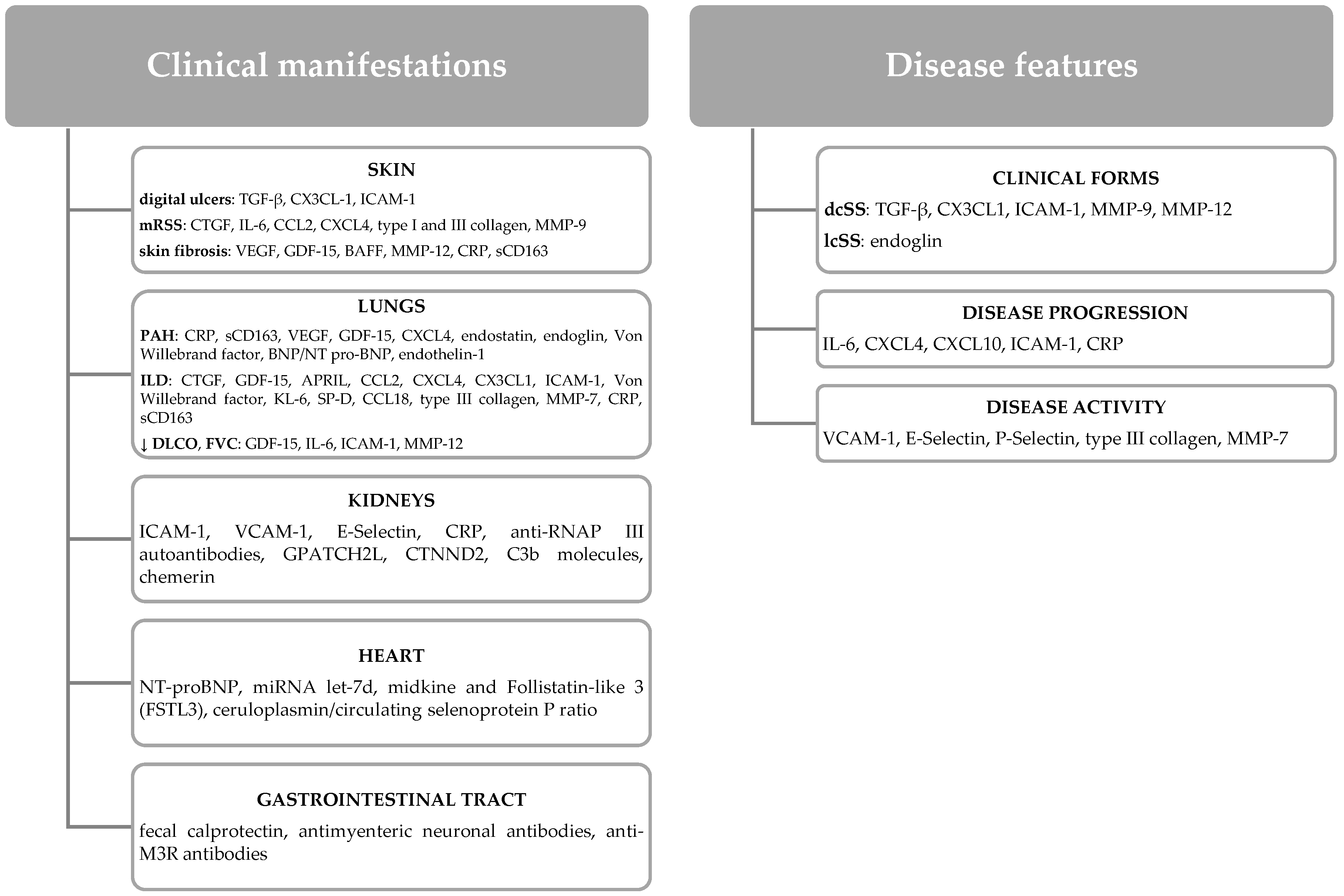
Figure 1. Potential biomarkers of organ involvement and disease features in SS. Key: TGF: transforming growth factor; GDF-15: growth differentiation factor 15; BAFF: B-cell-activating factor belonging to the tumor necrosis factor family; APRIL: a proliferation-inducing ligand; MMP: matrix metalloproteinases; BNP: brain natriuretic peptide; NT-proBNP: N-terminal-pro hormone BNP; CTG: connective tissue growth factor; mRSS: modified Rodnan total skin thickness score; ILD: interstitial lung disease; IL-6: interleukin 6; DLCO: diffusing capacity of carbon monoxide; PAH: pulmonary arterial hypertension; ICAM-1: intercellular adhesion molecule 1; dcSSc: diffuse cutaneous systemic sclerosis; VEGF: vascular endothelial growth factor; lcSSc: limited cutaneous systemic sclerosis; KL-6: krebs von den Lungen-6; SP-D: surfactant protein-D; CCL2: monocyte chemoattractant protein-1; CXCL4: platelet factor 4; VCAM-1: vascular cell adhesion molecule-1; CRP: C-reactive protein; sCD163: soluble CD163; anti-RNAP III antibodies: anti-RNA polymerase III antibodies; anti-M3R antibodies: anti-human muscarinic receptor M3 antibodies; miRNA let-7d: micro RNA let-7d; GPATCH2L: G-patch domain-containing protein 2-like; CTNND2: catenin delta 2.
Biomedicines 10 00318 g001
Figure 2. The intracellular signaling pathways of IL-6 and the main negative feedback loops that block the production of its biological effects. Key: IL-6: interleukin-6; IL-6R: interleukin-6 receptor; sIL-6R: soluble interleukin-6 receptor; gp130: glycoprotein 130; Jak: janus kinase; STAT: signal transducer and activator of transcription proteins; SOCS3: cytokine 3 signaling suppressor; PIAS3: protein inhibitor of activated STAT 3; ADAM: disintegrin metalloproteases; MCP: monocyte chemoattractant protein-1.
Biomedicines 10 00318 g002
Figure 3. The action of IL-6 on various cells and its local and systemic manifestations. Key: IL-6: interleukin 6; CD: cluster of differentiation; CRP: C-reactive protein; RANKL: activator receptor of nuclear factor kappa-B ligand; VEGF: vascular endothelial growth factor; Treg: regulatory T cells.
Biomedicines 10 00318 g003
Figure 4. Systemic harmful effects of IL-6 on the skin, cardiopulmonary, gastrointestinal and renal system. Key: IL-6, interleukin-6; ADAM17: disintegrin metalloproteases 17; PAH: pulmonary arterial hypertension; DLCO: diffusing capacity of lung for carbon monoxide; FVC: forced vital capacity.
Biomedicines 10 00318 g004
Table 1. Data regarding the clinical efficacy of Tocilizumab therapy in SS patients.
AuthorNumber of PatientsClinical and Paraclinical ParametersFollow-Up PeriodResults
Shima et al., 2010
[117]
2mRSS6 months
  • skin thickening improvement (mRSS decrease)
Elhai et al., 2013
[119]
27Polyarthritis
Myopathy
mRSS
5 months
  • joint/muscle damage improvement
  • skin thickening improvement (mRSS decrease)
Khanna et al., 2016
[114]
87mRSS
FVC
6 months
  • skin thickening improvement (mRSS decrease)
  • smaller decrease in FVC
Khanna et al., 2017 [122]78mRSS
FVC
24 months
  • skin thickening improvement (mRSS decrease)
  • lung function stabilization
Khanna et al., 2018 [115]210mRSS
FVC
12 months
  • skin thickening improvement (mRSS decrease)
  • lung function stabilization
Denton et al., 2018
[116]
12mRSS6 months
  • skin thickening improvement (mRSS decrease)
  • decreased skin collagen fiber production
Zacay et al., 2018
[118]
16Arthritis/arthralgia
Myalgia/myositis
FVC
mRSS
8 months
  • significant improvement in arthritis/arthralgia
  • muscle enzymes normalization
  • skin thickening improvement (mRSS decrease)
  • lung capacity improvement (FVC increase)
Shima et al., 2019
[63]
7mRSS6 months
  • skin thickening improvement (mRSS decrease) especially in cases with significant inflammatory syndrome and short disease duration
Narvaez et al., 2019 [124]9FVC
DLCO
6 min walking test
12 months
  • lung function stabilization and improvement (FVC increase) in cases refractory to corticosteroids or other immunosuppressants
Khanna et al. 2020
[121]
78FVC24 months
  • lung function stabilization / improvement (FVC increase)
Roofeh et al., 2021
[123]
136FVC12 months
  • lung function preservation according to the degree of pulmonary fibrosis
Khanna et al., 2021 [125]82mRSS
FVC
24 months
  • skin thickening improvement (mRSS decrease)
  • lung function improvement (FVC increase)
Adrovic et al., 2021 [130]9Juvenile SS
mRSS
DLCO
24 months
  • skin thickening improvement (mRSS decrease)
  • lung function improvement (DLCO increase)
Panopoulos et al., 2022 [126]21mRSS
Polyarthritis
FVC
PROs
12 months
  • skin thickening improvement (mRSS decrease)
  • lung function stabilization
  • joint damage improvement (Disease Activity Score 28 decrease)
  • improving patients’ quality of life
Key: mRSS: modified RODNAN skin score; DLCO: diffusing capacity of lung for carbon monoxide; FVC: forced vital capacity; PROs: patient-reported outcomes.
Publisher’s Note: MDPI stays neutral with regard to jurisdictional claims in published maps and institutional affiliations.