

Odontogenic Chronic Rhinosinusitis: Structured Histopathology Evidence in Different Patho-Physiological Mechanisms
 , ,
, ,  , , , and
Abstract
1. Introduction
2. Materials and Methods
2.1. Patients
2.2. Treatment
2.2.1. Transoral Approach
2.2.2. Transnasal Endoscopic Approach
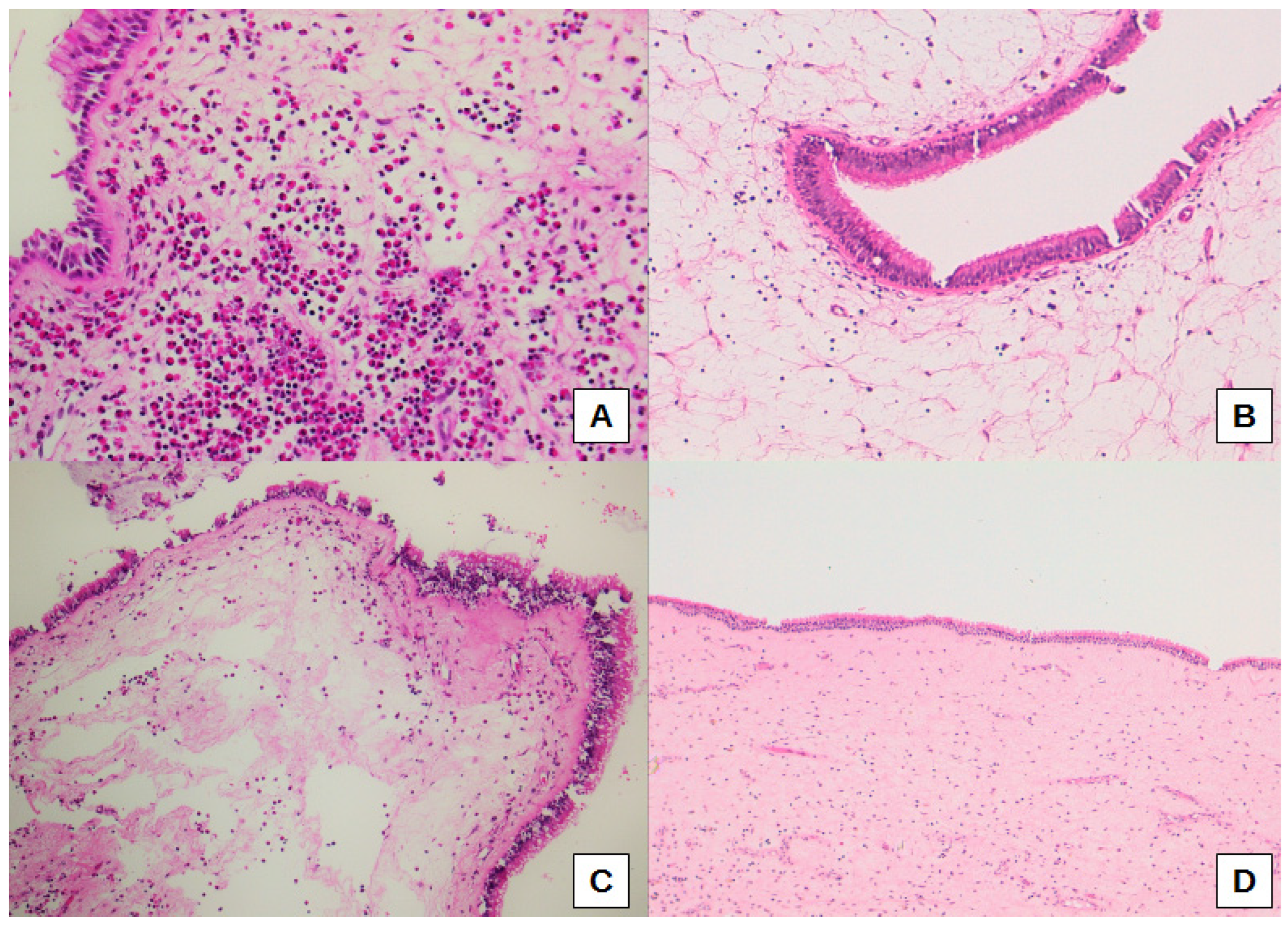
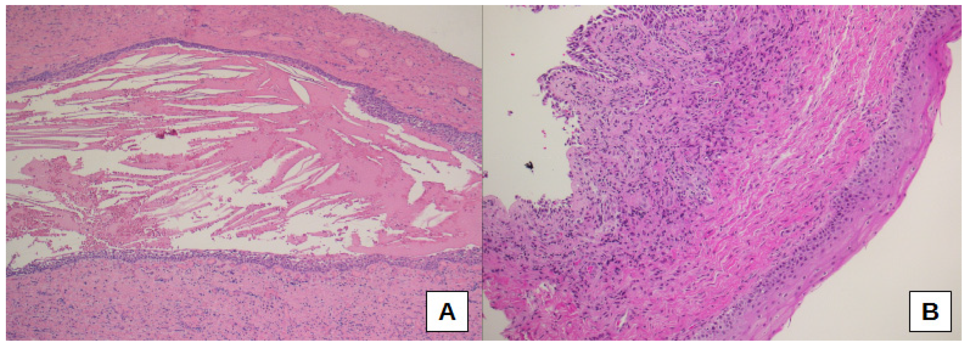
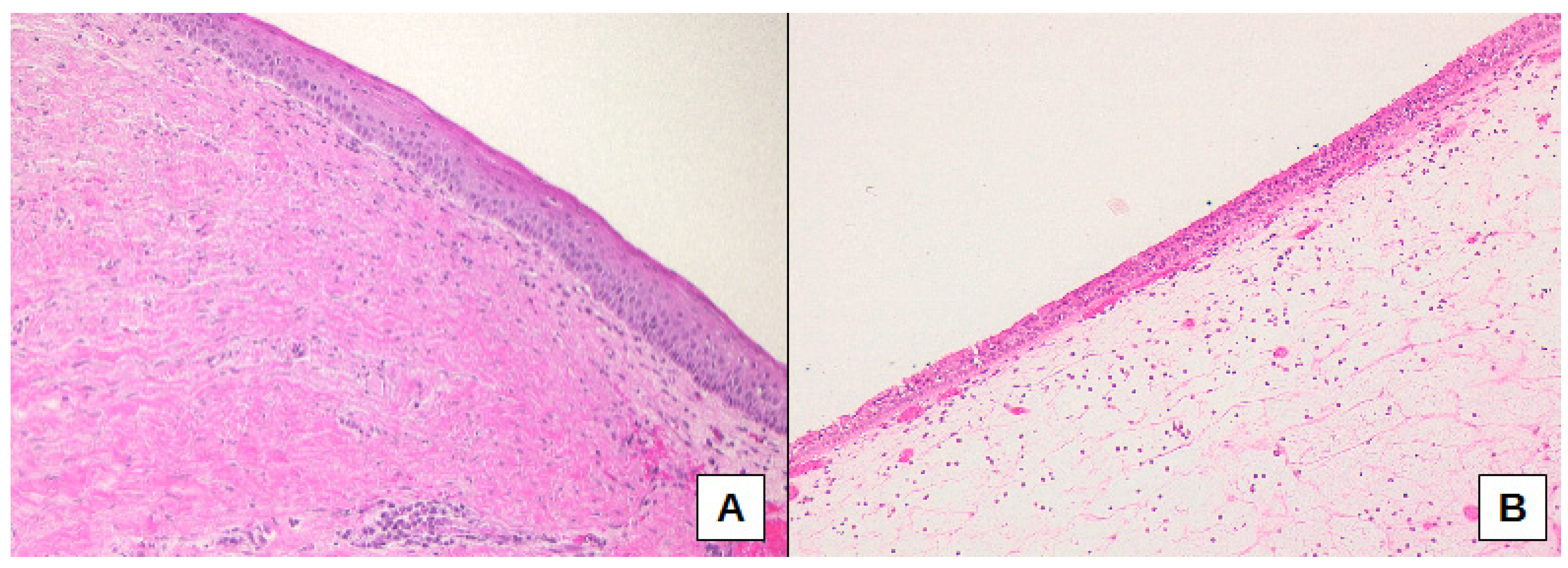
2.3. Histopathological Investigations
2.4. Statistical Analysis
3. Results
Structured Histopathology and oCRS Sub-Cohorts Stratified on Etiological Basis
4. Discussion
5. Conclusions
Author Contributions
Funding
Institutional Review Board Statement
Informed Consent Statement
Data Availability Statement
Acknowledgments
Conflicts of Interest
References
- Bauer, W.H. Maxillary sinusitis of dental origin. Am. J. Orthod Dentofacial Orthop. 1943, 29, 133–151. [Google Scholar] [CrossRef]
- Aukštakalnis, R.; Simonavičiūtė, R.; Simuntis, R. Treatment options for odontogenic maxillary sinusitis: A review. Stomatologija. 2018, 20, 22–26. [Google Scholar] [PubMed]
- Brescia, G.; Saia, G.; Apolloni, F.; Marioni, G. A novel nasal endoscopic approach for removing displaced dental implants from the maxillary sinus. Am. J. Otolaryngol. 2017, 38, 92–95. [Google Scholar] [CrossRef] [PubMed]
- Brescia, G.; Fusetti, S.; Apolloni, F.; Marioni, G.; Saia, G. Displaced dental materials in the maxillary sinus: An original series. Analysis and definition of a surgical decision-making process. Ann. Otol. Rhinol. Laryngol. 2019, 128, 177–183. [Google Scholar] [CrossRef] [PubMed]
- Workman, A.D.; Granquist, E.J.; Adappa, N.D. Odontogenic sinusitis: Developments in diagnosis, microbiology, and treatment. Curr. Opin. Otolaryngol. Head Neck Surg. 2018, 26, 27–33. [Google Scholar] [CrossRef] [PubMed]
- Little, R.E.; Long, C.M.; Loehrl, T.A.; Poetker, D.M. Odontogenic sinusitis: A review of the current literature. Laryngoscope Investig. Otolaryngol. 2018, 3, 110–114. [Google Scholar] [CrossRef] [PubMed]
- Raman, A.; Papagiannopoulos, P.; Kuhar, H.N.; Gattuso, P.; Batra, P.S.; Tajudeen, B.A. Histopathologic features of chronic sinusitis precipitated by odontogenic infection. Am. J. Rhinol. Allergy 2019, 33, 113–120. [Google Scholar] [CrossRef]
- Menditti, D.; Laino, L.; Di Domenico, M.; Troiano, G.; Guglielmotti, M.; Sava, S.; Mezzogiorno, A.; Baldi, A. Cysts and pseudocysts of the oral cavity: Revision of the literature and a new proposed classification. In Vivo 2018, 32, 999–1007. [Google Scholar] [CrossRef]
- Goyal, V.K.; Ahmad, A.; Turfe, Z.; Peterson, E.I.; Craig, J.R. Predicting odontogenic sinusitis in unilateral sinus disease: A prospective, multivariate analysis. Am. J. Rhinol. Allergy. 2021, 35, 164–171. [Google Scholar] [CrossRef]
- Brook, I. Sinusitis of odontogenic origin. Otolaryngol. Head Neck Surg. 2006, 135, 349–355. [Google Scholar] [CrossRef]
- Eggmann, F.; Connert, T.; Bühler, J.; Dagassan-Berndt, D.; Weiger, R.; Walter, C. Do periapical and periodontal pathologies affect Schneiderian membrane appearance? Systematic review of studies using cone-beam computed tomography. Clin. Oral Investig. 2017, 21, 1611–1630. [Google Scholar] [CrossRef] [PubMed]
- Vidal, F.; Coutinho, T.M.; Carvalho Ferreira, D.; Souza, R.C.; Gonçalves, L.S. Odontogenic sinusitis: A comprehensive review. Acta Odontol. Scand. 2017, 75, 623–633. [Google Scholar] [CrossRef] [PubMed]
- Snidvongs, K.; Lam, M.; Sacks, R.; Earls, P.; Kalish, L.; Phillips, P.S.; Pratt, E.; Harvey, R.J. Structured histopathology profiling of chronic rhinosinusitis in routine practice. Int. Forum Allergy Rhinol. 2012, 2, 376–385. [Google Scholar] [CrossRef] [PubMed]
- Kuhar, H.N.; Tajudeen, B.A.; Mahdavinia, M.; Gattuso, P.; Ghai, R.; Batra, P.S. Inflammatory infiltrate and mucosal remodeling in chronic rhinosinusitis with and without polyps: Structured histopathologic analysis. Int. Forum Allergy Rhinol. 2017, 7, 679–689. [Google Scholar] [CrossRef] [PubMed]
- Brescia, G.; Alessandrini, L.; Giacomelli, L.; Parrino, D.; Zanotti, C.; Tealdo, G.; Franz, L.; Carraro, V.; Barion, U.; Marioni, G. A classification of chronic rhinosinusitis with nasal polyps based on structured histopathology. Histopathology 2020, 76, 296–307. [Google Scholar] [CrossRef]
- Brescia, G.; Alessandrini, L.; Marioni, G. Structured histopathology for endotyping and planning rational treatment in chronic rhinosinusitis. Am. J. Otolaryngol. 2021, 42, 102795. [Google Scholar] [CrossRef]
- Lund, V.J.; Mackay, I.S. Staging in rhinosinusitus. Rhinology 1993, 31, 183–184. [Google Scholar]
- Manani, G.; Bacci, C.; Zanette, G.; Facco, E. Stato attuale della sedazione cosciente in odontoiatria/Contemporary state of sedation in dentistry. Dent. Cadmos 2012, 80, 357–369. [Google Scholar] [CrossRef]
- Nakayama, T.; Asaka, D.; Okushi, T.; Yoshikawa, M.; Moriyama, H.; Otori, N. Endoscopic medial maxillectomy with preservation of inferior turbinate and nasolacrimal duct. Am. J. Rhinol. Allergy 2012, 26, 405–408. [Google Scholar] [CrossRef]
- Omura, K.; Asaka, D.; Nayak, J.V.; Tanaka, Y. Transseptal access with crossing multiple incisions for improved pedicle control and septum preservation: “How I do it”. Am. J. Rhinol. Allergy 2017, 31, 139–141. [Google Scholar] [CrossRef]
- Omura, K.; Nomura, K.; Aoki, S.; Otori, N.; Tanaka, Y. Direct approach to the anterior and lateral part of the maxillary sinus with an endoscope. Auris Nasus Larynx 2019, 46, 871–875. [Google Scholar] [CrossRef] [PubMed]
- Shi, L.L.; Xiong, P.; Zhang, L.; Cao, P.P.; Liao, B.; Lu, X.; Cui, Y.H.; Liu, Z. Features of airway remodeling in different types of Chinese chronic rhinosinusitis are associated with inflammation patterns. Allergy 2013, 68, 101–109. [Google Scholar] [CrossRef]
- Brescia, G.; Alessandrini, L.; Frasconi, S.; Contro, G.; Frigo, A.C.; Marioni, G. Structured histopathology and laboratory evidence in nasal polyposis with different pathogenesis. Am. J. Otolaryngol. 2022, 44, 103649. [Google Scholar] [CrossRef]
- Lee, K.; Tai, J.; Lee, S.H.; Kim, T.H. Advances in the knowledge of the underlying airway remodeling mechanisms in chronic rhinosinusitis based on the endotypes: A review. Int. J. Mol. Sci. 2021, 22, 910. [Google Scholar] [CrossRef] [PubMed]
- Contro, G.; Brescia, G.; Alessandrini, L.; Barion, U.; Padoan, R.; Frigo, A.C.; Schiavon, F.; Marioni, G. Neutrophil infiltrates and eosinophil aggregates in chronic rhinosinusitis with nasal polyps and EGPA. Clin. Rheumatol. 2021, 40, 1949–1957. [Google Scholar] [CrossRef]
- Lombardi, C.; Berti, A.; Cottini, M. The emerging roles of eosinophils: Implications for the targeted treatment of eosinophilic-associated inflammatory conditions. Curr. Res. Immunol. 2022, 3, 42–53. [Google Scholar] [CrossRef] [PubMed]
- Brescia, G.; Alessandrini, L.; Parrino, D.; Franz, L.; Barion, U.; Marioni, G. Emerging contribution of histopathology to our understanding of chronic rhinosinusitis endotypes: Tissue eosinophil count and aggregates. Am. J. Rhinol. Allergy 2020, 34, 122–126. [Google Scholar] [CrossRef] [PubMed]
- Ramirez, G.A.; Yacoub, M.R.; Ripa, M.; Mannina, D.; Cariddi, A.; Saporiti, N.; Ciceri, F.; Castagna, A.; Colombo, G.; Dagna, L. Eosinophils from physiology to disease: A comprehensive review. Biomed. Res. Int. 2018, 2018. [Google Scholar] [CrossRef]
- Zitzmann, N.U.; Berglundh, T.; Marinello, C.P.; Lindhe, J. Experimental peri-implant mucositis in man. J. Clin. Periodontol. 2001, 28, 517–523. [Google Scholar] [CrossRef]
- Shafizadeh, M.; Amid, R.; Mahmoum, M.; Kadkhodazadeh, M. Histopathological characterization of peri-implant diseases: A systematic review and meta-analysis. Arch. Oral Biol. 2021, 132, 105288. [Google Scholar] [CrossRef]
- Gualini, F.; Berglundh, T. Immunohistochemical characteristics of inflammatory lesions at implants. J. Clin. Periodontol. 2003, 30, 14–18. [Google Scholar] [CrossRef] [PubMed]
- Obădan, F.; Crăiţoiu, Ş.; Manolea, H.O.; Hîncu, M.C.; Iacov-Crăiţoiu, M.M. The evaluation of the morphological evolution of the tissue integration of dental implants through conventional histology and immunohistochemistry techniques. Rom. J. Morphol. Embryol. 2018, 59, 851–859. [Google Scholar] [PubMed]
- Noskovicova, N.; Hinz, B.; Pakshir, P. Implant fibrosis and the underappreciated role of myofibroblasts in the foreign body reaction. Cells 2021, 10, 1794. [Google Scholar] [CrossRef] [PubMed]
- Mynatt, R.G.; Do, J.; Janney, C.; Sindwani, R. Squamous metaplasia and chronic rhinosinusitis: A clinicopathological study. Am. J. Rhinol. 2008, 22, 602–605. [Google Scholar] [CrossRef]


| Main Features | No. of Cases (%) |
|---|---|
| Sex | |
| Male | 24 (57.1) |
| Female | 18 (42.9) |
| Allergy/Asthma | |
| None | 35 (83.3) |
| Allergy | 2 (4.8) |
| Asthma | 2 (4.8) |
| Allergy and asthma | 3 (7.1) |
| Sinonasal polyps’ phenotype | |
| No | 21 (50.0) |
| Yes | 21 (50.0) |
| Total (42 Cases) | Radicular Cyst (11 Cases) | Dental Implants (9 Cases) | Other Tooth Diseases (22 Cases) | p-Value | |
|---|---|---|---|---|---|
| oCRS with nasal polyps | 21 (50.0) | 4 (36.4) | 6 (66.7) | 11 (50.0) | 0.4375 |
| oCRS without nasal polyps | 21 (50.0) | 7 (63.6) | 3 (33.3) | 11 (50.0) | |
| Unilateral polyposis | 19 (90.5) | 4 (100.0) | 5 (83.3) | 10 (90.9) | |
| Bilateral polyposis | 2 (9.5) | 0 (0.0) | 1 (16.7) | 1 (9.1) | |
| CT score | |||||
| Mean (SD) | 4.7 (3.4) | 4.5 (2.6) | 5.8 (1.9) | 4.1 (4.2) | 0.3423 |
| Median (Range) | 4.0 (1.0–12.0) | 5.0 (1.0–7.0) | 5.5 (4.0–9.0) | 2.0 (1.0–12.0) |
| No. of Cases (%) | Radicular Cyst (1) No. of Cases = 11 | Dental Implant (2) No. of Cases = 9 | Other Tooth Diseases (3) No. of Cases = 22 | Overall p-Value | Difference (95% CI) 2 vs. 1 | Difference (95% CI) 3 vs. 1 | Difference (95% CI) 2 vs. 3 | |
|---|---|---|---|---|---|---|---|---|
| Degree of inflammation | ||||||||
| 0 or 1 | 10 (23.8) | 3 (27.3) | 1 (11.1) | 6 (27.3) | 0.7106 | |||
| 2 or 3 | 32 (76.2) | 8 (72.7) | 8 (88.9) | 16 (72.7) | 16.2 (−25.1; 52.3) | 0.0 (−30.7; 36.3) | 16.2 (−22.8; 43. 2) | |
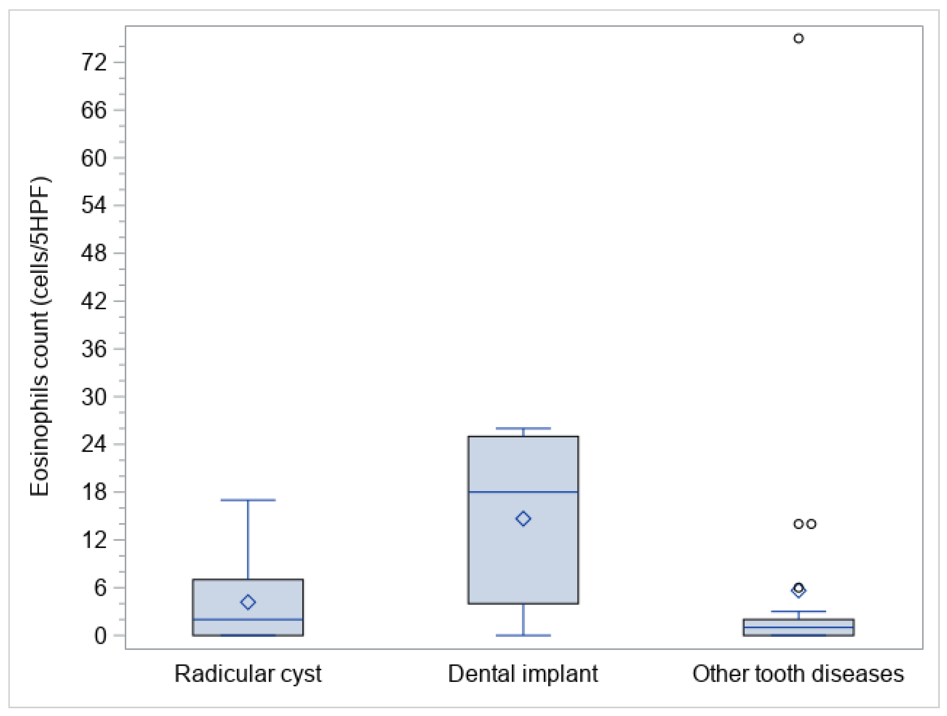
| Eosinophils count (cells/5HPF) | ||||||||
| Mean (SD) | 7.2 (13.3) | 4.2 (5.3) | 14.7 (10.7) | 5.6 (16.0) | 0.0118 | |||
| Median (Range) | 2.0 (0.0–75.0) | 2.0 (0.0–17.0) | 18.0 (0.0–26.0) | 1.0 (0.0–75.0) | 10 (0; 22) | 0 (−4; 1) | 11 (2; 22) | |
| Eosinophil aggregates | ||||||||
| No | 41 (97.6) | 11 (100.0) | 9 (100.0) | 21 (95.5) | 1.0000 | |||
| Yes | 1 (2.4) | 0 (0.0) | 0 (0.0) | 1 (4.5) | - | 4.5 (−24.5; 23.3) | −4.5 (−23.2; 30.0) | |
| Neutrophil infiltrate | ||||||||
| No | 16 (38.1) | 5 (45.5) | 2 (22.2) | 9 (40.9) | 0.5873 | |||
| Yes | 26 (61.9) | 6 (54.5) | 7 (77.8) | 13 (59.1) | 23.3 (−21.3; 62.6) | 4.6 (−31.1; 40.6) | 18.7 (−21.5; 49.1) | |
| Basal membrane thickening | ||||||||
| 0 or 1 | 37 (88.1) | 8 (72.7) | 9 (100.0) | 20 (90.9) | 0.1909 | |||
| 2 or 3 | 5 (11.9) | 3 (27.3) | 0 (0.0) | 2 (9.1) | −27.3 (−61.0; 9.9) | −18.2 (−51.8; 9.9) | −9.1 (−30.0; 25.5) | |
| Subepithelial edema | ||||||||
| 0 or 1 | 36 (85.7) | 8 (72.7) | 6 (66.7) | 22 (100.0) | 0.0099 | |||
| 2 or 3 | 6 (14.3) | 3 (27.3) | 3 (33.3) | 0 (0.0) | 6.1 (−32.2; 48.3) | −27.3 (−61.0; −4.1) | 33.3 (6.5; 70.1) | |
| Hyperplastic—papillary changes | ||||||||
| No | 33 (78.6) | 9 (81.8) | 6 (66.7) | 18 (81.8) | 0.6151 | |||
| Yes | 9 (21.4) | 2 (18.2) | 3 (33.3) | 4 (18.2) | 15.1 (−25.7; 54.8) | 0.0 (−33.7; 27.3) | 15.1 (−17.6; 52.0) | |
| Mucosal ulceration | ||||||||
| No | 32 (76.2) | 6 (54.5) | 7 (77.8) | 19 (86.4) | 0.1327 | |||
| Yes | 10 (23.8) | 5 (45.5) | 2 (22.2) | 3 (13.6) | −23.2 (−62.6; 21.3) | −31.8 (−64.5; 1.8) | 8.6 (−20.2; 47.4) | |
| Squamous metaplasia | ||||||||
| No | 30 (71.4) | 5 (45.5) | 9 (100.0) | 16 (72.7) | 0.0258 | |||
| Yes | 12 (28.6) | 6 (54.5) | 0 (0.0) | 6 (27.3) | −54.5 (−83.3; −14.9) | −27.3 (−61.0; 11.1) | −27.3 (−50.5; 9.3) | |
| Fibrosis | ||||||||
| No | 22 (52.4) | 4 (36.4) | 8 (88.9) | 10 (45.5) | 0.0408 | |||
| Yes | 20 (47.6) | 7 (63.6) | 1 (11.1) | 12 (54.5) | −52.5 (−82.7; −8.1) | −9.1 (−42.6; 28.7) | −43.4 (−70.1; 2.1) | |
| Fungal elements | ||||||||
| No | 39 (92.9) | 10 (90.9) | 9 (100.0) | 20 (90.9) | 1.0000 | |||
| Yes | 3 (7.1) | 1 (9.1) | 0 (0.0) | 2 (9.1) | −9.1 (−41.3; 25.2) | 0.0 (−32.9; 22.9) | −9.1 (−30.0; 25.5) | |
| Charcot–Leyden crystals | ||||||||
| No | 42 (100.0) | 11 (100.0) | 9 (100.0) | 22 (100.0) | - | |||
| Yes | 0 (0.0) | 0 (0.0) | 0 (0.0) | 0 (0.0) | ||||
| Globet cells hyperplasia | ||||||||
| 0 or 1 | 30 (71.4) | 7 (63.6) | 5 (55.6) | 18 (81.8) | 0.2875 | |||
| 2 or 3 | 12 (28.6) | 4 (36.4) | 4 (44.4) | 4 (18.2) | 8.1 (−38.0; 50.2) | −18.2 (−53.1; 13.6) | 26.2 (−9.6; 61.9) | |
Publisher’s Note: MDPI stays neutral with regard to jurisdictional claims in published maps and institutional affiliations. |
© 2022 by the authors. Licensee MDPI, Basel, Switzerland. This article is an open access article distributed under the terms and conditions of the Creative Commons Attribution (CC BY) license (https://creativecommons.org/licenses/by/4.0/).
Share and Cite
Brescia, G.; Alessandrini, L.; Bacci, C.; Bissolotti, G.; Fedrigo, M.; Contro, G.; Frasconi, S.; Boccuto, M.G.; Calcavecchia, A.; Frigo, A.C.; et al. Odontogenic Chronic Rhinosinusitis: Structured Histopathology Evidence in Different Patho-Physiological Mechanisms. Biomedicines 2022, 10, 2768. https://doi.org/10.3390/biomedicines10112768
Brescia G, Alessandrini L, Bacci C, Bissolotti G, Fedrigo M, Contro G, Frasconi S, Boccuto MG, Calcavecchia A, Frigo AC, et al. Odontogenic Chronic Rhinosinusitis: Structured Histopathology Evidence in Different Patho-Physiological Mechanisms. Biomedicines. 2022; 10(11):2768. https://doi.org/10.3390/biomedicines10112768
Chicago/Turabian StyleBrescia, Giuseppe, Lara Alessandrini, Christian Bacci, Guido Bissolotti, Marny Fedrigo, Giacomo Contro, Samuele Frasconi, Maria Grazia Boccuto, Arianna Calcavecchia, Anna Chiara Frigo, and et al. 2022. "Odontogenic Chronic Rhinosinusitis: Structured Histopathology Evidence in Different Patho-Physiological Mechanisms" Biomedicines 10, no. 11: 2768. https://doi.org/10.3390/biomedicines10112768
APA StyleBrescia, G., Alessandrini, L., Bacci, C., Bissolotti, G., Fedrigo, M., Contro, G., Frasconi, S., Boccuto, M. G., Calcavecchia, A., Frigo, A. C., Barion, U., Fusetti, S., Angelini, A., & Marioni, G. (2022). Odontogenic Chronic Rhinosinusitis: Structured Histopathology Evidence in Different Patho-Physiological Mechanisms. Biomedicines, 10(11), 2768. https://doi.org/10.3390/biomedicines10112768