Review

Activation of STAT and SMAD Signaling Induces Hepcidin Re-Expression as a Therapeutic Target for β-Thalassemia Patients

by
Hanan Kamel M. Saad
1,
Alawiyah Awang Abd Rahman
2,
Azly Sumanty Ab Ghani
2,
Wan Rohani Wan Taib
1,
Imilia Ismail
1,
Muhammad Farid Johan
3,
Abdullah Saleh Al-Wajeeh
4 and
Hamid Ali Nagi Al-Jamal
1,*
1
School of Biomedicine, Faculty of Health Sciences, Universiti Sultan Zainal Abidin, Kuala Nerus 21300, Terengganu, Malaysia
2
Pathology Department, Hospital Sultanah Nur Zahirah, Kuala Terengganu 20400, Terengganu, Malaysia
3
Department of Haematology, School of Medical Sciences, Universiti Sains Malaysia, Kubang Kerian 16150, Kelatan, Malaysia
4
Anti-Doping Lab Qatar, Doha 27775, Qatar
*
Author to whom correspondence should be addressed.
Biomedicines 2022, 10(1), 189; https://doi.org/10.3390/biomedicines10010189
Submission received: 16 November 2021 / Revised: 11 January 2022 / Accepted: 12 January 2022 / Published: 17 January 2022
(This article belongs to the Section Cell Biology and Pathology)

Abstract

:
Iron homeostasis is regulated by hepcidin, a hepatic hormone that controls dietary iron absorption and plasma iron concentration. Hepcidin binds to the only known iron export protein, ferroportin (FPN), which regulates its expression. The major factors that implicate hepcidin regulation include iron stores, hypoxia, inflammation, and erythropoiesis. When erythropoietic activity is suppressed, hepcidin expression is hampered, leading to deficiency, thus causing an iron overload in iron-loading anemia, such as β-thalassemia. Iron overload is the principal cause of mortality and morbidity in β-thalassemia patients with or without blood transfusion dependence. In the case of thalassemia major, the primary cause of iron overload is blood transfusion. In contrast, iron overload is attributed to hepcidin deficiency and hyperabsorption of dietary iron in non-transfusion thalassemia. Beta-thalassemia patients showed marked hepcidin suppression, anemia, iron overload, and ineffective erythropoiesis (IE). Recent molecular research has prompted the discovery of new diagnostic markers and therapeutic targets for several diseases, including β-thalassemia. In this review, signal transducers and activators of transcription (STAT) and SMAD (structurally similar to the small mothers against decapentaplegic in Drosophila) pathways and their effects on hepcidin expression have been discussed as a therapeutic target for β-thalassemia patients. Therefore, re-expression of hepcidin could be a therapeutic target in the management of thalassemia patients. Data from 65 relevant published experimental articles on hepcidin and β-thalassemia between January 2016 and May 2021 were retrieved by using PubMed and Google Scholar search engines. Published articles in any language other than English, review articles, books, or book chapters were excluded.

1. Thalassemia Syndrome

Thalassemia is an inherited autosomal recessive blood disorder that can be divided into either alpha (α) or beta (β) depending on the affected α or β globin chain [1]. The adult hemoglobin (HbA) consists of two α and two β (α2β2) chains in each HbA molecule [2,3]. Alpha-thalassemia occurs if one or more of the four alleles that code for α globin is missing or damaged [4]. On the other hand, β-thalassemia is caused by mutation in the β globin gene leading to a reduction in β globin or production of abnormal hemoglobin.

2. Beta-Thalassemia

Notably, β-thalassemia is highly reported in the Mediterranean and Southeast Asian countries as one of the most common genetic disorders [5]. Nevertheless, a lack of information on knowledge, attitudes, and practices towards β-thalassemia poses a challenge in many countries, including Malaysia [5]. This disease can be categorized into β-thalassemia major, intermedia, or minor [6]. β-thalassemia major and intermedia are genetically homozygous or heterozygous (β0 and β+), whereas β-thalassemia minor is usually heterozygous [6].
The β globin chain is encoded by the β globin gene (HBB), located on chromosome 11 at the short arm position 15.4 [7,8]. Individuals with β-thalassemia major and intermedia inherit the mutation in both copies of the HBB gene, affecting normal β globin chain production [7]. The clinical features of β-thalassemia major are revealed as early as the first two years of the patient’s life and are usually connected to transfusional iron overload, whereas the clinical presentation for β-thalassemia intermedia occurs later in life [2]. In contrast, β-thalassemia minor or trait has one mutation in the HBB gene and is described as a carrier [6]. They are usually symptomless, with a hypochromic microcytic blood picture and mild anemia, and can potentially increase in severity with malnutrition [6].
Beta-thalassemia can be present alongside other diseases associated with an abnormal β globin chain, such as the hemoglobin E (HbE) disease, exhibiting severe anemia [2]. HbE is a hemoglobin (Hb) variant caused by a single base substitution of glutamic acid to lysine at position 26 of the globin chain, commonly found in Southeast Asia [9]. It can be classified into three types: heterozygous, homozygous, or compound heterozygous [9]. When the HbE trait is coinherited with β-thalassemia, it is called compound heterozygous, a condition known as HbE/β-thalassemia, which resembles homozygous β0-thalassemia clinically and hematologically [10].
The phenotypic heterogeneity of HbE/β-thalassemia can range from mild asymptomatic anemia to a severe form that requires regular blood transfusion [11]. A study in Sri Lanka revealed that the HbE/β-thalassemia phenotype is unstable during the first 10 years of life but gradually stabilizes as the patient gets older [11]. This condition is caused by various changes in anemia and erythroid expansion progression during their early life [11]. However, the lack of knowledge on the classification of the disease severity will affect the understanding of HbE/β-thalassemia clinical progression with age [11].

3. Iron

Iron is an essential trace element found abundantly in the environment but is not readily available for uptake since it oxidizes when in contact with oxygen, thus making it highly insoluble [12]. The majority of iron in the body is found in red blood cells and is used to produce Hb during erythropoiesis [13,14]. In addition, trace amounts of iron are bound to enzyme effectiveness, such as cytochromes and those involved in the Krebs cycle [12]. Besides, iron can exist as non-hem compounds, such as ferritin and hemosiderin, primarily found in the spleen, bone marrow, and liver (see Figure 1) [12].
Iron in the form of ferrous ion (Fe2+) is an essential component of the Hb that binds with oxygen from the lungs before being transported to other parts of the body [15]. Furthermore, iron bound to cytochromes is vital for the electron transfer chain. It is reversibly recycled in the form of Fe2+ and ferric ion (Fe3+), aiding in energy production in the form of adenosine triphosphate [12]. On top of that, iron bound to peroxidases can convert potentially harmful hydrogen peroxide to water [16,17]. Other iron functions include deoxyribonucleic acid (DNA) replication, DNA repair, and cell signaling [15].

3.1. Iron Metabolism

The primary iron absorption takes place in the intestine, a crucial process in regulating the iron level in the body [18]. Iron is internalized as Fe2+ by divalent metal transporter 1 (DMT1) [19]. Initially, iron in food is in the form of Fe3+ and must undergo reduction via agents resembling vitamin C before it can be successfully absorbed [20,21]. Later, Fe2+ is bound by apoferritin in the intestinal cells, where it undergoes subsequent oxidation by extracellular protein, ceruloplasmin, to form Fe3+, which is bound by ferritin [22]. After that, apotransferrin helps iron absorption into the blood, where apotransferrin turns into transferrin by binding to two Fe3+ [23]. Transferrin carries iron in the plasma and releases it to organs such as bone marrow, where red blood cells are produced [24]. After about four months, iron is recycled from senescent red blood cells by the spleen, liver, and macrophages, ready to be reused [25]. The conservation of iron is critical because dietary iron is just enough to replace small losses [25]. Mice with iron overload were able to eliminate a significant amount of iron through the digestive tract by an unknown mechanism [26]. Most lost iron can be found in the epithelial and red blood cells excreted in the urine or feces [27]. An adult male is known to lose an average of 1 mg of iron per day, while women lose 1.4 mg per day through the menstrual cycle [28,29].

3.2. Iron Overload in β-Thalassemia

The liver contains approximately 70% of the body iron, thus being the most affected organ during iron overload [30]. Hepcidin discovery as a key regulator of iron metabolism is revolutionary in understanding the mechanism of iron overload in β-thalassemia patients [31,32]. It is a peptide hormone first isolated from human urine and encoded by the hepcidin antimicrobial peptide (HAMP) gene [25]. When there is an increase in iron level in the body, the hepatocytes are stimulated to release more hepcidin into the bloodstream [33]. Hepcidin is also a crucial negative regulator of iron absorption into the body through the intestine by internalizing and degrading the iron exporter, ferroportin (FPN), at the duodenal surface [31]. It blocks the flow of iron into the bloodstream from the iron storage cells and recycling within macrophages (Figure 2) [34]. Additionally, hepcidin is highly dependent on transferrin receptor 2 (TfR2) and hereditary hemochromatosis protein (HFE), a human homeostatic iron regulator protein encoded by the HFE gene [35]. A mutation or deficiency in TfR2 and HFE results in hepcidin deficiency, leading to increased iron absorption or enhanced iron release from macrophages [35].
The frequent measurement of iron is vital for the effective management of β-thalassemia [30]. Although direct estimation of liver iron concentration is the most accurate method to define iron overload in patients, it remains an aggressive procedure [30]. Thus, there is a dire need for a noninvasive method to accurately measure iron storage in the body [30]. Iron overload in the body can be measured using biochemical parameters, such as serum ferritin, hepatic liver concentration, urinary iron excretion, and total iron-binding capacity (TIBC) [36]. However, these parameters give variable results and are inaccurate for iron overload reflection. Currently, serum ferritin estimation is considered the most suitable to reflect iron storage in the body [30]; recently, magnetic resonance imaging (MRI) estimates tissue iron concentration indirectly by detecting the paramagnetic influences of stored iron [37].
According to research in India, 87.4% of 72 patients of β-thalassemia major and intermedia showed a very high ferritin level due to poor iron chelation, which makes patients more susceptible to iron overload complications [30]. The first dysregulation of hepcidin in β-thalassemia was reported using a mouse model [38]. Iron overload is proven to be less dominant in controlling the hepcidin expression compared to IE (Figure 3) [38]. Another study revealed that mice with β-thalassemia major and intermedia showed IE and iron deposition within organs, which is associated with suppressed expression of hepcidin and an increased level of FPN [39]. Even without blood transfusion, 63.8% of HbE/β-thalassemia patients are proven to develop iron overload with serum ferritin (200 to 400 ng/mL) [40,41].

3.2.1. Toxicity Effect of Iron Overload

Excessive iron absorption will result in iron accumulation, damaging vital organs, such as the liver and heart [25]. Iron is a pro-oxidant that induces oxidative stress, contributing to lipid peroxidation, atherosclerosis, DNA damage, carcinogenesis, and neurodegenerative diseases [12]. Moreover, the iron level in the body needs to be controlled for the benefit of resistance towards infection [42]. Bacteria grow faster and form biofilms more readily when iron increases in the body [42]. Therefore, patients with iron overload will be more susceptible to a wide range of intracellular and blood pathogens [42]. Based on a study conducted in Thailand, women with HbE/β-thalassemia demonstrated lower iron usage but higher iron absorption than controls [43]. In HbE/β-thalassemia patients, iron overload is a major problem, requiring regular blood transfusion for their survival [10]. Thus, therapeutic iron chelation therapy is vital in reducing the high iron level [44]. Nevertheless, iron chelators cause severe side effects, such as nausea, diarrhea, dizziness, and elevated liver enzymes, gastrointestinal disorders, and arthralgia [45].

3.2.2. Pathophysiology of Iron Overload in β-Thalassemia

The capacity of the transferrin iron transport system is saturated in β-thalassemia patients, causing the non-transferrin bound iron (NTBI) and labile plasma iron (LPI) to circulate in plasma and eventually be deposited into susceptible cells [30]. The NTBI enters cells through different cellular channels, such as the L-type voltage-dependent Ca2+ channel (LVDCC), a promiscuous divalent cation transporter [46], and Zip14, a member of the SLC39A zinc transporter family [47]. Increased iron and labile cellular iron storage result from long-term uptake and accumulation of the NTBI and labile iron pool (LIP) [48], affecting the heart, liver, and endocrine system [49].
When the cellular labile iron pool exceeds the cell ability to synthesize new ferritin molecules, a critical concentration of reactive oxygen species (ROS) is reached. The production of ROS by the NTBI metabolism plays a crucial role in cellular dysfunction, apoptosis, and necrosis [50]. Various ROS, particularly hydroxyl radicals, enhance lipid peroxidation and organelle damage, resulting in cell death [51] and fibrogenesis mediated by transforming growth factor β1 (TGF-β1) [52,53]. Apart from that, iron overload increases the risk of infection, a major cause of death in β-thalassemia patients [54,55]. Autophagy is crucial in eliminating oxidized proteins and damaged mitochondria. Its activation is higher in erythroblasts of HbE/β-thalassemia patients compared to normal control erythroblasts [56]. ROS may promote a higher level of autophagy in HbE/β-thalassemia erythroblasts, thus intensifying apoptosis and IE in HbE/β-thalassemia patients [56]. HbE/β-thalassemia patients with iron overload demonstrate decreased FPN expression compared to the healthy control [57]. Excess iron can contribute to IE (Figure 3). Growth differentiation factor 15 (GDF15) and twisted-gastrulation 1 (TWSG1) protein have been reported to suppress hepcidin synthesis and enhance iron absorption in β-thalassemia patients [58,59,60].

4. Hepcidin Expression in β-Thalassemia

Hepcidin expression in thalassemia was first reported in a mouse model of severe anemia (C57BI/6 Hbbth3/+) [38]. Furthermore, a decline in serum hepcidin levels has been reported in HbE/β-thalassemia patients, β-thalassemia trait, and HbE trait carriers [61]. The decreased serum hepcidin levels in β-thalassemia patients are associated with the downregulation of hepcidin expression in liver cells, resulting in continuous absorption of dietary iron that leads to iron overload [58]. In individuals with thalassemia major and intermedia, liver hepcidin mRNA expression is inversely associated with soluble transferrin receptor (sTfR) and erythropoietin (EPO), but not with iron storage [62]. Suppression of hepcidin in HbE/β-thalassemia patients is linked to increased iron loading, saturated iron-binding proteins, and organ damage [61]. Moreover, hepcidin suppression with enhanced iron absorption was found in the β-thalassemia trait [63].

4.1. Hepcidin Regulation in β-Thalassemia

Hepcidin is suppressed in β-thalassemia patients with increasing iron absorption in response to the iron demand by erythroblasts due to tissue hypoxia EPO production and anemia [64]. During the differentiation process, several hepcidin inhibitors are released from erythroblasts to regulate hepcidin expression in β-thalassemia. GDF15 serum level is inversely correlated with hepcidin expression in hepatocytes of thalassemia patients [65]. Meanwhile, TWSG1 was upregulated in the bone marrow, spleen, and liver of mice with β-thalassemia major and intermedia, associated with hepcidin suppression and absence of bone morphogenetic protein (BMP). Additionally, human hepatocytes’ TWSG1 indirectly suppressed hepcidin expression through inhibition of BMP-mediated signaling [66].
Erythroferrone (ERFE) hormone functions as a negative regulator of hepcidin synthesis. Elevated ERFE expression is associated with increased erythropoietin and hepcidin suppression in mice models with thalassemia intermediate during stress erythropoiesis. The ERFE-deficient mice failed to suppress hepcidin after hemorrhage and erythropoietin administration [67]. Therefore, increased iron absorption in β-thalassemia is most likely attributed to increased ERFE expression and other hypoxia-related molecules that suppress hepcidin synthesis or increase FPN expression [39,68].

4.2. Regulatory Effect of Hepcidin Transcription

Hepcidin is regulated by various stimuli, such as inflammation, plasma iron, anemia, and hypoxia. Its expression is inversely correlated with serum ferritin and induced by iron loading and inflammation. Hepcidin dysregulation is the underlying cause of several iron disorders. Erythropoietic activity is the main regulator of hepcidin transcription by stimulating erythropoiesis and increasing iron absorption via hepcidin downregulation. Chromatin immunoprecipitation analysis showed that the binding of CCAT enhancer binding protein (C/EBPa) to the hepcidin promoter was reduced after EPO supplementation. This indicates C/EBPa effects on hepcidin transcription in response to erythropoiesis stimulation [69]. Apart from that, erythropoietin levels increased under hypoxic conditions, involving hypoxia-inducible factor (HIF) in hepcidin regulation [58,66]. Furthermore, higher erythropoiesis activity and GDF15 are responsible for low hepcidin levels instead of high EPO levels [70].
GDF15, TWSG1, and ERFE have been reported as suppressors of hepcidin in β-thalassemia and other iron-containing anemia [58]. GDF15 was initially thought to be a macrophage inhibitory cytokine but it was later proven that its increase indirectly contributes to iron overload in cancer patients and those with sideropenic anemia by downregulating hepcidin expression and increasing iron absorption [71,72]. The tumor suppressor p53 drives GDF15, and its expression in the human body increases under stressful conditions, such as hypoxia, cancer, and tissue damage [73,74]. In addition, pregnancy is associated with low serum hepcidin levels in animal models and humans [75], which positively correlates with GDF15. In contrast, hepcidin is negatively correlated with EPO and hemojuvelin (HJV) during pregnancy [76]. Mutant TFR2 and HJV were associated with hepcidin suppression after hemorrhage and high levels of ERFE mRNA in the th3/+ β-thalassemia mouse model. The significance of ERFE needs to be further evaluated in different conditions of IE and iron loading anemia [77,78].
TWSG1 is higher in immature red cell precursors and mice with β-thalassemia. This erythrokine inhibits hepcidin transcription by inhibiting the BMP 2/4 pathway of SMAD 1/5/8 phosphorylation [66]. Atonal basic helix–loop–helix (bHLH) transcription factor 8 (ATOH8) has been identified as a candidate for activation of liver hepcidin transcription [79]. Hypoxia, hemolysis, hypotransferrinemia, and erythropoietin treatment enhanced erythropoiesis activity and decreased ATOH8 levels in mice. However, erythropoiesis inhibitors increased ATOH8 levels, suggesting the interference between erythropoiesis and hepcidin regulation [79].
Inflammatory cytokines mainly induce hepcidin transcription by activating the STAT3 signaling pathway [80]. The BMP–SMAD signaling pathway also plays an essential role in regulating hepcidin transcription. Binding of BMPs (BMP2,4,5,6) to type I or type II serine or threonine kinase receptors leads to intracellular R-SMADS (SMAD1, 5 and 8) phosphorylation, which, in turn, binds to SMAD4 (Co-SMAD) to promote its nucleus translocation, thus activating the hepcidin transcription. Furthermore, iron management in the body activates BMP/SMAD and hepcidin signaling [81]. Andriopoulos et al. reported that BMP6 physically interacts with HJV and induces hepcidin to lower serum iron in mice [82]. HFE is also involved in hepcidin pathway regulation [83,84]. Mutations in HFE genes involved in the regulation of iron homeostasis cause type I hereditary hemochromatosis (HH) [85]. Additionally, an HFE-deficient mouse develops an iron overload phenotype similar to type I HH in humans [84]. These findings suggest that HFE positively modulates hepcidin expression [86]. Besides, HFE interacts with transferrin receptor 1 (TfR1) and contends with the receptor’s transferrin (Tf) binding site [87], resulting in the activation of downstream signaling pathways, such as the mitogen-activated protein kinase (MAPK) pathway [81]. Moreover, the crosstalk between the activated MAPK pathway and the BMP/SMAD pathway enhances hepcidin expression [88].
BMP6 expression is positively associated with the liver iron [89], and its binding to the BMP receptor activates SMAD1/5/8 phosphorylation and upregulates hepcidin expression. HJV is required for the activation of the BMP/SMAD pathway [90,91]. TfR2 interacts with BMP and HJV, which induces TfR2/HFE complex and BMP signaling, resulting in hepcidin expression [92]. USF1/USF2 are involved in hepcidin transcription by interacting with E-boxes present in the hepcidin promoter [93]. Many pathways, such as Ras/Raf, MAPK, and mammalian target of rapamycin (mTOR) signaling pathways, are closely related to the regulation of hepcidin expression [94]. The hepcidin-mediated autoregulation pathway is bound to STAT and inhibits hepcidin expression [95]. The mechanism of hepcidin inhibition during iron deficiency was elucidated in mice. Phenotypic characteristics include the gradual loss of body hair with microcytic anemia and high hepcidin levels, leading to reduced iron absorption [96]. TMPRSS6 plays a critical role for regulating iron metabolism and iron homeostasis. It interacts with HJV and BMP/SMAD signaling to regulate the hepcidin expression [97].

4.3. Hepcidin Therapeutics in β-Thalassemia

The current treatment of iron overload in β-thalassemia patients includes the administration of iron chelators, such as deferiprone, deferasirox, and desferrioxamine [98,99]. Chelation therapy is recommended in patients with serum ferritin greater than 1000 ng/mL [100]. The direct scavenging of LPI and NTBI by chelators helps prevent adverse sequelae of iron overload [101]. On the other hand, splenectomy has been recommended when the transfusion requirement increases and worsens anemia [102]. Besides, the allogenic hematopoietic stem cell transplantation can also be a therapeutic option for hereditary β-thalassemia, but 60% of patients lack suitable donors, thus increasing the risk of developing transplant-related complications [103].
The correlation between iron overload and hepcidin has led to new approaches that target the disease pathophysiology, aiming to reduce iron overload and IE [104]. A previous study on β-thalassemia mice indicated that a rise in hepcidin level lowers iron bioavailable to erythroblasts, resulting in decreased heme synthesis and improved erythroid precursor and reticulocyte survival [105]. Furthermore, decreasing hepcidin levels in thalassemia leads to iron overload and restores hepcidin to normal and, hence, is a novel therapeutic approach for thalassemia patients [61]. The ligand of the BMP6 receptor is involved in hepcidin regulation and transcription [106]. Meanwhile, transferrin is a limiting factor and restricts iron availability for erythropoiesis [107]. TMPRSS6 is a negative regulator of hepcidin, and its depletion using small interfering ribonucleic acid siRNA increased hepcidin mRNA and improved erythropoiesis in a β-thalassemia mouse model [108]. Furthermore, the SiRNA therapy decreases TMPRSS6 expression, thus increasing hepcidin expression and improving the incidence of disease-related thalassemia [109]. Moreover, it is reported that the combined administration of iron chelator deferiprone for RNAi targeting TMPRSS6 can significantly reduce iron content in the liver and increase the efficiency of erythropoiesis in β-thalassemia mice [101,110].
Fibroblast growth factor 23 (FGF23) is a recently discovered hormone that regulates calcium (Ca) and phosphate (P) metabolism [111,112]. It is a 251-amino-acid protein with a molecular weight of 26KDa that is synthesized and secreted by osteoblasts [113,114]. Several studies have emphasized the interaction between iron (Fe) and FGF23 [115,116]. Hepcidin binds to FPN and internalizes to destroy FPN in the proteasome [117,118]. In HbE/β-thalassemia patients, serum levels of FGF23 are incredibly high [119]. Therefore, the direct effect of human FGF23 on the expression of hepcidin and FPN in HepG2 cells was investigated, demonstrating that the upregulation of hepcidin was associated with a significant FPN downregulation [120]. Thus, FGF23 expression is considered a key regulator of hepcidin expression [121,122].

5. Signaling Pathways

5.1. JAK/STAT Signaling Pathway

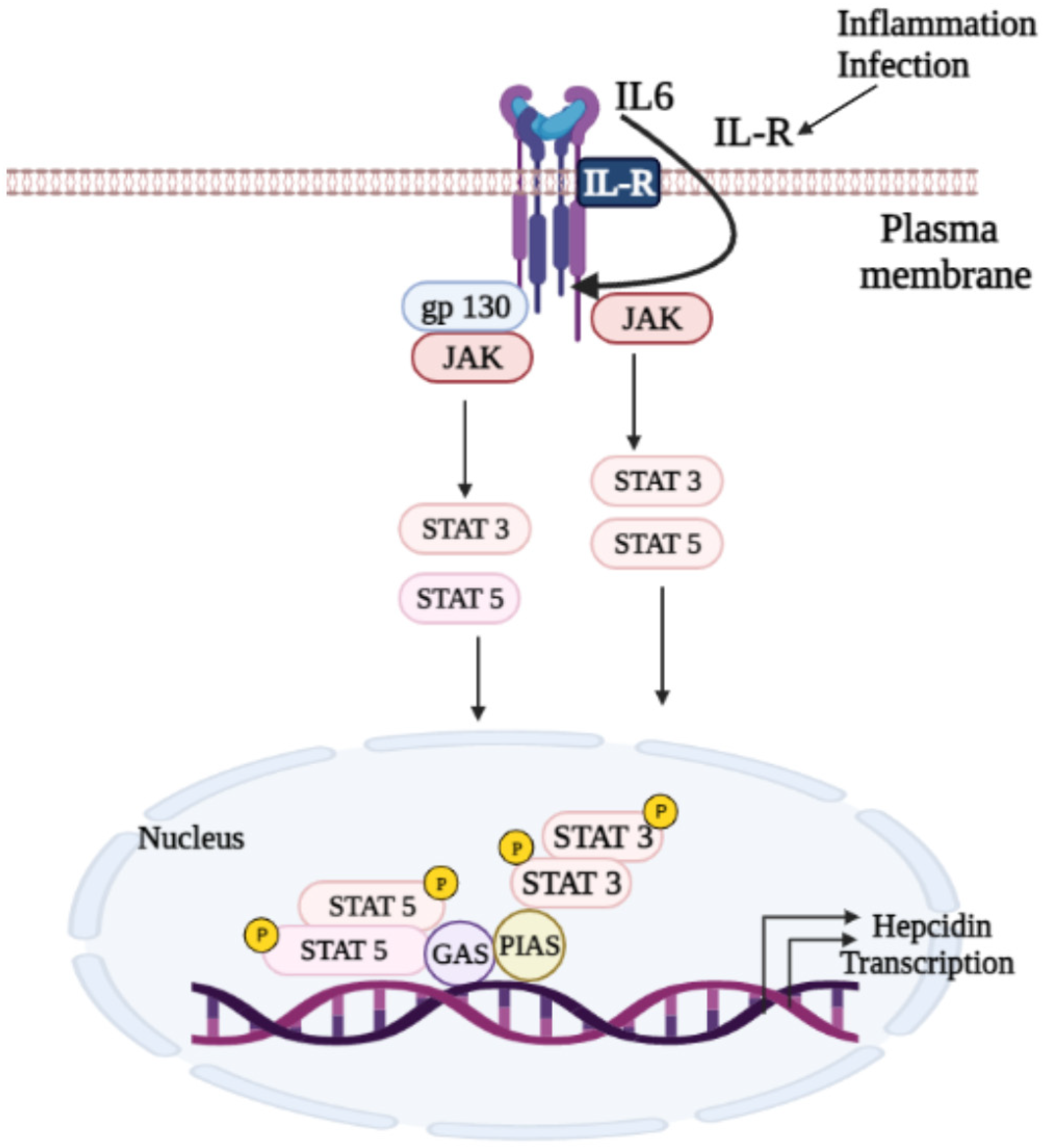
The JAK/STAT signaling pathway is one of the most important signaling cascades that regulates various cellular biological activities, including cell growth, differentiation, and hematopoiesis [79,123]. There are four members of the JAK family: JAK1, JAK2, JAK3, and tyrosine kinase 2 (TYK2) [124,125]. JAKs activate their downstream targets, STATs, a family of transcription factors consisting of seven members in mammals: STATs 1–4, STAT5A, STAT5B, and STAT6 [124]. Phosphorylation of STAT by JAK or Src kinases give rise to STAT dimerization and nuclear translocation to enhance target gene transcription (see Figure 4) [126].

Biological Roles of JAK/STAT Signaling Pathway

Many studies have reported that JAK/STAT signaling is aberrantly activated in hepatocellular carcinoma (HCC), dysregulating their downstream target genes associated with proliferation, immune, invasion, and metastasis [127,128]. Hepcidin transcription is regulated by BMP/SMAD and JAK/STAT pathways in response to inflammatory mediators and erythropoietic pathways [129,130]. The JAK/STAT signaling pathway is crucial in inflammation-induced hepcidin expression [127]. The discovery of JAK2 as a vital mediator of IE in β-thalassemia suggests that the use of small organic molecules, such as desferrioxamine (DFO) and deferasirox (DFX) chelators, for iron depletion in HH patients and JAK2 inhibition may reduce IE [131]. The reduced erythropoiesis indirectly increases serum hepcidin, reducing intestinal iron absorption and overload [39].

6. TGF-β/SMAD Signaling

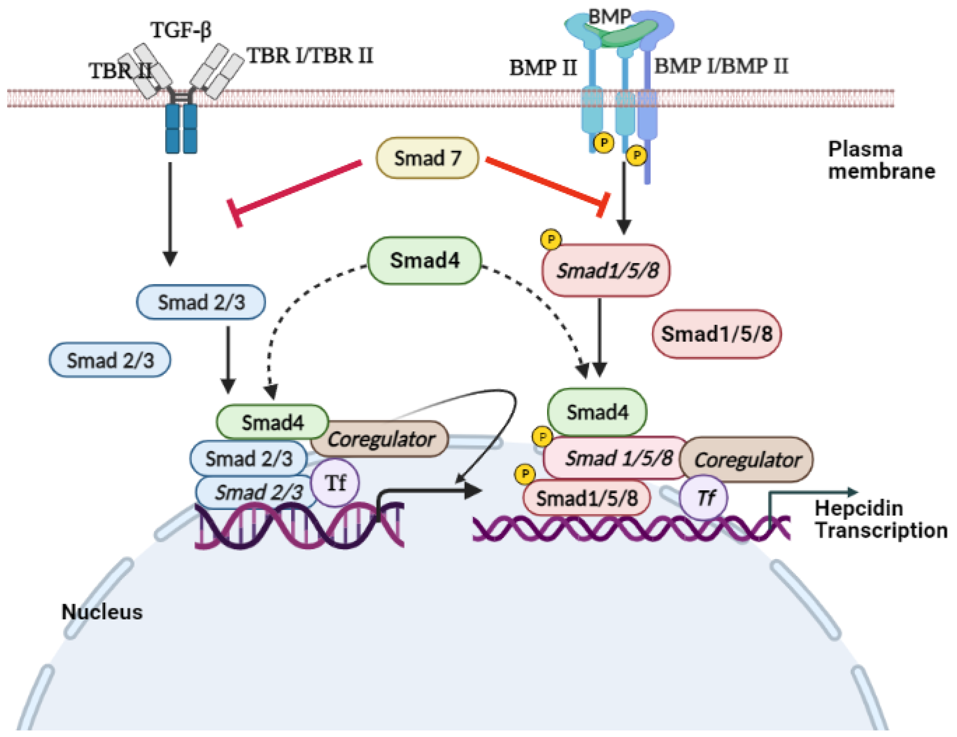
SMADs are proteins that are activated by the transforming growth factor β (TGF-β), BMP signaling, to mediate cell proliferation and differentiation [132,133]. Endosome-associated Fab1 (yeast orthologue of PIKfyve, YOTB, vesicle transport protein (Vac1), and EEA1 (FYVE zinc finger domain)-domain protein (endofin)) influences hepcidin expression by regulating SMAD1/5/8 phosphorylation [134]. STAT and SMAD signaling regulate hepcidin expression [135]. Ablation of SMAD4, specifically in the liver, triggers an iron overload in multiple organs due to decreased levels of liver hepcidin [136]. However, SMAD7 acts as an effective inhibitor of hepcidin mRNA expression through a negative regulation effect on TGF-β and BMP/SMAD signaling [137,138]. TGF-β is the prototypical ligand of the TGF-β superfamily, which signals during activation of serine/threonine receptor kinases. This superfamily is subdivided into the TGFβ/activin branch and BMP/growth and differentiation factor (GDF) branch.
TGF-β is expressed in most cell types and translated into a proprotein that is proteolytically cleaved into a noncovalently linked mature TGF-β and latency-associated protein (LAP) [139,140]. The active TGF-β ligand is a 25 kDa dimer, covalently linked by bisulfide bonds between cysteine residues of each monomeric peptide [139,140].
Various mechanisms are used to regulate the bioavailability of TGF-β in vivo. Once the bioavailable TGF-β reaches the target cell’s surface, it bonds with the homodimer of TGF-β type II receptor (TβRII) [141]. The TGF-β–TβRII complex provides a structural interface that forms a stable complex with the homodimer of the TGF-β type I receptor (TβRI) [132]. Subsequently, the active receptor–ligand complex is a heterotetrametric complex composed of TGFβ dimer and homodimer of TβRII and TβRI. In the active receptor complex, TβRII is constitutively activated and stimulates the transphosphorylation of TβRI [132,142]. In the TGF-β pathway, SMAD2 and SMAD3 are receptor-regulated effector proteins (R-SMADs) phosphorylated by activated TβRI on the C-terminal SSXS motif, leading to the nuclear accumulation of R-SMAD [132].
The activated receptor complex bound to the ligand is internalized by endocytosis [143]. Internalization of cell surface receptors can occur through clathrin-mediated or caveolae-mediated endocytosis [144].
Upon ligand stimulation, the SMADs accumulate in the nucleus as R-SMAD/CO-SMAD complex, leading to a decrease in their nuclear export rate [145,146]. The SMAD complex binds to DNA with other transcription factors and interacts with the general transcription machinery to regulate the expression of target genes (Figure 5) [147].
Signaling pathways such as STAT and SMAD regulate the expression of hepcidin. Therefore, it is hypothesized that STAT and SMAD could be dephosphorylated in thalassemia cases, including HbE/β-thalassemia patients, resulting in hepcidin downregulation with the presence of iron accumulation.

7. Conclusions

In summary, iron homeostasis dysregulation has a dominant physiological effect on transfusion-dependent and transfusion-independent β-thalassemia patients. Thus, understanding the expression of hepcidin and its regulation in β-thalassemia patients is vital in developing rational therapeutic interventions to provide safe, effective, and lifelong treatment options for their management. Therefore, recovery of hepcidin in β-thalassemia patients through the activation of STAT 3, STAT 5, SMAD 1/5/8, and SMAD 4 signaling could be a potential therapeutic target for managing iron overload (Figure 6). Therefore, it is highly recommended for future preclinical and clinical studies to evaluate the related risks and benefits of hepcidin-targeted treatment approaches.

Author Contributions

Conceptualization, H.A.N.A.-J. and H.K.M.S.; literature search, H.K.M.S.; writing original draft. Supervision was provided by H.A.N.A.-J., H.K.M.S.; review and editing, H.A.N.A.-J., A.A.A.R., A.S.b.A.G., W.R.W.T., I.I., M.F.J., A.S.A.-W. All authors have read and agreed to the published version of the manuscript.

Funding

The manuscript was funded by Universiti Sultan Zainal Abidin (grant no. UniSZA/2017/DPU/41(R0018-R3450)).

Institutional Review Board Statement

Not applicable.

Informed Consent Statement

Not applicable.

Data Availability Statement

Not applicable.

Conflicts of Interest

The authors declare no conflict of interest.

References

  1. Ayyash, H.; Sirdah, M. Hematological and biochemical evaluation of β-thalassemia major (βTM) patients in Gaza Strip: A cross-sectional study. IJHS 2018, 12, 18. [Google Scholar]
  2. Fibach, E.; Rachmilewitz, E.A. Pathophysiology and treatment of patients with beta-thalassemia—An update. F1000Research 2017, 6, 1–12. [Google Scholar] [CrossRef] [Green Version]
  3. Aziz, N.; Taib, W.; Kharolazaman, N.; Ismail, I.; Al-Jamal, H.; Jamil, W.; Esa, E.; Ibrahim, H. Evidence of New Intragenic HBB Haplotypes Model for the Prediction of Beta-Thalassemia in the Malaysian Population. Res. Sq. 2021; preprint. [Google Scholar] [CrossRef]
  4. Bender, M.; Yusuf, C.; Davis, T.; Dorley, M.C.; del Pilar Aguinaga, M.; Ingram, A.; Chan, M.S.; Ubaike, J.C.; Hassell, K.; Ojodu, J. Newborn Screening Practices and Alpha-Thalassemia Detection—United States, 2016. Morb. Mortal. Wkly. Rep. 2020, 69, 1269. [Google Scholar] [CrossRef]
  5. Wong, L.P.; George, E.; Tan, J.-A.M.A. Public perceptions and attitudes toward thalassaemia: Influencing factors in a multi-racial population. BMC Public Health 2011, 11, 193. [Google Scholar] [CrossRef] [Green Version]
  6. Galanello, R.; Origa, R. Beta-thalassemia. Orphanet J. Rare Dis. 2010, 5, 11. [Google Scholar] [CrossRef] [Green Version]
  7. Thein, S.L. Molecular basis of β thalassemia and potential therapeutic targets. Blood Cell Mol. Dis. 2018, 70, 54–65. [Google Scholar] [CrossRef] [PubMed]
  8. Abdullah, U.Y.; Ibrahim, H.M.; Mahmud, N.B.; Salleh, M.Z.; Teh, L.K.; Noorizhab, M.N.F.b.; Zilfalil, B.A.; Jassim, H.M.; Wilairat, P.; Fucharoen, S. Genotype-Phenotype Correlation of β-Thalassemia in Malaysian Population: Toward Effective Genetic Counseling. Hemoglobin 2020, 44, 184–189. [Google Scholar] [CrossRef] [PubMed]
  9. Pani, K.; Sharma, S.; Murari, M.; Yadav, M.; Phadke, S.; Agarwal, S. Clinico-hematological Profile of Hb E-β Thalassemia-Prospective Analysis in a tertiary Care Centre. J. Assoc. Phys. India 2018, 66, 42–45. [Google Scholar]
  10. Fucharoen, S.; Weatherall, D.J. The hemoglobin E thalassemias. Cold Spring Harb. Perspect. Med 2012, 2, a011734. [Google Scholar] [CrossRef]
  11. Olivieri, N.F.; Pakbaz, Z.; Vichinsky, E. Hb E/beta-thalassaemia: A common & clinically diverse disorder. Indian J. Med. Res. 2011, 134, 522. [Google Scholar]
  12. Abbaspour, N.; Hurrell, R.; Kelishadi, R. Review on iron and its importance for human health. J. Res. Med. Sci. 2014, 19, 164. [Google Scholar]
  13. Cohen-Solal, A.; Leclercq, C.; Deray, G.; Lasocki, S.; Zambrowski, J.-J.; Mebazaa, A.; de Groote, P.; Damy, T.; Galinier, M. Iron deficiency: An emerging therapeutic target in heart failure. Heart 2014, 100, 1414–1420. [Google Scholar] [CrossRef] [PubMed] [Green Version]
  14. Soetan, K.; Olaiya, C.; Oyewole, O. The importance of mineral elements for humans, domestic animals and plants—A review. Afr. J. Food. Sci. 2010, 4, 200–222. [Google Scholar]
  15. Evstatiev, R.; Gasche, C. Iron sensing and signalling. Gut 2012, 61, 933–952. [Google Scholar] [CrossRef] [PubMed]
  16. Kurz, T.; Eaton, J.W.; Brunk, U.T. The role of lysosomes in iron metabolism and recycling. Int. J. Biochem. Cell Biol 2011, 43, 1686–1697. [Google Scholar] [CrossRef] [PubMed] [Green Version]
  17. Halliwell, B. Biochemical mechanisms accounting for the toxic action of oxygen on living organisms: The key role of superoxide dismutase. Cell Biol. Int. 1978, 2, 113–128. [Google Scholar] [CrossRef]
  18. Mastrogiannaki, M.; Matak, P.; Keith, B.; Simon, M.C.; Vaulont, S.; Peyssonnaux, C. HIF-2α, but not HIF-1α, promotes iron absorption in mice. J. Clin. Investig. 2009, 119, 1159–1166. [Google Scholar] [CrossRef] [PubMed]
  19. Mackenzie, B.; Garrick, M.D. Iron Imports. II. Iron uptake at the apical membrane in the intestine. Am. J. Physiol. Gastrointest. Liver Physiol. 2005, 289, G981–G986. [Google Scholar] [CrossRef] [PubMed] [Green Version]
  20. Rahfiludin, M.Z.; Arso, S.P.; Joko, T.; Asna, A.F.; Murwani, R.; Hidayanti, L. Plant-based Diet and Iron Deficiency Anemia in Sundanese Adolescent Girls at Islamic Boarding Schools in Indonesia. J. Nutr. Metab. 2021, 2021, 6469883. [Google Scholar] [CrossRef]
  21. Timoshnikov, V.A.; Kobzeva, T.V.; Polyakov, N.E.; Kontoghiorghes, G.J. Redox interactions of vitamin C and iron: Inhibition of the pro-oxidant activity by deferiprone. Int. J. Mol. Sci. 2020, 21, 3967. [Google Scholar] [CrossRef]
  22. Zhao, K.; Wu, C.; Yao, Y.; Cao, L.; Zhang, Z.; Yuan, Y.; Wang, Y.; Pei, R.; Chen, J.; Hu, X. Ceruloplasmin inhibits the production of extracellular hepatitis B virions by targeting its middle surface protein. J. Gen. Virol. 2017, 98, 1410–1421. [Google Scholar] [CrossRef]
  23. Al Bratty, M.; Alhazmi, H.A.; Javed, S.A.; Rehman, Z.U.; Najmi, A.; El-Sharkawy, K.A. Rapid Screening and Estimation of Binding Constants for Interactions of Fe3+ with Two Metalloproteins, Apotransferrin and Transferrin, Using Affinity Mode of Capillary Electrophoresis. J. Spectrosc. 2021, 2021, 6987454. [Google Scholar] [CrossRef]
  24. Olaniyan, M.F.; Adepoju, D.B. Assessment of plasma iron, transferrin alanine, and aspartate transaminase in amoxicillin overdose supplemented with raw cucumber juice. J. Health Res. Rev. 2019, 6, 17. [Google Scholar] [CrossRef]
  25. Ganz, T. Hepcidin, a key regulator of iron metabolism and mediator of anemia of inflammation. Blood 2003, 102, 783–788. [Google Scholar] [CrossRef] [PubMed] [Green Version]
  26. Musumeci, M.; Maccari, S.; Massimi, A.; Stati, T.; Sestili, P.; Corritore, E.; Pastorelli, A.; Stacchini, P.; Marano, G.; Catalano, L. Iron excretion in iron dextran-overloaded mice. Blood Transfus. 2014, 12, 485. [Google Scholar] [PubMed]
  27. Mercadante, C.J.; Prajapati, M.; Parmar, J.H.; Conboy, H.L.; Dash, M.E.; Pettiglio, M.A.; Herrera, C.; Bu, J.T.; Stopa, E.G.; Mendes, P. Gastrointestinal iron excretion and reversal of iron excess in a mouse model of inherited iron excess. Haematologica 2019, 104, 678. [Google Scholar] [CrossRef]
  28. Ofojekwu, M.-J.N.; Nnanna, O.U.; Okolie, C.E.; Odewumi, L.A.; Isiguzoro, I.O.; Lugos, M.D. Hemoglobin and serum iron concentrations in menstruating nulliparous women in Jos, Nigeria. Lab. Med. 2013, 44, 121–124. [Google Scholar] [CrossRef] [Green Version]
  29. Li, J.; Gao, Q.; Tian, S.; Chen, Y.; Ma, Y.; Huang, Z. Menstrual blood loss and iron nutritional status in female undergraduate students. Wei Sheng Yan Jiu J. Hyg. Res. 2011, 40, 204–205. [Google Scholar]
  30. Mishra, A.K.; Tiwari, A. Iron overload in Beta thalassaemia major and intermedia patients. Maedica 2013, 8, 328. [Google Scholar]
  31. Liu, J.; Sun, B.; Yin, H.; Liu, S. Hepcidin: A promising therapeutic target for iron disorders: A systematic review. Maedica 2016, 95. [Google Scholar] [CrossRef] [PubMed]
  32. Ganz, T.; Nemeth, E. Hepcidin and iron homeostasis. Biochim. Biophys. Acta (BBA)—Mol. Cell Res. 2012, 1823, 1434–1443. [Google Scholar] [CrossRef] [PubMed] [Green Version]
  33. Nemeth, E. Hepcidin in β-thalassemia. Ann. N. Y. Acad. Sci. 2010, 1202, 31. [Google Scholar] [CrossRef] [PubMed]
  34. Gardenghi, S.; Ramos, P.; Follenzi, A.; Rao, N.; Rachmilewitz, E.A.; Giardina, P.J.; Grady, R.W.; Rivella, S. Hepcidin and Hfe in iron overload in β-thalassemia. Ann. N. Y. Acad. Sci. 2010, 1202, 221. [Google Scholar] [CrossRef] [Green Version]
  35. Nemeth, E.; Roetto, A.; Garozzo, G.; Ganz, T.; Camaschella, C. Hepcidin is decreased in TFR2 hemochromatosis. Blood 2005, 105, 1803–1806. [Google Scholar] [CrossRef] [Green Version]
  36. Leecharoenkiat, K.; Lithanatudom, P.; Sornjai, W.; Smith, D.R. Iron dysregulation in beta-thalassemia. Asian. Pac. J. Trop. Med. 2016, 9, 1035–1043. [Google Scholar] [CrossRef] [Green Version]
  37. Hankins, J.S.; McCarville, M.B.; Loeffler, R.B.; Smeltzer, M.P.; Onciu, M.; Hoffer, F.A.; Li, C.-S.; Wang, W.C.; Ware, R.E.; Hillenbrand, C.M. R2* magnetic resonance imaging of the liver in patients with iron overload. Blood J. Am. Soc. Hematol. 2009, 113, 4853–4855. [Google Scholar] [CrossRef]
  38. Adamsky, K.; Weizer, O.; Amariglio, N.; Breda, L.; Harmelin, A.; Rivella, S.; Rachmilewitz, E.; Rechavi, G. Decreased hepcidin mRNA expression in thalassemic mice. Br. J. Hematol. 2004, 124, 123–124. [Google Scholar] [CrossRef]
  39. Gardenghi, S.; Marongiu, M.F.; Ramos, P.; Guy, E.; Breda, L.; Chadburn, A.; Liu, Y.; Amariglio, N.; Rechavi, G.; Rachmilewitz, E.A. Ineffective erythropoiesis in β-thalassemia is characterized by increased iron absorption mediated by down-regulation of hepcidin and up-regulation of ferroportin. Blood J. Am. Soc. Hematol. 2007, 109, 5027–5035. [Google Scholar] [CrossRef]
  40. Chowdhury, P.K.; Saha, M.; Basu, A.; Chowdhury, D.; Jena, R.k. Profile of Iron Overload in Nontransfusion Dependent Hb E Beta Thalassaemia Patients-Is It Different? Blood 2015, 126, 4557. [Google Scholar] [CrossRef]
  41. Chowdhury, P.; Saha, M.; Karpurkayastha, S.; Chowdhury, D.; Jena, R. Estimation of Iron Overload-Implications of its Non-linear Correlation. J. Blood Disord. Transfus. 2016, 7, 2. [Google Scholar] [CrossRef]
  42. Collins, H.L. The role of iron in infections with intracellular bacteria. Immunol. Lett. 2003, 85, 193–195. [Google Scholar] [CrossRef]
  43. Zimmermann, M.B.; Fucharoen, S.; Winichagoon, P.; Sirankapracha, P.; Zeder, C.; Gowachirapant, S.; Judprasong, K.; Tanno, T.; Miller, J.L.; Hurrell, R.F. Iron metabolism in heterozygotes for hemoglobin E (HbE), α-thalassemia 1, or β-thalassemia and in compound heterozygotes for HbE/β-thalassemia. Am. J. Clin. Nutr. 2008, 88, 1026–1031. [Google Scholar] [CrossRef] [PubMed] [Green Version]
  44. Shander, A.; Cappellini, M.; Goodnough, L. Iron overload and toxicity: The hidden risk of multiple blood transfusions. Vox. Sang. 2009, 97, 185–197. [Google Scholar] [CrossRef]
  45. Sharma, A.; Arora, E.; Singh, H. Hypersensitivity reaction with deferasirox. J. Pharmacol. Pharmacother. 2015, 6, 105. [Google Scholar] [CrossRef] [Green Version]
  46. Oudit, G.Y.; Sun, H.; Trivieri, M.G.; Koch, S.E.; Dawood, F.; Ackerley, C.; Yazdanpanah, M.; Wilson, G.J.; Schwartz, A.; Liu, P.P. L-type Ca 2+ channels provide a major pathway for iron entry into cardiomyocytes in iron-overload cardiomyopathy. Nat. Med. 2003, 9, 1187–1194. [Google Scholar] [CrossRef]
  47. Liuzzi, J.P.; Aydemir, F.; Nam, H.; Knutson, M.D.; Cousins, R.J. Zip14 (Slc39a14) mediates non-transferrin-bound iron uptake into cells. Proc. Natl. Acad. Sci. USA 2006, 103, 13612–13617. [Google Scholar] [CrossRef] [Green Version]
  48. Cabantchik, Z.I. Labile iron in cells and body fluids: Physiology, pathology, and pharmacology. Front. Pharmacol. 2014, 5, 45. [Google Scholar] [CrossRef] [Green Version]
  49. Murphy, C.J.; Oudit, G.Y. Iron-overload cardiomyopathy: Pathophysiology, diagnosis, and treatment. J. Card. Fail. 2010, 16, 888–900. [Google Scholar] [CrossRef]
  50. Bresgen, N.; Eckl, P.M. Oxidative stress and the homeodynamics of iron metabolism. Biomolecules 2015, 5, 808–847. [Google Scholar] [CrossRef]
  51. Olivieri, N.F.; Brittenham, G.M. Iron-chelating therapy and the treatment of thalassemia. Blood J. Am. Soc. Hematol. 1997, 89, 739–761. [Google Scholar] [CrossRef] [Green Version]
  52. Zhuang, S.; Demirs, J.T.; Kochevar, I.E. p38 mitogen-activated protein kinase mediates bid cleavage, mitochondrial dysfunction, and caspase-3 activation during apoptosis induced by singlet oxygen but not by hydrogen peroxide. Int. J. Biol. Chem. 2000, 275, 25939–25948. [Google Scholar] [CrossRef] [Green Version]
  53. Lin, T.; Mak, N.; Yang, M. MAPK regulate p53-dependent cell death induced by benzo [a] pyrene: Involvement of p53 phosphorylation and acetylation. Toxicology 2008, 247, 145–153. [Google Scholar] [CrossRef]
  54. BORGNA-PIGNATTI, C.; Rugolotto, S.; De Stefano, P.; Piga, A.; Di Gregorio, F.; Gamberini, M.R.; Sabato, V.; Melevendi, C.; Cappellini, M.D.; Verlato, G. Survival and disease complications in thalassemia major. Ann. N. Y. Acad. Sci. 1998, 850, 227–231. [Google Scholar] [CrossRef]
  55. Farmakis, D.; Giakoumis, A.; Aessopos, A.; Polymeropoulos, E. Pathogenetic aspects of immune deficiency associated with ß thalassemia. Med. Sci. Monit. 2003, 9, RA19–RA22. [Google Scholar] [PubMed]
  56. Lithanatudom, P.; Wannatung, T.; Leecharoenkiat, A.; Svasti, S.; Fucharoen, S.; Smith, D.R. Enhanced activation of autophagy in β-thalassemia/Hb E erythroblasts during erythropoiesis. Ann. Hematol. 2011, 90, 747–758. [Google Scholar] [CrossRef] [PubMed]
  57. Sornjai, W.; Jaratsittisin, J.; Khungwanmaythawee, K.; Svasti, S.; Fucharoen, S.; Lithanatudom, P.; Smith, D.R. Dysregulation of ferroportin gene expression in β 0-thalassemia/Hb E disease. Ann. Hematol. 2016, 95, 387–396. [Google Scholar] [CrossRef]
  58. Tanno, T.; Bhanu, N.V.; Oneal, P.A.; Goh, S.-H.; Staker, P.; Lee, Y.T.; Moroney, J.W.; Reed, C.H.; Luban, N.L.; Wang, R.-H. High levels of GDF15 in thalassemia suppress expression of the iron regulatory protein hepcidin. Nat. Med. 2007, 13, 1096–1101. [Google Scholar] [CrossRef] [PubMed]
  59. Tanno, T.; Noel, P.; Miller, J.L. Growth differentiation factor 15 in erythroid health and disease. Curr. Opin. Hematol. 2010, 17, 184. [Google Scholar] [CrossRef] [PubMed]
  60. Casanovas, G.; Spasić, M.V.; Casu, C.; Rivella, S.; Strelau, J.; Unsicker, K.; Muckenthaler, M.U. The murine growth differentiation factor 15 is not essential for systemic iron homeostasis in phlebotomized mice. Haematologica 2013, 98, 444. [Google Scholar] [CrossRef] [PubMed] [Green Version]
  61. Saad, H.K.M.; Taib, W.R.W.; Ismail, I.; Johan, M.F.; Al-Wajeeh, A.S.; Al-jamal, H.A.N. Reduced hepcidin expression enhances iron overload in patients with HbE/β-thalassemia: A comparative cross-sectional study. Exp. Ther. Med. 2021, 22, 1–8. [Google Scholar] [CrossRef]
  62. Kattamis, A.; Papassotiriou, I.; Palaiologou, D.; Apostolakou, F.; Galani, A.; Ladis, V.; Sakellaropoulos, N.; Papanikolaou, G. The effects of erythropoetic activity and iron burden on hepcidin expression in patients with thalassemia major. Haematologica 2006, 91, 809–812. [Google Scholar]
  63. Jones, E.; Pasricha, S.-R.; Allen, A.; Evans, P.; Fisher, C.A.; Wray, K.; Premawardhena, A.; Bandara, D.; Perera, A.; Webster, C. Hepcidin is suppressed by erythropoiesis in hemoglobin E β-thalassemia and β-thalassemia trait. Blood J. Am. Soc. Hematol. 2015, 125, 873–880. [Google Scholar] [CrossRef] [Green Version]
  64. Pak, M.; Lopez, M.A.; Gabayan, V.; Ganz, T.; Rivera, S. Suppression of hepcidin during anemia requires erythropoietic activity. Blood 2006, 108, 3730–3735. [Google Scholar] [CrossRef] [PubMed]
  65. Weizer-Stern, O.; Adamsky, K.; Amariglio, N.; Levin, C.; Koren, A.; Breuer, W.; Rachmilewitz, E.; Breda, L.; Rivella, S.; Ioav Cabantchik, Z. Downregulation of hepcidin and haemojuvelin expression in the hepatocyte cell-line HepG2 induced by thalassaemic sera. Br. J. Hematol. 2006, 135, 129–138. [Google Scholar] [CrossRef] [PubMed]
  66. Tanno, T.; Porayette, P.; Sripichai, O.; Noh, S.-J.; Byrnes, C.; Bhupatiraju, A.; Lee, Y.T.; Goodnough, J.B.; Harandi, O.; Ganz, T. Identification of TWSG1 as a second novel erythroid regulator of hepcidin expression in murine and human cells. Blood J. Am. Soc. Hematol. 2009, 114, 181–186. [Google Scholar] [CrossRef] [PubMed]
  67. Kautz, L.; Jung, G.; Du, X.; Gabayan, V.; Chapman, J.; Nasoff, M.; Nemeth, E.; Ganz, T. Erythroferrone contributes to hepcidin suppression and iron overload in a mouse model of β-thalassemia. Blood J. Am. Soc. Hematol. 2015, 126, 2031–2037. [Google Scholar] [CrossRef] [Green Version]
  68. Muckenthaler, M.U.; Rivella, S.; Hentze, M.W.; Galy, B. A red carpet for iron metabolism. Cell J. 2017, 168, 344–361. [Google Scholar] [CrossRef] [Green Version]
  69. Pinto, J.P.; Ribeiro, S.; Pontes, H.; Thowfeequ, S.; Tosh, D.; Carvalho, F.; Porto, G. Erythropoietin mediates hepcidin expression in hepatocytes through EPOR signaling and regulation of C/EBPα. Blood J. Am. Soc. Hematol. 2008, 111, 5727–5733. [Google Scholar] [CrossRef]
  70. Liu, Q.; Davidoff, O.; Niss, K.; Haase, V.H. Hypoxia-inducible factor regulates hepcidin via erythropoietin-induced erythropoiesis. J. Clin. Investig. 2012, 122, 4635–4644. [Google Scholar] [CrossRef] [Green Version]
  71. Jiang, F.; Yu, W.-J.; Wang, X.-H.; Tang, Y.-T.; Guo, L.; Jiao, X.-Y. Regulation of hepcidin through GDF-15 in cancer-related anemia. Clin. Chim. Acta 2014, 428, 14–19. [Google Scholar] [CrossRef]
  72. YAŞAR, C.A.; Tarkun, P.; Ateşoğlu, E.B.; Eraldemir, F.C.; Özsoy, Ö.D.; Demirsoy, E.T.; Mehtap, Ö.; Gedük, A.; Hacihanifioğlu, A. The role of hepcidin, GDF15, and mitoferrin-1 in iron metabolism of polycythemia vera and essential thrombocytosis patients. Turk. J. Med. Sci. 2019, 49, 74–80. [Google Scholar]
  73. Osada, M.; Park, H.L.; Park, M.J.; Liu, J.-W.; Wu, G.; Trink, B.; Sidransky, D. A p53-type response element in the GDF15 promoter confers high specificity for p53 activation. Biochem. Biophys. Res. Commun. 2007, 354, 913–918. [Google Scholar] [CrossRef] [Green Version]
  74. Zimmers, T.A.; Jin, X.; Hsiao, E.C.; McGrath, S.A.; Esquela, A.F.; Koniaris, L.G. Growth differentiation factor-15/macrophage inhibitory cytokine-1 induction after kidney and lung injury. Shock 2005, 23, 543–548. [Google Scholar] [PubMed]
  75. Jenkitkasemwong, S.; Broderius, M.; Nam, H.; Prohaska, J.R.; Knutson, M.D. Anemic copper-deficient rats, but not mice, display low hepcidin expression and high ferroportin levels. J. Nutr. 2010, 140, 723–730. [Google Scholar] [CrossRef] [PubMed]
  76. Finkenstedt, A.; Widschwendter, A.; Brasse-Lagnel, C.; Theurl, I.; Hubalek, M.; Dieplinger, H.; Tselepis, C.; Ward, D.; Vogel, W.; Zoller, H. Hepcidin is correlated to soluble hemojuvelin but not to increased GDF15 during pregnancy. Blood Cell Mol. Dis. 2012, 48, 233–237. [Google Scholar] [CrossRef] [PubMed]
  77. Vokurka, M.; Krijt, J.; Šulc, K.; Nečas, E. Hepcidin mRNA levels in mouse liver respond to inhibition of erythropoiesis. Physiol. Res. 2006, 55, 667–674. [Google Scholar]
  78. Kautz, L.; Jung, G.; Valore, E.V.; Rivella, S.; Nemeth, E.; Ganz, T. Identification of erythroferrone as an erythroid regulator of iron metabolism. Nat. Genet. 2014, 46, 678–684. [Google Scholar] [CrossRef] [Green Version]
  79. Patel, N.; Varghese, J.; Masaratana, P.; Latunde-Dada, G.O.; Jacob, M.; Simpson, R.J.; McKie, A.T. The transcription factor ATOH 8 is regulated by erythropoietic activity and regulates HAMP transcription and cellular pSMAD 1, 5, 8 levels. Br. J. Hematol. 2014, 164, 586–596. [Google Scholar] [CrossRef] [Green Version]
  80. Wrighting, D.M.; Andrews, N.C. Interleukin-6 induces hepcidin expression through STAT3. Blood 2006, 108, 3204–3209. [Google Scholar] [CrossRef]
  81. Bayele, H.K.; Srai, S.K.S. Genetic variation in hepcidin expression and its implications for phenotypic differences in iron metabolism. Haematologica 2009, 94, 1185. [Google Scholar] [CrossRef] [Green Version]
  82. Andriopoulos Jr, B.; Corradini, E.; Xia, Y.; Faasse, S.A.; Chen, S.; Grgurevic, L.; Knutson, M.D.; Pietrangelo, A.; Vukicevic, S.; Lin, H.Y. BMP6 is a key endogenous regulator of hepcidin expression and iron metabolism. Nat. Genet. 2009, 41, 482–487. [Google Scholar] [CrossRef] [PubMed] [Green Version]
  83. Bridle, K.R.; Frazer, D.M.; Wilkins, S.J.; Dixon, J.L.; Purdie, D.M.; Crawford, D.H.; Subramaniam, V.N.; Powell, L.W.; Anderson, G.J.; Ramm, G.A. Disrupted hepcidin regulation in HFE-associated haemochromatosis and the liver as a regulator of body iron homoeostasis. Lancet 2003, 361, 669–673. [Google Scholar] [CrossRef]
  84. Zhou, X.Y.; Tomatsu, S.; Fleming, R.E.; Parkkila, S.; Waheed, A.; Jiang, J.; Fei, Y.; Brunt, E.M.; Ruddy, D.A.; Prass, C.E. HFE gene knockout produces mouse model of hereditary hemochromatosis. Proc. Natl. Acad. Sci. USA 1998, 95, 2492–2497. [Google Scholar] [CrossRef] [PubMed] [Green Version]
  85. Feder, J.; Gnirke, A.; Thomas, W.; Tsuchihashi, Z.; Ruddy, D.; Basava, A.; Dormishian, F.; Domingo, R.; Ellis, M.; Fullan, A. A novel MHC class I–like gene is mutated in patients with hereditary haemochromatosis. Nat. Genet. 1996, 13, 399–408. [Google Scholar] [CrossRef] [PubMed]
  86. Schmidt, P.J.; Toran, P.T.; Giannetti, A.M.; Bjorkman, P.J.; Andrews, N.C. The transferrin receptor modulates Hfe-dependent regulation of hepcidin expression. Cell Metab. 2008, 7, 205–214. [Google Scholar] [CrossRef] [Green Version]
  87. Lebrón, J.A.; Bennett, M.J.; Vaughn, D.E.; Chirino, A.J.; Snow, P.M.; Mintier, G.A.; Feder, J.N.; Bjorkman, P.J. Crystal structure of the hemochromatosis protein HFE and characterization of its interaction with transferrin receptor. Cell J. 1998, 93, 111–123. [Google Scholar] [CrossRef] [Green Version]
  88. Ramey, G.; Deschemin, J.-C.; Vaulont, S. Cross-talk between the mitogen activated protein kinase and bone morphogenetic protein/hemojuvelin pathways is required for the induction of hepcidin by holotransferrin in primary mouse hepatocytes. Haematologica 2009, 94, 765–772. [Google Scholar] [CrossRef] [Green Version]
  89. Kautz, L.; Meynard, D.; Besson-Fournier, C.; Darnaud, V.; Al Saati, T.; Coppin, H.; Roth, M.-P. BMP/Smad signaling is not enhanced in Hfe-deficient mice despite increased Bmp6 expression. Blood 2009, 114, 2515–2520. [Google Scholar] [CrossRef]
  90. Nili, M.; Shinde, U.; Rotwein, P. Soluble repulsive guidance molecule c/hemojuvelin is a broad spectrum bone morphogenetic protein (BMP) antagonist and inhibits both BMP2-and BMP6-mediated signaling and gene expression. Int. J. Biol. Chem. 2010, 285, 24783–24792. [Google Scholar] [CrossRef] [Green Version]
  91. Babitt, J.L.; Huang, F.W.; Wrighting, D.M.; Xia, Y.; Sidis, Y.; Samad, T.A.; Campagna, J.A.; Chung, R.T.; Schneyer, A.L.; Woolf, C.J. Bone morphogenetic protein signaling by hemojuvelin regulates hepcidin expression. Nat. Genet. 2006, 38, 531. [Google Scholar] [CrossRef]
  92. D’Alessio, F.; Hentze, M.W.; Muckenthaler, M.U. The hemochromatosis proteins HFE, TfR2, and HJV form a membrane-associated protein complex for hepcidin regulation. J. Hepatol. 2012, 57, 1052–1060. [Google Scholar] [CrossRef]
  93. Bayele, H.K.; McArdle, H.; Srai, S.K. Cis and trans regulation of hepcidin expression by upstream stimulatory factor. Blood 2006, 108, 4237–4245. [Google Scholar] [CrossRef] [Green Version]
  94. Mleczko-Sanecka, K.; Roche, F.; Rita da Silva, A.; Call, D.; D’Alessio, F.; Ragab, A.; Lapinski, P.E.; Ummanni, R.; Korf, U.; Oakes, C. Unbiased RNAi screen for hepcidin regulators links hepcidin suppression to proliferative Ras/RAF and nutrient-dependent mTOR signaling. Blood J. Am. Soc. Hematol. 2014, 123, 1574–1585. [Google Scholar] [CrossRef] [Green Version]
  95. Pandur, E.; Sipos, K.; Grama, L.; Nagy, J.; Poór, V.S.; Sétáló Jr, G.; Miseta, A.; Fekete, Z. Prohepcidin binds to the HAMP promoter and autoregulates its own expression. Biochem. J. 2013, 451, 301–311. [Google Scholar] [CrossRef] [Green Version]
  96. Du, X.; She, E.; Gelbart, T.; Truksa, J.; Lee, P.; Xia, Y.; Khovananth, K.; Mudd, S.; Mann, N.; Moresco, E.M.Y. The serine protease TMPRSS6 is required to sense iron deficiency. Science 2008, 320, 1088–1092. [Google Scholar] [CrossRef] [PubMed] [Green Version]
  97. Silvestri, L.; Pagani, A.; Nai, A.; De Domenico, I.; Kaplan, J.; Camaschella, C. The serine protease matriptase-2 (TMPRSS6) inhibits hepcidin activation by cleaving membrane hemojuvelin. Cell Metab. 2008, 8, 502–511. [Google Scholar] [CrossRef] [PubMed] [Green Version]
  98. Poggi, M.; Sorrentino, F.; Pugliese, P.; Smacchia, M.P.; Daniele, C.; Equitani, F.; Terlizzi, F.; Guitarrini, M.R.; Monti, S.; Maffei, L. Longitudinal changes of endocrine and bone disease in adults with β-thalassemia major receiving different iron chelators over 5 years. Ann. Hematol. 2016, 95, 757–763. [Google Scholar] [CrossRef] [PubMed]
  99. Farmaki, K.; Tzoumari, I.; Pappa, C. Oral chelators in transfusion-dependent thalassemia major patients may prevent or reverse iron overload complications. Blood Cell Mol. Dis. 2011, 47, 33–40. [Google Scholar] [CrossRef]
  100. Poggiali, E.; Cassinerio, E.; Zanaboni, L.; Cappellini, M.D. An update on iron chelation therapy. Blood Transfus. 2012, 10, 411. [Google Scholar]
  101. Schmidt, P.J.; Racie, T.; Westerman, M.; Fitzgerald, K.; Butler, J.S.; Fleming, M.D. Combination therapy with a T mprss6 RNA i-therapeutic and the oral iron chelator deferiprone additively diminishes secondary iron overload in a mouse model of β-thalassemia intermedia. Am. J. Hematol. 2015, 90, 310–313. [Google Scholar] [CrossRef] [PubMed] [Green Version]
  102. Sripichai, O.; Munkongdee, T.; Kumkhaek, C.; Svasti, S.; Winichagoon, P.; Fucharoen, S. Coinheritance of the different copy numbers of α-globin gene modifies severity of β-thalassemia/Hb E disease. Ann. Hematol. 2008, 87, 375–379. [Google Scholar] [CrossRef] [PubMed]
  103. Lucarelli, G.; Isgrò, A.; Sodani, P.; Gaziev, J. Hematopoietic stem cell transplantation in thalassemia and sickle cell anemia. Cold Spring Harb. Perspect. Med 2012, 2, a011825. [Google Scholar] [CrossRef] [PubMed]
  104. Camaschella, C.; Nai, A. Ineffective erythropoiesis and regulation of iron status in iron loading anaemias. Br. J. Hematol. 2016, 172, 512–523. [Google Scholar] [CrossRef] [Green Version]
  105. Gardenghi, S.; Ramos, P.; Roy, C.N.; Andrews, N.C.; Nemeth, E.; An, X.; Narla, M.; Ginzburg, Y.; Rachmilewitz, E.A.; Giardina, P. Hepcidin as a Therapeutic Tool to Limit Iron Overload and Improve Anemia In β-Thalassemia. Blood 2010, 116, 1009. [Google Scholar] [CrossRef]
  106. Wu, X.; Yung, L.-M.; Cheng, W.-H.; Yu, P.B.; Babitt, J.L.; Lin, H.Y.; Xia, Y. Hepcidin regulation by BMP signaling in macrophages is lipopolysaccharide dependent. PLoS ONE 2012, 7, e44622. [Google Scholar] [CrossRef]
  107. Nai, A.; Lidonnici, M.R.; Rausa, M.; Mandelli, G.; Pagani, A.; Silvestri, L.; Ferrari, G.; Camaschella, C. The second transferrin receptor regulates red blood cell production in mice. Blood J. Am. Soc. Hematol. 2015, 125, 1170–1179. [Google Scholar] [CrossRef] [Green Version]
  108. Finberg, K.E.; Heeney, M.M.; Campagna, D.R.; Aydınok, Y.; Pearson, H.A.; Hartman, K.R.; Mayo, M.M.; Samuel, S.M.; Strouse, J.J.; Markianos, K. Mutations in TMPRSS6 cause iron-refractory iron deficiency anemia (IRIDA). Nat. Genet. 2008, 40, 569–571. [Google Scholar] [CrossRef] [Green Version]
  109. Bou-Fakhredin, R.; Bazarbachi, A.-H.; Chaya, B.; Sleiman, J.; Cappellini, M.D.; Taher, A.T. Iron overload and chelation therapy in non-transfusion dependent thalassemia. Int. J. Mol. Sci. 2017, 18, 2778. [Google Scholar] [CrossRef] [Green Version]
  110. Casu, C.; Aghajan, M.; Oikonomidou, P.R.; Guo, S.; Monia, B.P.; Rivella, S. Combination of Tmprss6-ASO and the iron chelator deferiprone improves erythropoiesis and reduces iron overload in a mouse model of beta-thalassemia intermedia. Haematologica 2016, 101, e8. [Google Scholar] [CrossRef] [Green Version]
  111. Shimada, T.; Hasegawa, H.; Yamazaki, Y.; Muto, T.; Hino, R.; Takeuchi, Y.; Fujita, T.; Nakahara, K.; Fukumoto, S.; Yamashita, T. FGF-23 is a potent regulator of vitamin D metabolism and phosphate homeostasis. J. Bone Miner. Res. 2004, 19, 429–435. [Google Scholar] [CrossRef] [PubMed] [Green Version]
  112. Yokota, H.; Raposo, J.F.; Chen, A.; Jiang, C.; Ferreira, H.G. Evaluation of the role of FGF23 in mineral metabolism. Gene Regul. Syst. Biol. 2009, 3, GRSB-S2990. [Google Scholar] [CrossRef] [PubMed]
  113. Hu, M.C.; Shiizaki, K.; Kuro-o, M.; Moe, O.W. Fibroblast growth factor 23 and Klotho: Physiology and pathophysiology of an endocrine network of mineral metabolism. Annu. Rev. Physiol. 2013, 75, 503–533. [Google Scholar] [CrossRef] [Green Version]
  114. Ramon, I.; Kleynen, P.; Body, J.-J.; Karmali, R. Fibroblast growth factor 23 and its role in phosphate homeostasis. Eur. J. Endocrinol. 2010, 162, 1. [Google Scholar] [CrossRef] [Green Version]
  115. Lewerin, C.; Ljunggren, Ö.; Nilsson-Ehle, H.; Karlsson, M.K.; Herlitz, H.; Lorentzon, M.; Ohlsson, C.; Mellström, D. Low serum iron is associated with high serum intact FGF23 in elderly men: The Swedish MrOS study. Bone Res. 2017, 98, 1–8. [Google Scholar] [CrossRef]
  116. Kanbay, M.; Vervloet, M.; Cozzolino, M.; Siriopol, D.; Covic, A.; Goldsmith, D.; Solak, Y. Novel faces of fibroblast growth factor 23 (FGF23): Iron deficiency, inflammation, insulin resistance, left ventricular hypertrophy, proteinuria and acute kidney injury. Calcif. Tissue Int. 2017, 100, 217–228. [Google Scholar] [CrossRef]
  117. Aschemeyer, S.; Qiao, B.; Stefanova, D.; Valore, E.V.; Sek, A.C.; Ruwe, T.A.; Vieth, K.R.; Jung, G.; Casu, C.; Rivella, S. Structure-function analysis of ferroportin defines the binding site and an alternative mechanism of action of hepcidin. Blood J. Am. Soc. Hematol. 2018, 131, 899–910. [Google Scholar] [CrossRef]
  118. Nemeth, E.; Tuttle, M.S.; Powelson, J.; Vaughn, M.B.; Donovan, A.; Ward, D.M.; Ganz, T.; Kaplan, J. Hepcidin regulates cellular iron efflux by binding to ferroportin and inducing its internalization. Science 2004, 306, 2090–2093. [Google Scholar] [CrossRef] [PubMed] [Green Version]
  119. Saki, F.; Salehifar, A.; Kassaee, S.R.; Omrani, G.R. Association of vitamin D and FGF23 with serum ferritin in hypoparathyroid thalassemia: A case control study. BMC Nephrol. 2020, 21, 482. [Google Scholar] [CrossRef]
  120. Charytan, D.M.; Pai, A.B.; Chan, C.T.; Coyne, D.W.; Hung, A.M.; Kovesdy, C.P.; Fishbane, S. Considerations and challenges in defining optimal iron utilization in hemodialysis. J. Am. Soc. Nephrol. J. 2015, 26, 1238–1247. [Google Scholar] [CrossRef] [PubMed] [Green Version]
  121. Pasricha, S.-R.; Frazer, D.M.; Bowden, D.K.; Anderson, G.J. Transfusion suppresses erythropoiesis and increases hepcidin in adult patients with β-thalassemia major: A longitudinal study. Blood J. Am. Soc. Hematol. 2013, 122, 124–133. [Google Scholar] [CrossRef] [Green Version]
  122. Hendy, O.M.; Allam, M.; Allam, A.; Attia, M.H.; El Taher, S.; Eldin, M.M.; Ali, A. Hepcidin levels and iron status in beta-thalassemia major patients with hepatitis C virus infection. Egypt. J. Immunol. 2010, 17, 33–44. [Google Scholar]
  123. Al-Jamal, H.A.N.; Johan, M.F.; Jusoh, S.A.M.; Ismail, I.; Taib, W.R.W. Re-expression of bone marrow proteoglycan-2 by 5-azacytidine is associated with STAT3 inactivation and sensitivity response to imatinib in resistant CML cells. Asian Pac. J. Cancer Prev. APJCP 2018, 19, 1585. [Google Scholar]
  124. Leonard, W.J.; O’Shea, J.J. Jaks and STATs: Biological implications. Annu. Rev. Immunol. 1998, 16, 293–322. [Google Scholar] [CrossRef] [PubMed] [Green Version]
  125. Minegishi, Y.; Saito, M.; Morio, T.; Watanabe, K.; Agematsu, K.; Tsuchiya, S.; Takada, H.; Hara, T.; Kawamura, N.; Ariga, T. Human tyrosine kinase 2 deficiency reveals its requisite roles in multiple cytokine signals involved in innate and acquired immunity. Immunity 2006, 25, 745–755. [Google Scholar] [CrossRef] [PubMed] [Green Version]
  126. Levy, D.E.; Darnell, J. Stats: Transcriptional control and biological impact. Nat. Rev. Mol. Cell Biol. 2002, 3, 651–662. [Google Scholar] [CrossRef] [PubMed]
  127. Hin Tang, J.J.; Hao Thng, D.K.; Lim, J.J.; Toh, T.B. JAK/STAT signaling in hepatocellular carcinoma. Hepat. Oncol. 2020, 7, HEP18. [Google Scholar] [CrossRef] [PubMed] [Green Version]
  128. Han, T.; Wan, Y.; Wang, J.; Zhao, P.; Yuan, Y.; Wang, L.; She, Y.; Broering, R.; Lu, M.; Ye, L. Set7 Facilitates Hepatitis C Virus Replication via Enzymatic Activity–Dependent Attenuation of the IFN-Related Pathway. J. Immunol. 2015, 194, 2757–2768. [Google Scholar] [CrossRef] [PubMed] [Green Version]
  129. Maliken, B.D.; Nelson, J.E.; Kowdley, K.V. The hepcidin circuits act: Balancing iron and inflammation. Hepatology 2011, 53, 1764–1766. [Google Scholar] [CrossRef] [Green Version]
  130. Darshan, D.; Anderson, G.J. Interacting signals in the control of hepcidin expression. Biometals 2009, 22, 77–87. [Google Scholar] [CrossRef] [PubMed]
  131. Melchiori, L.; Gardenghi, S.; Rivella, S. β-thalassemia: HiJAKing ineffective erythropoiesis and iron overload. Adv. Hematol. 2010, 2010, 938640. [Google Scholar] [CrossRef] [Green Version]
  132. Shi, Y.; Massagué, J. Mechanisms of TGF-β signaling from cell membrane to the nucleus. Cell J. 2003, 113, 685–700. [Google Scholar] [CrossRef] [Green Version]
  133. Massagué, J. TGF-β signaling in development and disease. FEBS Lett. 2012, 586, 1833. [Google Scholar] [CrossRef] [PubMed] [Green Version]
  134. Goh, J.B.; Wallace, D.F.; Hong, W.; Subramaniam, V.N. Endofin, a novel BMP-SMAD regulator of the iron-regulatory hormone, hepcidin. Sci. Rep. 2015, 5, 13986. [Google Scholar] [CrossRef] [PubMed] [Green Version]
  135. Wang, R.-H.; Li, C.; Xu, X.; Zheng, Y.; Xiao, C.; Zerfas, P.; Cooperman, S.; Eckhaus, M.; Rouault, T.; Mishra, L. A role of SMAD4 in iron metabolism through the positive regulation of hepcidin expression. Cell Metab. 2005, 2, 399–409. [Google Scholar] [CrossRef] [PubMed] [Green Version]
  136. Chen, J.; Chloupkova, M. Abnormal iron uptake and liver cancer. Cancer Biol. Ther. 2009, 8, 1699–1708. [Google Scholar] [CrossRef] [PubMed] [Green Version]
  137. Mleczko-Sanecka, K.; Casanovas, G.; Ragab, A.; Breitkopf, K.; Müller, A.; Boutros, M.; Dooley, S.; Hentze, M.W.; Muckenthaler, M.U. SMAD7 controls iron metabolism as a potent inhibitor of hepcidin expression. Blood J. Am. Soc. Hematol. 2010, 115, 2657–2665. [Google Scholar] [CrossRef] [PubMed] [Green Version]
  138. Spasić, M.V.; Sparla, R.; Mleczko-Sanecka, K.; Migas, M.C.; Breitkopf-Heinlein, K.; Dooley, S.; Vaulont, S.; Fleming, R.E.; Muckenthaler, M.U. Smad6 and Smad7 are co-regulated with hepcidin in mouse models of iron overload. Biochim. Biophys. Acta Mol. Basis Dis. 2013, 1832, 76–84. [Google Scholar] [CrossRef] [Green Version]
  139. Annes, J.P.; Munger, J.S.; Rifkin, D.B. Making sense of latent TGFβ activation. J. Cell Sci. 2003, 116, 217–224. [Google Scholar] [CrossRef] [Green Version]
  140. Blobe, G.C.; Schiemann, W.P.; Lodish, H.F. Role of transforming growth factor β in human disease. N. Engl. J. Med. 2000, 342, 1350–1358. [Google Scholar] [CrossRef]
  141. Massague, J. TGF-β signal transduction. Annu. Rev. Biochem. 1998, 67, 753–791. [Google Scholar] [CrossRef] [PubMed]
  142. Salma, U.; Xue, M.; Ali Sheikh, M.S.; Guan, X.; Xu, B.; Zhang, A.; Huang, L.; Xu, D. Role of transforming growth factor-β1 and smads signaling pathway in intrauterine adhesion. Mediat. Inflamm. 2016, 2016, 4158287. [Google Scholar] [CrossRef] [PubMed] [Green Version]
  143. Chen, Y.-G. Endocytic regulation of TGF-β signaling. Cell Res 2009, 19, 58–70. [Google Scholar] [CrossRef] [PubMed]
  144. Di Guglielmo, G.M.; Le Roy, C.; Goodfellow, A.F.; Wrana, J.L. Distinct endocytic pathways regulate TGF-β receptor signalling and turnover. Nat. Cell Biol. 2003, 5, 410–421. [Google Scholar] [CrossRef]
  145. Clarke, D.C.; Betterton, M.; Liu, X. Systems theory of Smad signalling. IEEE Proc. Syst. Biol. 2006, 153, 412–424. [Google Scholar] [CrossRef] [Green Version]
  146. Schmierer, B.; Hill, C.S. Kinetic analysis of Smad nucleocytoplasmic shuttling reveals a mechanism for transforming growth factor β-dependent nuclear accumulation of Smads. Mol.Cell. Biol. 2005, 25, 9845–9858. [Google Scholar] [CrossRef] [Green Version]
  147. Massagué, J.; Seoane, J.; Wotton, D. Smad transcription factors. Genes Dev. 2005, 19, 2783–2810. [Google Scholar] [CrossRef] [Green Version]
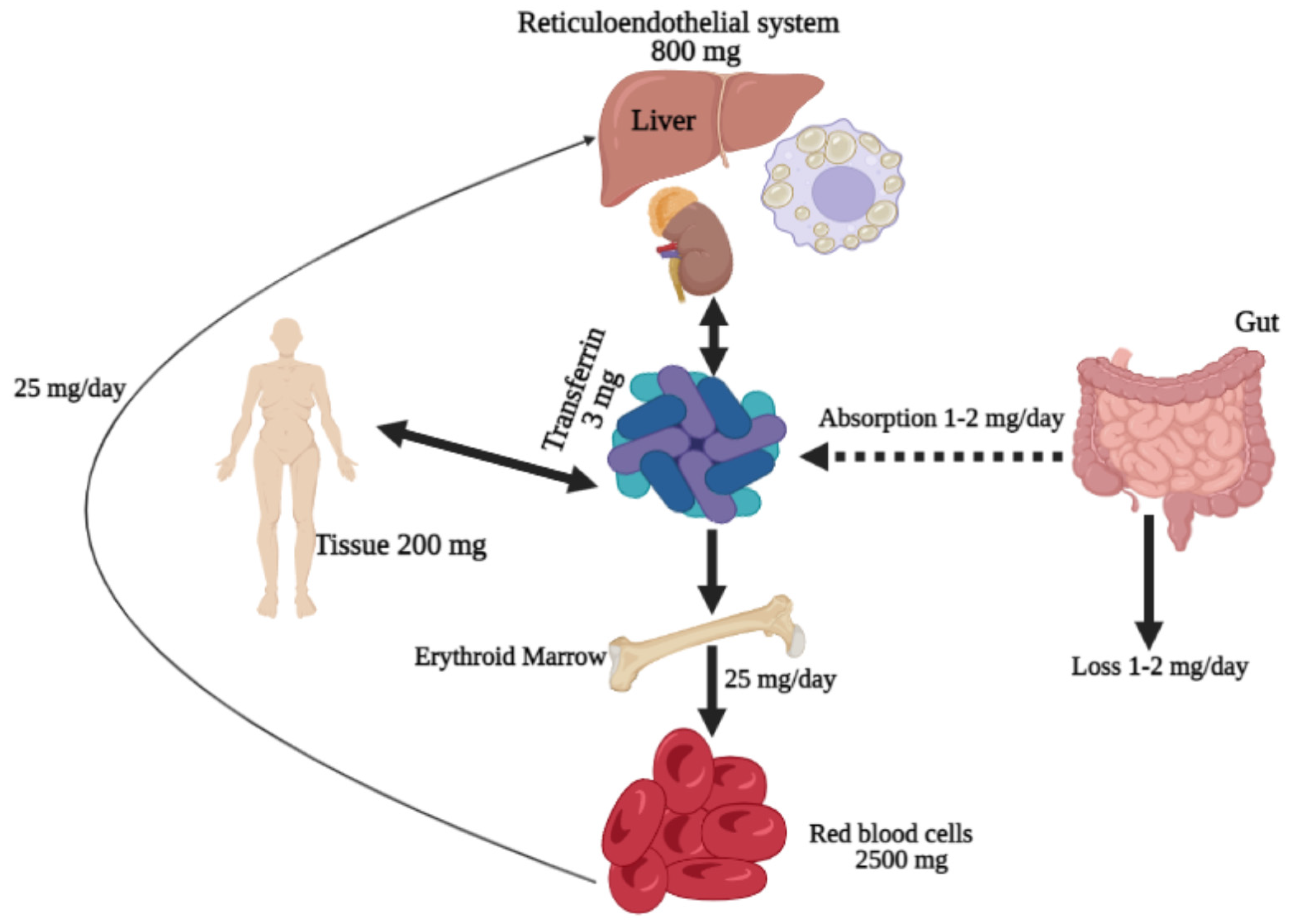
Figure 1. Iron distribution in the human body. Iron is absorbed by the duodenum enterocytes and into the plasma, where transferrin delivers it to bone marrow for Hb synthesis by erythroid precursors and erythrocytes or to muscles for myoglobin synthesis. Excess iron in the blood circulation can be stored as ferritin molecules in the liver or macrophages. The regular daily iron loss (1–2 mg) occurs mainly through blood loss (hemorrhage or menstruation).
Biomedicines 10 00189 g001
Figure 2. Hepcidin regulation on iron homeostasis: hepcidin synthesis is regulated at the transcriptional level by multiple stimuli. Hepcidin transcription increased with rising intra–extracellular iron concentrations and inflammation. In contrast, hepcidin production is suppressed in response to higher erythropoietic activity. Iron concentration in plasma is regulated by hepcidin through controlling FPN concentrations in iron exporting cells (duodenal, enterocytes, hepatocytes, and macrophages from liver and spleen). : resulting in or enhances expression and ⊥: reduced expression.
Biomedicines 10 00189 g002
Figure 3. Mechanism of iron dysregulation in β-thalassemia syndrome. Affected patients experience anemia because of IE and shortened red blood cell (RBCs) survival. This condition induces erythropoietin production, leading to enhanced erythropoiesis. The dramatic increase in erythroid expansion activates the erythroid factors, including GDF15, TWSG1, and ERFE secretion. Excessive erythroid factors suppress hepcidin expression in liver cells, resulting in iron overload due to increased iron absorption from duodenal enterocytes, an increase in iron from hepatocytes and the reticuloendothelial system. : resulting in and : suppresses expression.
Biomedicines 10 00189 g003
Figure 4. Several regulatory pathways, including JAK/STAT in hepcidin transcription. The activation of JAKs after ligand–receptor coupling stimulates phosphorylation of STATs, followed by STAT dimerization and nucleus translocation to activate hepcidin transcription. ➔: Activation/Nucleus translocation.
Biomedicines 10 00189 g004
Figure 5. Signal transduction of TGF-β and BMP. The binding of TGF-β to the TβRII dimer allows the ligand to bind to the TβRI dimer and stimulate TβRI kinase activity. In SMAD-mediated TGF-β signal transduction, TβRI phosphorylates cytoplasmic SMAD2 and SMAD3, which interact with SMAD4 after dissociating from TβRI. The two receptors activate the trimeric complex of SMAD2 and SMAD3, and a SMAD4 then enters the nucleus, where it interacts with the DNA-binding transcription factor (TF) and coregulators of the target gene. Similarly, the BMP signals run parallel to the TGF-β signals. In response to the binding of the BMP ligand to the BMPRII heteromeric receptor complex and BMPRI transmembrane kinase, receptor-activated SMAD1 and SMAD5 bind to SMAD4 and are transported to the nucleus to activate or inhibit transcription of hepcidin. ➔: Activation and ⊥: Inhibit.
Biomedicines 10 00189 g005
Figure 6. Regulation of STAT and SMAD signaling pathway on hepcidin expression.
Biomedicines 10 00189 g006
Publisher’s Note: MDPI stays neutral with regard to jurisdictional claims in published maps and institutional affiliations.