Non-Enzymatic Electrochemical Glucose Sensors Based on Metal Oxides and Sulfides: Recent Progress and Perspectives

Abstract
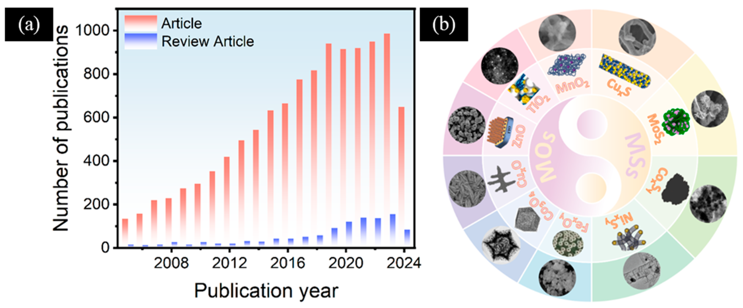
1. Introduction
2. MO-Based Non-Enzymatic Electrochemical Glucose Sensors
2.1. Iron-Based Oxides
2.2. Cobalt-Based Oxides
2.3. Copper-Based Oxides
2.4. Zinc-Based Oxides
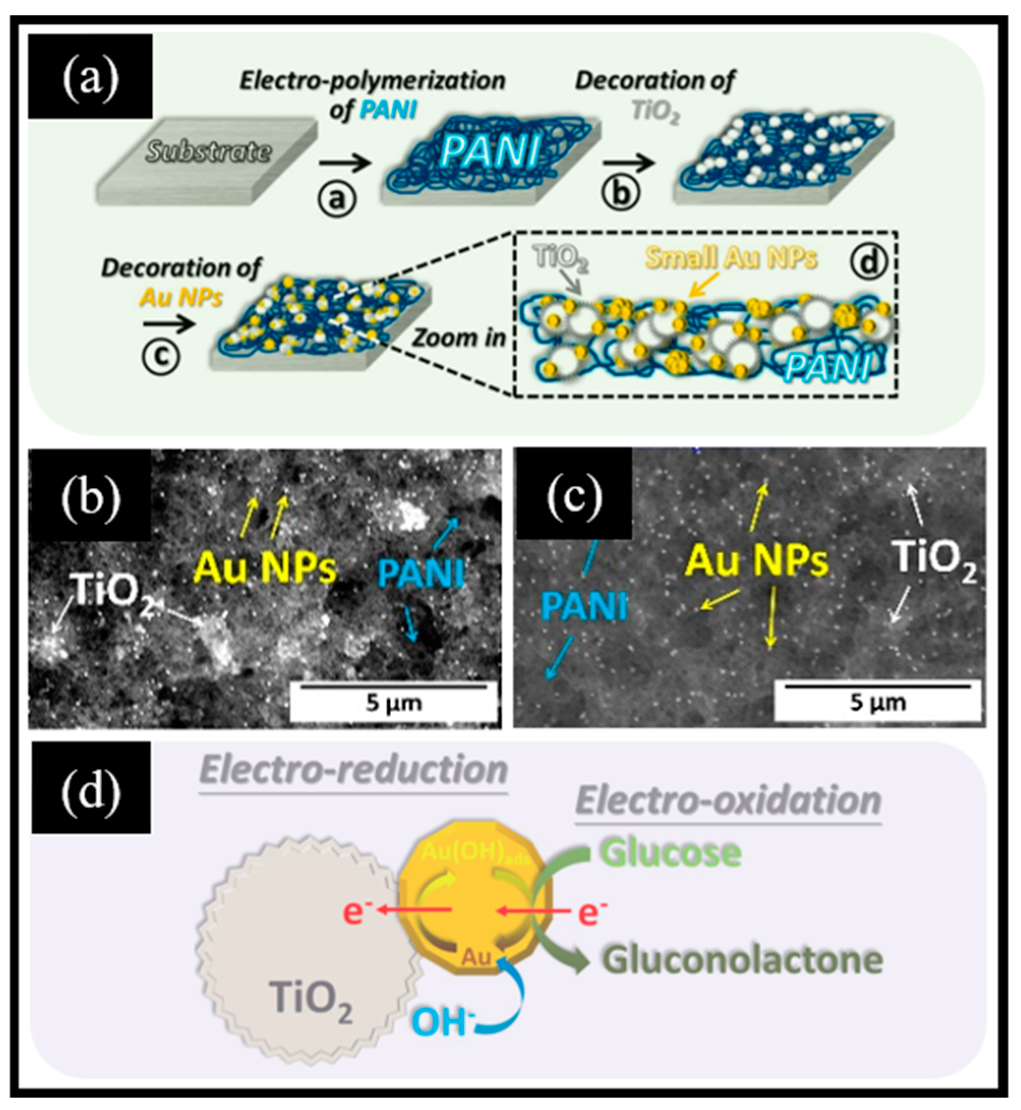
2.5. Titanium-Based Oxides
2.6. Manganese-Based Oxides
3. MS-Based Non-Enzymatic Electrochemical Glucose Sensors
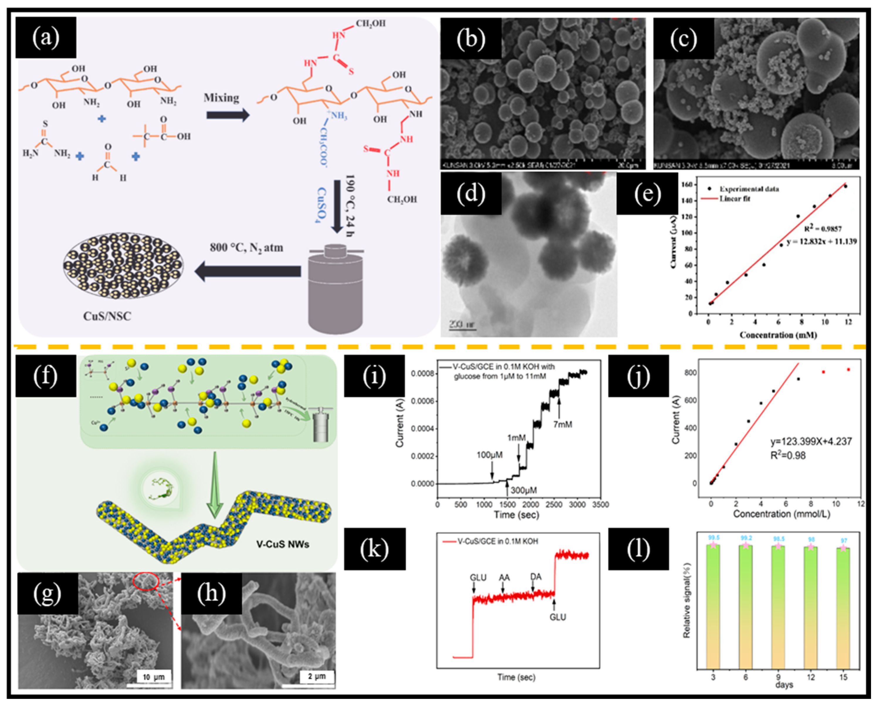
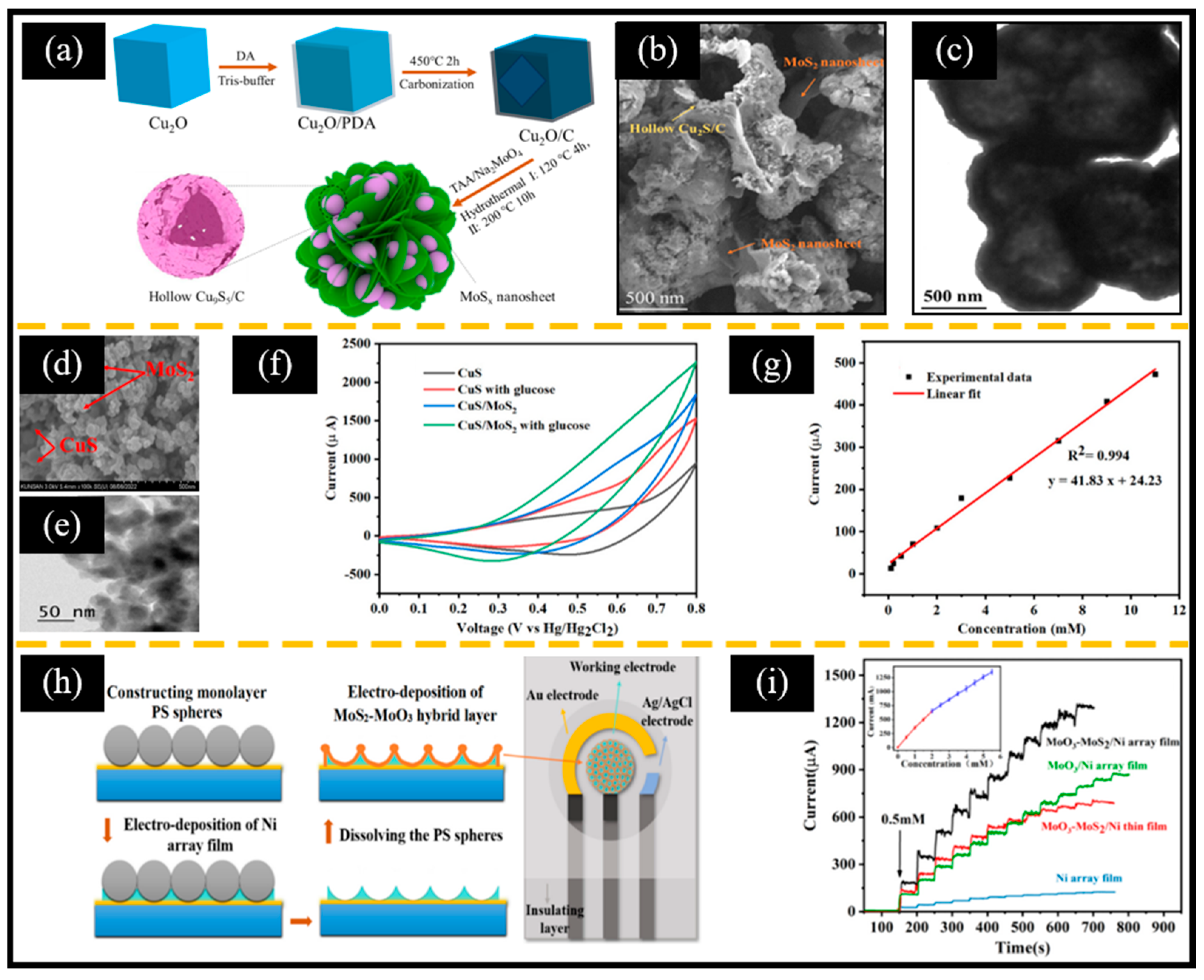
3.1. Copper-Based Sulfides
3.2. Molybdenum-Based Sulfides
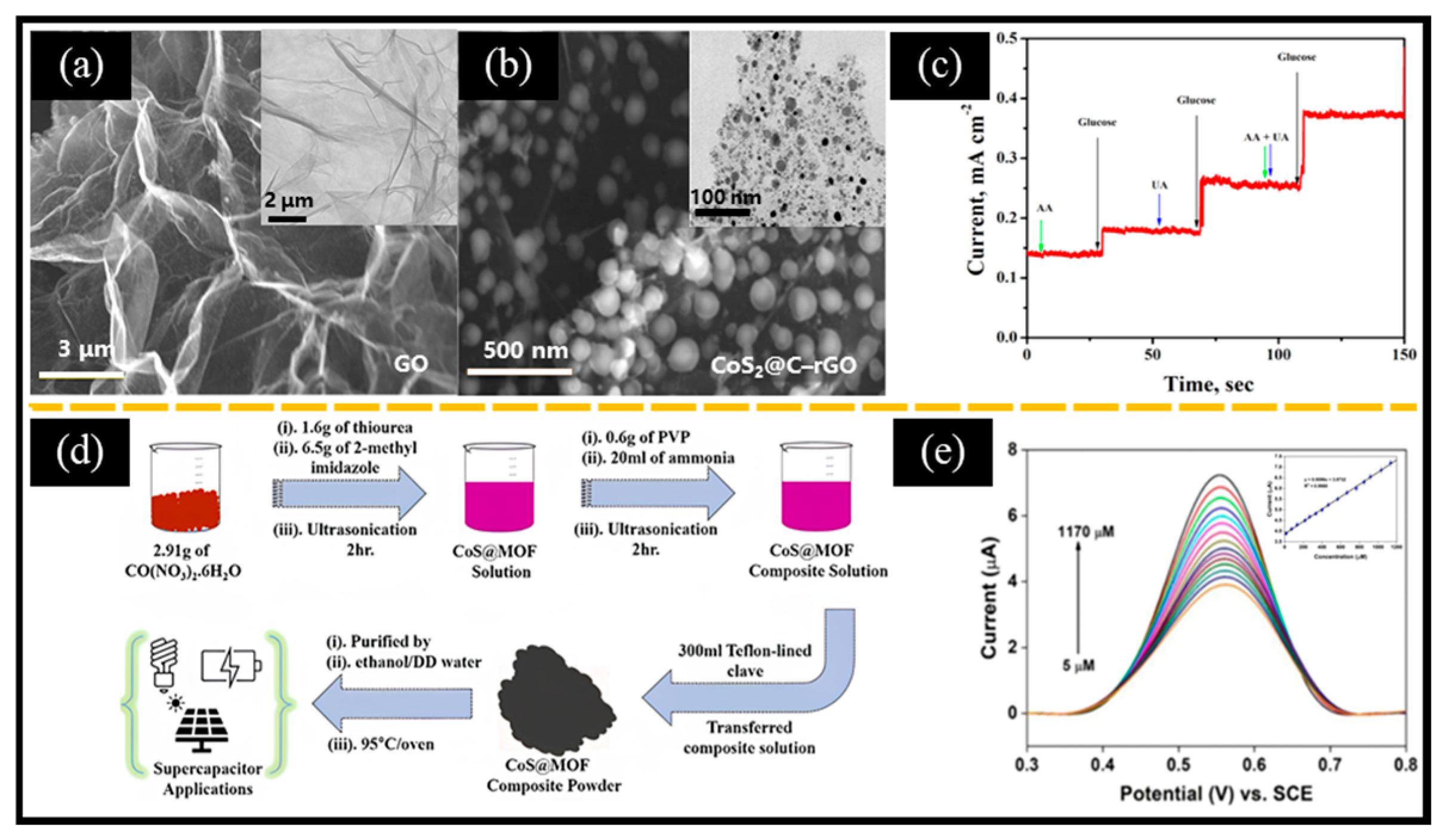
3.3. Cobalt-Based Sulfides
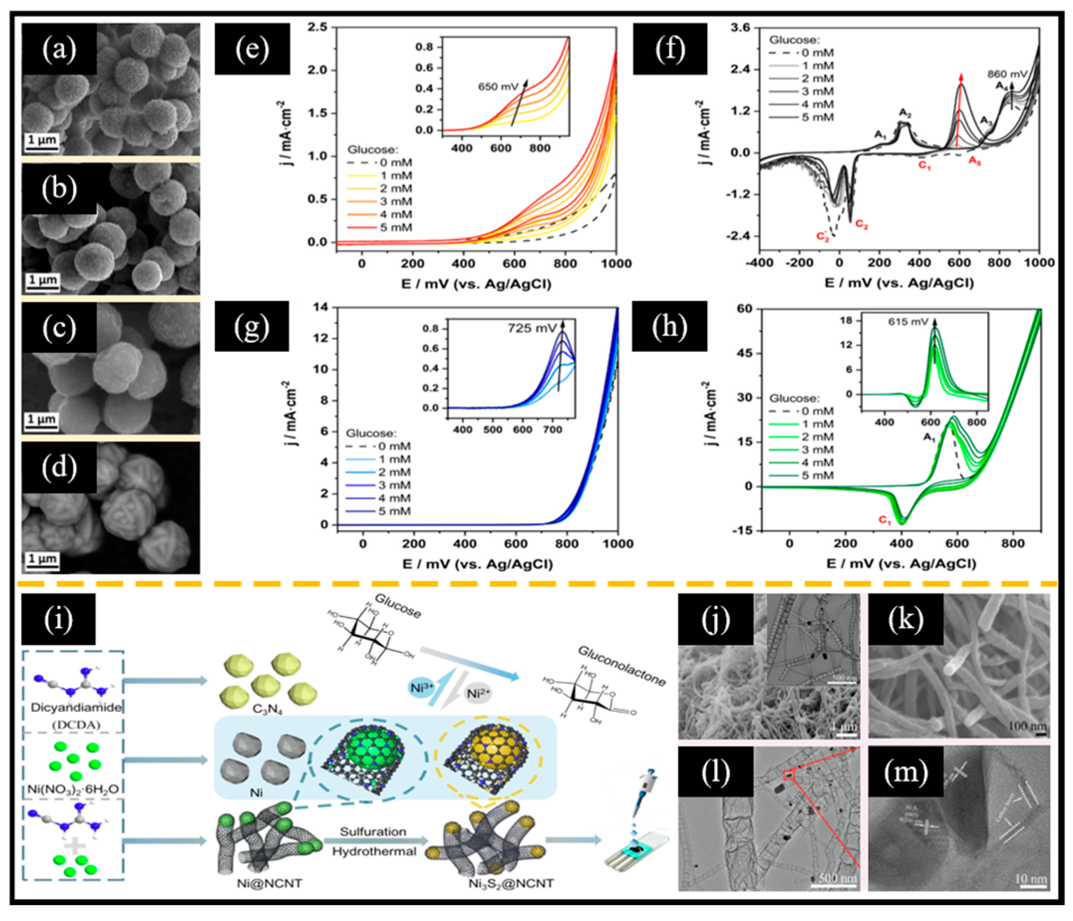
3.4. Nickel-Based Sulfides
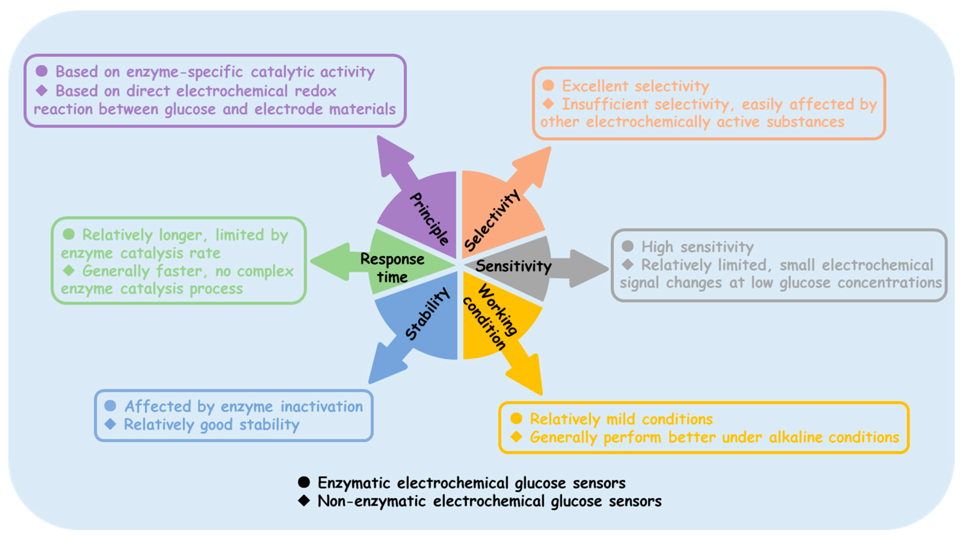
4. Challenges Faced by Non-Enzymatic Electrochemical Glucose Sensors in Practical Applications
- (i)
- Insufficient selectivity: Non-enzyme electrochemical glucose sensors rely on the electrochemical oxidation of glucose. However, other electrochemically active substances present in complex biological samples and real-world detection environments, such as ascorbic acid (AA), uric acid (UA), and dopamine, may also undergo reactions on the electrode surface, generating interference signals that affect the accurate detection of glucose. Therefore, the selectivity of non-enzyme electrochemical glucose sensors remains a significant challenge.
- (ii)
- Sensitivity requires further improvement: In certain applications involving low glucose concentrations, such as blood glucose monitoring in diabetic patients after insulin administration or cell cultures sensitive to glucose levels, non-enzyme glucose sensors may fail to provide accurate detection.
- (iii)
- Limited working environment: A large number of studies have shown that common MOs and MSs exhibit good catalytic activity in alkaline environments; however, their catalytic activity significantly decreases in neutral or acidic environments.
- (iv)
- Process optimization issues: In this review, we found that some MOs perform better after high-temperature annealing, while MSs typically involve a sulfide reaction between the metal and sulfur source. Therefore, synthesizing these materials in the laboratory generally requires high-temperature equipment, such as muffle furnaces, vacuum tube furnaces, and electric ovens. Simplifying the synthesis process would facilitate the promotion of large-scale applications.
- (i)
- Nanoconfinement effect: The fixed size of nanopores provides a strategy for excluding the influence of certain interfering substances. Benedetti et al. [149] recently reported a conductive mesoporous carbon shell-coated Au nanoparticle used to simulate the 3D structure of enzymes. The separated nanopores created the surface of Au, which not only creates an alkaline environment for non-enzymatic detection locally, but also eliminates the interference from Cl−, UA, AA, and proteins. This non-enzymatic electrochemical glucose sensor based on the nanoconfinement effect enables glucose detection in whole blood.
- (ii)
- Defect engineering: Defect engineering has been demonstrated to be an effective strategy to enhance the catalytic activity of nanomaterials. Zhong et al. [150] obtained Ni(OH)2 nanosheets with varying defect (oxygen vacancy) concentrations by treating the product with Ar plasma for different times. Interestingly, defective Ni(OH)2 exhibited better selectivity than pristine Ni(OH)2.
- (iii)
- Design electrode materials with a stronger glucose molecule adsorption capability based on theoretical calculations: In principle, the stronger the electrode material’s adsorption capability for glucose molecules, the more beneficial it is for reducing the diffusion energy barrier and accelerating reaction kinetics. Furthermore, the glucose molecule adsorption layer formed on the electrode surface hinders other interfering substances from approaching the electrode surface to some extent, thereby reducing their adsorption and reaction on the electrode.
- (iv)
- Intelligent back-propagation (BP) neural network: Recently, Zhou et al. [151] innovatively introduced an intelligent BP neural network into electrochemical microarrays to improve the selectivity of non-enzymatic electrochemical sensors. They prepared three non-enzymatic electrodes (NiO/Pt, Ni(OH)2/Au, and Ni(OH)2/Pt) by electrodeposition and dripping, and then integrated them into a single electrochemical unit. This strategy overcame the overlapping oxidation peaks of glucose and lactate for the first time and can identify multiple biomarkers, which will help promote the commercialization of non-enzymatic glucose sensors in the future.
5. Summary and Prospectives
Author Contributions
Funding
Institutional Review Board Statement
Informed Consent Statement
Data Availability Statement
Conflicts of Interest
References
- Hwang, D.W.; Lee, S.; Seo, M.; Chung, T.D. Recent advances in electrochemical non-enzymatic glucose sensors-A review. Anal. Chim. Acta 2018, 1033, 1–34. [Google Scholar] [CrossRef] [PubMed]
- Du, Y.T.; Zhang, X.Y.; Liu, P.; Yu, D.G.; Ge, R.L. Electrospun nanofiber-based glucose sensors for glucose detection. Front. Chem. 2022, 10, 944428. [Google Scholar] [CrossRef]
- Senjam, S.S. Diabetes and diabetic retinopathy: The growing public health concerns in India. Lancet Global Health 2024, 12, e727–e728. [Google Scholar] [CrossRef] [PubMed]
- Shi, F.; Xu, J.M.; Hu, Z.F.; Ren, C.L.; Xue, Y.D.; Zhang, Y.C.; Li, J.; Wang, C.Y.; Yang, Z.J. Bird nest-like zinc oxide nanostructures for sensitive electrochemical glucose biosensor. Chin. Chem. Lett. 2021, 32, 3185–3188. [Google Scholar] [CrossRef]
- Shu, Y.; Shang, Z.J.; Su, T.; Zhang, S.H.; Lu, Q.; Xu, Q.; Hu, X.Y. A highly flexible Ni-Co MOF nanosheet coated Au/PDMS film based wearable electrochemical sensor for continuous human sweat glucose monitoring. Analyst 2022, 147, 1440. [Google Scholar] [CrossRef]
- Cho, N.H.; Shaw, J.E.; Karuranga, S.; Huang, Y.; da Rocha Fernandes, J.D.; Ohlrogge, A.W.; Malanda, B. IDF Diabetes Atlas: Global estimates of diabetes prevalence for 2017 and projections for 2045. Diabetes Res. Clin. Pract. 2018, 138, 271–281. [Google Scholar] [CrossRef]
- Wang, J. Electrochemical glucose biosensors. Chem. Rev. 2008, 108, 814–825. [Google Scholar] [CrossRef]
- Kiang, S.W.; Kuan, J.W.; Kuan, S.S.; Guilbault, G.G. An immobilized hexokinase enzyme stirrer for a simple and economical assay of plasma glucose. Clin. Chim. Acta 1977, 78, 495–498. [Google Scholar] [CrossRef]
- Pham, K.; Rieta, R.; Zikos, T.; Bowen, R.; Park, W. Su1350 Analytical performance of a routinely utilized serum/plasma spectrophotometric method for glucose quantification in pancreatic cyst fluid. Gastroenterology 2016, 160, S500. [Google Scholar] [CrossRef]
- Hoshino, T.; Takahashi, Y.; Suzuki, M. Application of high-performance liquid chromatography in establishing an accurate index of blood glucose control. J. Chromatogr. A 1990, 515, 531–536. [Google Scholar] [CrossRef]
- Liu, Y.; Lv, R.; Sun, S.Y.; Tan, D.Y.; Dong, F.Q.; Golubev, Y.A.; Nie, X.Q.; Kotova, O.B.; Liu, J.; Wang, K. High-performance cascade nanoreactor based on halloysite nanotubes-integrated enzyme-nanozyme microsystem. Chin. Chem. Lett. 2022, 33, 807–811. [Google Scholar] [CrossRef]
- Zhang, Q.; Li, P.P.; Wu, J.; Peng, Y.; Pang, H. Pyridine-regulated lamellar nickel-based metal-organic framework (Ni-MOF) for nonenzymatic electrochemical glucose sensor. Adv. Sci. 2023, 10, 2304102. [Google Scholar] [CrossRef] [PubMed]
- Tong, P.H.; Wang, J.J.; Hu, X.L.; James, T.D.; He, X.P. Metal-organic framework (MOF) hybridized gold nanoparticles as a bifunctional nanozyme for glucose sensing. Chem. Sci. 2023, 14, 7762–7769. [Google Scholar] [CrossRef] [PubMed]
- Fang, W.; Ding, L.Y.; Zhang, Y.M.; Li, H.J. Prism SPR glucose sensor based on gold nanoparticle/gold film coupling enhanced SPR. IEEE Sens. J. 2023, 23, 12477–12484. [Google Scholar] [CrossRef]
- Zhang, X.H.; Sun, B.; Zhang, Y.Y.; Zhang, Q.F.; Akhtar, M.H.; Li, M.; Gu, Y.W.; Yu, C. Portable smartphone-assisted ratiometric fluorescence sensor for visual detection of glucose. Anal. Chim. Acta 2023, 1260, 341173. [Google Scholar] [CrossRef]
- Saha, T.; Caño, R.D.; Mahato, K.; De la Paz, E.; Chen, C.R.; Ding, S.C.; Yin, L.; Wang, J. Wearable electrochemical glucose sensors in diabetes management: A comprehensive review. Chem. Rev. 2023, 123, 7854–7889. [Google Scholar] [CrossRef]
- Teymourian, H.; Barfidokht, A.; Wang, J. Electrochemical glucose sensors in diabetes management: An updated review (2010–2020). Chem. Soc. Rev. 2020, 49, 7671–7709. [Google Scholar] [CrossRef]
- Gao, Y.K.; Zhang, C.M.; Yang, Y.X.; Yang, N.; Lu, S.C.; You, T.T.; Yin, P.G. A high sensitive glucose sensor based on Ag nanodendrites/Cu mesh substrate via surface-enhanced Raman spectroscopy and electrochemical analysis. J. Alloys Compd. 2021, 863, 158758. [Google Scholar] [CrossRef]
- He, C.H.; Asif, M.; Liu, Q.Q.; Xiao, F.; Liu, H.F.; Xia, B.Y. Noble metal construction for electrochemical nonenzymatic glucose detection. Adv. Mater. Technol. 2023, 8, 2200272. [Google Scholar] [CrossRef]
- Xia, Y.P.; Shi, F.; Liu, R.X.; Zhu, H.B.; Liu, K.; Ren, C.L.; Li, J.; Yang, Z.J. In situ electrospinning MOF-derived highly dispersed α-cobalt confined in nitrogen-doped carbon nanofibers nanozyme for biomolecule monitoring. Anal. Chem. 2024, 96, 1345–1353. [Google Scholar] [CrossRef]
- Zeng, J.Y.; Yang, Y.T.; Lei, X.Y.; Deng, J.A.; Hu, N.; Yang, J. Tuning Co/Ni ratio in Co-Ni bimetallic hybrid for electrochemical detection of glucose. Chemosensors 2024, 12, 38. [Google Scholar] [CrossRef]
- Singh, K.; Maurya, K.K.; Malviya, M. Review of electrochemical sensors and biosensors based on first-row transition metals, their oxides, and noble metals nanoparticles. J. Anal. Test. 2024, 8, 143–159. [Google Scholar] [CrossRef]
- Li, N.; Li, Q.; Yuan, M.J.; Guo, X.T.; Zheng, S.S.; Pang, H. Synthesis of Co0.5Mn0.1Ni0.4C2O4·nH2O micropolyhedrons: Multi-metal synergy for high-performance glucose catalysis. Chem. Asian J. 2019, 14, 2259–2265. [Google Scholar] [CrossRef] [PubMed]
- Yang, Z.Y.; Bai, X.; Zhu, S.Y.; Qi, C.C. Synthesis of porous Co3S4 for enhanced voltammetric nonenzymatic determination of glucose. Microchim. Acta 2020, 187, 98. [Google Scholar] [CrossRef]
- Bano, M.; Naikoo, G.A.; BaOmar, F.; Rather, J.A.; Hassan, I.U.; Sheikh, R.A.; Kannan, P.; Tambuwala, M.M. Revolutionizing glucose monitoring: Enzyme-free 2D-MoS2 nanostructures for ultra-sensitive glucose sensors with real-time health-monitoring capabilities. ACS Omega 2024, 9, 20021–20029. [Google Scholar] [CrossRef]
- Xia, Y.Y.; Su, T.; Mi, Z.Y.; Feng, Z.Y.; Hong, Y.W.; Hu, X.Y.; Shu, Y. Wearable electrochemical sensor based on bimetallic MOF coated CNT/PDMS film electrode via a dual-stamping method for real-time sweat glucose analysis. Anal. Chim. Acta 2023, 1278, 341754. [Google Scholar] [CrossRef]
- Ramu, J.; Ramasundaram, S.; Yellappa, S.; Gunamalai, L.; Kamilya, T.; Afzal, M.; Jeffery, A.A.; Oh, T.H.; Mahanthappa, M.; Vishwanath, R.S. Carbon-supported nickel/nickel oxide nanohybrid composite as a high-performance sensor for electrochemical non-enzymatic glucose detection. New J. Chem. 2024, 48, 13814–13824. [Google Scholar] [CrossRef]
- Huu Do, H.; Kim, S.Y.; Le, Q.V. Development of non-precious metal oxide-based electrodes for enzyme-free glucose detection: A review. Microchem. J. 2023, 193, 109202. [Google Scholar] [CrossRef]
- Franceschini, F.; Payo, M.R.; Schouteden, K.; Ustarroz, J.; Locquet, J.P.; Taurino, I. MBE grown vanadium oxide thin films for enhanced non-enzymatic glucose sensing. Adv. Funct. Mater. 2023, 33, 2304037. [Google Scholar] [CrossRef]
- Sun, F.C.; Wang, X.Z.; You, Z.H.; Xia, H.H.; Wang, S.T.; Jia, C.P.; Zhou, Y.; Zhang, J. Sandwich structure confined gold as highly sensitive and stable electrochemical non-enzymatic glucose sensor with low oxidation potential. J. Mater. Sci. Technol. 2022, 123, 113–122. [Google Scholar] [CrossRef]
- Hilal, M.; Xie, W.F.; Yang, W. Straw-sheaf-like Co3O4 for preparation of an electrochemical non-enzymatic glucose sensor. Microchim. Acta 2022, 189, 364. [Google Scholar] [CrossRef] [PubMed]
- Yang, K.; Cheng, S.S.; Yao, Z.Q.; Li, S.J.; Yang, Y.T. Dumbbell shaped nanocomposite Co3O4/CeO2 derived from metal-organic frameworks (MOFs) as an excellent non-enzymatic glucose sensor. Solid State Sci. 2024, 150, 107498. [Google Scholar] [CrossRef]
- Yuan, M.J.; Guo, X.T.; Pang, H. Derivatives (Cu/CuO, Cu/Cu2O, and CuS) of Cu superstructures reduced by biomass reductants. Mater. Today Chem. 2021, 21, 100519. [Google Scholar] [CrossRef]
- Gu, J.W.; Xu, Y.X.; Li, Q.; Pang, H. Porous Ni/NiO nanohybrids for electrochemical catalytic glucose oxidation. Chin. Chem. Lett. 2021, 32, 2017–2020. [Google Scholar] [CrossRef]
- Zhou, Y.J.; Wei, W.; Lei, W.L.; Li, F.D.; Shu, J.X.; Deng, Z.X.; Hui, W.Y.; Zhao, Y.M.; Shan, C.S. Synergistically enhancing electrocatalysis and non-enzymatic sensing for glucose by iridium single-atom/nickel oxide/N-doped graphene. Anal. Bioanal. Chem. 2024, 416, 6011–6019. [Google Scholar] [CrossRef]
- Mahar, I.A.; Tahira, A.; Parveen, M.; Hulio, A.A.; Ibupoto, Z.A.; Bhatti, M.A.; Dawi, E.; Nafady, A.; Alshammari, R.H.; Vigolo, B.; et al. Glucose sensing via green synthesis of NiO-SiO2 composites with citrus lemon peel extract. J. Mater. Sci. Mater. Electron. 2024, 35, 490. [Google Scholar] [CrossRef]
- Liu, Y.H.; Young, S.J.; Hsien, C.Y.; Chu, Y.L.; Wang, Z.H.; Chang, S.J. Improved non-enzymatic glucose sensors of ZnO nanorods by adsorb Pt nanoparticles. IEEE Trans. Nanotechnol. 2024, 23, 303–310. [Google Scholar] [CrossRef]
- Sharma, S.; Ganeshan, S.K.; Kundu, S.; Chappanda, K.N. Effect of doping on TiO2 nanotubes based electrochemical sensors: Glucose sensing as a case study. IEEE Trans. Nanotechnol. 2021, 20, 185–193. [Google Scholar] [CrossRef]
- Zhu, J.L.; Peng, X.; Nie, W.; Wang, Y.J.; Gao, J.W.; Wen, W.; Selvaraj, J.N.; Zhang, X.H.; Wang, S.F. Hollow copper sulfide nanocubes as multifunctional nanozymes for colorimetric detection of dopamine and electrochemical detection of glucose. Biosens. Bioelectron. 2019, 141, 111450. [Google Scholar] [CrossRef]
- Medhi, A.; Giri, M.K.; Mohanta, D. Non-enzymatic approach of H2O2 and glucose sensing using NiO-MoS2-derived electrochemical sensor. Bull. Mater. Sci. 2024, 47, 223. [Google Scholar] [CrossRef]
- Sharma, K.P.; Shin, M.; Awasthi, G.P.; Yu, C. One-pot hydrothermal synthesis of CuS/CoS composite for electrochemical non-enzymatic glucose sensor. Curr. Appl. Phys. 2023, 56, 126–134. [Google Scholar] [CrossRef]
- Arivazhagan, M.; Santhosh, Y.M.; Maduraiveeran, G. Non-enzymatic glucose detection based on NiS nanoclusters@NiS nanosphere in human serum and urine. Micromachines 2021, 12, 403. [Google Scholar] [CrossRef] [PubMed]
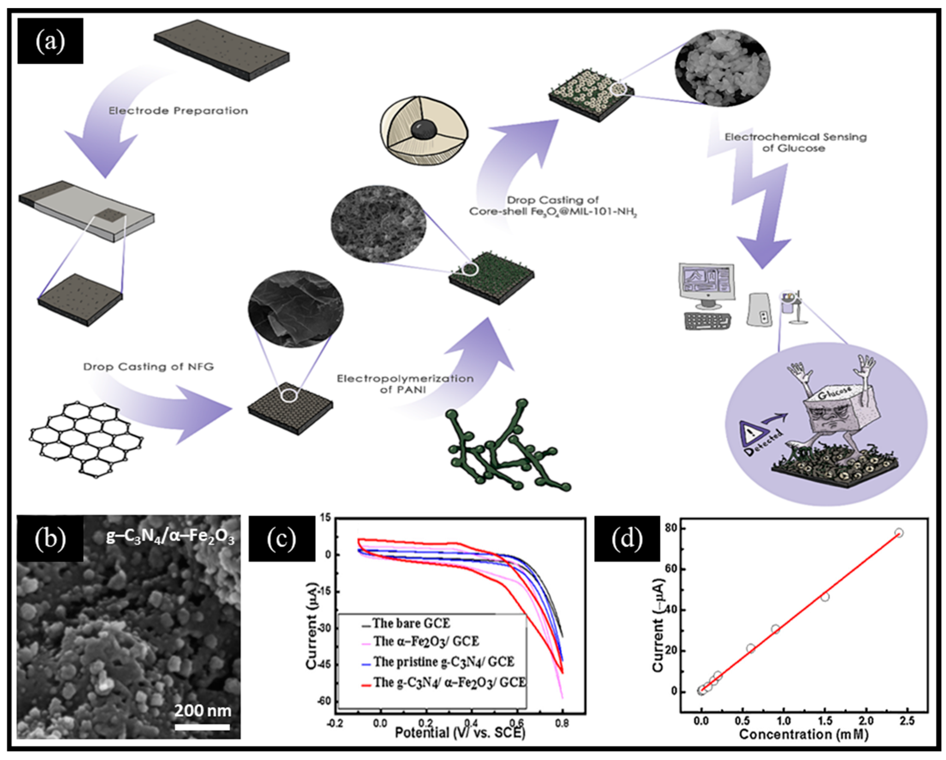
- Ghaffarirad, M.A.; Sabahi, A.; Golshani, Z.; Manteghi, F.; Ghaffarinejad, A. Non-enzymatic glucose electrochemical sensor based on nitrogen-doped graphene modified with polyaniline and Fe3O4@MIL-101-NH2 nano framework. Inorg. Chem. Commun. 2024, 159, 111812. [Google Scholar] [CrossRef]
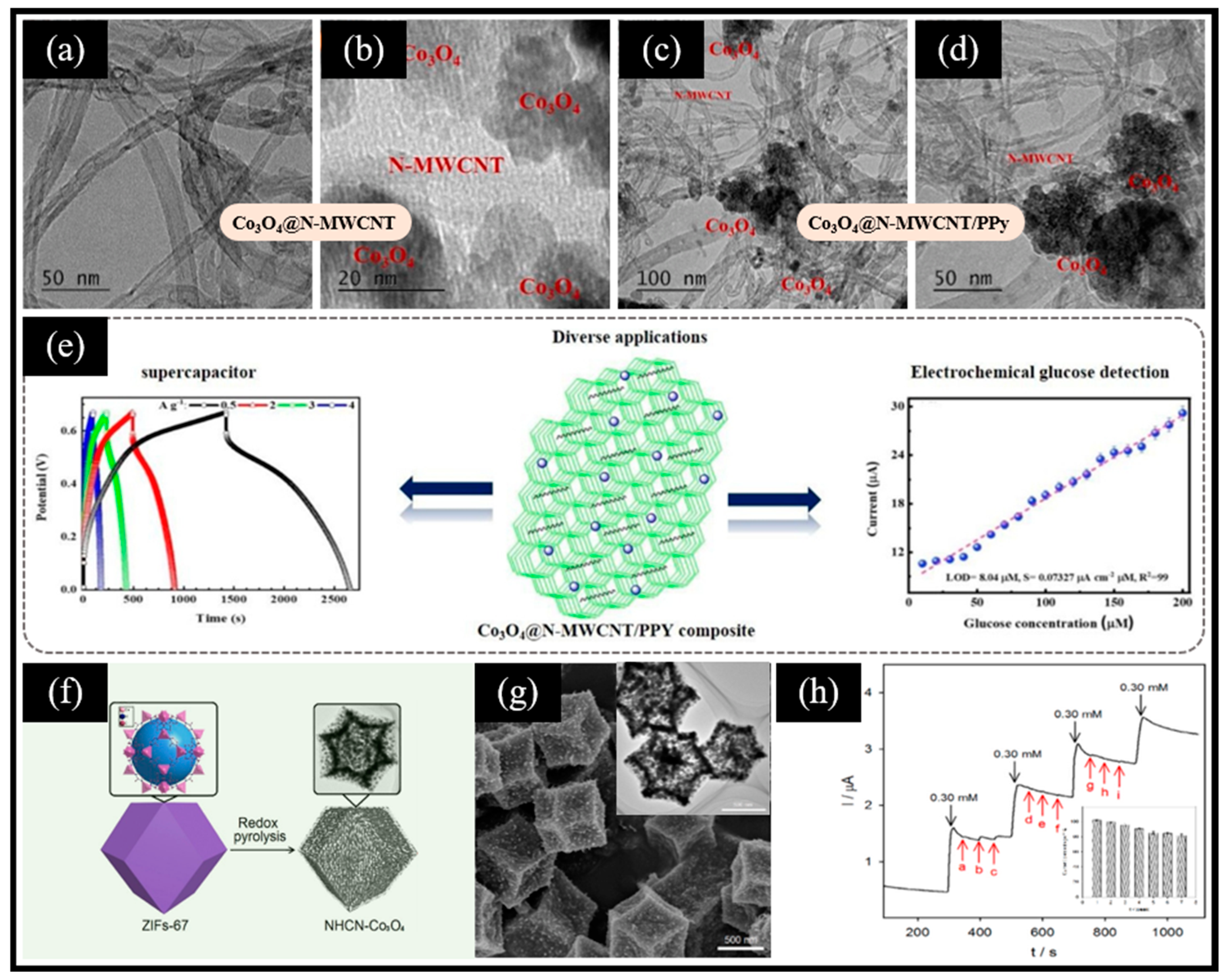
- Ouyang, Y.C.; Zheng, X.R.; Li, Q.X.; Ye, N.B.; Mo, G.Q. ZIFs derived polyhedron with cobalt oxide nanoparticles as novel nanozyme for the biomimetic catalytic oxidation of glucose and non-enzymatic sensor. Anal. Chim. Acta 2022, 1209, 339839. [Google Scholar] [CrossRef]
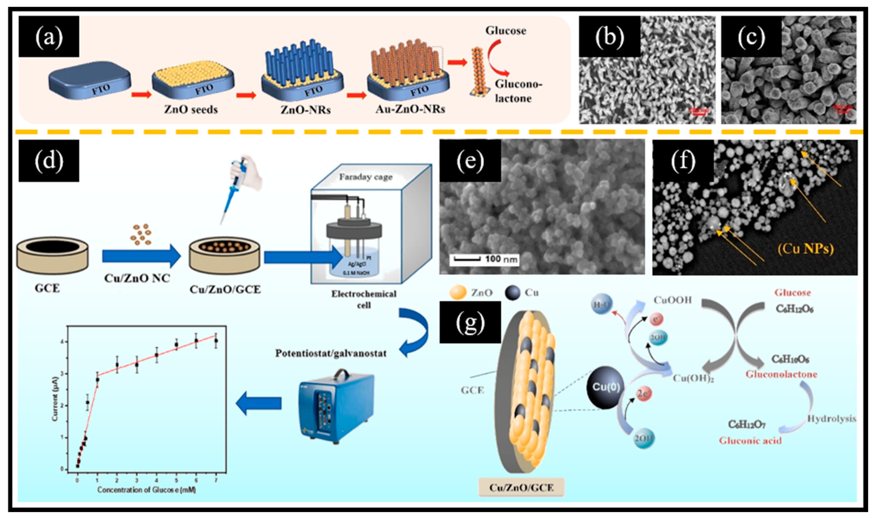
- Awais, A.; Arsalan, M.; Qiao, X.J.; Wang, Y.H.; Sheng, Q.L.; Yue, T.L.; He, Y.P. Facial synthesis of highly efficient non-enzymatic glucose sensor based on vertically aligned Au-ZnO NRs. J. Electroanal. Chem. 2021, 895, 115424. [Google Scholar] [CrossRef]
- Chiu, W.T.; Chang, T.F.M.; Sone, M.; Mita, A.T.; Toshiyoshi, H. Electrocatalytic activity enhancement of Au NPs-TiO2 electrode via a facile redistribution process towards the non-enzymatic glucose sensors. Sens. Actuat. B Chem. 2020, 319, 128279. [Google Scholar] [CrossRef]
- Okamoto, K.; Kawakami, H.; Chien, Y.A.; Kurioka, T.; Chiu, W.T.; Chakraborty, P.; Nakamoto, T.; Hsu, Y.J.; Sone, M.; Chang, T.F.M. Gold/MnO2 particles decorated on electrodeposited polyaniline toward non-enzymatic electrochemical sensor for glucose. Micro Nano Eng. 2023, 18, 100175. [Google Scholar] [CrossRef]
- He, L.; Ma, C.Y.; Lv, G.; Liu, J.; Li, R.Z. Study on the preparation of vine-like CuS nanowires for non-enzymatic glucose sensing application. Mater. Lett. 2024, 361, 136029. [Google Scholar] [CrossRef]
- Cao, L.H.; Yu, H.; Deng, B.H.; Li, Z.H.; Ma, L. MoSx nanosheets supported hollow Cu9S5/C for electrochemical detection of glucose in human serum and beverages. Microchem. J. 2024, 205, 111238. [Google Scholar] [CrossRef]
- Ramesh, S.; Ahmed, A.T.A.; Haldorai, Y.; Kakani, V.; Bathula, C.; Kim, H.S. Cobalt sulfide@cobalt-metal organic frame works materials for energy storage and electrochemical glucose detection sensor application. J. Alloys Compd. 2023, 967, 171760. [Google Scholar] [CrossRef]
- Li, G.F.; Xie, G.Q.; Chen, D.; Gong, C.; Chen, X.; Zhang, Q.; Pang, B.L.; Zhang, Y.C.; Li, C.J.; Hu, J.; et al. Facile synthesis of bamboo-like Ni3S2@NCNT as efficient and stable electrocatalysts for non-enzymatic glucose detection. Appl. Surf. Sci. 2022, 585, 152683. [Google Scholar] [CrossRef]
- Godarzi, S.M.; Khodadadi, A.A.; Naseh, M.V.; Mortazavi, Y. Highly stable and selective non-enzymatic glucose biosensor using carbon nanotubes decorated by Fe3O4 nanoparticles. J. Electrochem. Soc. 2014, 161, B19–B25. [Google Scholar] [CrossRef]
- Ghosh, A.; Ganguly, D.; Sundara, R. A new approach to in-situ uniform growth of Fe3O4 nanoparticles over thermally exfoliated rGO sheet for the non-enzymatic and enzymatic detection of glucose. J. Electroanal. Chem. 2021, 895, 115386. [Google Scholar] [CrossRef]
- Xu, J.S.; Sun, Y.T.; Zhang, J. Solvothermal synthesis of Fe3O4 nanospheres for high-performance electrochemical non-enzymatic glucose sensor. Sci. Rep. 2020, 10, 16026. [Google Scholar] [CrossRef] [PubMed]
- Zhou, F.; Wang, J.Y.; Tang, Y.M.; Song, X.H.; Zhou, W.R.; Li, Y.; Gao, F. Enhanced sensing performance of flexible non-enzymatic electrochemical glucose sensors using hollow Fe3O4 nanospheres of controllable morphologies. Ceram. Int. 2024, 50, 38009–38021. [Google Scholar] [CrossRef]
- Hernández-Ramírez, D.; Mendoza-Huizar, L.H.; Galán-Vidal, C.A.; Aguilar-Lira, G.Y.; Álvarez-Romero, G.A. Development of a non-enzymatic glucose sensor based on Fe2O3 nanoparticles-carbon paste electrodes. J. Electrochem. Soc. 2022, 169, 067507. [Google Scholar] [CrossRef]
- Luo, L.; Cui, J.W.; Wang, Y.; Wang, Y.; Zheng, H.M.; Qin, Y.Q.; Shu, X.; Yu, D.B.; Zhang, Y.; Wu, Y.C. Synthesis of NiO/Fe2O3 nanocomposites as substrate for the construction of electrochemical biosensors. J. Solid State Electrochem. 2018, 22, 1763–1770. [Google Scholar] [CrossRef]
- Abrori, S.A.; Septiani, N.L.W.; Nugraha; Anshori, I.; Suyatman; Suendo, V.; Yuliarto, B. Metal-Organic-Framework FeBDC-Derived Fe3O4 for non-enzymatic electrochemical detection of glucose. Sensors 2020, 20, 4891. [Google Scholar] [CrossRef]
- Liu, L.; Wang, J.X.; Wang, C.Y.; Wang, G.X. Facile synthesis of graphitic carbon nitride/nanostructured α-Fe2O3 composites and their excellent electrochemical performance for supercapacitor and enzyme-free glucose detection applications. Appl. Surf. Sci. 2016, 390, 303–310. [Google Scholar] [CrossRef]
- Qin, Y.N.; Sun, Y.J.; Li, Y.J.; Li, C.J.; Wang, L.; Guo, S.J. MOF derived Co3O4/N-doped carbon nanotubes hybrids as efficient catalysts for sensitive detection of H2O2 and glucose. Chin. Chem. Lett. 2020, 31, 774–778. [Google Scholar] [CrossRef]
- Lu, J.H.; Cheng, C.; Cao, Y.; Hou, X.H.; Li, H.J.; Yin, X.M. MOFs-derived core-shell structured NiCo2O4 NWs@Co3O4 NPs for non-enzymatic glucose detection. Ceram. Int. 2023, 49, 23958–23966. [Google Scholar] [CrossRef]
- Zhang, Y.; Zhao, Y.C.; Liu, Y.Q.; Yang, Y.Q.; Liu, X. Assist of multi-walled carbon nanotubes toward metal-organic framework chamfer cube-derived rambutan-like cobalt oxide@hollow chain and its application for non-enzymatic glucose sensing. Appl. Surf. Sci. 2023, 624, 157155. [Google Scholar] [CrossRef]
- Mohammadpour-Haratbar, A.; Mosallanejad, B.; Zare, Y.; Rhee, K.Y.; Park, S.J. Co3O4 nanoparticles embedded in electrospun carbon nanofibers as free-standing nanocomposite electrodes as highly sensitive enzyme-free glucose biosensors. Rev. Adv. Mater. Sci. 2022, 61, 744–755. [Google Scholar] [CrossRef]
- Dong, Q.C.; Wang, X.D.; Willis, W.S.; Song, D.H.; Huang, Y.K.; Zhao, J.; Li, B.K.; Lei, Y. Nitrogen-doped hollow Co3O4 nanofibers for both solid-state pH sensing and improved non-enzymatic glucose sensing. Electroanal. 2019, 31, 678–687. [Google Scholar] [CrossRef]
- Xiong, L.Y.; Kim, Y.J.; Seo, W.C.; Lee, H.K.; Yang, W.C.; Xie, W.F. High-performance non-enzymatic glucose sensor based on Co3O4/rGO nanohybrid. Rare Met. 2023, 42, 3046–3053. [Google Scholar] [CrossRef]
- Mustafa, A.; Alsafari, I.A.; Somaily, H.H.; Yousaf, S.; Din, M.I.; Rahman, J.; Shahid, M.; Ashraf, M.; Warsi, M.F. Fabrication, characterization of NiO-Co3O4/rGO based nanohybrid and application in the development of non-enzymatic glucose sensor. Phys. B Condens. 2023, 648, 414404. [Google Scholar] [CrossRef]
- Yin, Z.H.; He, S.S.; Li, Y.W.; Dai, W.J.; Wang, H.; He, R.H.; Tang, K.; Xiao, Y.M.; Wang, S.B.; Gao, J.; et al. Self-supported carbon electrodes with a carbon membrane and Co3O4 nanosheets for high-performance enzymeless glucose detection and supercapacitors. ACS Appl. Nano Mater. 2023, 6, 6208–6220. [Google Scholar] [CrossRef]
- Manoj, D.; Aziz, A.; Muhammad, N.; Wang, Z.P.; Xiao, F.; Asif, M.; Sun, Y. Integrating Co3O4 nanocubes on MXene anchored CFE for improved electrocatalytic activity: Freestanding flexible electrode for glucose sensing. J. Environ. Chem. Eng. 2022, 10, 108433. [Google Scholar] [CrossRef]
- Maghsoudi, S.; Mohammadi, A. Reduced graphene oxide nanosheets decorated with cobalt oxide nanoparticles: A nonenzymatic electrochemical approach for glucose detection. Synth. Met. 2020, 269, 116543. [Google Scholar] [CrossRef]
- Ramesh, S.; Karuppasamy, K.; Haldorai, Y.; Sivasamy, A.; Kim, H.S.; Kim, H.S. Hexagonal nanostructured cobalt oxide@nitrogen doped multiwalled carbon nanotubes/polypyyrole composite for supercapacitor and electrochemical glucose sensor. Colloid. Surface. B 2021, 205, 111840. [Google Scholar] [CrossRef]
- Muqaddas, S.; Javed, M.; Nadeem, S.; Asghar, M.A.; Haider, A.; Ahmad, M.; Ashraf, A.R.; Nazir, A.; Iqbal, M.; Alwadai, N.; et al. Carbon nanotube fiber-based flexible microelectrode for electrochemical glucose sensors. ACS Omega 2023, 8, 2272–2280. [Google Scholar] [CrossRef] [PubMed]
- Shao, Z.T.; Gao, Q.Y.; Sun, S.M.; Wu, L.L.; Feng, W. MOF-derived CuO/CNT for high sensitivity and fast response glucose sensing. Sens. Actuat. B Chem. 2024, 398, 134713. [Google Scholar] [CrossRef]
- Wang, S.Z.; Zheng, M.; Zhang, X.; Zhuo, M.P.; Zhou, Q.Q.; Su, Y.; Zheng, M.; Yuan, G.T.; Wang, Z.S. Flowerlike CuO/Au nanoparticle heterostructures for nonenzymatic glucose detection. ACS Appl. Nano Mater. 2021, 4, 5808–5815. [Google Scholar] [CrossRef]
- Xiang, D.; Zhao, L.P.; Wang, Y.; Li, X.Z.; Sun, X.; Yang, J.K.; Liu, H.; Wan, C. A highly sensitive non-enzymatic glucose electrochemical sensor electrode material of CuO nanospheres/activated carbon composites. Russ. J. Electrochem. 2023, 59, 1194–1205. [Google Scholar] [CrossRef]
- Carinelli, S.; Salazar-Carballo, P.A.; Melián, J.E.D.; García-García, F. Porous copper oxide thin film electrodes for non-enzymatic glucose detection. Chemosensors 2023, 11, 260. [Google Scholar] [CrossRef]
- Kuzhandaivel, H.; Paramasivam, K.; Manickam, S.; Nallathambi, K.S. Nickel-doped CuO/Cu/Cu2O nanocomposite as an efficient electrode for electrochemical non-enzymatic glucose sensor and asymmetric supercapacitor. J. Appl. Electrochem. 2023, 53, 1869–1886. [Google Scholar] [CrossRef]
- He, Z.Y.; Tang, X.; Zhang, Y.; Yu, H.M.; Zou, Z.R.; Huang, K.; Xue, K.; Xiong, X.L. Rapid preparation of 3D ultra-thin CuO nanosheets by dielectric barrier discharge microplasma for non-enzymatic detection of glucose. Catal. Lett. 2022, 152, 3517–3525. [Google Scholar] [CrossRef]
- Abd-Elrahim, A.G.; Chun, A.G. Room-temperature fabrication of a heterostructure Cu2O@CuO nanosheet electrocatalyst for non-enzymatic detection of glucose and H2O2. J. Electroanal. Chem. 2022, 924, 116874. [Google Scholar] [CrossRef]
- Patil, A.S.; Patil, R.T.; Lohar, G.M.; Fulari, V.J. Facile synthesis of CuO nanostructures for non-enzymatic glucose sensor by modified SILAR method. Appl. Phys. A 2021, 127, 101. [Google Scholar] [CrossRef]
- Wei, C.H.N.; Zou, X.; Liu, Q.M.; Li, S.X.; Kang, C.X.; Xiang, W. A highly sensitive non-enzymatic glucose sensor based on CuS nanosheets modified Cu2O/CuO nanowire arrays. Electrochim. Acta 2020, 334, 135630. [Google Scholar] [CrossRef]
- Gopal, T.S.; James, J.T.; Gunaseelan, B.; Ramesh, K.; Raghavan, V.; Malathi, C.J.A.; Amarnath, K.; Kumar, V.G.; Rajasekaran, S.J.; Pandiaraj, S.; et al. MXene-embedded porous carbon-based Cu2O nanocomposites for non-enzymatic glucose sensors. ACS Omega 2024, 9, 8448–8456. [Google Scholar]
- Fan, B.Y.; Spindler, B.D.; Zhao, W.Y.; Chan, H.; Wang, Z.; Kim, M.; Chipangura, Y.; Bühlmann, P.; Stein, A. Comparison of Copper(II) oxide nanostructures with different morphologies for nonenzymatic glucose sensing. ACS Appl. Nano Mater. 2023, 6, 1475–1486. [Google Scholar] [CrossRef]
- Zohaa; Arif, D.; Hassan, M.; Abdullah, M.; Miran, W.; Nasir, M.A.; Batool, S.; Baig, M.A.; Liaqat, U. An electrochemical sensor based on copper oxide nanoparticles loaded on a mesoporous MCM-41 for non-enzymatic detection of glucose. Ceram. Int. 2024, 50, 12614–12620. [Google Scholar] [CrossRef]
- Ngo, N.T.M.; Tran, T.B.Q.; Tran, M.K.; Bui, L.A.T.; Doan, V.H.T. Synthesis of Au/Cu2O/graphene quantum dots nanocomposites and its application for glucose oxidation. J. Chem. Sci. 2024, 136, 13. [Google Scholar]
- Yang, Z.; Yang, H.M.; Wang, W.; Zhao, H.Y.; Meng, P.R.; Xie, Y.X.; Sun, Y. A flexible electrochemical sensor for simultaneous determination of glucose (Glu) and ethanol (Eth) using ZnO and Pd nanoparticles. J. Appl. Electrochem. 2023, 53, 2013–2023. [Google Scholar] [CrossRef]
- Weston, M.; Kuchel, R.P.; Ciftci, M.; Boyer, C.; Chandrawati, R. A polydiacetylene-based colorimetric sensor as an active use-by date indicator for milk. J. Colloid Interface Sci. 2020, 572, 31–38. [Google Scholar] [CrossRef]
- Myndrul, V.; Coy, E.; Babayevska, N.; Zahorodna, V.; Balitskyi, V.; Baginskiy, I.; Gogotsi, O.; Bechelany, M.; Giardi, M.T.; Iatsunskyi, I. MXene nanoflakes decorating ZnO tetrapods for enhanced performance of skin-attachable stretchable enzymatic electrochemical glucose sensor. Biosens. Bioelectron. 2022, 207, 114141. [Google Scholar] [CrossRef]
- Morais, M.; Marques, A.C.; Ferreira, S.H.; Pinheiro, T.; Pimentel, A.; Macedo, P.; Martins, R.; Fortunato, E. Visible photoluminescent zinc oxide nanorods for label-free nonenzymatic glucose detection. ACS Appl. Nano Mater. 2022, 5, 4386–4396. [Google Scholar] [CrossRef]
- Lin, Y.; Wu, J.W.; Tan, X.C.; Huang, K.J. Electrochemiluminescence sensor based on graphdiyne quantum dots functionalized MOF-derived ZnO with self-enhancing effect for the detection of radical scavenging drugs. Sens. Actuat. B Chem. 2023, 382, 133541. [Google Scholar] [CrossRef]
- Mai, H.H.; Janssens, E. Au nanoparticle-decorated ZnO nanorods as fluorescent non-enzymatic glucose probe. Microchim. Acta 2020, 187, 577. [Google Scholar] [CrossRef]
- Ali, F.; Mari, R.H.; Dawi, E.; Tahira, A.; Halepoto, I.A.; Mahar, I.A.; Rind, N.A.; Nangraj, N.A.; Bhatti, M.A.; Almani, K.F.; et al. Development of highly sensitive and selective electrochemical glucose sensors based on the modification of the surface, structural, and morphological properties of ZnO using vitamin-B complexes. J. Electrochem. Soc. 2024, 171, 077501. [Google Scholar] [CrossRef]
- Ahmed, S.; Ashraf, M.; Yousaf, S.; Alsafari, I.A.; Akhtar, M.; Shahid, M.; Somaily, H.H.; Warsi, M.F. High-performance, non-enzymatic glucose sensor based on Fe and Cu doped ZnO/rGO based nanocomposite. Mater. Chem. Phys. 2023, 297, 127335. [Google Scholar] [CrossRef]
- Hussein, B.A.; Tsegaye, A.A.; Shifera, G.; Taddesse, A.M. A sensitive non-enzymatic electrochemical glucose sensor based on a ZnO/Co3O4/reduced graphene oxide nanocomposite. Sens. Diagn. 2023, 2, 347. [Google Scholar] [CrossRef]
- Sharma, A.; Agrawal, A.; Pandey, G.; Kumar, S.; Awasthi, K.; Awasthi, A. Carbon nano-onion-decorated ZnO composite-based enzyme-less electrochemical biosensing approach for glucose. ACS Omega 2022, 7, 37748–37756. [Google Scholar] [CrossRef]
- Young, S.J.; Liu, Y.H.; Chu, Y.L.; Huang, J.Z. Nonenzymatic glucose sensors of ZnO nanorods modified by Au nanoparticles. IEEE Sens. J. 2023, 23, 12503–12510. [Google Scholar] [CrossRef]
- Golli, A.E.; Echabaane, M.; Dridi, C. Development of an electrochemical nanoplatform for non-enzymatic glucose sensing based on Cu/ZnO nanocomposite. Mater. Chem. Phys. 2022, 280, 125844. [Google Scholar] [CrossRef]
- Yao, P.; Yu, S.H.; Shen, H.F.; Yang, J.; Min, L.F.; Yang, Z.J.; Zhu, X.S. A TiO2-SnS2 nanocomposite as a novel matrix for the development of an enzymatic electrochemical glucose biosensor. New J. Chem. 2019, 43, 16748–16752. [Google Scholar] [CrossRef]
- Li, M.Z.; Zhang, Y.; Li, K.; Shang, Y.N.; Zhang, Y.Z.; Kan, Y.J.; Jiao, Z.Y.; Han, Y.Y.; Cao, X.Q. In situ regeneration of catalyst for Fenton-like degradation by photogenerated electron transportation: Characterization, performance and mechanism comparison. Chin. Chem. Lett. 2025, 36, 109885. [Google Scholar] [CrossRef]
- Yadav, M.; Lata, S.; Singh, G. Revisiting some recently developed conducting polymer@metal oxide nanostructures for electrochemical sensing of vital biomolecules: A review. J. Anal. Test. 2022, 6, 274–295. [Google Scholar] [CrossRef]
- Lu, C.Y.; Ma, Y.Y.; Cao, Y.; Huang, Q.G.; Wang, J.L. Fluorine doping-induced oxygen vacancy-rich TiO2 via contact activation for signal boosting in electrochemical sensing. Chem. Eng. J. 2023, 468, 143598. [Google Scholar] [CrossRef]
- Yang, Z.; Xu, W.; Yan, B.D.; Wu, B.Q.; Ma, J.X.; Wang, X.H.; Qiao, B.; Tu, J.C.; Pei, H.; Chen, D.L.; et al. Gold and platinum nanoparticle-functionalized TiO2 nanotubes for photoelectrochemical glucose sensing. ACS Omega 2022, 7, 2474–2483. [Google Scholar] [CrossRef] [PubMed]
- Shen, B.; Wu, Q.; Fan, Y.P.; Wu, H.P.; Li, X.M.; Zhao, X.F.; Wang, Y.W.; Ding, S.J.; Zhang, J. TiO2@Ag nanozyme enhanced electrochemiluminescent biosensor coupled with DNA nanoframework-carried emitters and enzyme-assisted target recycling amplification for ultrasensitive detection of microRNA. Chem. Eng. J. 2022, 445, 136820. [Google Scholar] [CrossRef]
- Caglar, A.; Aktas, N.; Kivrak, H. Tailoring cadmium composition on titanium dioxide to achieve enhanced photocatalytic glucose fuel cell anode performance. ACS Appl. Energy Mater. 2021, 4, 12298–12309. [Google Scholar] [CrossRef]
- Wu, P.L.; Fan, J.W.; Tai, Y.Z.; He, X.; Zheng, D.D.; Yao, Y.C.; Sun, S.J.; Ying, B.W.; Luo, Y.; Hu, W.C.; et al. Ag@TiO2 nanoribbon array: A high-performance sensor for electrochemical non-enzymatic glucose detection in beverage sample. Food Chem. 2024, 447, 139018. [Google Scholar] [CrossRef]
- Kang, Y.; Ren, X.L.; Li, Y.J.; Yu, Z.M. Ni-coated diamond-like carbon-modified TiO2 nanotube composite electrode for electrocatalytic glucose oxidation. Molecules 2022, 27, 5815. [Google Scholar] [CrossRef]
- Jeong, H.; Yoo, J.; Park, S.; Lu, J.L.; Park, S.; Lee, J. Non-enzymatic glucose biosensor based on highly pure TiO2 nanoparticles. Biosensors 2021, 11, 149. [Google Scholar] [CrossRef]
- Kumar, B.; Sinha, S.K. Nanostructured Cu2O deposited on TiO2 nanotube arrays for ultra-sensitive non-enzymatic glucose electrochemical biosensor. Ionics 2023, 29, 793–805. [Google Scholar] [CrossRef]
- Chen, L.J.; Gao, H.Y.; Bai, Y.; Wei, W.; Wang, J.F.; Fakhri, G.E.; Wang, M.Y. Colorimetric biosensing of glucose in human serum based on the intrinsic oxidase activity of hollow MnO2 nanoparticles. New J. Chem. 2020, 44, 15066–15070. [Google Scholar] [CrossRef]
- Seifi, A.; Afkhami, A.; Madrakian, T. Improved MnO2 based electrode performance arising from step by step heat treatment during electrodeposition of MnO2 for determination of paracetamol, 4-aminophenol, and 4-nitrophenol. Sci. Rep. 2024, 14, 26577. [Google Scholar] [CrossRef]
- Gricar, E.; Radic, J.; Genorio, B.; Kolar, M. Highly sensitive and selective graphene nanoribbon based enzymatic glucose screen-printed electrochemical sensor. Sensors 2022, 22, 9590. [Google Scholar] [CrossRef]
- Juang, F.R.; Wang, T.M. Surfactant-free synthesis of self-assembled CuO spheres composited with MnO2 nanorods for non-enzymatic glucose detection. Physica E Low Dimens. Syst. Nanostruct. 2021, 134, 114831. [Google Scholar] [CrossRef]
- Huang, M.; Feng, S.; Yang, C.; Wen, F.; He, D.; Jiang, P. Construction of an MnO2 nanosheet array 3D integrated electrode for sensitive enzyme-free glucose sensing. Anal. Methods 2021, 13, 1247–1254. [Google Scholar] [CrossRef] [PubMed]
- Jadhav, S.B.; Malavekar, D.B.; Kale, S.B.; Sabale, S.R.; Patil, U.M.; Lokhande, C.D.; Pawaskar, P.N. Reliable glucose sensing properties of electrodeposited vertically aligned manganese oxide thin flm electrode. Appl. Phys. A 2021, 127, 391. [Google Scholar] [CrossRef]
- Yin, Z.Y.; Allado, K.; Sheardy, A.T.; Ji, Z.W.; Arvapalli, D.; Liu, M.X.; He, P.; Zeng, X.P.; Wei, J.J. Mingled MnO2 and Co3O4 binary nanostructures on well-aligned electrospun carbon nanofibers for nonenzymatic glucose oxidation and sensing. Cryst. Growth Des. 2021, 21, 1527–1539. [Google Scholar] [CrossRef]
- Wang, S.; Zhang, D.; Li, B.; Zhang, C.; Du, Z.G.; Yin, H.M.; Bi, X.F.; Yang, S.B. Ultrastable in-plane 1T-2H MoS2 heterostructures for enhanced hydrogen evolution reaction. Adv. Energy Mater. 2018, 8, 1801345. [Google Scholar] [CrossRef]
- Li, X.; Li, Q.; Yan, W.J.; Fan, B.B.; Wang, Z.D. In-situ controllable electrodeposition of NiSx nanostructures coupled with polypyrrole for enhanced hydrogen evolution reaction. Int. J. Hydrogen Energy 2024, 51, 443–451. [Google Scholar] [CrossRef]
- Liu, Y.; Jin, H.J.; Zou, W.T.; Guo, R. Protein-mediated sponge-like copper sulfide as an ingenious and efficient peroxidase mimic for colorimetric glucose sensing. RSC Adv. 2020, 10, 28819–28826. [Google Scholar] [CrossRef]
- Zhang, Y.; Gao, Y.M.; Zhang, S.; Huang, Y.Y.; Wei, Y.; Cai, H.S.; Jia, Z.F.; Su, X.D. Au-wrapped CuS-interlaced chain structure for ultrafast response and high sensitive non-enzymatic glucose sensor. J. Electrochem. Soc. 2023, 170, 057509. [Google Scholar] [CrossRef]
- Zhou, X.; Ling, S.Q.; Lin, Z.H.; Wu, Y.X.; Bao, J.; Liu, X. Low-applied potential non-enzymatic glucose sensor based on ultrathin 2-D CuS nanowall arrays. Electroanalysis 2021, 33, 2204–2215. [Google Scholar] [CrossRef]
- Cao, W.B.; Guo, T.; Ding, Y.G.; Hu, Y.M.; Liu, D. Substantially boosting the performance of non-enzymatic glucose sensing: Introducing a novel CuS nanosheet anchored on hollow Ni(OH)2 nanosphere. Appl. Surf. Sci. 2023, 634, 157650. [Google Scholar] [CrossRef]
- Yang, T.; Zhang, W.N.; Wu, J.S.; Liu, S.; Zhao, Y. Core-shell composite of CuS nanocages and NiCo layered double hydroxide nanosheets with modulated electron structure as “two birds with one Stone” glucose oxidation electrocatalysts. Appl. Surf. Sci. 2022, 600, 154205. [Google Scholar] [CrossRef]
- Choi, M.; Lee, G.; Lee, Y.L.; Lee, H.; Yang, J.H.; Jang, H.; Han, H.; Kang, M.S.; Yoo, S.; Jang, A.R.; et al. Transferable, highly crystalline covellite membrane for multifunctional thermoelectric systems. InfoMat 2024, 6, e12626. [Google Scholar] [CrossRef]
- Cho, D.; Lee, G.; Lee, Y.L.; Cho, A.; Lee, S.; Kim, G.H.; Park, G.; Jang, S.; Choi, M.; Shim, Y.S.; et al. Ultrathin copper monosulfide films for an optically semitransparent, highly selective ammonia chemosensor. ACS Appl. Mater. Interfaces 2024, 16, 60530. [Google Scholar] [CrossRef]
- Mai, L.N.T.; Tran, T.H.; Bui, Q.B.; Nhac-Vu, H.T. A novel nanohybrid of gold nanoparticles anchored copper sulfide nanosheets as sensitive sensor for nonenzymatic glucose detection. Colloid. Surface. A 2019, 582, 123936. [Google Scholar] [CrossRef]
- Sharma, K.P.; Shin, M.; Awasthi, G.P.; Poudel, M.B.; Kim, H.J.; Yu, C. Chitosan polymer matrix-derived nanocomposite (CuS/NSC) for non-enzymatic electrochemical glucose sensor. Int. J. Biol. Macromol. 2022, 206, 708–717. [Google Scholar] [CrossRef]
- Abidi, I.H.; Giridhar, S.P.; Tollerud, J.O.; Limb, J.; Waqar, M.; Mazumder, A.; Mayes, E.L.H.; Murdoch, B.J.; Xu, C.; Bhoriya, A.; et al. Oxygen driven defect engineering of monolayer MoS2 for tunable electronic, optoelectronic, and electrochemical devices. Adv. Funct. Mater. 2024, 34, 2402402. [Google Scholar] [CrossRef]
- Hu, Z.F.; Xu, R.; Yu, S.H.; Li, J.; Yang, Z.J. Facile synthesis of a nanorod-like MoS2 nanostructure for sensitive electrochemical biosensing application. Analyst 2020, 145, 7864–7869. [Google Scholar] [CrossRef]
- Huang, T.X.; Cong, X.; Wu, S.S.; Wu, J.B.; Bao, Y.F.; Cao, M.F.; Wu, L.W.; Lin, M.L.; Tan, P.H.; Ren, B. Visualizing the structural evolution of individual active sites in MoS2 during electrocatalytic hydrogen evolution reaction. Nat. Catal. 2024, 7, 646–654. [Google Scholar] [CrossRef]
- Kumar, B.A.; Ramalingam, G.; Al Omari, S.A.B.; Bakenov, Z.; Sangaraju, S.; Sudhakar, S. Efficient processed carbon Soot@MoS2 hybrid Bi-functional electrode for dye-sensitized solar cell and asymmetric supercapacitor devices. Nano Mater. Sci. 2024, 6, 484–494. [Google Scholar] [CrossRef]
- Sharma, K.P.; Shin, M.; Awasthi, G.P.; Cho, S.; Yu, C. One-step hydrothermal synthesis of CuS/MoS2 composite for use as an electrochemical non-enzymatic glucose sensor. Heliyon 2024, 10, e23721. [Google Scholar] [CrossRef]
- Luo, Y.Y.; Xiang, Y.J.; Qin, L.R.; Zhao, J.W.; He, H.M.; Liu, Y.X. Screen-printed electrode modified with MoO3-MoS2/Ni porous array for sensitive non-enzymatic glucose sensor. Sensor. Actuat. A Phys. 2023, 364, 114817. [Google Scholar] [CrossRef]
- Li, J.; Liu, Y.T.; Tang, X.; Xu, L.J.; Min, L.F.; Xue, Y.D.; Hu, X.Y.; Yang, Z.J. Multiwalled carbon nanotubes coated with cobalt(II) sulfide nanoparticles for electrochemical sensing of glucose via direct electron transfer to glucose oxidase. Microchim. Acta 2020, 187, 80. [Google Scholar] [CrossRef]
- Naderi, L.; Shahrokhian, S.; Amini, M.K.; Kahnamouei, M.H. Comparison of electrocatalytic performance of CuCo2O4 nanorods and nanospheres decorated with Co3S4 nanosheets for electrochemical sensing of hydrogen peroxide and glucose in human serum. ACS Appl. Nano Mater. 2023, 6, 2755–2769. [Google Scholar] [CrossRef]
- Sridhar, V.; Park, H. Carbon encapsulated cobalt sulfide nano-particles anchored on reduced graphene oxide as high capacity anodes for sodium-ion batteries and glucose sensor. J. Alloys Compd. 2018, 764, 490–497. [Google Scholar] [CrossRef]
- Qi, Y.L.; Hu, Y.A.; Wu, X.C.; Wu, W.X.; Bao, J.; Yang, H.S.; Zhao, J.Y.; Hou, C.J.; Huo, D.Q. Self-supporting flexible enzyme-free sensor based on CoS-PPy-CP for glucose detection. J. Electrochem. Soc. 2021, 168, 107507. [Google Scholar] [CrossRef]
- Gharani, M.; Bahari, A.; Ghasemi, S. MOF-derived MoS2@Co3S4 nanoparticles modified with Ag as efficient sensor for glucose determination. Synth. Met. 2023, 293, 117252. [Google Scholar] [CrossRef]
- Tan, Z.; Huang, Y.L.; Wang, S.Q.; Feng, C.Q.; Sun, Z.G.; Wu, H.M.; Zhang, Y.Q. Production of Ni7S6/NiO hybrids as a highly sensitive amperometric sensor for glucose. Ionics 2019, 25, 3961–3969. [Google Scholar] [CrossRef]
- Kuznowicz, M.; Rebis, T.; Jedrzak, A.; Nowaczyk, G.; Jesionowski, T. Facile fabrication of a selective poly(caffeic acid)@MWCNT-Ni(OH)2 hybrid nanomaterial and its application as a non-enzymatic glucose sensor. Chemosensors 2023, 11, 452. [Google Scholar] [CrossRef]
- Ensafi, A.A.; Mirzaii, F.; Nasr-Esfahani, P.; Rezaei, B. Ni3S2 supported on porous ball-milled silicon, a highly selective electrochemical sensor for glucose determination. Electroanal. 2020, 32, 1707–1716. [Google Scholar] [CrossRef]
- El-Wekil, M.M.; Hayallah, A.M.; Abdelgawad, M.A.; Abourehab, M.A.S.; Shahin, R.Y. Nanocomposite of gold nanoparticles@nickel disulfide-plant derived carbon for molecularly imprinted electrochemical determination of favipiravir. J. Electroanal. Chem. 2022, 922, 116745. [Google Scholar] [CrossRef]
- Arivazhagan, M.; Maduraiveeran, G. Ultra-fine nickel sulfide nanoclusters@nickel sulfide microsphere as enzyme-free electrode materials for sensitive detection of lactic acid. J. Electroanal. Chem. 2020, 874, 114465. [Google Scholar] [CrossRef]
- Sunil Kumar Naik, T.S.; Saravanan, S.; Sri Saravana, K.N.; Pratiush, U.; Ramamurthy, P.C. A non-enzymatic urea sensor based on the nickel sulfide/graphene oxide modified glassy carbon electrode. Mater. Chem. Phys. 2020, 245, 122798. [Google Scholar] [CrossRef]
- Kannan, P.; Chen, F.S.; Jiang, H.S.; Wang, H.; Wang, R.F.; Subramanian, P.; Ji, S. Hierarchical core-shell structured Ni3S2/NiMoO4 nanowires: A high-performance and reusable electrochemical sensor for glucose detection. Analyst 2019, 144, 4925–4934. [Google Scholar] [CrossRef]
- Kim, S.; Lee, S.H.; Cho, M.; Lee, Y. Solvent-assisted morphology confinement of a nickel sulfide nanostructure and its application for non-enzymatic glucose sensor. Biosens. Bioelectron. 2016, 85, 587–595. [Google Scholar] [CrossRef]
- Mazurków, J.M.; Kusior, A.; Mikuła, A.; Radecka, M. Predicting sensitivity to glucose in metal sulfides: A structural and surface characterization study. J. Alloys Compd. 2024, 1004, 175749. [Google Scholar] [CrossRef]
- Meng, A.; Hong, X.C.; Zhang, H.Q.; Tian, W.L.; Li, Z.J.; Sheng, L.Y.; Li, Q.D. Nickel sulfide nanoworm network architecture as a binder-free high-performance non-enzymatic glucose sensor. Microchim. Acta 2021, 188, 34. [Google Scholar] [CrossRef]
- Qian, X.; Ko, A.; Li, H.F.; Liao, C.Z. Flexible non-enzymatic glucose strip for direct non-invasive diabetic management. Microchem. J. 2024, 197, 109818. [Google Scholar] [CrossRef]
- Osuna, V.; Martínez, E.P.A.; Dominguez, R.B.; Rios, A.V. A Review on the advances in nanomaterials for electrochemical non-enzymatic glucose sensors working in physiological conditions. Chemosensors 2024, 12, 159. [Google Scholar] [CrossRef]
- Benedetti, T.M.; Somerville, S.V.; Wordsworth, J.; Yamamoto, Y.; Schuhmann, W.; Tilley, R.D.; Gooding, J.J. An artificial enzyme: How nanoconfinement allows the selective electrochemical detection of glucose directly in whole blood. Adv. Funct. Mater. 2024, 34, 2400322. [Google Scholar] [CrossRef]
- Zhong, X.; He, Z.Y.; Chen, H.J.; Chen, Y. Enhancing the non-enzymatic glucose detection performance of Ni(OH)2 nanosheets via defect engineering. Surf. Interfaces 2021, 25, 101234. [Google Scholar] [CrossRef]
- Zhou, Z.Z.; Wang, L.J.; Wang, J.; Liu, C.H.; Xu, T.L.; Zhang, X.J. Machine learning with neural networks to enhance selectivity of nonenzymatic electrochemical biosensors in multianalyte mixtures. ACS Appl. Mater. Interfaces 2022, 14, 52684–52690. [Google Scholar] [CrossRef] [PubMed]
- Zhu, J.; Liu, S.B.; Hu, Z.H.; Zhang, X.Z.; Yi, N.; Tang, K.R.; Dexheimer, M.G.; Lian, X.J.; Wang, Q.; Yang, J.; et al. Laser-induced graphene non-enzymatic glucose sensors for on body measurements. Biosens. Bioelectron. 2021, 193, 113606. [Google Scholar] [CrossRef] [PubMed]
- Shamili, C.; Pillai, A.S.; Saisree, S.; Chandran, A.; Varma, M.R.; Peethambharan, S.K. All-printed wearable biosensor based on MWCNT-iron oxide nanocomposite ink for physiological level detection of glucose in human sweat. Biosens. Bioelectron. 2024, 258, 116358. [Google Scholar] [CrossRef] [PubMed]
- Shrestha, D.; Kang, K.; Nayaju, T.; Bacirhonde, P.M.; Maharjan, B.; Park, C.H.; Kim, C.S. Co1.22xNixO4/fMWCNTs-hybrid nanocomposite-based self-adhesive wearable non-enzymatic electrochemical sensor for continuous glucose monitoring in sweat. Colloid. Surface. A 2024, 686, 133361. [Google Scholar] [CrossRef]
- Kim, C.H.; Lee, D.H.; Youn, J.; Lee, H.; Jeong, J. Simple and cost-effective microfabrication of flexible and stretchable electronics for wearable multi-functional electrophysiological monitoring. Sci. Rep. 2021, 11, 14823. [Google Scholar] [CrossRef]

| Nanomaterial | Electrolyte | Sensitivity | Linear Range | LOD | Real Sample | Ref. |
|---|---|---|---|---|---|---|
| Fe3O4@Au@CoFe-LDH | 1.0 M KOH | 6342 µA mM−1 cm−2 | 37.5 μM–15.64 mM | 12.7 μM | / | [30] |
| Fe3O4 nanospheres | 0.1 M NaOH | 96.1 µA mM−1 cm−2 38.2 µA mM−1 cm−2 | 0 mM–10 mM 3 mM–18 mM | 19.2 µM | Apple/watermelon/pear juice | [55] |
| NiO/Fe2O3 | 0.05 M PBS (pH = 7.0) | 230.5 µA mM−1 cm−2 | 50 μM–2.867 mM | 3.9 μM | Human serum | [57] |
| GS/NFG/PANI/Fe3O4@MIL-101-NH2 | 0.02 M PBS | 61.183 µA µM−1 cm−2 | 0.5 μM–25 mM | 0.3 μM | Human plasma and serum | [43] |
| Co3O4/CeO2 | 0.1 M NaOH | 790.746 µA mM−1 cm−2 | 83.75 μM–2.796 mM | 5.5 μM | Fruit juice | [32] |
| Co3O4NPs@HCC-MWCNTs | 0.1 M NaOH | 1261 µA mM−1 cm−2 | 0.5 μM–0.1 mM | 43.9 nM | Human serum | [62] |
| Co3O4 NSs | 0.1 M NaOH | 2787 µA mM−1 cm−2 | 1 μM–1.1 mM | 0.1 μM | / | [66] |
| NHCN-Co3O4 | 0.1 M NaOH | 12.9 µA mM−1 cm−2 | 1.0 μM–32 mM | 0.2 μM | Human serum | [44] |
| CuO/CNT | 0.1 M NaOH | 4340 µA mM−1 cm−2 | 0.5 µM–1 mM | 0.355 μM | Human urine and beverages | [72] |
| CuO/AC | 0.1 M NaOH | 2073.6 µA mM−1 cm−2 | 0.2 μM–2.4 mM | 0.1 μM | / | [74] |
| CuO@MCM-41 | 0.1 M NaOH | 17.23 mA mM−1 cm−2 | 83 μM–1.5 mM | 16 nM | / | [83] |
| Au/Cu2O/GQDs | 0.1 M PBS (pH = 7.4) | 32.5 µA µM−1 cm−2 | 1 nM–1 M | 70 nM | / | [84] |
| Pt/ZnO NRs | 0.1 M NaOH | 32.0527 µA mM−1 cm−2 | 0 mM–8 mM | / | / | [37] |
| B12-derived ZnO | 0.1 M NaOH | 78.88 mA mM cm−2 | 1 mM–10 mM | 5 μM | Human whole blood | [91] |
| Au/ZnO NRs | 0.1 M NaOH | 182.96 µA mM−1 cm−2 | 0 mM–8 mM | / | / | [95] |
| Ag@TiO2 | 0.1 M NaOH | 19106 µA mM−1 cm−2 4264 µA mM−1 cm−2 | 1 μM–1 mM 1 mM–4 mM | 0.18 μM | Energy beverage and beer | [104] |
| Ni-DLC/TiO2 nanotube | 0.5 M NaOH | 1063.78 µA mM−1 cm−2 | 0.99 mM–22.97 mM | 0.53 μM | / | [105] |
| Ti6Al4V-TNTs/Cu2O NPs | 0.1 M NaOH | 101.65 µA mM−1 cm−2 | 5 mM–35 mM | 0.655 mM | Orange juice | [107] |
| N-htGONR/MnO2 | 0.1 M PBS (pH = 7.4) | 82.05 µA mM−1 cm−2 | 50 μM–5 mM | 8 μM | Beer | [110] |
| AuNPs-MnO2/PANI | 0.1 M KOH | 13.1 µA mM−1 cm−2 | 0.5 mM–10 mM | / | / | [47] |
| Nanomaterial | Electrolyte | Sensitivity | Linear Range | LOD | Real Sample | Ref. |
|---|---|---|---|---|---|---|
| CuS-NWAs | 0.1 M NaOH | 2610 µA mM−1 cm−2 | 0.5 µM–560 µM | 17.5 nM | Human serum | [119] |
| H-Ni(OH)2@CuS | 0.1 M NaOH | 2738.57 µA mM−1 cm−2 | 10 µM–6.64 mM | 3.3 µM | Human serum | [120] |
| CuS NCs@Ni1Co2 LDHs | 0.1 M NaOH | 2236.4 µA mM−1 cm−2 | 1 µM–4.6 mM | 0.18 µM | Human serum | [121] |
| CuS/NSC | 0.1 M NaOH | 13.62 mA mM−1 cm−2 | 160 µM–11.76 mM | 2.72 µM | Human serum | [125] |
| V-CuS NWs | 0.1 M KOH | 2518 µA mM−1 cm−2 | 1 µM–7 mM | 0.13 µM | / | [48] |
| NiO NS-MoS2 | 0.1 M NaOH | 1880 µA mM−1 cm−2 | 5 µM–370 μM | 3.53 µM | Artificial urine and ORS | [40] |
| Cu9S5/C/MoSx | 0.1 M NaOH | 2528.6 µA mM−1 cm−2 1185.7 µA mM−1 cm−2 | 0.5 μM–1.082 mM 1.082 mM–8.332 mM | 0.12 μM | Human serum and beverages | [49] |
| CuS/MoS2 | 0.1 M NaOH | 252.7 µA mM−1 cm−2 | 0.1 mM–11 mM | 1.52 μM | Human serum | [130] |
| MoS2-MoO3/Ni | 0.2 M NaOH | 2278.2 µA mM−1 cm−2 1421.1 µA mM−1 cm−2 | 0.5 μM–2 mM 2 mM–5.5 mM | 0.2 μM | Glucose drinks and serum | [131] |
| Co3S4 | 0.2 M NaOH | 346.7 µA mM−1 cm−2 | 2 μM–1.11 mM | 0.17 μM | / | [24] |
| CuS/CoS | 0.1 M NaOH | 314.85 µA mM−1 cm−2 | 0.1 mM–11 mM | 1.71 μM | Human serum | [41] |
| Co3S4/CuCo2O4 | 0.1 M NaOH | 1062.5 µA mM−1 cm−2 512.5 µA mM−1 cm−2 | 1 μM–0.405 mM 405 μM–5.03 mM | 2.1 μM | Human serum and seawater | [133] |
| CoS@Co-MOF | 0.1 M KOH | 4.6 μA µM−1 cm−2 | 5 μM–1.17 mM | 0.11 μM | Human serum | [50] |
| Ag/MoS2@Co3S4 | 0.1 M NaOH | 546.8 µA mM−1 cm−2 | 0.2 μM–30 μM | 0.08 μM | / | [136] |
| NC-NiS@NS-NiS | 1.0 M KOH | 54.6 µA mM−1 cm−2 | 20 μM–5 mM | 8.3 nM | Human urine and serum | [42] |
| Ni7S6/NiO | 0.1 M NaOH | 7.10 µA mM−1 cm−2 | 90 μM–3.12 mM | 0.3 μM | Human serum | [137] |
| Ni-Ni3S2/NiMoO4 | 0.5 M NaOH | 10.49 µA μM−1 cm−2 | 0 μM–0.24 mM | 55 nM | Human serum | [143] |
| Ni3S2@NCNT | 0.1 M NaOH | 1447.64 µA mM−1 cm−2 | 0.46 μM–3.19 mM | 0.14 μM | Artificial sweat | [51] |
| Ni3S2 NWs/PEDOT-rGO HFs | 0.1 M NaOH | 2123 µA mM−1 cm−2 | 15 μM–9105 μM | 0.48 μM | Human serum | [146] |
Disclaimer/Publisher’s Note: The statements, opinions and data contained in all publications are solely those of the individual author(s) and contributor(s) and not of MDPI and/or the editor(s). MDPI and/or the editor(s) disclaim responsibility for any injury to people or property resulting from any ideas, methods, instructions or products referred to in the content. |
© 2025 by the authors. Licensee MDPI, Basel, Switzerland. This article is an open access article distributed under the terms and conditions of the Creative Commons Attribution (CC BY) license (https://creativecommons.org/licenses/by/4.0/).
Share and Cite
Zhu, H.; Shi, F.; Peng, M.; Zhang, Y.; Long, S.; Liu, R.; Li, J.; Yang, Z. Non-Enzymatic Electrochemical Glucose Sensors Based on Metal Oxides and Sulfides: Recent Progress and Perspectives. Chemosensors 2025, 13, 19. https://doi.org/10.3390/chemosensors13010019
Zhu H, Shi F, Peng M, Zhang Y, Long S, Liu R, Li J, Yang Z. Non-Enzymatic Electrochemical Glucose Sensors Based on Metal Oxides and Sulfides: Recent Progress and Perspectives. Chemosensors. 2025; 13(1):19. https://doi.org/10.3390/chemosensors13010019
Chicago/Turabian StyleZhu, Haibing, Feng Shi, Maoying Peng, Ye Zhang, Sitian Long, Ruixin Liu, Juan Li, and Zhanjun Yang. 2025. "Non-Enzymatic Electrochemical Glucose Sensors Based on Metal Oxides and Sulfides: Recent Progress and Perspectives" Chemosensors 13, no. 1: 19. https://doi.org/10.3390/chemosensors13010019
APA StyleZhu, H., Shi, F., Peng, M., Zhang, Y., Long, S., Liu, R., Li, J., & Yang, Z. (2025). Non-Enzymatic Electrochemical Glucose Sensors Based on Metal Oxides and Sulfides: Recent Progress and Perspectives. Chemosensors, 13(1), 19. https://doi.org/10.3390/chemosensors13010019