Article

A Zinc Oxide Nanorod-Based Electrochemical Aptasensor for the Detection of Tumor Markers in Saliva

1
Institute of Medical Engineering, Department of Biophysics, School of Basic Medical Sciences, Health Science Center, Xi’an Jiaotong University, Xi’an 710061, China
2
Jiashan JunYuan New Material Sci&Tech Co., Ltd., Jiashan 314100, China
*
Authors to whom correspondence should be addressed.
These authors contributed equally to this work.
Chemosensors 2024, 12(10), 203; https://doi.org/10.3390/chemosensors12100203
Submission received: 31 August 2024 / Revised: 24 September 2024 / Accepted: 30 September 2024 / Published: 2 October 2024
(This article belongs to the Special Issue Chemical and Biosensors Based on Metal-Organic Frames (MOFs))

Abstract

:
Biosensors have emerged as a promising tool for the early detection of oral squamous cell carcinoma (OSCC) due to their rapid, sensitive, and specific detection of cancer biomarkers. Saliva is a non-invasive and easy-to-obtain biofluid that contains various biomarkers of OSCC, including the carcinoembryonic antigen (CEA). In this study, an electrochemical aptasensor for the detection of CEA in saliva has been developed towards the diagnosis and early screening of OSCC. This aptasensor utilized a CEA-sensitive aptamer as sensitive elements. A fluorine-doped Tin Oxide (FTO) chip with a surface modification of a zinc oxide nanorod was employed as a transducer. Electrochemical measurements were carried out to detect the responsive signals originating from the specific binding between aptamers and CEAs. The measurement results indicated that this aptasensor was responsive to different concentrations of CEA ranging from 1 ng/mL to 80 ng/mL in a linear relationship. The limit of detection (LOD) was 0.75 ng/mL. This aptasensor also showed very good specificity and regenerative capability. Stability testing over a 12-day period showed excellent performance of this aptasensor. All the results demonstrated that this aptasensor has great potential to be used for the detection of CEA in the saliva of OSCC patients. This aptasensor provides a promising method for the rapid detection of CEA with convenience, which has great potential to be used as a new method for clinical diagnoses and early screening of OSCC.

1. Introduction

Oral squamous cell carcinoma (OSCC) is one of the most common head and neck malignancies, accounting for more than 90% of all oral cancers [1,2]. Because the early symptoms of OSCC are not obvious or individuals are even asymptomatic, most OSCC is not detected and diagnosed until the late stage, and the mortality rate is high [3]. Survey data from the National Cancer Institute (NCI) show that the 5-year survival rate for patients with OCSCC is about 63% overall, with 83% in the early stage and 38% in the late stage [4,5]. Therefore, an early and accurate diagnosis of OSCC is of great significance to improve the survival rate of patients and reduce the mortality rate.
At present, OSCC is mainly diagnosed by oral examination and tissue biopsy [6]. As the gold standard in diagnoses, tissue biopsy has irreplaceable value, but it is not suitable for the early diagnosis of OSCC due to its invasive nature, high cost, and possible infection close to normal tissues [6,7,8,9]. In recent years, liquid biopsy has shown important clinical value in the early diagnosis of tumors through the detection and analysis of biomarkers [10,11,12]. The carcinoembryonic antigen (CEA) is one of the most widely used tumor biomarkers. Studies have shown that the increase in CEA concentration in blood and saliva is related to the occurrence and development of OSCC, which can be used for the early identification, efficacy judgment, and prognosis monitoring of OSCC [13,14]. Compared with blood testing, saliva testing has the advantages of non-invasive and painless sampling, simplicity and convenience, and low risk of contamination, and is more suitable for point-of-care testing (POCT) [15,16]. In addition, because saliva is more stable than blood, there are fewer interfering substances, and it is in direct contact with cancerous tissue, so the accuracy and specificity of the test are also higher [6,17]. Traditional CEA detection methods include an enzyme-linked immunosorbent assay (ELISA), fluorescence spectrometry, colorimetry, a chemiluminescence immunoassay, etc., which have many shortcomings such as low sensitivity and selectivity, time consumption, complex operation, and high cost [18,19,20]. Therefore, it is particularly important and essential to develop a sensitive, accurate, simple, rapid, and low-cost CEA detection method towards early diagnoses and screening of OSCC.
Electrochemical biosensors have great prospects for CEA detection due to their high sensitivity, convenience, and significant specificity [21,22]. For example, antibody-based electrochemical immunosensors have been widely studied in the determination of CEA. Although they are specific, they have disadvantages such as high cost and poor stability [23,24,25]. In contrast, aptamers are specially modified oligonucleotides, which have the advantages of a small size, high affinity, strong stability, non-immunogenicity, easy preparation, low cost of synthesis, and modification. Therefore, electrochemical biosensors based on aptamers have attracted much attention [26,27,28].
In recent years, nanomaterials have been widely used in the field of biosensors because of their superior physical and chemical properties [29,30,31]. Among them, zinc oxide (ZnO) nanomaterials have attracted much attention in the field of electrochemical biosensors due to their good biocompatibility, electron transfer properties, large specific surface area, and non-toxicity, which effectively improve the sensitivity and responsiveness of biosensors [32,33]. In order to continuously optimize the performance of ZnO nanosensors, 3D ZnO nanostructures have attracted extensive attention in recent years [34,35,36]. The three-dimensional structure of ZnO nanocrystals has a higher specific surface area, which can effectively increase the contact area between the tested substance and the semiconductor material, and improve the electron transfer rate and the fixing efficiency of the aptamer [33,37,38]. To effectively monitor CEA levels in saliva, aptamers need to be fixed to substrates such as carbon nanotubes and metal–organic frameworks [39,40]. In recent years, gold (Au), silver (Ag), and other metal nanoparticles (NPs) have been widely used to fix aptamers on the surface of sensor electrodes because of their good electrical conductivity and biocompatibility, large surface area, and easy synthesis [41,42]. Studies have shown that 3D biosensors based on 3D ZnO nanostructures and gold nanoparticles can effectively improve the sensitivity of detection [43].
In this study, an electrochemical aptasensor based on an aptamer-modified fluorine-doped Tin Oxide (FTO)-ZnO-Au structure was developed to detect CEA in saliva for the early diagnosis and screening of OSCC. Firstly, the FTO-ZnO-Au structure was formed by modifying ZnO nanorods with gold nanoparticles, and the signal amplification system of the aptamer-based biosensor was constructed. Then, the 3′-SH CEA-sensitive aptamer was covalently immobilized onto the surface of the electrode gold nanoparticle by the Au-S bond to achieve the high-efficient coupling of sensitive elements with transducers. Electrochemical impedance spectroscopy (EIS) and cyclic voltammetry (CV) measurement were employed to characterize the aptasensor preparation process as well as the performance testing of this biosensor for CEA detection. This electrochemical biosensor based on the FTO-ZnO-Au structure utilizes the three-dimensional structure constructed by ZnO-NRs (zinc oxide nanorods) to bind more aptamer molecules, and the highly specific binding of the aptamer to CEA also facilitates rapid and inexpensive CEA detection. The obtained results demonstrated the good performance of this electrochemical biosensor for CEA detection. This biosensor provides a new method for clinical diagnoses and early screening of OSCC by realizing the rapid detection of CEA in a rapid and convenient manner. It is worthwhile to note that FTO chips and CEA-sensitive aptamers used in this study are only for the demonstration of the technical feasibility of the novel approach for CEA detection in saliva.

2. Materials and Methods

2.1. Materials and Reagents

The following reagents were used as received: 6-mercapto-1-hexanol (MCH), tris-(2-carboxyethyl) phosphine hydrochloride (TCEP), and bovine serum albumin (BSA) (purchased from Sigma-Aldrich, St. Louis, MO, USA). We also used the AREG Antigen (AREG-Ag), C-reactive protein (CRP), and the carcinoembryonic antigen (CEA) (purchased from Sangon Biotech, Shanghai, China). The wash buffer was phosphate buffer saline (PBS) (0.1 M, pH 7.4). We used sulfuric acid (H2SO4), hydrochloric acid (HCl), sodium hydroxide (NaOH), hydrogen peroxide (H2O2), potassium ferricyanide (K3[Fe(CN)6]), potassium ferrocyanide (K4[Fe(CN)6]), and potassium chloride (KCl) (purchased from Sinopharm Chemical Reagent Co, Ltd., Shanghai, China). All reagents were analytical grade and used directly without further purification.
The CEA-sensitive aptamer sequence was chosen according to previous research [44]; it had high affinity and specificity, and was modified at the 3-terminus with a thiol group for the purpose of immobilization on the gold surface. The CEA-sensitive aptamer sequence was as follows: 3′-SH-ATACC AGCTT ATTCA ATT-5′. The aptamers were dissolved in a PBS buffer (pH 7.4) to certain concentrations for further experiments.

2.2. Preparation of FTO-ZnO-Au Structure

ZnO NRs were prepared by a hydrothermal method onto the FTO substrate. A thin film of ZnO was deposited onto the surface of FTO by radio frequency (RF) spurting as a seeding layer. The hydrothermal deposition was carried out in a solution of an equal volume of the same concentration of 0.02 M Zn(NO3)2 and methenamine in a sealed beaker. The surface onto which the arrays were expected to grow was put downward, with temperature for the growth at 95 °C for 2 h. After growth, the substrate was removed from the container and rinsed with deionized water thoroughly and blown-dry with N2 for the deposition of a layer of Au by evaporation. Then, the prepared FTO-ZnO-Au structure could be used as an electrochemical electrode for further experiments.

2.3. Immobilization of Aptamer

To covalently immobilize a CEA-sensitive aptamer onto the gold surface of an electrochemical electrode, TCEP offers a good linkage between the gilded groups and the aptamer. To achieve this, a CEA-sensitive aptamer (10 μM, 18 μL), TCEP solution (10 mM, 3 μL), and NaAc solution (500 mM, 2 μL) were mixed, and the solution was stirred for 1 h at room temperature, allowing for the full reaction.
Prior to aptasensor preparation, the bare FTO-ZnO-Au electrodes (specs of 1 cm × 1 cm) were pretreated for cleaning. Firstly, the electrodes were ultrasonically washed in a 1 M NaOH solution for 30 min. Then, the electrodes were rinsed with ultrapure water and dried in a stream of nitrogen gas. Afterwards, the electrodes were ultrasonically washed in a 1 M HCl solution for 5 min and rinsed with ultrapure water and dried in a stream of nitrogen gas. Next, the electrodes were ultrasonically washed in a piranha solution (H2O2:H2SO4 = 1:3, 1 mL/3 mL) for 8 min. Finally, the electrodes were rinsed with ultrapure water and dried in a stream of nitrogen gas.
The preparation process of the aptasensor is schematically shown in Figure 1. First, 20 μL of the previously prepared CEA-sensitive aptamer solution was added dropwise onto the surface of pretreated electrodes with the FTO-ZnO-Au structure, which was incubated for 12 h at room temperature. Then, the electrode surface was rinsed with the PBS buffer (PH 7.4) to remove the aptamers that were not bound to the gold surface, and dried naturally at room temperature. After that, the previously prepared MCH solution (1 mM, 10 μL) was added dropwise onto the modified electrode surface, which was incubated for 30 min at room temperature to seal the non-specific binding sites. Then, the excess MCH on the electrode surface was removed with the PBS buffer (PH 7.4) to obtain an electrochemical aptasensor suitable for the detection of CEA. The prepared aptasensors were stored in a refrigerator at 4 °C for future use.

2.4. Electrical Measurement

The three-electrode system is a commonly used system in electrochemical experiments, especially in electroanalytical chemistry. In this study, the electrochemical measurement circuit adopted a three-electrode system, including a zinc oxide nanorod gold electrode as the working electrode, a silver/silver chloride electrode as the reference electrode, and a Pt wire as the counter-electrode. The electrochemical measurements were performed on an electrochemical workstation (CHI600E, Chenhua, Shanghai, China), with a signal generator update rate of 10 MHz. When the scanning speed of cyclic voltammetry was 1000 V/s, the potential increment was only 0.1 mV. When the scanning speed was 5000 V/s, the potential increment was 1 mV. The data acquisition used was two synchronous 16-bit high-resolution, low-noise analog-to-digital converters, and the maximum speed for dual-channel simultaneous sampling was 1 MHz. The scanning voltage of the CV parameter was set to −0.4~0.8 V and the scanning speed was 0.1 V/s. The electrochemical impedance signal acquisition system used a 32-bit high-precision, high-resolution analog/digital signal converter with high-dynamic-range technology and a scanning voltage range of 14 V. The frequency range of the EIS parameter was set to 100 kHz~100 mHz.
Electrical measurements were carried out in electrolytes containing 5 mM K3[Fe(CN)6], 5 mM K4[Fe(CN)6], and 0.1 M KCl. Firstly, the electrochemical characterization of bare FTO-ZnO-Au electrodes, adapter-modified electrodes, and sealed electrodes were measured separately. Next, the electrode surface was rinsed with the PBS buffer (PH 7.4) and dried naturally at room temperature. Then, a certain concentration gradient of the CEA solution (1 ng/mL, 5 ng/mL, 20 ng/mL, 50 ng/mL, 60 ng/mL, 80 ng/mL) was added dropwise onto the electrode surface, which was incubated for 1 h at room temperature. The electrochemical measurement of different CEA concentrations were performed separately. After that was the electrochemical characterization of the electrodes, which were hydrolyzed with trypsin for 5 min at room temperature. Finally, we performed the electrochemical characterization of the electrodes, which were incubated with 20 ng/mL CRP, BSA, and Areg-Ag to test the specificity of this aptasensor.

2.5. Saliva Sample Collection

All subjects brushed their teeth after dinner one day before sampling, then abstained from eating and drinking and any oral cleaning measures until saliva samples were collected in the morning of the second day. Saliva sample: Ask the subject not to cough up sputum before sample collection and use non-irritating methods to obtain the saliva sample, and ask the patient to hold the saliva collection container, sit still, lower their head, open their mouth slightly, open their eyes, and tilt their head slightly forward; swallowing is avoided and the position is held for 5 min, so that saliva naturally flows into the 15 mL sterile centrifuge tube, and the collected amount is at least 5 mL (saliva does not contain impurities such as blood, food residue, and sputum); otherwise, the above steps should be repeated. All the collected samples were grouped and numbered, frozen in liquid nitrogen, and sent to a laboratory within 2 h, and stored in a refrigerator at −80 °C.

2.6. Signal Analysis

Accurate resistance values on the surface of the working electrode chip under different testing environments were analyzed using ZView 3.1 software to fit and merge the measured electrochemical impedance spectra. We performed linear fitting and created plot fitting curves for the resistance of electrodes incubated with different concentrations of CEA using SPSS 13.0. The electrochemical impedance map and cyclic voltammetry map were all plotted using Origin 8.0.

3. Results and Discussion

3.1. Characterization of Prepared FTO-ZnO-Au Structure

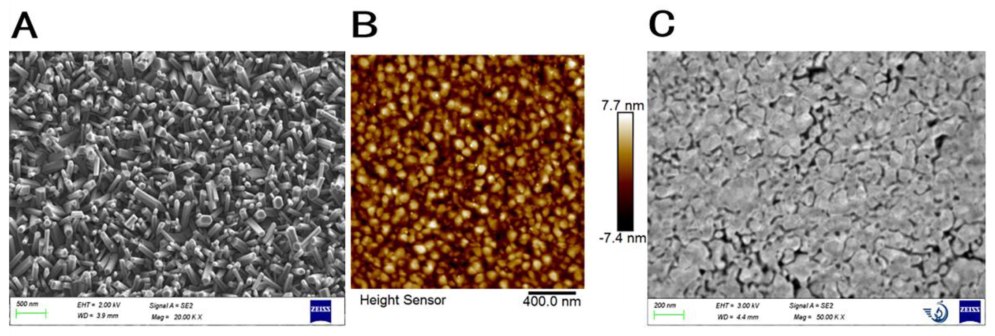
The FTO-ZnO-Au structure was utilized as a transducer for the development of the electrochemical aptasensor towards CEA detection. The surface with ZnO-NRs could improve the coupling efficiency between the transducer surface and aptamers. This could consequently improve the sensing performance for target detection. The surface gold layer was employed to facilitate the immobilization of thiol-modified aptamers onto the sensor surface via the strong Au-S bond. As a result, it is crucial to characterize the surface morphology of the FTO-ZnO-Au structure. In this study, an atomic force microscope (AFM) and scanning electron microscope (SEM) were employed to characterize the electrode surface morphology. The characterization resulting images are shown in detail in Figure 2. As shown in Figure 2A, the SEM result indicated that ZnO-NRs are uniformly and densely distributed on the electrode surface. ZnO-NRs grow approximately vertically, with a hexagonal cross-section and a diameter of ~75 nm. From the AFM result shown in Figure 2B, it is indicated that ZnO-NRs grow uniformly, which further demonstrate the nanostructure on the sensor surface. Figure 2C shows SEM images of the surface of ZnO-NRs after the deposition of a thin layer of chromium and gold. We can see that the chromium and gold layers are evenly distributed on the surface of ZnO-NRs by magnetron sputtering, and the morphology of ZnO-NRs can still be observed, which indicate that the three-dimensional structure of ZnO NRs is still retained on the electrode surface after gold deposition. All the results demonstrate that the FTO-ZnO-Au structure has been successfully prepared and is suitable for further experiments.

3.2. Electrochemical Characterization of Aptasensor Preparation

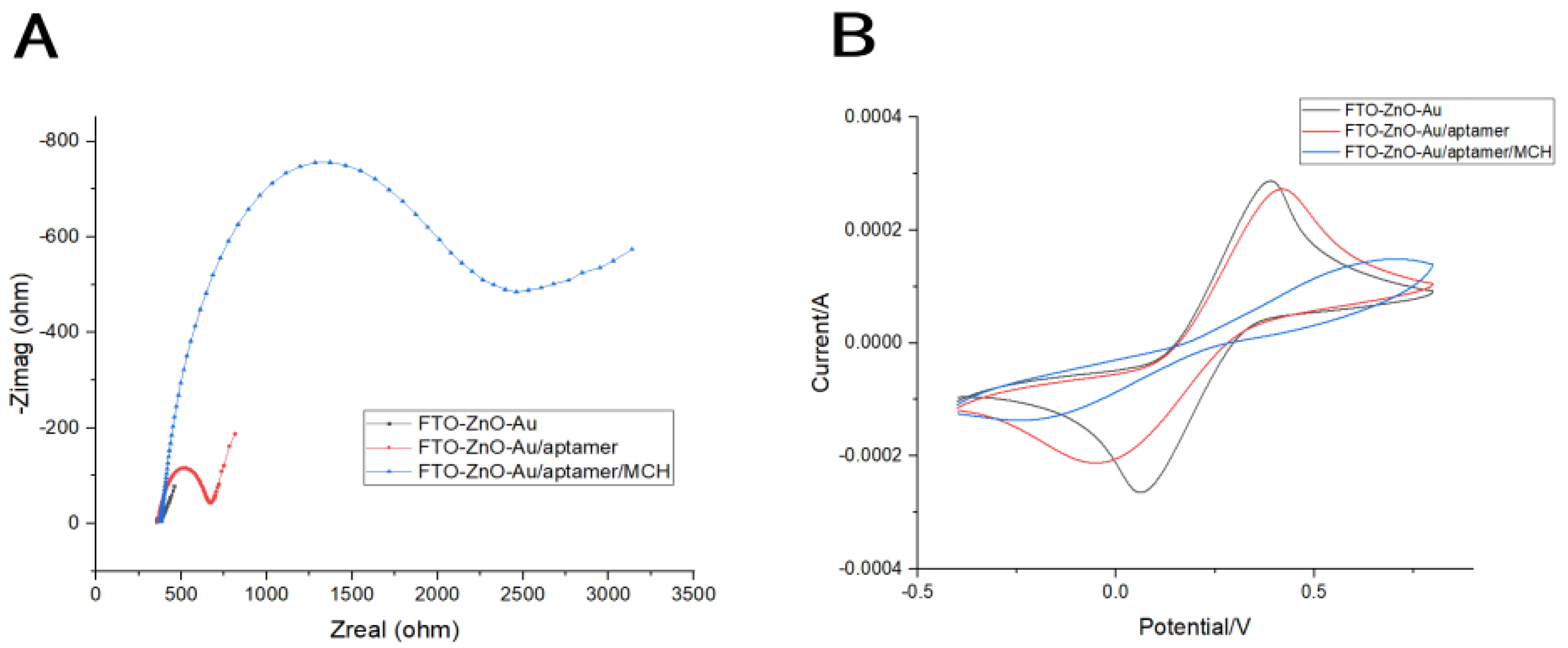
The electrochemical aptasensors were constructed by assembling CEA-sensitive aptamers on the electrode surface, and then treated with 1 mM MCH to block the non-specific sites on the surface of the aptamer-modified electrode (Figure 1). Electrochemical impedance spectroscopy (EIS) and cyclic voltammetry (CV) were performed to describe its electrochemical characterization. For the Nyquist diagram, the semicircle diameter of the electrochemical impedance spectroscopy equals the surface electron transfer resistance (Rct). As shown in Figure 3A, the bare FTO-ZnO-Au electrode showed a very small semicircle domain, which has almost no effect on electron transfer, exhibiting a very fast electron transfer process of [Fe(CN)6]3−/4−. When the aptamer was immobilized onto the electrode surface via the Au-S bond, the Rct value increased significantly. This is mainly due to the negatively charged aptamer acting as an electrostatic barrier in the [Fe(CN)6]3−/4− system, hindering the electron transfer on the chip surface, which leads to an increase in the Rct value. After MCH blocks the aptamer-modified electrode surface, the electron surface defects are repaired, which makes the electron surface layer more compact and further hinders the electron transfer, with a consequent further increase in the Rct value.
Cyclic voltammograms (CVs) were also carried out to describe the electrochemical characterization of the surface modification process. As shown in Figure 3B, the peak current decreased with the connections of aptamers and the closure of MCH and increase in the peak-to-peak separation. This is mainly caused by the electrostatic repulsion between negative charges of the DNA aptamer and the anionic redox probe and the formation of the compact layer by the modification of MCH, which together results in the hampering of the interfacial electron transfer. The CV results were consistent to the EIS measurement results, further confirming the successful fabrication of the sensing interface.

3.3. Optimization of Measurement Conditions

To achieve the best detection performance, we optimized the relevant experimental parameters such as CEA-sensitive aptamer concentrations and incubation time of the CEA-sensitive aptamer. We measure the adhesion of the aptamer to the electrode surface by measuring the electrode surface resistance with EIS. The more CEA aptamers attached onto the electrode surface, the greater Rct value that could be detected.
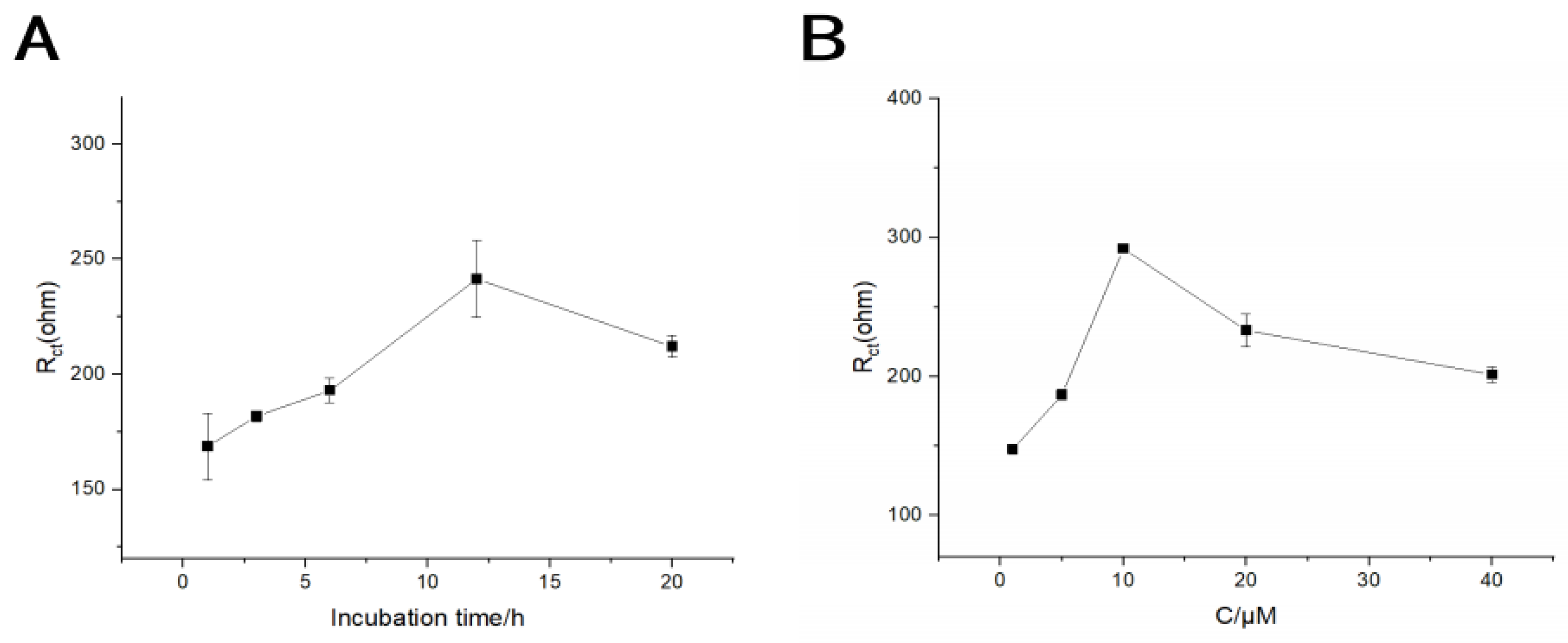
Optimizing the time of the incubate CEA-sensitive aptamer was an important step in building the electrochemical aptasensors. As we can see from the curves shown in Figure 4A, when we use the CEA-sensitive aptamer with a concentration of 5 μM for incubation, the peak Rct value first increased and then decreased with the CEA-sensitive aptamer incubation time increasing from 1 h to 20 h. Meanwhile, an excessive amount of incubation time might have a negative impact on the aptamer attachment onto the electrode surface. Therefore, in this study, 12 h was selected as the appropriate incubation time. Observing the trends depicted in Figure 4B, it was evident that the peak Rct value also initially increased and later leveled off with the increase in CEA-sensitive aptamer concentrations from 1 μM to 40 μM. This indicated that the number of binding sites for the CEA-sensitive aptamer on the electrode surface had reached saturation at the concentration of 10 μM. Therefore, a concentration of 10 μM for the CEA-sensitive aptamer was deemed suitable for optimal performance.

3.4. Analytical Performance Testing of Electrochemical Aptasensor

When different concentrations of CEA were added under optimal conditions, as shown in Figure 5A, the electrode impedance increased with increasing CEA concentrations. The linear regression equation was Rct(ohm) = 27.68 × C[CEA] + 1335.69 (ng/mL) (R2 = 0.9986). The linear range between the electrode impedance and CEA concentrations was from 1 to 80 ng/mL (Figure 5B), with a limit of detection (LOD) estimated to be 0.75 ng/mL (S/N = 3). Typically, the salivary CEA concentration in healthy individuals is found to be minimal (0~3 ng/mL), whereas in cancer patients, the CEA concentration is significantly elevated (>5 ng/mL), correlating with the presence of malignancies [45]. A prior investigation employing the enzyme-linked immunosorbent assay (ELISA) has reported salivary CEA levels of 42.6 ± 21.1 ng/mL in OSCC patients, contrasting with the levels of 22.6 ± 22.1 ng/mL observed in the control cohort [46]. Then, Table 1 summarizes the detection performance of the developed electrochemical aptasensor and the previously reported CEA biosensors. The developed electrochemical aptasensor using the FTO-ZnO-Au nanostructure showed exceptional analytical performance, and was characterized by a wide linear range, a lower LOD, and an exemplary linear correlation. In addition, compared with the anti-CEA antibody-based biosensors, the developed biosensor adopted an aptamer-based strategy to effectively improve the accuracy and reliability of detection.

3.5. Selectivity, Reproducibility, Stability, and Regeneration of the Aptasensor

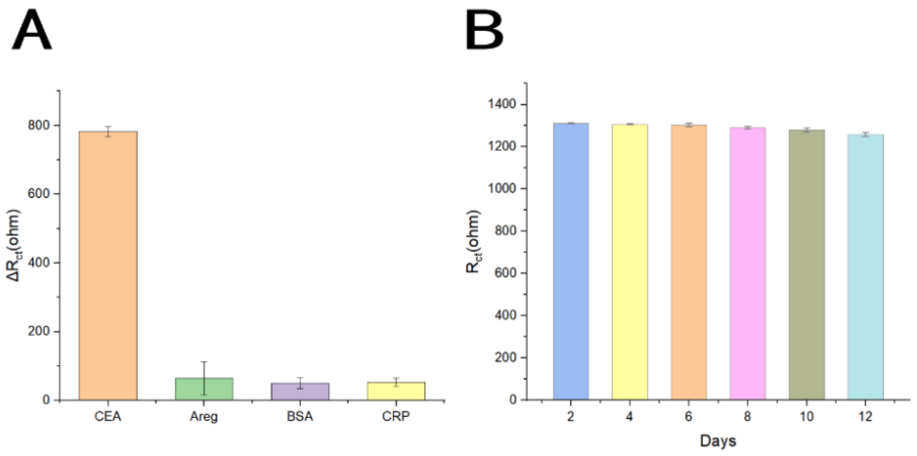
To examine the selectivity of this electrochemical aptasensor, we measured other proteins present in human saliva, such as amphiregulin (Areg), C-reactive protein (CRP), Interleukin-8 (IL-8), and bovine serum albumin (BSA), which is commonly used for blocking. As shown in Figure 6A, only CEA showed significant responses, and other interferers only had subtle signal changes. These similar signal changes indicated that the interfering substances had little effect on the sensor, which proved that the developed aptasensor has high specificity, which can be used for the specific detection of CEA. Secondly, we prepared two batches of six electrodes under the same conditions to detect 5 and 50 ng/mL CEA. The results were basically consistent, and the relative standard deviations (RSDs) were 2.44 and 1.83%, respectively. The detection results showed that this aptasensor had good reproducibility. At the same time, the stability of this biosensor was also investigated (Figure 6B). The results showed that after storage of 12 days at 4 °C, compared with the current test signal in the initial experiment, the signal change rate was 94.52%. Therefore, this aptasensor could be stored stably for at least 12 days. The reasons that make the sensor stable are as follows. Firstly, the gold plating on the electrode surface has good biocompatibility and does not damage the aptamer activity. Secondly, the aptamer is firmly bound to the electrode surface through the Au-SH bond and falling off is not easy. Finally, the steric hindrance effect provided by the three-dimensional structure of ZnO-NRs will also prevent the aptamer from detaching from the electrode surface to a certain extent.
The regeneration of the electrode surface is of great significance for reusable biosensors. In this study, we selected trypsin to digest CEA specifically bound to the aptamer, changing its structure and thus separating it from the aptamer to achieve the purpose of sensor regeneration, and achieved good results. The resistance of the sensor chip incubated with 5 and 60 ng/mL CEA for 5 min by trypsin digestion decreased to 67.04% of the original resistance, and the resistance increased to 89.81% of the original resistance after the same concentration of CEA was incubated again, indicating that the state of the chip surface does not return to the state of unbound protein after digestion, and the activity of the aptamer is affected to a certain extent.

3.6. Anti-Interference Performance and Preliminary Analysis of Human Saliva Samples

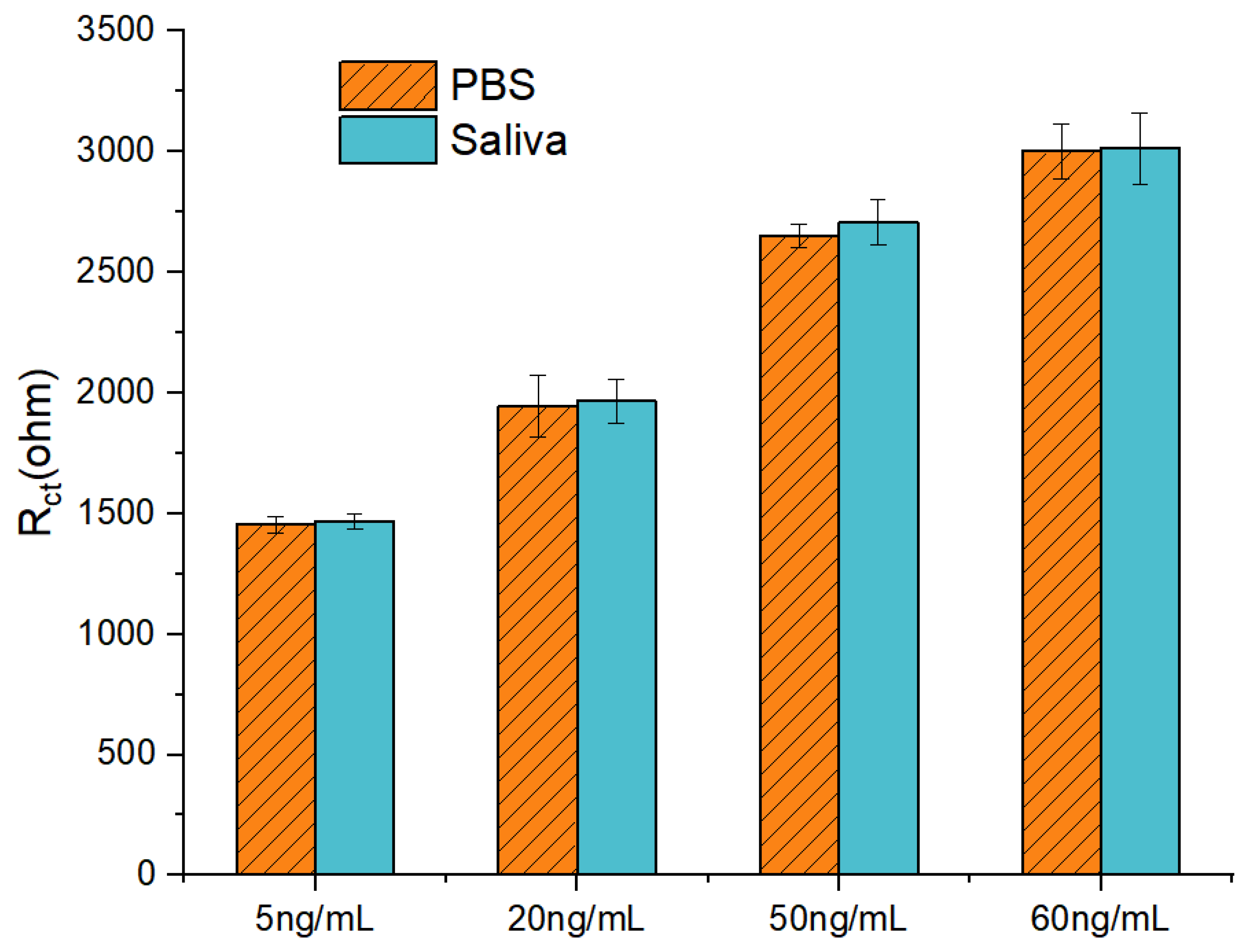
In order to explore the electrode detection performance in the complex saliva environment, we studied the saliva samples of healthy people. We measured the impedance values of CEA concentrations of 5 ng/mL, 20 ng/mL, 50 ng/mL, and 60 ng/mL in 10% healthy human saliva, and compared them with those of corresponding concentrations measured in 0.1 M PBS. As shown in Figure 7, when the CEA concentration is 5 ng/mL, the error of the impedance value measured in the saliva sample is 0.73%; when the CEA concentration is 20 ng/mL, the error of the impedance value measured in the saliva sample is 1.15%; when the CEA concentration is 50ng/mL, the error of the impedance value measured in the saliva sample is 2.07%. When the CEA concentration was 60ng/mL, the impedance error was 0.41%. The above results indicate that the electrode has good anti-interference performance in a complex salivary environment, and has a significant signal for CEA detection.

4. Conclusions

This study developed a simple and highly sensitive aptasensor for detecting the salivary biomarker CEA in OSCC. This aptasensor is based on an FTO-ZnO-Au composite structure, where the corresponding ligands are immobilized on the electrode surface for target protein capture and signal detection. The combination of ZnO-NRs’ electrode and ligands offers multiple advantages, including a high sensitivity, specificity, stability, and ease of fabrication. EIS and CV electrochemical characterization confirmed that the aptasensor was successfully constructed and could effectively capture the target protein CEA. The results showed good linearity in the range of 1~80 ng/mL, with a LOD of 0.75 ng/mL. Experimentally determined tiny responses of AREG, CRP, and BSA to the aptasensor ensure that the aptamer sensor achieves the specific detection of CEA in complex environments. Additionally, consistent detection results were obtained from multiple batches of sensors, confirming their reproducibility. Stability testing over a 12-day period showed excellent performance of the aptasensor. In addition, we found that trypsin had a good digestion effect on the aptamer-bound CEA, but at the same time, it also had a certain effect on the activity of the aptamer, making it less effective for electrode regeneration. This indicated that this aptasensor has great potential for application in the detection of a tumor marker in saliva, and a class of non-invasive, portable, and highly sensitive OSCC detection devices is expected to be developed based on this aptasensor.

Author Contributions

J.L. (Junrong Li): Conceptualization, Investigation, Methodology, Project administration, Writing—Original draft preparation, Writing—Review and editing; Y.D.: Conceptualization, Data curation, Investigation, Methodology, Software, Visualization, Writing—Review and editing; Y.S.: Conceptualization, Data curation, Formal analysis, Methodology, Software, Visualization; Z.L.: Conceptualization, Formal analysis, Methodology, Validation, Visualization; J.L. (Jun Lin): Formal analysis, Investigation, Methodology, Software, Writing—Review and editing; R.C.: Data curation, Investigation, Methodology, Software, Validation; M.W.: Investigation, Methodology, Validation, Visualization; Y.T.: Data curation, Investigation, Methodology, Software; X.Z.: Investigation, Methodology, Resources; Z.Q.: Conceptualization, Data curation, Investigation, Methodology, Software, Validation, Writing—Review and editing; L.D.: Conceptualization, Funding acquisition, Resources, Supervision, Validation, Writing—Review and editing; C.W.: Conceptualization, Funding acquisition, Project administration, Resources, Supervision, Writing—Review and editing. All authors have read and agreed to the published version of the manuscript.

Funding

This research was funded by the National Natural Science Foundation of China (Grant Nos. 32071370, 32271427, and 32471433), and the S&T Program of Hebei (Grant No.: 22321902D).

Institutional Review Board Statement

Not applicable.

Informed Consent Statement

Not applicable.

Data Availability Statement

The data presented in this study are available on request from the corresponding author due to privacy.

Conflicts of Interest

Author Xiaolin Zong was employed by the company Jiashan JunYuan New Material Sci&Tech Co., Ltd. The remaining authors declare that the research was conducted in the absence of any commercial or financial relationships that could be construed as a potential conflict of interest.

References

  1. Badwelan, M.; Muaddi, H.; Ahmed, A.; Lee, K.T.; Tran, S.D. Oral Squamous Cell Carcinoma and Concomitant Primary Tumors, What Do We Know? A Review of the Literature. Curr. Oncol. 2023, 30, 3721–3734. [Google Scholar] [CrossRef] [PubMed]
  2. Bugshan, A.; Farooq, I. Oral squamous cell carcinoma: Metastasis, potentially associated malignant disorders, etiology and recent advancements in diagnosis. F1000Research 2020, 9, 229. [Google Scholar] [CrossRef] [PubMed]
  3. Li, Q.; Hu, Y.; Zhou, X.; Liu, S.; Han, Q.; Cheng, L. Role of Oral Bacteria in the Development of Oral Squamous Cell Carcinoma. Cancers 2020, 12, 2797. [Google Scholar] [CrossRef]
  4. Al Feghali, K.A.; Ghanem, A.I.; Burmeister, C.; Chang, S.S.; Ghanem, T.; Keller, C.; Siddiqui, F. Impact of smoking on pathological features in oral cavity squamous cell carcinoma. J. Cancer Res. Ther. 2019, 15, 582–588. [Google Scholar] [CrossRef]
  5. Imbesi Bellantoni, M.; Picciolo, G.; Pirrotta, I.; Irrera, N.; Vaccaro, M.; Vaccaro, F.; Squadrito, F.; Pallio, G. Oral Cavity Squamous Cell Carcinoma: An Update of the Pharmacological Treatment. Biomedicines 2023, 11, 1112. [Google Scholar] [CrossRef]
  6. Chakraborty, D.; Natarajan, C.; Mukherjee, A. Advances in oral cancer detection. Adv. Clin. Chem. 2019, 91, 181–200. [Google Scholar] [CrossRef]
  7. Khurshid, Z.; Zafar, M.S.; Khan, R.S.; Najeeb, S.; Slowey, P.D.; Rehman, I.U. Role of Salivary Biomarkers in Oral Cancer Detection. Adv. Clin. Chem. 2018, 86, 23–70. [Google Scholar] [CrossRef]
  8. Abati, S.; Bramati, C.; Bondi, S.; Lissoni, A.; Trimarchi, M. Oral Cancer and Precancer: A Narrative Review on the Relevance of Early Diagnosis. Int. J. Environ. Res. Public Health 2020, 17, 9160. [Google Scholar] [CrossRef]
  9. Goldoni, R.; Scolaro, A.; Boccalari, E.; Dolci, C.; Scarano, A.; Inchingolo, F.; Ravazzani, P.; Muti, P.; Tartaglia, G. Malignancies and Biosensors: A Focus on Oral Cancer Detection through Salivary Biomarkers. Biosensors 2021, 11, 396. [Google Scholar] [CrossRef]
  10. Jayanthi, V.; Das, A.B.; Saxena, U. Recent advances in biosensor development for the detection of cancer biomarkers. Biosens. Bioelectron. 2017, 91, 15–23. [Google Scholar] [CrossRef]
  11. Cho, S.; Yang, H.C.; Rhee, W.J. Simultaneous multiplexed detection of exosomal microRNAs and surface proteins for prostate cancer diagnosis. Biosens. Bioelectron. 2019, 146, 111749. [Google Scholar] [CrossRef] [PubMed]
  12. Wu, L.L.; Zhang, Z.L.; Tang, M.; Zhu, D.L.; Dong, X.J.; Hu, J.; Qi, C.B.; Tang, H.W.; Pang, D.W. Spectrally Combined Encoding for Profiling Heterogeneous Circulating Tumor Cells Using a Multifunctional Nanosphere-Mediated Microfluidic Platform. Angew. Chem. Int. Ed. Engl. 2020, 59, 11240–11244. [Google Scholar] [CrossRef] [PubMed]
  13. Zheng, J.; Sun, L.; Yuan, W.; Xu, J.; Yu, X.; Wang, F.; Sun, L.; Zeng, Y. Clinical value of Naa10p and CEA levels in saliva and serum for diagnosis of oral squamous cell carcinoma. J. Oral Pathol. Med. 2018, 47, 830–835. [Google Scholar] [CrossRef]
  14. Rajguru, J.P.; Mouneshkumar, C.D.; Radhakrishnan, I.C.; Negi, B.S.; Maya, D.; Hajibabaei, S.; Rana, V. Tumor markers in oral cancer: A review. J. Fam. Med. Prim. Care 2020, 9, 492–496. [Google Scholar] [CrossRef]
  15. Zarrin, P.S.; Jamal, F.I.; Roeckendorf, N.; Wenger, C. Development of a Portable Dielectric Biosensor for Rapid Detection of Viscosity Variations and Its In Vitro Evaluations Using Saliva Samples of COPD Patients and Healthy Control. Healthcare 2019, 7, 11. [Google Scholar] [CrossRef]
  16. Senf, B.; Yeo, W.-H.; Kim, J.-H. Recent Advances in Portable Biosensors for Biomarker Detection in Body Fluids. Biosensors 2020, 10, 127. [Google Scholar] [CrossRef]
  17. Li, Y.; Hu, S.; Chen, C.; Alifu, N.; Zhang, X.; Du, J.; Li, C.; Xu, L.; Wang, L.; Dong, B. Opal photonic crystal-enhanced upconversion turn-off fluorescent immunoassay for salivary CEA with oral cancer. Talanta 2023, 258, 124435. [Google Scholar] [CrossRef]
  18. Qin, W.; Wang, K.; Xiao, K.; Hou, Y.; Lu, W.; Xu, H.; Wo, Y.; Feng, S.; Cui, D. Carcinoembryonic antigen detection with “Handing”-controlled fluorescence spectroscopy using a color matrix for point-of-care applications. Biosens. Bioelectron. 2017, 90, 508–515. [Google Scholar] [CrossRef]
  19. Rouhi, J.; Kakooei, S.; Sadeghzadeh, S.M.; Rouhi, O.; Karimzadeh, R. Highly efficient photocatalytic performance of dye-sensitized K-doped ZnO nanotapers synthesized by a facile one-step electrochemical method for quantitative hydrogen generation. J. Solid. State Electrochem. 2020, 24, 1599–1606. [Google Scholar] [CrossRef]
  20. Wang, K.; Yang, J.; Xu, H.; Cao, B.; Qin, Q.; Liao, X.; Wo, Y.; Jin, Q.; Cui, D. Smartphone-imaged multilayered paper-based analytical device for colorimetric analysis of carcinoembryonic antigen. Anal. Bioanal. Chem. 2020, 412, 2517–2528. [Google Scholar] [CrossRef]
  21. Wang, L.; Xiong, Q.; Xiao, F.; Duan, H. 2D nanomaterials based electrochemical biosensors for cancer diagnosis. Biosens. Bioelectron. 2017, 89, 136–151. [Google Scholar] [CrossRef] [PubMed]
  22. Zhang, C.; Zhang, S.; Jia, Y.; Li, Y.; Wang, P.; Liu, Q.; Xu, Z.; Li, X.; Dong, Y. Sandwich-type electrochemical immunosensor for sensitive detection of CEA based on the enhanced effects of Ag NPs@CS spaced Hemin/rGO. Biosens. Bioelectron. 2019, 126, 785–791. [Google Scholar] [CrossRef] [PubMed]
  23. Paniagua, G.; Villalonga, A.; Eguílaz, M.; Vegas, B.; Parrado, C.; Rivas, G.; Díez, P.; Villalonga, R. Amperometric aptasensor for carcinoembryonic antigen based on the use of bifunctionalized Janus nanoparticles as biorecognition-signaling element. Anal. Chim. Acta 2019, 1061, 84–91. [Google Scholar] [CrossRef] [PubMed]
  24. Shamsuddin, S.H.; Gibson, T.D.; Tomlinson, D.C.; McPherson, M.J.; Jayne, D.G.; Millner, P.A. Reagentless Affimer- and antibody-based impedimetric biosensors for CEA-detection using a novel non-conducting polymer. Biosens. Bioelectron. 2021, 178, 113013. [Google Scholar] [CrossRef]
  25. Song, Y.; Chen, K.; Li, S.; He, L.; Wang, M.; Zhou, N.; Du, M. Impedimetric aptasensor based on zirconium-cobalt metal-organic framework for detection of carcinoembryonic antigen. Mikrochim. Acta 2022, 189, 338. [Google Scholar] [CrossRef]
  26. Hashkavayi, A.B.; Raoof, J.B.; Ojani, R. Preparation of Epirubicin Aptasensor Using Curcumin as Hybridization Indicator: Competitive Binding Assay between Complementary Strand of Aptamer and Epirubicin. Electroanalysis 2017, 30, 378–385. [Google Scholar] [CrossRef]
  27. Byun, J. Recent Progress and Opportunities for Nucleic Acid Aptamers. Life 2021, 11, 193. [Google Scholar] [CrossRef]
  28. Zhao, H.; Ming, T.; Tang, S.; Ren, S.; Yang, H.; Liu, M.; Tao, Q.; Xu, H. Wnt signaling in colorectal cancer: Pathogenic role and therapeutic target. Mol. Cancer 2022, 21, 123. [Google Scholar] [CrossRef]
  29. Li, J.; Yang, F.; Chen, X.; Fang, H.; Zha, C.; Huang, J.; Sun, X.; Mohamed Ahmed, M.B.; Guo, Y.; Liu, Y. Dual-ratiometric aptasensor for simultaneous detection of malathion and profenofos based on hairpin tetrahedral DNA nanostructures. Biosens. Bioelectron. 2023, 227, 114853. [Google Scholar] [CrossRef]
  30. Maral, M.; Erdem, A. Carbon Nanofiber-Ionic Liquid Nanocomposite Modified Aptasensors Developed for Electrochemical Investigation of Interaction of Aptamer/Aptamer–Antisense Pair with Activated Protein C. Biosensors 2023, 13, 458. [Google Scholar] [CrossRef]
  31. Zhang, X.-W.; Du, L.; Liu, M.-X.; Wang, J.-H.; Chen, S.; Yu, Y.-L. All-in-one nanoflare biosensor combined with catalyzed hairpin assembly amplification for in situ and sensitive exosomal miRNA detection and cancer classification. Talanta 2024, 266, 125145. [Google Scholar] [CrossRef]
  32. Lv, S.; Zhang, K.; Zhu, L.; Tang, D. ZIF-8-Assisted NaYF4:Yb,Tm@ZnO Converter with Exonuclease III-Powered DNA Walker for Near-Infrared Light Responsive Biosensor. Anal. Chem. 2020, 92, 1470–1476. [Google Scholar] [CrossRef] [PubMed]
  33. Que, M.; Lin, C.; Sun, J.; Chen, L.; Sun, X.; Sun, Y. Progress in ZnO Nanosensors. Sensors 2021, 21, 5502. [Google Scholar] [CrossRef] [PubMed]
  34. Shetti, N.P.; Bukkitgar, S.D.; Reddy, K.R.; Reddy, C.V.; Aminabhavi, T.M. ZnO-based nanostructured electrodes for electrochemical sensors and biosensors in biomedical applications. Biosens. Bioelectron. 2019, 141, 111417. [Google Scholar] [CrossRef] [PubMed]
  35. Xia, Y.; Chen, Y.; Tang, Y.; Cheng, G.; Yu, X.; He, H.; Cao, G.; Lu, H.; Liu, Z.; Zheng, S.Y. Smartphone-Based Point-of-Care Microfluidic Platform Fabricated with a ZnO Nanorod Template for Colorimetric Virus Detection. ACS Sens. 2019, 4, 3298–3307. [Google Scholar] [CrossRef] [PubMed]
  36. Sinha, K.; Chakraborty, B.; Chaudhury, S.S.; Chaudhuri, C.R.; Chattopadhyay, S.K.; Das Mukhopadhyay, C. Selective, Ultra-Sensitive, and Rapid Detection of Serotonin by Optimized ZnO Nanorod FET Biosensor. IEEE Trans. NanoBiosci. 2022, 21, 65–74. [Google Scholar] [CrossRef]
  37. Wang, Z.L. From nanogenerators to piezotronics—A decade-long study of ZnO nanostructures. MRS Bull. 2012, 37, 814–827. [Google Scholar] [CrossRef]
  38. Khan, M.; Nagal, V.; Masrat, S.; Tuba, T.; Alam, S.; Bhat, K.S.; Wahid, I.; Ahmad, R. Vertically Oriented Zinc Oxide Nanorod-Based Electrolyte-Gated Field-Effect Transistor for High-Performance Glucose Sensing. Anal. Chem. 2022, 94, 8867–8873. [Google Scholar] [CrossRef]
  39. Lv, M.; Zhou, W.; Tavakoli, H.; Bautista, C.; Xia, J.; Wang, Z.; Li, X. Aptamer-functionalized metal-organic frameworks (MOFs) for biosensing. Biosens. Bioelectron. 2021, 176, 112947. [Google Scholar] [CrossRef]
  40. Wang, Z.; Dai, L.; Yao, J.; Guo, T.; Hrynsphan, D.; Tatsiana, S.; Chen, J. Enhanced adsorption and reduction performance of nitrate by Fe–Pd–Fe3O4 embedded multi-walled carbon nanotubes. Chemosphere 2021, 281, 130718. [Google Scholar] [CrossRef]
  41. Chen, Y.; Wang, A.-J.; Yuan, P.-X.; Luo, X.; Xue, Y.; Feng, J.-J. Three dimensional sea-urchin-like PdAuCu nanocrystals/ferrocene-grafted-polylysine as an efficient probe to amplify the electrochemical signals for ultrasensitive immunoassay of carcinoembryonic antigen. Biosens. Bioelectron. 2019, 132, 294–301. [Google Scholar] [CrossRef] [PubMed]
  42. Jouyandeh, M.; Sajadi, S.M.; Seidi, F.; Habibzadeh, S.; Munir, M.T.; Abida, O.; Ahmadi, S.; Kowalkowska-Zedler, D.; Rabiee, N.; Rabiee, M.; et al. Metal nanoparticles-assisted early diagnosis of diseases. OpenNano 2022, 8, 100104. [Google Scholar] [CrossRef]
  43. Kim, H.-M.; Park, J.-H.; Lee, S.-K. Fiber optic sensor based on ZnO nanowires decorated by Au nanoparticles for improved plasmonic biosensor. Sci. Rep. 2019, 9, 17371. [Google Scholar] [CrossRef] [PubMed]
  44. Luo, C.; Wen, W.; Lin, F.G.; Zhang, X.H.; Gu, H.S.; Wang, S.F. Simplified aptamer-based colorimetric method using unmodified gold nanoparticles for the detection of carcinoma embryonic antigen. RSC Adv. 2015, 5, 10994–10999. [Google Scholar] [CrossRef]
  45. Joshi, S.; Kallappa, S.; Kumar, P.; Shukla, S.; Ghosh, R. Simple diagnosis of cancer by detecting CEA and CYFRA 21-1 in saliva using electronic sensors. Sci. Rep. 2022, 12, 15421. [Google Scholar] [CrossRef]
  46. Honarmand, M.H.; Farhad-Mollashahi, L.; Nakhaee, A.; Nehi, M. Salivary Levels of ErbB2 and CEA in Oral Squamous Cell Carcinoma Patients. Asian Pac. J. Cancer Prev. 2016, 17, 77–80. [Google Scholar] [CrossRef]
  47. Xu, Z.; Wang, C.; Ma, R.; Sha, Z.; Liang, F.; Sun, S. Aptamer-based biosensing through the mapping of encoding upconversion nanoparticles for sensitive CEA detection. Analyst 2022, 147, 3350–3359. [Google Scholar] [CrossRef]
  48. Shahbazi, N.; Hosseinkhani, S.; Ranjbar, B. A facile and rapid aptasensor based on split peroxidase DNAzyme for visual detection of carcinoembryonic antigen in saliva. Sens. Actuators B Chem. 2017, 253, 794–803. [Google Scholar] [CrossRef]
  49. Yu, Q.; Wang, X.; Duan, Y. Capillary-Based Three-Dimensional Immunosensor Assembly for High-Performance Detection of Carcinoembryonic Antigen Using Laser-Induced Fluorescence Spectrometry. Anal. Chem. 2014, 86, 1518–1524. [Google Scholar] [CrossRef]
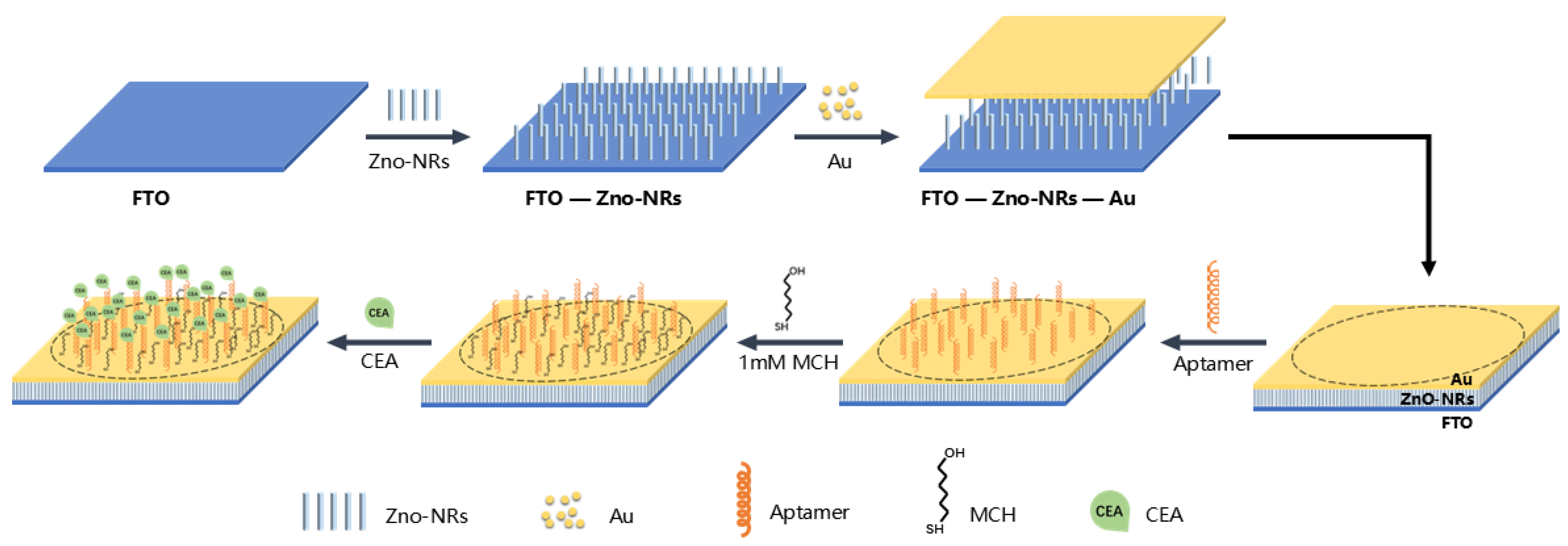
Figure 1. Schematic diagram showing preparation process of electrochemical aptasensors based on FTO-ZnO-Au structure and CEA-sensitive aptamer.
Chemosensors 12 00203 g001
Figure 2. (A) The SEM characterization of ZnO−NRs; (B) the AFM characterization of FTO−ZnO−Au; (C) an SEM image shows the surface of ZnO−NRs after the deposition of a thin layer of gold.
Chemosensors 12 00203 g002
Figure 3. (A) The Nyquist plots and (B) CVs of the preparation steps of the electrochemical aptasensor based on the CEA-sensitive aptamer.
Chemosensors 12 00203 g003
Figure 4. Optimization of aptasensor preparation conditions. (A) Influence of incubation time of CEA-sensitive aptamer (5 μM); incubation time of aptamer is 1 h, 3 h, 6 h, 12 h, and 20 h. (B) Influence of concentrations of CEA-sensitive aptamer (incubating for 12 h); concentrations of CEA-sensitive aptamer are 1 μM, 5 μM, 10 μM, 20 μM, and 40 μM.
Chemosensors 12 00203 g004
Figure 5. (A) EIS responses of the electrochemical aptasensor for the detection of different concentrations of CEA. (B) The linear calibration plot between the electrode impedance and CEA concentrations.
Chemosensors 12 00203 g005
Figure 6. (A) Selectivity of the aptasensor against CEA (20 ng/mL), Areg (50 ng/mL), BSA (50 ng/mL), and CRP (20 ng/mL). (B) Stability of the aptasensor for 12 days of storage.
Chemosensors 12 00203 g006
Figure 7. The impedance values of CEA concentrations of 5 ng/mL, 20 ng/mL, 50 ng/mL, and 60 ng/mL were detected in 0.1 M PBS and 10% saliva.
Chemosensors 12 00203 g007
Table 1. Comparison of analytical methods for the detection of CEA.
Analytical MethodSensitive ElementsAnalytesLinear Ranges (ng/mL)LOD (ng/mL)R2ReferenceReal Sample Test
FluoroimmunosensorAnti-CEA antibodyCEA1 × 10−1~2.5
2.5~20
1.0 × 10−10.988
0.996
[17]Yes
AptasensorAptamerCEA2 × 10−2~6.03.25 × 10−10.962[47]No
AptasensorAptamerCEA1~501.00.990[48]Yes
FluoroimmunosensorAnti-CEA antibodyCEA7 × 10−1~805.5 × 10−30.993[49]Yes
AptasensorAptamerCEA1~807.5 × 10−10.999This workYes
Disclaimer/Publisher’s Note: The statements, opinions and data contained in all publications are solely those of the individual author(s) and contributor(s) and not of MDPI and/or the editor(s). MDPI and/or the editor(s) disclaim responsibility for any injury to people or property resulting from any ideas, methods, instructions or products referred to in the content.