Review

Recent Advances in Quartz Crystal Microbalance Biosensors Based on the Molecular Imprinting Technique for Disease-Related Biomarkers

Department of Chemistry, Hacettepe University, Ankara 06800, Turkey
*
Author to whom correspondence should be addressed.
Chemosensors 2022, 10(3), 106; https://doi.org/10.3390/chemosensors10030106
Submission received: 10 February 2022 / Revised: 4 March 2022 / Accepted: 8 March 2022 / Published: 10 March 2022

Abstract

:
The molecular imprinting technique is a quickly developing field of interest regarding the synthesis of artificial recognition elements that enable the specific determination of target molecule/analyte from a matrix. Recently, these smart materials can be successfully applied to biomolecule detection in biomimetic biosensors. These biosensors contain a biorecognition element (a bioreceptor) and a transducer, like their biosensor analogs. Here, the basic difference is that molecular imprinting-based biosensors use a synthetic recognition element. Molecular imprinting polymers used as the artificial recognition elements in biosensor platforms are complementary in shape, size, specific binding sites, and functionality to their template analytes. Recent progress in biomolecular recognition has supplied extra diagnostic and treatment methods for various diseases. Cost-effective, more robust, and high-throughput assays are needed for monitoring biomarkers in clinical settings. Quartz crystal microbalance (QCM) biosensors are promising tools for the real-time and quick detection of biomolecules in the past two decades A quick, simple-to-use, and cheap biomarkers detection technology based on biosensors has been developed. This critical review presents current applications in molecular imprinting-based quartz crystal microbalance biosensors for the quantification of biomarkers for disease monitoring and diagnostic results.

Graphical Abstract

1. Introduction

Quartz crystal microbalance (QCM) based on piezoelectric phenomena has arisen as a significant biosensing system employing label-free and real-time biorecognition mechanism, which enables the detection of a broad variety of biomolecules such as proteins, nucleic acids, peptides, oligonucleotides, hormones, etc. [1,2,3]. QCM is a reliable, low-cost, and sensitive biosensing tool with a short detection time, which offers improvement in early and accurate clinical diagnosis, and which could facilitate the disease-treatment process [4]. Thus, the detection and quantification of disease-related biomarkers in blood, urine, saliva, etc., play a crucial role. The early detection of typical biomarkers related to different types of cancers and chronic diseases such as rheumatoid arthritis and Alzheimer’s disease, and infectious diseases such as SARS, Ebola, and Zika, will aid in the struggle with these diseases. The biomarkers include bacteria, viruses, and their residues, such as proteins and nucleic acids, and antibodies that act against the pathogens [5,6,7].
The conventional analytical methods, including enzyme-linked immunosorbent assay, immunofluorescence, radioimmunoassay, polymerase chain reactions, etc., are commonly utilized for the diagnosis of many diseases. However, these techniques require a multi-step procedure, expensive instrumentation, expert operators, and they lack onsite applicability; these disadvantages lead to high cost, a remarkable delay in sample collection, and time consumption [8,9]. Accordingly, there is a significant demand for sensitive, specific, cost-efficient, rapid, equipment-free, and on-site applicable systems for accurate diagnoses [10].
In recent years, biosensors have emerged as one of the most researched topics with label-free and real-time potential applications. Biosensors, as analytical tools, convert the biochemical/biological responses into a measurable output signal [11]. Thus, they do not require extra processing steps or chemicals during sample collection (or sampling) and signal output; they are novel, promising analytical tools for quantitative, fast, on-site, and economic biorecognition measurements [12,13].
A biosensor involves a sensing component with a physicochemical transducer that can be electrochemical, optical, piezoelectric, thermal, magnetic, etc. [14]. QCM technology is a sensor system that incorporate a piezoelectric material, which relies on the piezoelectric effect of quartz crystals. A QCM-based device offers a bulk thickness-shear-mode mass-sensing acoustic wave that responds to any changes in the resonant frequency related to the mass accumulated on the surface of a quartz crystal resonator (any piezoelectric material) [15,16,17]. QCM-based biosensors have emerged as efficient tools to detect and quantify disease-related biomarkers [4]. The integration of molecularly imprinted materials as biorecognition units with QCM-based biosensors provides these sensing platforms with some significant advantages, such as notably long-term storage stability, potential reusability, and resistance to environmental conditions such as temperature and pH changes.
The molecular imprinting technique is a creative approach that enables the building of artificial biorecognition sites that mimic native biological structures, such as antibodies, enzymes, etc. The novelty of this approach relies on the polymerization of a functional monomer with excess crosslinking agents in the presence of the target analyte [18,19,20]. Several review papers have focused on MIP-based sensors. The principles of MIPs and microfluidic systems and the integration of MIPs with microfluidic systems for point-of-care applications have been summarized [21]. The research field has highlighted MIP-based electrochemical, mass-sensitive, and optical sensors and their advantages [22,23]. A comprehensive review of recent reports in the environmental and biomedical fields, with a focus on electrochemical and optical signaling mechanisms, has been presented [24].
This review article presents an overview of QCM systems based on the molecular imprinting technique as promising in providing biosensing applications for disease biomarkers., The accurate, rapid, and on-site detection process, with high sensitivity and specificity, as provided by QCM systems based on the molecular imprinting approach, has gained attention as a novel promising diagnostic platform. The biosensing of disease biomarkers via QCM systems based on the molecular imprinting approach is examined, and the outcome and future aspects of molecular imprinting-based QCM biosensors as an accurate diagnostic approach for disease biomarkers are evaluated. The general scheme of this review is as follows: an explanation of biosensor devices is provided in Section 2; a general overview of QCM biosensor technology is explained in Section 2.1; a wide review of the molecular imprinting technique is provided in Section 3; the performances and characteristics of biomarker detection applications are discussed in Section 4; and the conclusions and a discussion of future perspectives are added in Section 5.

2. Biosensor Devices

Biosensors are analytic tools that detect biomolecules and are employed in clinical settings for the determination of disease-related markers in biological fluids for the monitoring of disease and the discovery of drugs [25,26]. A sensor is an analytical device that works to detect a particular analyte in a matrix sample [27]. Biosensors are excellent devices comprised of biological and physico-chemical components to detect a target analyte by producing a measurable signal [28,29,30]. These promising tools, with real-time and label-free application potential, have recently attracted attention as hypersensitive early-detection devices for a broad variety of molecules [31]. The biosensor history began with Nelson and Griffin in 1916 [32], based on protein immobilization onto a solid substrate [31]. The main components of a biosensor are the biorecognition element (often called the receptor), which binds with the target molecule (or analyte), and the transducer, which then transmits a molecular recognition incident as a measurable signal (Figure 1).
Figure 1’s schematics illustrate the components of a classic biosensor platform: (i) the receptor (the biorecognition element) specifically interacts with the analyte or molecule of interest; (ii) a specific biological incident occurs and provides an increase to a signal collected by the transducer; (iii) the transducer device signal is converted to a measurable electronic signal; and (iv) computer software forms a meaningful physical parameter [33]. Biosensors can be categorized into different transducer groups for the generation of the output signal, including optical, electrochemical, gravimetric, magnetic, and piezoelectric transducers [34,35,36,37]. The selection of a transduction device is primarily based on the natural and physicochemical features of the surface layer, which change when interacting with the target molecule [38].
Piezoelectric materials that act as an electromechanical transducers are fitted for use as biosensors and actuators in tools and are good structures [39,40]. The piezoelectric, mass-sensitive, or acoustic wave tools present label-free and quick detection of molecules due to mass, which is the main feature of each molecule. These transducers include (i) the surface acoustic wave (SAW) devices [41,42]; (ii) the bulk acoustic wave (BAW) devices and quartz crystal microbalance (QCM) devices [43,44,45,46]; and (iii) the shear transverse wave (STW) devices [47], which are constantly combining with various bioreceptor elements for biomedical diagnosis and molecular recognition [48]. Quartz crystal microbalance biosensors are needed for the determination of a broad variety of biomolecules. They are effective analytical devices [49,50,51]. Compared to traditional methods, they provide label-free and real-time detection, easy use with modern technologies, portable size, high sensitivity, low cost, and basic data analysis [52,53,54].
It is very important to use accurate and timely detection methods to prevent the progression of a disease and stop the chain of transmission with early diagnosis. Traditional assays are often time-consuming and expensive. It is necessary to develop clinically sensitive, rapid, and cost-effective diagnostic methods. QCM biosensors are one of these technologies. QCM technologies have emerged as a robust biosensing platform due to their label-free mechanism that provides the detection and quantification of a broad variety of biomolecules [4].

2.1. QCM Technology

The AT-cut quartz crystal is a typical QCM electrode. The Sauerbrey equation displays the mass sensitivity of the quartz crystal electrode: a rise in mass collected on the quartz surface results in a decrease in the resonant frequency of the oscillator in the gas phase, called the Sauerbrey effect [55,56,57].
According to Sauerbrey,
S a = Δ f Δ m = 2 / ρ v   f 0 2 / n     ( Hz   cm 2   ng 1 )
where Δm is the mass deposited per unit area on the crystal surface (g), Δf is the change in resonant frequency shift (Hz), v is the velocity of the wave (m/s), ρ is the quartz density (2.648 g/cm3), and n is the harmonic number (n = 1, 3, …). Also, the limit of detection (LOD) of a QCM biosensor can be calculated as,
Δ m m i n = Δ f m i n / S a  
where Δm represents the change in mass on the electrode surface, and Δf is the time-dependent change in the frequency of the material in the oscillating circuit.
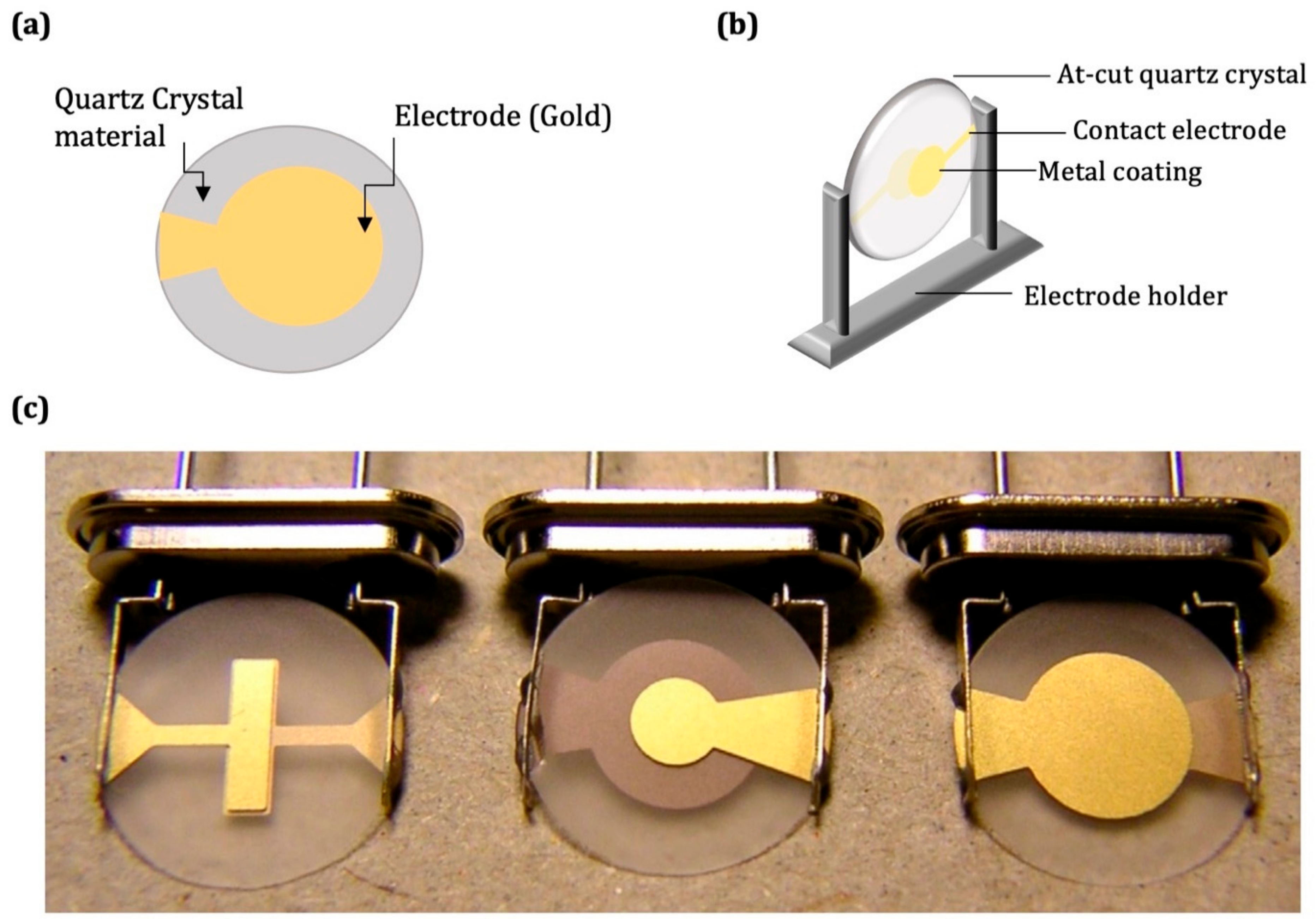
The quartz crystal electrode’s top view is displayed in Figure 2a. The lower and upper piezoelectric electrodes operate the resonation of the piezoelectric biosensor (Figure 2b). A schematic diagram of quartz crystal and three different quartz crystals (AT-cut 10 MHz) are provided in Figure 2c [58,59].
Piezoelectric materials include ceramics, crystals, polymers such as polyvinylidene fluoride, and piezoelectric composites [60]. Anisotropic materials such as poly(vinylidene fluoride) (PVDF) [61,62], zinc oxide (ZnO) [63], aluminum nitride (AlN) [64,65], barium titanate (BaTiO3) [66], quartz (SiO2) [67,68], and lead titanate (PbTiO3) [69,70] are mainly utilized as biosensor materials [71]. The piezoelectric biosensor vehicle, based on a label-free technique, has achieved a big advance [72] and has been successfully carried out in applications of biomolecule detection. Therefore, the QCM biosensor is the most popular in various fields, such as food control [73], environmental monitoring [74], drug function mechanisms [75,76], etc. Piezoelectric biosensors, which are mass-based chemical sensors (e.g., QCM), have important properties, such as high sensitivity and selectivity, ease of use, cost-effectiveness, stability, portability, and simplicity. Several methods can be applied to design QCM-based biosensor surfaces for various application areas. The QCM is also a sensitive and universal device for measuring concentrations of various gases (e.g., aldehydes) in the air [77,78,79]. The broad uses of QCM-based biosensors have been referred to in published articles concerning the detection of various molecules, including proteins [80], enzymes [81], peptides [82], drugs [83], vitamins [84], metals [85], pesticides [86], biomarkers [87], antibiotics [88], bacteria [89], alcohols [90], aldehyde [91], furanic compounds [92], etc. Mass-sensitive-based QCM biosensors are commonly employed for the detection of disease-related biomarkers.

3. Molecular Imprinting Technique

The ability to specifically bind and recognize complementary substrates present in the complex matrix is fundamental for molecular recognition. Selective binding takes place by various secondary interactions, including hydrophobic interactions, electrostatic interactions, hydrogen bonds, and weak metal chelates. Molecular imprinting is a technique that mimics the recognition events observed in biomolecular recognition processes [93]. The molecular imprinting technique (MIT) has been broadly used in different areas due to its smart and unique properties of application universality, recognition specificity, and structure predictability [94]. MIT has quickly advanced, and this technique has become one of the most critical technologies in preparing artificial recognition materials [95,96,97,98]. Molecularly imprinted polymers (MIPs) have received significant attention as promising alternatives to natural recognition elements. In general, MIPs are synthesized through self-assembly of target/template molecules, complementary functional monomers, and suitable cross-linker monomers (Figure 3) [99]. MIPs receive considerable attention, due to their recognition specificity, structure predictability, and application universality, as well as their simplicity, inexpensiveness, and robustness [22,100].
The MIT can be performed in different steps. However, the common synthesis steps are as follows: (i) a template or pre-complex of the target molecule is prepared, which is covalently or non-covalently bound to the functional monomer pre-complex; (ii) the polymerization step is initiated with the appropriate promoter pair; (iii) the regeneration step, in which the template is removed (desorbed) from the polymer matrix; and (iv) the final step, when the MIP interacts with the sample containing the template molecule and template molecule-specific recognition sites are created. The MIT’s general approach is shown in Figure 3 [101,102].
In general, there are three methods for the molecular imprinting technique to act as a recognition mechanism: metal-coordinating imprinting, covalent imprinting, and non-covalent imprinting. Molecular imprinting techniques can generally be categorized into three approaches: surface imprinting, epitope imprinting, and bulk imprinting [103]. Pioneering studies with covalent imprinting and non-covalent imprinting techniques were reported for the first time by Wulff and Mosbach.
MIPs are also notable for their perfect physical and chemical stability compared to the biorecognition element [104,105,106,107,108,109,110]. These properties have provided for the application of MIPs in different areas, including purification [111], separation sciences [112], decontamination [113], food safety [114], chemical biosensing [115], microfluidic chip device [116], immunoassays [117], therapy [118], drug delivery [119], and cell imaging [120].
The effect of the nature and the volume of the solvent on the analyte desorption (or elution) is a very critical step. With this objective, different solvent mixtures and volumes are put in contact with the MIPs. Various elution solutions are employed as desorption agents to break the covalent or non-covalent interactions. The traditional elution solvent is a particular ratio of methanol/acetic acid mixture, buffer, and NaCl solution for template removal. The elution solution should not produce impurities and should not destroy the polymer structure. Therefore, it is necessary to find new elution materials for the removal of target template molecules. Finally, optimal elution and adsorption time can be developed for this purpose. The non-covalent imprinting technique overcomes the limitations of the covalent imprinting method, which requires a difficult desorption process with a hard eluent through non-covalent interactions. Moreover, the surface imprinting technique provides a fast adsorption/elution rate, good stability, and an easy separation characteristic [121,122,123,124].
The prepared MIPs have demonstrated highly selective and sensitive recognition capabilities, which have great application potential in real and complex samples [125]. To define the success of the imprinting coefficient and the magnitude of the imprinting effect, the synthesis process of MIPs is accompanied by the synthesis of a control polymer called the non-imprinted polymer (NIP), which is generally prepared without a template analyte to check whether any specific cavities for the template analyte have been formed. Using merit figures, which contain an imprinting factor (IF) and selectivity coefficient, the imprinting operation can be categorized as successful or not. The IF and selectivity coefficient is employed to determine the magnitude of imprinting and should be greater than one (i.e., IF > 1) for any successful imprinting. IFs are dependent on the concentration of the template analyte [126].
The difference between the initial amount of template analyte in the solution and the amount in the final solution leads to the determination of the equilibrium binding capacity (Q) or the distribution coefficient (Kd), as per Equations (3) and (4):
Q = C i C f m V
K d = ( C i C f ) V m × C f
where Ci is the initial concentration of the analyte, Cf is the equilibrium concentration of the analyte in solution, m is the mass of the polymer, and V is the volume of the solution.
IF is defined by Equations (5) and (6):
I F = Q M I P Q N I P
I F = K d M I P K d N I P
where QMIP is the amount of analyte bound by MIP, QNIP is the amount of analyte bound by NIP, Kd(MIP) is the distribution coefficient of MIP, and Kd(NIP) is the distribution coefficient of NIP.
Likewise, selectivity is defined by selectivity factor (α), selectivity coefficient (k), or relative selectivity coefficient (k′), as determined by Equation (7):
k = k M I P k N I P
The selectivity coefficient (SC) is also calculated, according to the following Equation (8) [127]:
S C = I F t e m p l a t e   a n a l y t e I F o t h e r   a n a l y t e s
The main advantages of MIPs are their high affinity and selectivity for the template analyte chosen in the molecular imprinting technique procedure [128]. MIPs have resistance to elevated temperature and pressure, higher strength, physical robustness, and inertness towards bases, acids, and organic solvents [129]. Additionally, they are inexpensive to synthesize, and the storage life of the polymers can be very great, maintaining their specific recognition capacity at room temperature for several years [130]. MIPs prepared in a solid phase are very stable, with long operational and shelf lives [107]. Shelf-life studies conducted at different periods were evaluated differently for each MIP fabrication [131]. As a result, the stable and reusable properties have increased the long shelf-life usability of MIPs. Some studies reported the shelf life of MIPs is a minimum of 6 weeks [132] and a maximum of 6 [133] to 12 months [134] for MIP-based biosensors stored at 25 °C. This method is considered a versatile and hopeful technique that can recognize both biological and chemical analytes, including nucleotides [135], amino acids [136], enzymes [137], proteins [138], phosphoproteins [139], viruses [140], bacteria [141], pollutants [142], dyes [143], food poisons [144], pesticides [145], and toxins [146,147].
Disease-related imprinted biomarkers have attracted important interest due to their excellent stability, simplicity, quickness, high selectivity, eco-friendliness, and low cost [94]. Here, we aim to examine the recent advances of MIP-based QCM biosensors for the detection of disease-related biomarkers, focusing on imprinting applications. Various imprinting methods for biomarkers-imprinted polymer preparation are comprehensively summarized.

4. QCM Biosensors Based on Molecular Imprinting for Biomarkers

With the coronavirus (COVID-19) pandemic globally, people are making increased and more urgent demands in relation to disease prevention, diagnosis, and treatment. The detection and determination of disease-related biomarkers help to provide a diagnosis before a disease becomes incurable in later stages. In recent years, MIP-based QCM biosensors have been significantly researched as promising analytic tools in several respects, such as use in clinical analysis, providing specificity, providing fast responses, offering desired portability and low cost. This critical review aims to offer readers a critical overview of the recent significant success in MIP-based QCM biosensor tools in the detection of biomarkers, as indicated in chosen publications from 2010 to 2022.

4.1. Protein

Glycoprotein detection holds significant potential for the early diagnosis of different diseases [148]. Zhang et al. developed an oligomer-immobilized QCM biosensor for the detection of glycoprotein. In the present study, a Stanford Research Systems (USA) QCM biosensor platform was used. First, the oligomer was prepared as follows: 10 mL of a monomer solution containing a combination of acrylamide (AAm), glycidyl methacrylate (GMA), lauryl methacrylate (LMA), 4-Vinylphenylboronic acid (VPBA) poly(ethylene glycol), and monomethacrylate (PEGMA) were prepared. Polymerization was started by using 2,2′-Azobisisobutyronitrile (AIBN) as the initiator, and 2-mercaptoethanol was used as a chain transfer agent. Polymerization conditioning was carried out at 65 °C temperature for 27 h. Then, the cysteamine-modified gold QCM chip was cleaned using piranha solution. Oligomer and ovalbumin (OVA) complexes were prepared at a 1:20 volume ratio. The co-assembly and immobility of oligomer and OVA were conducted by dipping at 37 °C for 5 h. The protein concentration range was from 1 × 10⁻⁷ g/mL to 1 × 10⁻⁴ g/mL. Bovine serum albumin (BSA), lysozyme (LYZ) and horseradish peroxidase (HRP) were chosen for the selectivity studies. In the same data processing, the frequency shifts of OVA were over three times higher than those of other proteins [149].
Immunoglobulin M (IgM) has an important role in the first reply of the immune system to foreign antigens. The IgM amount can be utilized to predict the immune function in human serum and is a significant factor for diagnosing rheumatoid arthritis, acute and chronic hepatitis, hepatocirrhosis, and malignant plasma cell tumors. As a result, the selective and sensitive detection of IgM is essential. Diltemiz et al. designed a QCM biosensor based on molecularly imprinted polymers for the detection of IgM and mannose molecules. The methacryloylamidophenylboronic acid (MAPBA) and the mannose were utilized as functional monomers and as a template, respectively. First, QCM chips were modified with 2-propene-1-thiol to form mannose-binding regions onto a QCM electrode bare gold layer. The ethylene glycol dimethacrylate (EGDMA) was utilized as a cross-linker, and AIBN was used as an initiator. Thereafter, a MAPBA/mannose pre-complex mixture, EGDMA, and an AIBN monomer system were dropped onto the QCM chip surface. Finally, the mixture was kept on the QCM chip surface under UV light for coating a molecularly imprinted film. The bare QCM chip and the modified QCM chip were characterized by Atomic Force Microscopy (AFM). Non-imprinted-based QCM chip was also prepared for imprinting factor calculation. The detection of the mannose and IgM solutions was carried out at a broad concentration range, from 0.01 mM to 0.1 mM [150].
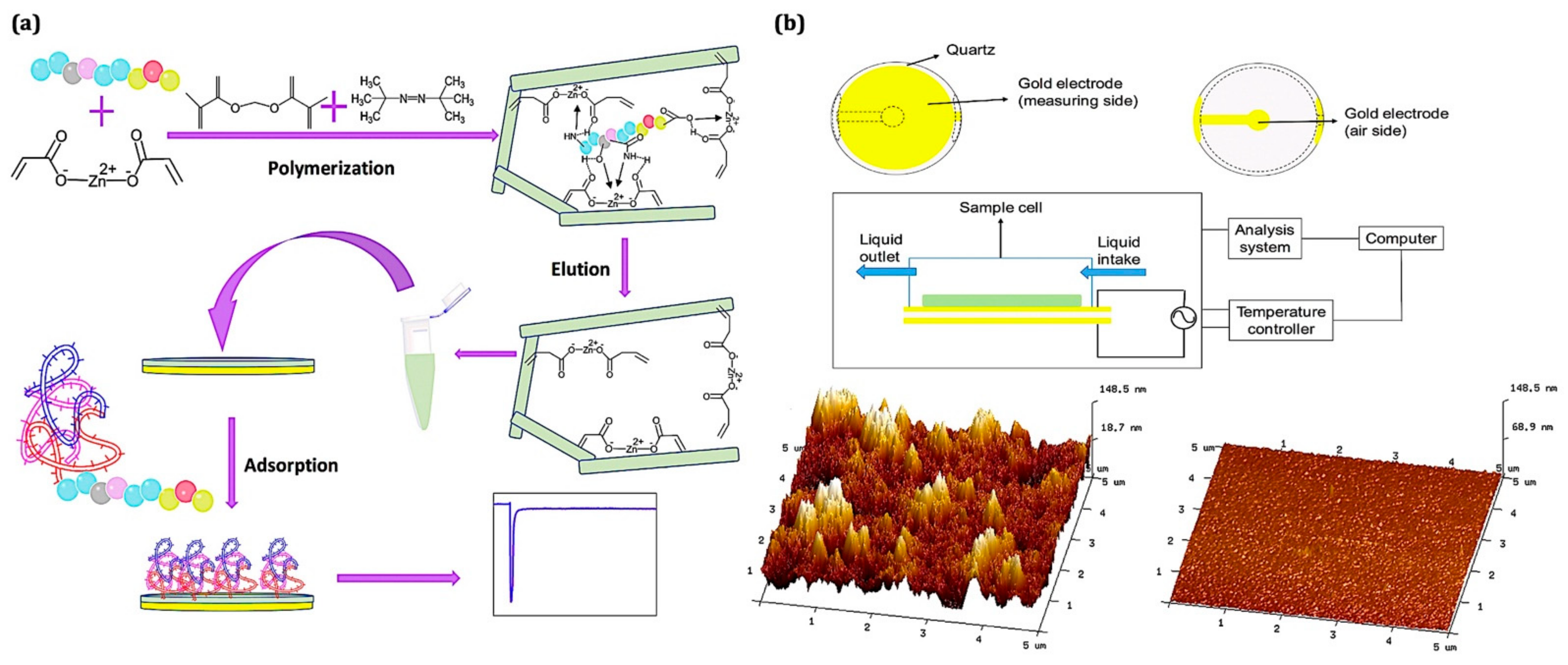
The quantitative detection of biological macromolecules is of major importance in medicine, diagnostics, and biology. Human serum albumin (HSA) is an essential component and the most-abundant plasma macromolecule in plasma (∼60%). A changing HSA concentration can be a signal of multiple myeloma or coronary heart disease. A low HSA concentration is a marker of cirrhosis, chronic hepatitis, and liver failure. As a result, the detection of HSA levels is of great significance. Ma et al. developed an epitope imprinting-based coated QCM biosensor for human serum albumin detection. The epitope MIP was synthesized by using the C-terminus epitope of human serum albumin as the template molecule and zinc acrylate as the functional monomer in N,N-dimethylformamide (DMF). An epitope non-imprinted QCM sensor was also prepared for imprinting factor calculation. The prepared epitope-imprinted polymer-coated QCM biosensor displayed a linear range for HSA between 0.050 and 0.500 μg/mL. The limit of detection value was 0.026 μg/mL. It was reported that the imprinting factor was calculated as 6.9. The results showed that the epitope-MIP had a perfect imprinting effect on the HSA template molecule. The epitope-imprinting QCM biosensor design procedure and the QCM chip characterization results are given in Figure 4 [151].
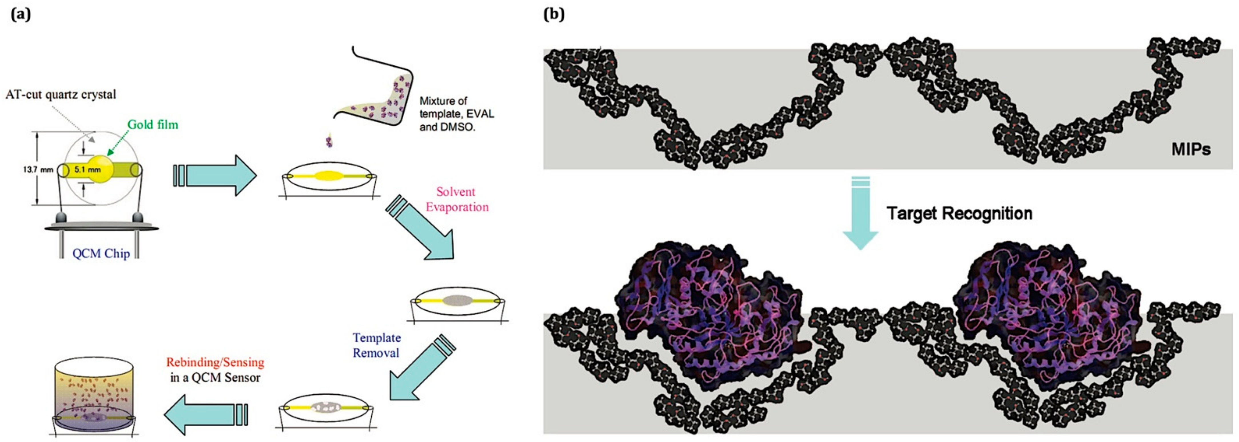
Human saliva as a diagnostic fluid can provide a non-invasive, safe, simple, and inexpensive approach for disease detection. Lee et al. developed a MIP thin film-coated QCM biosensor for sensing digestive protein, including lysozyme, lipase, and amylase. The non-covalent recognition of template proteins by the MIP technique was also examined. They reported that incorporating MIPs in a QCM is a highly selective approach for the real-time sensing of salivary proteins on the molecularly imprinted nanofilm. The limits of detection were as low as ∼pM for those salivary proteins [152]. The design of the amylase-imprinted polymer and the possible recognition mechanism of amylase on a QCM biosensor chip is provided in Figure 5.
Epitope technology, as an alternate technique to related-target molecules, has been suitably used as template molecules to produce the specific recognition sites for proteins. Gupta et al. developed an epitope-imprinting technique for the detection of a target epitope sequence using electrochemical quartz crystal microbalance (EQCM). The fbp A protein template sequence present in Neisseria meningitidis (N. meningitidis) bacteria were selected. Thiol chemistry was employed to form the self-assembled monolayer (SAM) onto the bare surface of the EQCM electrode to orient the peptide sequence. The benzyl methacrylate and 3-sulfopropyl methacrylate potassium-salt were employed as multiple monomers. N,N-methylene-bis-acrylamide as cross-linker and azo-isobutyronitrile was used as cross linker and initiator, respectively. Multiple monomers were chosen to provide various noncovalent interactions. The limit of detection (LOD) was found to be 1.39 ng/mL. The epitope molecularly imprinted/nonimprinted imprinting factor was found to be 12.27 [153].
The α1-Acid glycoprotein (α1-AGp) is the crucial plasma protein in its role as a marker for various illnesses when generated in high amounts from 1.2 mg/mL. Nasrullah et al. reported the MIP-Cu2O-decorated reduced graphene oxide (Cu2O-rGO) hybrid coatings on the QCM biosensor for detection of α1-AGp. The high selective coatings based on the MIP matrix were fabricated with boronate-affinity and Cu2O-rGO nanomaterials. The MIP-based biosensor can successfully detect 150–200 ng/mL of α1-AGp in spiked human serum samples. They reported that MIP-Cu2O-rGO-based QCM biosensors can be successfully employed for accurate and label-free detection of α1-AGp. The limit of detection was found to be 0.25 ng/mL. They also prepared non-imprinted polymer for comparison with the sensor response [154].

4.2. Bacteria

There is an urgent need for the design of sensitive, selective, and quick biosensors for the detection of bacteria in areas such as food analysis, health care, security, and environmental monitoring. Diseases related to pathogenic bacteria are a continuing major public health problem [155]. Latif et al. reported the use of a MIP-based QCM biosensor for the detection of Escherichia coli (E. coli) bacteria and spores (Bacillus subtilis). The biomimetic QCM biosensor was prepared using the bulk molecular imprinting technique. The QCM biosensor results were characterized with AFM analysis and the bacteria cells adhering to the sensor surface coatings were counted. Bacteria and spores detection was carried out by using dual channel QCMs with a fundamental frequency of 10 MHz [156]. In another study, Yılmaz et al. prepared whole cell-imprinted QCM biosensors used for the detection of E. coli. They carried out kinetic studies with an INFICON Acquires Maxtek Inc. with a Research Quartz Crystal Microbalance (RQCM). The QCM chips were characterized by the contact angle, AFM analysis, ellipsometry, and scanning electron microscope (SEM) devices. The bacteria concentration range was 0.5–3.0 McFarland standard. The limit of detection value was calculated as 3.72 × 10⁵ CFU/mL [157]. Guha et al. designed a nano-MIP-based quartz crystal resonator (QCR) biosensor for the detection of small molecules, including N-hexanoyl-L-homoserine lactone (C6-HSL). The detection of a small molecule is of broad interest in industrial and clinical applications. The nano-MIPs were synthesized using the solid-phase molecular imprinting technique [158]. The QCR experiment system is provided in Figure 6a.
Mycobacterium leprae is a bacterium causing leprosy. It is crucial that a fast and real-time diagnostic tool be developed. Kushwaha et al. designed an epitope-imprinted nanoparticles-modified EQCM biosensor for the sensing of M. leprae bacteria. The epitope of mycobacterium leprae LDIYTTLARDMAAIP (LP-15: template analyte) was derived computationally. The preparation of imprinted and non-imprinted nanoparticles and the design of the biosensor were carried out in four steps. The limit of detection and the limit of quantification values of the EQCM biosensor was found to be 0.161 nM and 0.536 nM, respectively. They reported that a selective diagnostic device for the bacterium causing leprosy is effectively produced in a facile manner. This approach can also widen clinical access, and effective population monitoring can be made feasible. A schematic diagram of an LP-15-imprinted EQCM biosensor is provided in Figure 6b [159].
A novel biomimetic QCM biosensor for the selective and real-time detection of E. coli bacteria was developed by Cornelis et al. Polyurethane layers were coated onto a stainless-steel chip surface using a surface imprinting technique. Biomimetic biosensors demonstrated a wide concentration range from 102 CFU/mL up to 106 CFU/mL in both a buffer solution and apple juice. They were examined in cross-sensitivity studies by testing the biosensor with related members of the Enterobacteriaceae family [160].
The overoxidized polypyrrole (OPPy) containing a MIP film-coated QCM biosensor for the quick and specific detection of bacilliform bacteria (P. aeruginosa) was reported by Tokonami et al. The PPy film imprinted with bacilli was coated on a QCM biosensor chip. The limit of detection for template bacilli without any bacterial pretreatment was found to be 103 CFU/mL within three minutes. The modified QCM electrode surface was characterized by SEM analysis. A schematic diagram of the QCM electrode and a top view of the MIP thin film surface is provided in Figure 7. They reported that this MIP film-based QCM biosensor can be employed in various applications in which quick detection of bacteria is needed, including clinical point-of-care testing, food safety risk assessment, and real-time environmental monitoring [161].

4.3. Virus

While old viruses are getting stronger day by day, new viruses may cause epidemics. Therefore, the continuous updating of existing biodetection systems is needed to overcome the ever-increasing challenges of virus diagnosis. Wangchareansak et al. designed a molecularly imprinted polymer-coated QCM biosensor for detection of an inactivated strain of influenza A H5N1 [162]. Hemagglutinin (HA) and neuraminidase (NA) are the two main proteins found on the H5N1 surface. Methacrylic acid and methyl-methacrylate were used as functional monomer and monomer, respectively. H5N1 was used as a template molecule. Sample injection was performed with a buffer solution, pH 7.2.
Lu et al. developed a selective biosensor based on the epitope-imprinting technique for HIV type 1, to detect glycoprotein 41 (gp41) [163]. A synthetic peptide with 35 amino acid residues analogous to residues 579–613 of HIV-1 gp41 and dopamine were utilized as a template and a functional monomer, respectively. The limit of detection for gp41 was 2 ng/mL. Direct detection of gp41 was achieved using a MIP-based piezoelectric biosensor in human urine samples. They also reported that this biosensor is comparable to the enzyme-linked immunosorbent assay analysis.
Dengue virus is a newly emerging disease and poses a serious problem in the world. The clinical symptoms of dengue hemorrhagic fever and dengue fever are atypical. There are still no effective vaccines or medicines available to prevent or cure diseases caused by the dengue virus. A short response time, high accuracy, and labor-free processes are crucial to the early detection of dengue fever. The epitope approach to synthesizing molecular imprinting polymer film onto a biosensor chip surface was described by Dar-Fu Tai et al. Molecular imprinting polymer (MIP) film was synthesized onto a QCM biosensor in the presence of a pentadecapeptide, and a 15-mer peptide was used as a linear epitope of the NS1 protein [164].
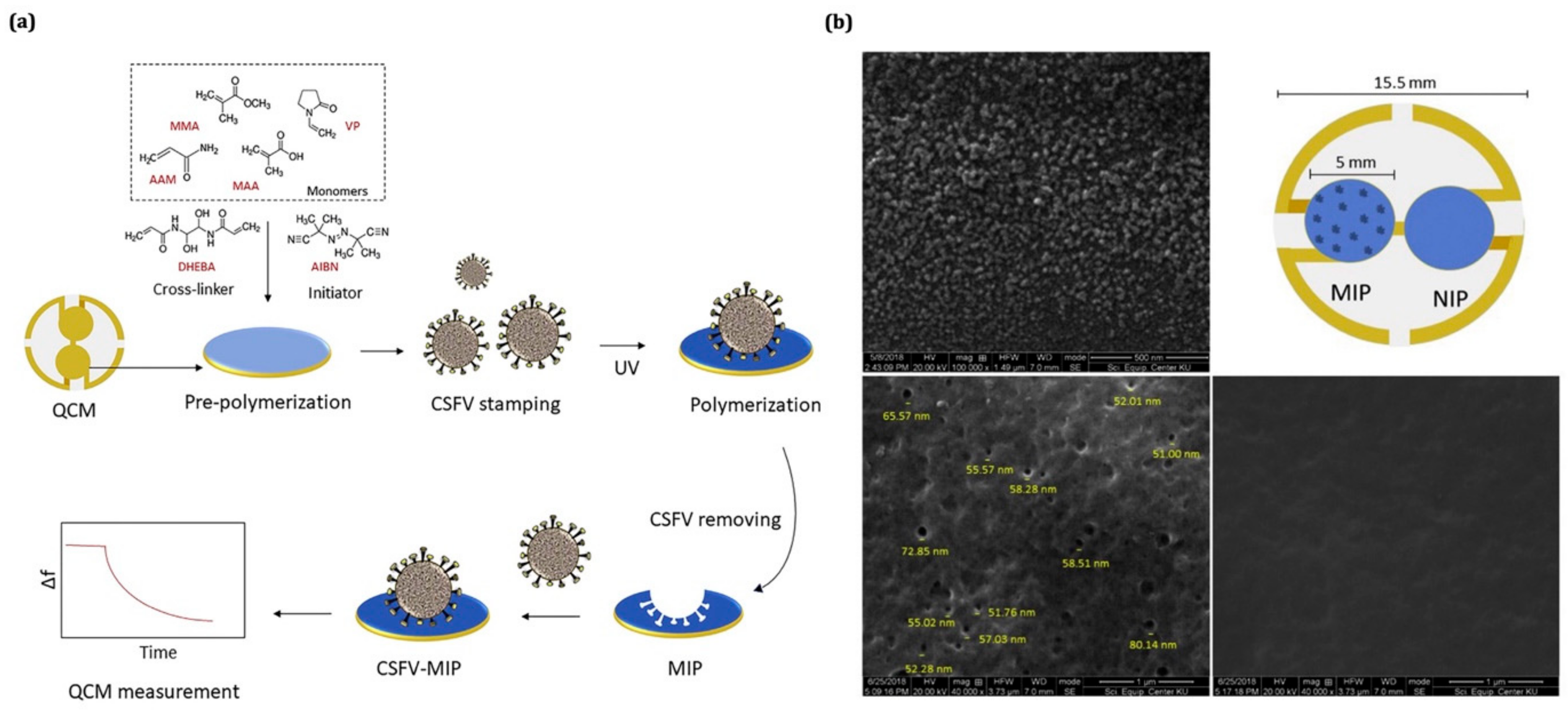
Classical swine fever virus (CSFV) is a highly contagious and fatal viral disease in pigs caused by the virus of the same name. A QCM biosensor for CSFV detection using a molecularly imprinted polymer receptor was developed by Klangprapan et al. The polymer film surface was characterized by SEM analysis, and the average diameter of the cavity was 59 nm onto the polymer film. This result was suitable for CSFV particles. The concentration range was between 4–21 μg/mL for CSFV. The limit of detection was 1.7 μg/mL. The reusability of the MIP-QCM biosensor has been tested three times with 21 μg/mL of CSFV samples. They compared the MIP-based QCM biosensor responses with other viruses, such as the pseudorabies virus (PRV) and the respiratory syndrome virus (PRRSV), which demonstrated that CSFV-MIP biosensors bind CSFV with imprinting factors of 62 over the PRV and 2 over the PRRSV. The designs of the CSFV-imprinted QCM biosensor procedure and the SEM image of MIP and NIP ACM biosensors are shown in Figure 8 [165].
Jenik et al. designed a QCM biosensor based on molecular imprinting polymer for human rhinovirus (HRV) and the foot-and-mouth disease virus (FMDV), which are two types of picornaviruses. The polyurethane MIPs were pre-polymerized at 70 °C for ∼10 min in the presence of catalytic amounts of pyridine. The prepolymer mixture was coated onto a QCM surface. The QCM biosensor electrode was characterized by AFM analysis for HRV. The mass effect on the QCM was an excellent signal as a −270 Hz frequency shift for 100 μg/mL HRV. The selectivity factor was also reported at ∼9 for HRV [166].
In the last two decades, QCM biosensors have developed rapidly and are capable of detecting biochemical properties and interactions at low scales. As mentioned, major efforts have been made to achieve rapid, direct analysis, high specificity, and high sensitivity in the detection of biomarkers. The combination with the molecular imprinting technique has made these sensors more popular. The creation of selective recognition zones on the sensor surface provides a great advantage in this area. Early detection steps of QCM biosensors with a low limit of detection are among the most important points. The reusability of MIP biosensors is important for practical sensing applications and the construction of detection platforms. The designed QCM biosensor has been reported to be reused with reproducible data. The multiple uses of MIP-based QCM biosensors with repetitive elution (regeneration of the imprinted cavity) and binding cycles does not put at risk the sensitivity and selectivity of the sensing matrix, in the absence of a significant loss of detection limits. However, the QCM biosensors’ progress faces many challenges and problems in some areas, including piezoelectric materials, surface modifications, and portable systems. Despite the increasing attention and the broad research carried out on the development of biosensors in several areas, such as medical, food, security, and environmental applications, only a small number of these sensing devices are commercially available [167,168]. The lack of biological receptors that are expensive to manufacture and stable for storage has hindered the commercial success of biosensors. This critical problem becomes especially significant when designing sensing devices meeting the requirements of clinical diagnostics. MIP technology may ensure a sustainable alternative to solving these noteworthy problems. In this last section, the performance of MIP-based QCM biosensors is highlighted and discussed in Table 1.

5. Conclusions

The advancement and investigation of biosensors are among the most important scientific fields at the crossroad of the biochemical and engineering sciences. Quartz crystal microbalance biosensors are application tools that can be utilized for the detection and determination of a broad variety of biological molecules and biomarkers. Over the past two decades, QCM biosensor technology has become a popular tool for quick, highly sensitive, and selective biomolecule detection. Various QCM biosensors have already been designed with analysis methods integrated with other analytical instruments. As demonstrated by an extensive literature review, molecular imprinting-based QCM biosensors provide label-free, direct, and quick real-time detection of template molecules or analytes compared to expensive devices. A QCM biosensor is also a portable and easy-to-use tool. Throughout this review paper, we have presented the principles of molecular imprinting technology, overviewed biosensors, and studied QCM biosensor systems. QCM biosensors have been designed for several target analytes, from proteins to cells, by combining this molecular imprinting technology with different recognition materials, such as polymer thin film and nanoparticles.
In this critical review paper, we presented the exemplary applications of MIP-based QCM biosensors in the label-free detection of protein, bacteria, and virus species, as preferred micro- and nano-sized biomolecules, respectively. The significant features of QCM systems based on the molecular imprinting approach is a promising alternative trend for biosensing applications, such as healthcare and diagnosis. MIP design is perhaps the crucial step in the process of QCM biosensor development because it must match the requirements of high selectivity and sensitivity for a template analyte. As a result, the straightforward engineering, fine-tuning, and the facility of integration into the standard industrial procedure of MIPs make them perfect applicants for recognition elements. Even though MIPs have been employed in a large variety of biosensing platforms, there are still some critical technological problems and challenges to be addressed. As a recent trend, the miniaturization of biosensors deserves special attention, due to the increasing number of applications available for smartphones. It is necessary to develop the progress of highly versatile diagnostic tools ready to be employed in clinical analysis directly at patients’ bedsides. Since MIP-based QCM biosensors applied in the clinical area are still developing, there are many more processes that are required to be done to obtain the building and marketing of MIP-based QCM biosensor platforms.

Author Contributions

Conception and design, S.A., E.Ö. and A.D.; Writing—original draft preparation, review, and editing, S.A., E.Ö. and A.D.; methodology, visualization, and supervision, S.A., E.Ö. and A.D. All authors have read and agreed to the published version of the manuscript.

Funding

This research received no external funding.

Institutional Review Board Statement

Not applicable.

Informed Consent Statement

Not applicable.

Data Availability Statement

Not applicable.

Conflicts of Interest

The authors declare no conflict of interest.

References

  1. Skládal, P. Piezoelectric Biosensors. Trends Anal. Chem. 2016, 79, 127–133. [Google Scholar] [CrossRef]
  2. Ferreira, G.N.M.; Da-Silva, A.-C.; Tome, B. Acoustic Wave Biosensors: Physical Models and Biological Applications of Quartz Crystal Microbalance. Trends Biotechnol. 2009, 27, 689–697. [Google Scholar] [CrossRef]
  3. Zhang, J.; Zhang, X.; Wei, X.; Xue, Y.; Wan, H. Recent Advances in Acoustic Wave Biosensors for the Detection of Disease-Related Biomarkers: A Review. Anal. Chim. Acta 2021, 1164, 338321. [Google Scholar] [CrossRef] [PubMed]
  4. Jean, H.; Saha, T.; Tey, T.; Siang, W.; Wei, C. Quartz Crystal Microbalance-Based Biosensors as Rapid Diagnostic Devices for Infectious Diseases. Biosens. Bioelectron. 2020, 168, 112513. [Google Scholar] [CrossRef]
  5. Kelley, S.O. What Are Clinically Relevant Levels of Cellular and Biomolecular Analytes? ACS Sens. 2017, 2, 193–197. [Google Scholar] [CrossRef] [PubMed]
  6. Rossetti, C.; Świtni, M.A.; Halvorsen, T.G.; Cormack, P.A.G.; Sellergren, B.; Reubsaet, L. Automated Protein Biomarker Analysis: On-Line Extraction of Clinical Samples by Molecularly Imprinted Polymers. Sci. Rep. 2017, 7, 44298. [Google Scholar] [CrossRef]
  7. Kelley, S.O. Advancing Ultrasensitive Molecular and Cellular Analysis Methods to Speed and Simplify the Diagnosis of Disease. Acc. Chem. Res. 2017, 50, 503–507. [Google Scholar] [CrossRef]
  8. Giamblanco, N.; Conoci, S.; Russo, D.; Marletta, G. Single-Step Label-Free Hepatitis B Virus Detection by a Piezoelectric Biosensor. RSC Adv. 2015, 5, 38152–38158. [Google Scholar] [CrossRef]
  9. Ragavan, K.V.; Kumar, S.; Swaraj, S.; Neethirajan, S. Advances in Biosensors and Optical Assays for Diagnosis and Detection of Malaria. Biosens. Bioelectron. 2018, 105, 188–210. [Google Scholar] [CrossRef]
  10. Kosack, C.S.; Page, A.; Klatser, P.R. A Guide to Aid the Selection of Diagnostic Tests. Bull World Health Organ. 2017, 95, 639–645. [Google Scholar] [CrossRef]
  11. Orazio, P.D. Biosensors in Clinical Chemistry—2011 Update. Clin. Chim. Acta 2011, 412, 1749–1761. [Google Scholar] [CrossRef]
  12. Tothill, I.E. Biosensors for Cancer Markers Diagnosis. Semin. Cell Dev. Biol. 2009, 20, 55–62. [Google Scholar] [CrossRef]
  13. Mascini, M.; Tombelli, S. Biosensors for Biomarkers in Medical Diagnostics. Biomarkers 2008, 13, 637–657. [Google Scholar] [CrossRef]
  14. Whitcombe, M.J.; Chianella, I.; Larcombe, L.; Piletsky, S.A.; Noble, J.; Horgan, A. The Rational Development of Molecularly Imprinted Polymer-Based Sensors for Protein Detection. Chem. Soc. Rev. 2011, 40, 1547–1571. [Google Scholar] [CrossRef] [Green Version]
  15. Montagut, Y.; Garcia, J.V.; Jimenez, Y.; March, C.; Montoya, A.; Arnau, A. Validation of a Phase-Mass Characterization Concept and Interface for Acoustic Biosensors. Sensors 2011, 11, 4702–4720. [Google Scholar] [CrossRef]
  16. Özgür, E.; Parlak, O.; Beni, V.; Turner, A.P.F.; Uzun, L. Bioinspired Design of a Polymer-Based Biohybrid Sensor Interface. Sens. Actuators B Chem. 2017, 251, 674–682. [Google Scholar] [CrossRef]
  17. Marx, K.A. Quartz Crystal Microbalance: A Useful Tool for Studying Thin Polymer Films and Complex Biomolecular Systems at the Solution—Surface Interface. Biomacromolecules 2003, 4, 1009–1120. [Google Scholar] [CrossRef] [Green Version]
  18. Pan, J.; Chen, W.; Ma, Y.; Pan, G. Molecularly Imprinted Polymers as Receptor Mimics for Selective Cell Recognition. Chem. Soc. Rev. 2018, 47, 5574–5587. [Google Scholar] [CrossRef]
  19. Pan, G.; Shinde, S.; Yeung, S.Y.; Jakštaitė, M.; Li, Q.; Wingren, A.G.; Sellergren, B. An Epitope Imprinted Biointerface with Dynamic Bioactivity for Modulating Cell-Biomaterial Interactions. Angew. Chem. Int. Ed. 2017, 56, 15959–15963. [Google Scholar] [CrossRef]
  20. Mosbach, K. Molecular Imprinting. Techniques 1994, 19, 9–14. [Google Scholar] [CrossRef]
  21. Saylan, Y.; Denizli, A. Molecularly Imprinted Polymer-Based Microfluidic Systems for Point-of-Care Applications. Micromachines 2019, 10, 766. [Google Scholar] [CrossRef] [Green Version]
  22. Leibl, N.; Haupt, K.; Gonzato, C.; Duma, L. Molecularly Imprinted Polymers for Chemical Sensing: A Tutorial Review. Chemosensors 2021, 9, 123. [Google Scholar] [CrossRef]
  23. Crapnell, R.D.; Dempsey-Hibbert, N.C.; Peeters, M.; Tridente, A.; Banks, C.E. Molecularly Imprinted Polymer Based Electrochemical Biosensors: Overcoming the Challenges of Detecting Vital Biomarkers and Speeding up Diagnosis. Talanta Open 2020, 2, 100018. [Google Scholar] [CrossRef]
  24. Kadhem, A.J.; Gentile, G.J.; de Cortalezzi, M.M.F. Molecularly Imprinted Polymers (Mips) in Sensors for Environmental and Biomedical Applications: A Review. Molecules 2021, 26, 6233. [Google Scholar] [CrossRef]
  25. Murphy, A.C.; Wechsler, M.E.; Peppas, N.A. Recent Advancements in Biosensing Approaches for Screening and Diagnostic Applications. Curr. Opin. Biomed. Eng. 2021, 19, 100318. [Google Scholar] [CrossRef]
  26. Rong, G.; Corrie, S.R.; Clark, H.A. In Vivo Biosensing: Progress and Perspectives. ACS Sens. 2017, 2, 327–338. [Google Scholar] [CrossRef]
  27. Rocchitta, G.; Spanu, A.; Babudieri, S.; Latte, G.; Madeddu, G.; Galleri, G.; Nuvoli, S.; Bagella, P.; Demartis, M.I.; Fiore, V.; et al. Enzyme Biosensors for Biomedical Applications: Strategies for Safeguarding Analytical Performances in Biological Fluids. Sensors 2016, 16, 780. [Google Scholar] [CrossRef] [Green Version]
  28. Akgönüllü, S.; Bakhshpour, M.; Denizli, A. Molecularly imprinted bionanomaterials and their biomedical applications. In Bionanomaterials Fundamentals and Biomedical Applications; Singh, R.P., Singh, K.R., Eds.; IOP Science: London, UK, 2021; pp. 9–27. ISBN 9780750337670. [Google Scholar]
  29. Rovira, J.; Domingo, J.L. Human Health Risks Due to Exposure to Inorganic and Organic Chemicals from Textiles: A Review. Environ. Res. 2019, 168, 62–69. [Google Scholar] [CrossRef]
  30. Mehrotra, P. Biosensors and Their Applications—A Review. J. Oral Biol. Craniofac. Res. 2016, 6, 153–159. [Google Scholar] [CrossRef] [Green Version]
  31. Jandas, P.J.; Prabakaran, K.; Luo, J.; Derry Holaday, M.G. Effective Utilization of Quartz Crystal Microbalance as a Tool for Biosensing Applications. Sens. Actuators A. Phys. 2021, 331, 113020. [Google Scholar] [CrossRef]
  32. Nelson, J.M.; Griffin, E.G. Adsorption of Invertase. J. Am. Chem. Soc. 1916, 38, 1109–1115. [Google Scholar] [CrossRef] [Green Version]
  33. Grieshaber, D.; MacKenzie, R.; Vörös, J.; Reimhult, E. Electrochemical Biosensors–Sensor Principles and Architectures. Sensors 2008, 8, 1400–1458. [Google Scholar] [CrossRef] [PubMed]
  34. Rezaei, Z.; Mahmoudifard, M. Pivotal Role of Electrospun Nanofibers in Microfluidic Diagnostic Systems-a Review. J. Mater. Chem. B 2019, 7, 4602–4619. [Google Scholar] [CrossRef] [PubMed]
  35. Qureshi, A.; Gurbuz, Y.; Niazi, J.H. Biosensors for Cardiac Biomarkers Detection: A Review. Sens. Actuators B Chem. 2012, 171–172, 62–76. [Google Scholar] [CrossRef] [Green Version]
  36. Tibuzzi, A.; Rea, G.; Pezzotti, G.; Esposito, D.; Johanningmeier, U.; Giardi, M.T. A New Miniaturized Multiarray Biosensor System for Fluorescence Detection. J. Phys. Condens. Matter 2007, 19, 395006. [Google Scholar] [CrossRef]
  37. Rahtuvanoğlu, A.; Akgönüllü, S.; Karacan, S.; Denizli, A. Biomimetic Nanoparticles Based Surface Plasmon Resonance Biosensors for Histamine Detection in Foods. ChemistrySelect 2020, 5, 5683–5692. [Google Scholar] [CrossRef]
  38. Heller, D.A.; Baik, S.; Eurell, T.E.; Strano, M.S. Single-Walled Carbon Nanotube Spectroscopy in Live Cells: Towards Long-Term Labels and Optical Sensors. Adv. Mater. 2005, 17, 2793–2799. [Google Scholar] [CrossRef]
  39. Xie, J.; Zhang, L.; Liu, B.; Bai, P.; Wang, C.; Xu, J.; Wang, H. Highly Selective Gas Sensor Based on Hydrophobic Silica Decorated with Trimethoxyoctadecylsilane. ACS Appl. Mater. Interfaces 2021, 13, 1956–1966. [Google Scholar] [CrossRef]
  40. Kim, K.; Ellis, J.E.; Howard, B.H.; Ohodnicki, P.R. Centimeter-Scale Pillared-Layer Metal—Organic Framework Thin Films Mediated by Hydroxy Double Salt Intermediates for CO 2 Sensor Applications. ACS Appl. Mater. Interfaces 2021, 13, 2062–2071. [Google Scholar] [CrossRef]
  41. Pan, Y.; Zhang, L.; Cao, B.; Xue, X.; Liu, W.; Zhang, C.; Wang, W. Effects of Temperature and Humidity on the Performance of a PECH Polymer Coated SAW Sensor. RSC Adv. 2020, 10, 18099–18106. [Google Scholar] [CrossRef]
  42. Jeng, M.J.; Sharma, M.; Li, Y.C.; Lu, Y.C.; Yu, C.Y.; Tsai, C.L.; Huang, S.F.; Chang, L.B.; Lai, C.S. Surface Acoustic Wave Sensor for C-Reactive Protein Detection. Sensors 2020, 20, 6640. [Google Scholar] [CrossRef]
  43. Temel, F. One Novel Calix[4]Arene Based QCM Sensor for Sensitive, Selective and High Performance-Sensing of Formaldehyde at Room Temperature. Talanta 2020, 211, 120725. [Google Scholar] [CrossRef]
  44. Haghighi, E.; Zeinali, S. Formaldehyde Detection Using Quartz Crystal Microbalance (QCM) Nanosensor Coated by Nanoporous MIL-101(Cr) Film. Microporous Mesoporous Mater. 2020, 300, 110065. [Google Scholar] [CrossRef]
  45. Jin, X.; Zhang, Y.P.; Li, D.M.; Ma, D.; Zheng, S.R.; Wu, C.H.; Li, J.Y.; Zhang, W.G. The Interaction of an Amorphous Metal-Organic Cage-Based Solid (AMOC) with MiRNA/DNA and Its Application on a Quartz Crystal Microbalance (QCM) Sensor. Chem. Commun. 2020, 56, 591–594. [Google Scholar] [CrossRef]
  46. Ghatak, B.; Banerjee, S.; Ali, S.B.; Das, N.; Tudu, B.; Pramanik, P.; Mukherji, S.; Bandyopadhyay, R. Development of a Low-Cost Portable Aroma Sensing System for Identifying Artificially Ripened Mango. Sens. Actuators A Phys. 2021, 331, 112964. [Google Scholar] [CrossRef]
  47. Mujahid, A.; Afzal, A.; Glanzing, G.; Leidl, A.; Lieberzeit, P.A.; Dickert, F.L. Imprinted Sol-Gel Materials for Monitoring Degradation Products in Automotive Oils by Shear Transverse Wave. Anal. Chim. Acta 2010, 675, 53–57. [Google Scholar] [CrossRef]
  48. Afzal, A.; Mujahid, A.; Schirhagl, R.; Bajwa, S.Z.; Latif, U.; Feroz, S. Gravimetric Viral Diagnostics: QCM Based Biosensors for Early Detection of Viruses. Chemosensors 2017, 5, 7. [Google Scholar] [CrossRef]
  49. Adamczyk, Z.; Sadowska, M.; Paulina, Z. Applicability of QCM - D for Quantitative Measurements of Nano- and Microparticle Deposition Kinetics: Theoretical Modeling and Experiments. Anal. Chem. 2020, 92, 15087–15095. [Google Scholar] [CrossRef]
  50. Adamczyk, Z.; Sadowska, M. Hydrodynamic Solvent Coupling E Ff Ects in Quartz Crystal Microbalance Measurements of Nanoparticle Deposition Kinetics. Anal. Chem. 2020, 92, 3896–3903. [Google Scholar] [CrossRef]
  51. Shan, Y.; Liu, L.; Liu, Y.; Harms, H.; Wick, L.Y. Effects of Electrokinetic Phenomena on Bacterial Deposition Monitored by Quartz Crystal Microbalance with Dissipation Monitoring. Environ. Sci. Technol. 2020, 54, 14036–14045. [Google Scholar] [CrossRef]
  52. Son, J.; Ji, S.; Kim, S.; Kim, S.; Kim, S.K.; Song, W.; Lee, S.S.; Lim, J.; An, K.; Myung, S. GC-like Graphene-Coated Quartz Crystal Microbalance Sensor with Microcolumns. ACS Appl. Mater. Interfaces 2021, 13, 4703–4710. [Google Scholar] [CrossRef]
  53. Wu, Y.; Zhang, Y.; Wang, K.; Luo, Z.; Xue, Z.; Gao, H.; Cao, Z.; Cheng, J.; Liu, C.; Zhang, L. Construction of Self-Assembled Polyelectrolyte/Cationic Microgel Multilayers and Their Interaction with Anionic Dyes Using Quartz Crystal. ACS Omega 2021, 6, 5764–5774. [Google Scholar] [CrossRef]
  54. Chinwangso, P.; Lee, H.J.; Lee, T.R. Self-Assembled Monolayers Generated from Unsymmetrical Partially Fluorinated Spiroalkanedithiols. Langmuir 2015, 31, 13341–13349. [Google Scholar] [CrossRef]
  55. Sauerbrey, G. Use of Quartz Crystals for Weighing Thin Layers and for Microweighing. Mag. Physic 1959, 155, 206–222. [Google Scholar]
  56. Chen, J.Y.; Penn, L.S.; Xi, J. Quartz Crystal Microbalance: Sensing Cell-Substrate Adhesion and Beyond. Biosens. Bioelectron. 2018, 99, 593–602. [Google Scholar] [CrossRef]
  57. Latif, U.; Can, S.; Hayden, O.; Grillberger, P.; Dickert, F.L. Sauerbrey and Anti-Sauerbrey Behavioral Studies in QCM Sensors—Detection of Bioanalytes. Sens. Actuators B. Chem. 2013, 176, 825–830. [Google Scholar] [CrossRef]
  58. Anderson, H.; Jönsson, M.; Vestling, L.; Lindberg, U.; Aastrup, T. Quartz Crystal Microbalance Sensor Design. I. Experimental Study of Sensor Response and Performance. Sens. Actuators B Chem. 2007, 123, 27–34. [Google Scholar] [CrossRef]
  59. Sullivan, C.K.O.; Guilbault, G.G. Commercial Quartz Crystal Microbalances—Theory and Applications. Biosens. Bioelectron. 1999, 14, 663–670. [Google Scholar] [CrossRef]
  60. Han, D.H.; Kang, L.H. Piezoelectric Properties of Paint Sensor According to Piezoelectric Materials. Funct. Compos. Struct. 2020, 2, 2–13. [Google Scholar] [CrossRef]
  61. Shindo, Y.; Domon, W.; Narita, F. Dynamic Bending of a Symmetric Piezoelectric Laminated Plate with a through Crack. Theor. Appl. Fract. Mech. 1998, 28, 175–182. [Google Scholar] [CrossRef]
  62. Fay, B.; Lewin, P.A.; Ludwig, G.; Sessler, G.M.; Yang, G. The influence of spatial polarization distribution on spot poled PVDF membrane hydrophone performance. Ultrasound Med Biol. 1992, 18, 625–635. [Google Scholar] [CrossRef]
  63. Yoshimura, H.N.; Molisani, A.L.; Narita, N.E.; Manholetti, J.L.A.; Cavenaghi, J.M. Mechanical Properties and Microstructure of Zinc Oxide Varistor Ceramics. Mater. Sci. Forum 2006, 530–531, 408–413. [Google Scholar] [CrossRef]
  64. Symposium, U. 1983 IEEE Ultrasonics Symposium Abstracts. IEEE Trans. Sonics Ultrason. 2012, 32, 78–122. [Google Scholar] [CrossRef]
  65. Kamiya, T. Calculation of Crystal Structures, Dielectric Constants and Piezoelectric Properties of Wurtzite-Type Crystals Using Ab-Initio Periodic Hartree-Fock Method. Jpn. J. Appl. Phys. 1996, 35, 4421. [Google Scholar] [CrossRef]
  66. Narita, F.; Shindo, Y. Piezoelectric Detection and Response Characteristics of Barium Titanate Unimorph Cantilevers Under AC Electric Fields. Int. J. Metall. Mater. Eng. 2015, 1, 10–13. [Google Scholar] [CrossRef] [Green Version]
  67. Hwa, L.G.; Lu, C.L.; Liu, L.C. Elastic Moduli of Calcium Alumino-Silicate Glasses Studied by Brillouin Scattering. Mater. Res. Bull. 2000, 35, 1285–1292. [Google Scholar] [CrossRef]
  68. Carr, P.H. Measurement of the Piezoelectric Constant of Quartz at Gigacycle Frequencies. J. Acoust. Soc. Am. 1967, 41, 75–83. [Google Scholar] [CrossRef]
  69. Kuma, S.; Woldemariam, M.M. Structural, Electronic, Lattice Dynamic, and Elastic Properties of SnTiO3 and PbTiO3 Using Density Functional Theory. Adv. Condens. Matter Phys. 2019, 2019, 317614. [Google Scholar] [CrossRef] [Green Version]
  70. Haun, M.J.; Furman, E.; Jang, S.J.; McKinstry, H.A.; Cross, L.E. Thermodynamic Theory of BiFeO 3 -PbTiO 3. J. Appl. Phys. 1987, 6, 3331. [Google Scholar] [CrossRef]
  71. Ravina; Dalal, A.; Mohan, H.; Prasad, M.; Pundir, C.S. Detection Methods for Influenza A H1N1 Virus with Special Reference to Biosensors: A Review. Biosci. Rep. 2020, 40, 1–18. [Google Scholar] [CrossRef] [Green Version]
  72. Perumal, V.; Hashim, U. Advances in Biosensors: Principle, Architecture and Applications. J. Appl. Biomed. 2014, 12, 1–15. [Google Scholar] [CrossRef]
  73. Pan, M.; Li, R.; Xu, L.; Yang, J.; Cui, X.; Wang, S. Reproducible Molecularly Imprinted Piezoelectric Sensor for Accurate and Sensitive Detection of Ractopamine in Swine and Feed Products. Sensors 2018, 18, 1870. [Google Scholar] [CrossRef] [Green Version]
  74. Lee, L.T.; Mohamed, M.A.; Yahya, I.; Kulothungan, J.; Muruganathan, M.; Mizuta, H. Comparison of Piezoelectric Energy Harvesting Performance Using Silicon and Graphene Cantilever Beam. Microsyst. Technol. 2018, 24, 3783–3789. [Google Scholar] [CrossRef]
  75. Rianjanu, A.; Roto, R.; Julian, T.; Hidayat, S.N.; Kusumaatmaja, A.; Suyono, E.A.; Triyana, K. Polyacrylonitrile Nanofiber-Based Quartz Crystal Microbalance for Sensitive Detection of Safrole. Sensors 2018, 18, 1150. [Google Scholar] [CrossRef] [Green Version]
  76. Battal, D.; Akgönüllü, S.; Yalcin, M.S.; Yavuz, H.; Denizli, A. Molecularly Imprinted Polymer Based Quartz Crystal Microbalance Sensor System for Sensitive and Label-Free Detection of Synthetic Cannabinoids in Urine. Biosens. Bioelectron. 2018. [Google Scholar] [CrossRef]
  77. Wasilewski, T.; Szulczyński, B.; Kamysz, W.; Gębicki, J.; Namieśnik, J. Evaluation of Three Peptide Immobilization Techniques on a Qcm Surface Related to Acetaldehyde Responses in the Gas Phase. Sensors 2018, 18, 3942. [Google Scholar] [CrossRef] [Green Version]
  78. Hardoyono, F.; Windhani, K. Identification and Detection of Bioactive Compounds in Turmeric (Curcuma LongaL.) Using a Gas Sensor Array Based on Molecularly Imprinted Polymer Quartz Crystal Microbalance. New J. Chem. 2021, 45, 17930–17940. [Google Scholar] [CrossRef]
  79. Shim, D.Y.; Chang, S.M.; Kim, J.M. Development of Fast Resettable Gravimetric Aromatic Gas Sensors Using Quartz Crystal Microbalance. Sens. Actuators B Chem. 2021, 329, 129143. [Google Scholar] [CrossRef]
  80. Meléndrez, D.; Hampitak, P.; Jowitt, T.; Iliut, M.; Vijayaraghavan, A. Development of an Open-Source Thermally Stabilized Quartz Crystal Microbalance Instrument for Biomolecule-Substrate Binding Assays on Gold and Graphene. Anal. Chim. Acta 2021, 1156. [Google Scholar] [CrossRef]
  81. Komorek, P.; Wałek, M.; Jachimska, B. Mechanism of Lysozyme Adsorption onto Gold Surface Determined by Quartz Crystal Microbalance and Surface Plasmon Resonance. Bioelectrochemistry 2020, 135, 107582. [Google Scholar] [CrossRef]
  82. Pizzoni, D.; Mascini, M.; Lanzone, V.; Del Carlo, M.; Di Natale, C.; Compagnone, D. Selection of Peptide Ligands for Piezoelectric Peptide Based Gas Sensors Arrays Using a Virtual Screening Approach. Biosens. Bioelectron. 2014, 52, 247–254. [Google Scholar] [CrossRef]
  83. Zhao, X.; He, Y.; Wang, Y.; Wang, S.; Wang, J. Hollow Molecularly Imprinted Polymer Based Quartz Crystal Microbalance Sensor for Rapid Detection of Methimazole in Food Samples. Food Chem. 2020, 309, 125787. [Google Scholar] [CrossRef]
  84. Li, B.; Hu, X.; Zhang, Q.; Peng, X.; Xiang, Y. Improved Piezoelectricity of Polylactide Using Vitamin B2 for Poling-Free Mechanical and Acoustic Nanogenerators. J. Mater. Sci. 2021, 56, 902–912. [Google Scholar] [CrossRef]
  85. Li, D.-M.; Zhang, S.-Y.; Wan, J.-Y.; Zhang, W.-Q.; Yan, Y.-L.; Tang, X.-H.; Zheng, S.-R.; Cai, S.; Zhang, W.-G. A New Hydrazone-Linked Covalent Organic Framework for Fe(Ⅲ) Detection by Fluorescence and QCM Technologies. CrystEngComm 2021, 23, 3594–3601. [Google Scholar] [CrossRef]
  86. Cervera-Chiner, L.; Juan-Borrás, M.; March, C.; Arnau, A.; Escriche, I.; Montoya, Á.; Jiménez, Y. High Fundamental Frequency Quartz Crystal Microbalance (HFF-QCM) Immunosensor for Pesticide Detection in Honey. Food Control 2018, 92, 1–6. [Google Scholar] [CrossRef] [Green Version]
  87. Su, L.; Zou, L.; Fong, C.C.; Wong, W.L.; Wei, F.; Wong, K.Y.; Wu, R.S.S.; Yang, M. Detection of Cancer Biomarkers by Piezoelectric Biosensor Using PZT Ceramic Resonator as the Transducer. Biosens. Bioelectron. 2013, 46, 155–161. [Google Scholar] [CrossRef]
  88. Cervera-Chiner, L.; Jiménez, Y.; Montoya, Á.; Juan-Borrás, M.; Pascual, N.; Arnau, A.; Escriche, I. High Fundamental Frequency Quartz Crystal Microbalance (HFF-QCMD) Immunosensor for Detection of Sulfathiazole in Honey. Food Control 2020, 115, 107296. [Google Scholar] [CrossRef]
  89. Buchatip, S.; Ananthanawat, C.; Sithigorngul, P.; Sangvanich, P.; Rengpipat, S.; Hoven, V.P. Detection of the Shrimp Pathogenic Bacteria, Vibrio Harveyi, by a Quartz Crystal Microbalance-Specific Antibody Based Sensor. Sens. Actuators B Chem. 2010, 145, 259–264. [Google Scholar] [CrossRef]
  90. Sankaran, S.; Panigrahi, S.; Mallik, S. Olfactory Receptor Based Piezoelectric Biosensors for Detection of Alcohols Related to Food Safety Applications. Sens. Actuators B Chem. 2011, 155, 8–18. [Google Scholar] [CrossRef]
  91. Wasilewski, T.; Szulczyński, B.; Wojciechowski, M.; Kamysz, W.; Gębicki, J. A Highly Selective Biosensor Based on Peptide Directly Derived from the HarmOBP7 Aldehyde Binding Site. Sensors 2019, 19, 4284. [Google Scholar] [CrossRef] [Green Version]
  92. Lin, W.L.; Lin, C.Y.; Tai, D.F. Preparation of a Molecularly Imprinted Film on Quartz Crystal Microbalance Chip for Determination of Furanic Compounds. Chemosensors 2021, 9, 338. [Google Scholar] [CrossRef]
  93. Refaat, D.; Aggour, M.G.; Farghali, A.A.; Mahajan, R.; Wiklander, J.G.; Nicholls, I.A.; Piletsky, S.A. Strategies for Molecular Imprinting and the Evolution of MIP Nanoparticles as Plastic Antibodies—Synthesis and Applications. Int. J. Mol. Sci. 2019, 20, 6304. [Google Scholar] [CrossRef] [PubMed] [Green Version]
  94. Jia, M.; Zhang, Z.; Li, J.; Ma, X.; Chen, L.; Yang, X. Molecular Imprinting Technology for Microorganism Analysis. Trends Anal. Chem. 2018, 106, 190–201. [Google Scholar] [CrossRef]
  95. Chen, L.; Wang, X.; Lu, W.; Wua, X.; Lia, J. Molecular Imprinting: Perspectives and Applications. Chem. Soc. Rev. 2016, 45, 2137–2211. [Google Scholar] [CrossRef]
  96. Culver, H.R.; Peppas, N.A. Protein-Imprinted Polymers: The Shape of Things to Come? Chem. Mater. 2017, 29, 5753–5761. [Google Scholar] [CrossRef]
  97. Zhang, N.; Zhang, N.; Xu, Y.; Li, Z.; Yan, C.; Mei, K.; Ding, M.; Ding, S.; Guan, P.; Qian, L.; et al. Molecularly Imprinted Materials for Selective Biological Recognition. Macromol. Rapid Commun. 2019, 40, 1900096. [Google Scholar] [CrossRef]
  98. Regan, B.; Boyle, F.; Kennedy, R.O.; Collins, D. Evaluation of Molecularly Imprinted Polymers for Point-of-Care Testing for Cardiovascular Disease. Sensors 2019, 19, 3485. [Google Scholar] [CrossRef] [Green Version]
  99. Ding, S.; Lyu, Z.; Niu, X.; Zhou, Y.; Liu, D.; Falahati, M.; Du, D.; Lin, Y. Integrating Ionic Liquids with Molecular Imprinting Technology for Biorecognition and Biosensing: A Review. Biosens. Bioelectron. 2020, 149, 111830. [Google Scholar] [CrossRef]
  100. Guo, X.; Li, J.; Arabi, M.; Wang, X.; Wang, Y.; Chen, L. Molecular-Imprinting-Based Surface-Enhanced Raman Scattering Sensors. ACS Sens. 2020, 5, 601–619. [Google Scholar] [CrossRef]
  101. Belbruno, J.J. Molecularly Imprinted Polymers. Chem. Rev. 2019, 119, 94–119. [Google Scholar] [CrossRef]
  102. Yang, K.; Li, S.; Liu, L.; Chen, Y.; Zhou, W.; Pei, J.; Liang, Z.; Zhang, L.; Zhang, Y. Epitope Imprinting Technology: Progress, Applications, and Perspectives toward Artificial Antibodies. Adv. Mater. 2019, 31, 1902048. [Google Scholar] [CrossRef]
  103. Ge, Y.; Turner, A.P.F. Too Large to Fit ? Recent Developments in Macromolecular Imprinting. Trends Biotechnol. 2008, 26, 218–224. [Google Scholar] [CrossRef] [Green Version]
  104. Hayden, O. One Binder to Bind Them All. Sensors 2016, 16, 1665. [Google Scholar] [CrossRef] [Green Version]
  105. Hayden, O.; Lieberzeit, P.A.; Blaas, D.; Dickert, F.L. Artificial Antibodies for Bioanalyte Detection—Sensing Viruses and Proteins. Adv. Funct. Mater. 2006, 16, 1269–1278. [Google Scholar] [CrossRef]
  106. Dickert, F.L. Molecular Imprinting and Functional Polymers for All Transducers and Applications. Sensors 2018, 18, 327. [Google Scholar] [CrossRef] [Green Version]
  107. Uzun, L.; Turner, A.P.F. Molecularly-Imprinted Polymer Sensors: Realising Their Potential. Biosens. Bioelectron. 2016, 76, 131–144. [Google Scholar] [CrossRef]
  108. Lowdon, J.W.; Diliën, H.; Singla, P.; Peeters, M.; Cleij, T.J.; van Grinsven, B.; Eersels, K. MIPs for Commercial Application in Low-Cost Sensors and Assays—An Overview of the Current Status Quo. Sens. Actuators B Chem. 2020, 325, 128973. [Google Scholar] [CrossRef]
  109. Ye, L.; Haupt, K. Molecularly Imprinted Polymers as Antibody and Receptor Mimics for Assays, Sensors and Drug Discovery. Anal. Bioanal. Chem. 2004, 378, 1887–1897. [Google Scholar] [CrossRef]
  110. Saylan, Y.; Akgönüllü, S.; Yavuz, H.; Ünal, S.; Denizli, A. Molecularly Imprinted Polymer Based Sensors for Medical Applications. Sensors 2019, 19, 1279. [Google Scholar] [CrossRef] [Green Version]
  111. Gómez-Arribas, L.N.; Urraca, J.L.; Benito-Penìa, E.; Moreno-Bondi, M.C. Tag-Specific Affinity Purification of Recombinant Proteins by Using Molecularly Imprinted Polymers. Anal. Chem. 2019, 91, 4100–4106. [Google Scholar] [CrossRef]
  112. Donato, L.; Drioli, E. Imprinted Membranes for Sustainable Separation Processes. Front. Chem. Sci. Eng. 2021. [Google Scholar] [CrossRef]
  113. Mohamed, S.; Balieu, S.; Petit, E.; Galas, L.; Schapman, D.; Hardouin, J.; Baati, R.; Estour, F. A Versatile and Recyclable Molecularly Imprinted Polymer as an Oxidative Catalyst of Sulfur Derivatives: A New Possible Method for Mustard Gas and v Nerve Agent Decontamination. Chem. Commun. 2019, 55, 13243–13246. [Google Scholar] [CrossRef]
  114. Ceylan Cömert, Ş.; Özgür, E.; Uzun, L.; Odabaşı, M. The Creation of Selective Imprinted Cavities on Quartz Crystal Microbalance Electrode for the Detection of Melamine in Milk Sample. Food Chem. 2022, 372. [Google Scholar] [CrossRef]
  115. Wackerlig, J.; Lieberzeit, P.A. Molecularly Imprinted Polymer Nanoparticles in Chemical Sensing—Synthesis, Characterisation and Application. Sens. Actuators B Chem. 2015, 207, 144–157. [Google Scholar] [CrossRef]
  116. Qi, J.; Li, B.; Wang, X.; Fu, L.; Luo, L.; Chen, L. Rotational Paper-Based Microfluidic-Chip Device for Multiplexed and Simultaneous Fluorescence Detection of Phenolic Pollutants Based on a Molecular-Imprinting Technique. Anal. Chem. 2018, 90, 11827–11834. [Google Scholar] [CrossRef]
  117. Muhammad, P.; Tu, X.; Liu, J.; Wang, Y.; Liu, Z. Molecularly Imprinted Plasmonic Substrates for Specific and Ultrasensitive Immunoassay of Trace Glycoproteins in Biological Samples. ACS Appl. Mater. Interfaces 2017, 9, 12082–12091. [Google Scholar] [CrossRef]
  118. Parisi, O.I.; Morelli, C.; Puoci, F.; Saturnino, C.; Caruso, A.; Sisci, D.; Trombino, G.E.; Picci, N.; Sinicropi, M.S. Magnetic Molecularly Imprinted Polymers (MMIPs) for Carbazole Derivative Release in Targeted Cancer Therapy. J. Mater. Chem. B 2014, 2, 6619–6625. [Google Scholar] [CrossRef]
  119. Kurczewska, J.; Cegłowski, M.; Pecyna, P.; Ratajczak, M.; Gajęcka, M.; Schroeder, G. Molecularly Imprinted Polymer as Drug Delivery Carrier in Alginate Dressing. Mater. Lett. 2017, 201, 46–49. [Google Scholar] [CrossRef]
  120. Piletsky, S.; Canfarotta, F.; Poma, A.; Bossi, A.M.; Piletsky, S. Molecularly Imprinted Polymers for Cell Recognition. Trends Biotechnol. 2020, 38, 368–387. [Google Scholar] [CrossRef] [PubMed]
  121. Chen, W.; Guo, Z.; Ding, Q.; Zhao, C.; Yu, H.; Zhu, X.; Fu, M.; Liu, Q. Magnetic-Graphene Oxide Based Molecular Imprinted Polymers for Selective Extraction of Glycoprotein at Physiological PH. Polymer 2021, 215, 123384. [Google Scholar] [CrossRef]
  122. Cao, J.; Wu, X.; Wang, L.; Shao, G.; Qin, B.; Wang, Z.; Wang, T.; Fu, Y. A Cellulose-Based Temperature Sensitivity Molecular Imprinted Hydrogel for Specific Recognition and Enrichment of Paclitaxel. Int. J. Biol. Macromol. 2021, 181, 1231–1242. [Google Scholar] [CrossRef]
  123. Lu, Z.; Du, X.; Sun, M.; Zhang, Y.; Li, Y.; Wang, X.; Wang, Y.; Du, H.; Yin, H.; Rao, H. Novel Dual-Template Molecular Imprinted Electrochemical Sensor for Simultaneous Detection of CA and TPH Based on Peanut Twin-like NiFe2O4/CoFe2O4/NCDs Nanospheres: Fabrication, Application and DFT Theoretical Study. Biosens. Bioelectron. 2021, 190, 113408. [Google Scholar] [CrossRef]
  124. Gao, R.; Hao, Y.; Zhang, L.; Cui, X.; Liu, D.; Zhang, M.; Tang, Y.; Zheng, Y. A Facile Method for Protein Imprinting on Directly Carboxyl-Functionalized Magnetic Nanoparticles Using Non-Covalent Template Immobilization Strategy. Chem. Eng. J. 2016, 284, 139–148. [Google Scholar] [CrossRef]
  125. Zhao, Q.Y.; Zhao, H.T.; Yang, X.; Zhang, H.; Dong, A.J.; Wang, J.; Li, B. Selective Recognition and Fast Enrichment of Anthocyanins by Dummy Molecularly Imprinted Magnetic Nanoparticles. J. Chromatogr. A 2018, 1572, 9–19. [Google Scholar] [CrossRef]
  126. Ndunda, E.N. Molecularly Imprinted Polymers—A Closer Look at the Control Polymer Used in Determining the Imprinting Effect: A Mini Review. J. Mol. Recognit. 2020, 33, e2855. [Google Scholar] [CrossRef]
  127. Kalogiouri, N.P.; Tsalbouris, A.; Kabir, A.; Furton, K.G.; Samanidou, V.F. Synthesis and Application of Molecularly Imprinted Polymers Using Sol–Gel Matrix Imprinting Technology for the Efficient Solid-Phase Extraction of BPA from Water. Microchem. J. 2020, 157, 104965. [Google Scholar] [CrossRef]
  128. Yao, T.; Gu, X.; Li, T.; Li, J.; Li, J.; Zhao, Z.; Wang, J.; Qin, Y.; She, Y. Enhancement of Surface Plasmon Resonance Signals Using a MIP/GNPs/RGO Nano-Hybrid Film for the Rapid Detection of Ractopamine. Biosens. Bioelectron. 2016. [Google Scholar] [CrossRef]
  129. Özgür, E.; Saylan, Y.; Bereli, N.; Türkmen, D.; Denizli, A. Molecularly Imprinted Polymer Integrated Plasmonic Nanosensor for Cocaine Detection. J. Biomater. Sci. Polym. Ed. 2020, 31, 1211–1222. [Google Scholar] [CrossRef]
  130. Vasapollo, G.; Sole, R.D.; Mergola, L.; Lazzoi, M.R.; Scardino, A.; Scorrano, S.; Mele, G. Molecularly Imprinted Polymers: Present and Future Prospective. Int. J. Mol. Sci. 2011, 12, 5008–5945. [Google Scholar] [CrossRef] [Green Version]
  131. Nezhadali, A.; Mojarrab, M. Computational Design and Multivariate Optimization of an Electrochemical Metoprolol Sensor Based on Molecular Imprinting in Combination with Carbon Nanotubes. Anal. Chim. Acta 2016, 924, 86–98. [Google Scholar] [CrossRef]
  132. Garcia-Cruz, A.; Ahmad, O.S.; Alanazi, K.; Piletska, E.; Piletsky, S.A. Generic Sensor Platform Based on Electro-Responsive Molecularly Imprinted Polymer Nanoparticles (e-NanoMIPs). Microsyst. Nanoeng. 2020, 6, 83. [Google Scholar] [CrossRef]
  133. Alanazi, K.; Garcia Cruz, A.; Di Masi, S.; Voorhaar, A.; Ahmad, O.S.; Cowen, T.; Piletska, E.; Langford, N.; Coats, T.J.; Sims, M.R.; et al. Disposable Paracetamol Sensor Based on Electroactive Molecularly Imprinted Polymer Nanoparticles for Plasma Monitoring. Sens. Actuators B Chem. 2021, 329, 129128. [Google Scholar] [CrossRef]
  134. Akgönüllü, S.; Battal, D.; Yalcin, M.S.; Yavuz, H.; Denizli, A. Rapid and Sensitive Detection of Synthetic Cannabinoids JWH-018, JWH-073 and Their Metabolites Using Molecularly Imprinted Polymer-Coated QCM Nanosensor in Artificial Saliva. Microchem. J. 2020, 153, 104454. [Google Scholar] [CrossRef]
  135. Bartold, K.; Pietrzyk-le, A.; Huynh, T.; Sosnowska, M.; Noworyta, K.; Lisowski, W.; Sannicolo, F.; Cauteruccio, S.; Licandro, E.; Souza, F.D.; et al. Programmed Transfer of Sequence Information into a Molecularly Imprinted Polymer for Hexakis (2,2′-Bithien-5-Yl) DNA Analogue Formation toward Single-Nucleotide-Polymorphism Detection. ACS Appl. Mater. Interfaces 2017, 9, 3948–3958. [Google Scholar] [CrossRef] [Green Version]
  136. Zhang, L.; Liu, Z.; Xiong, C.; Zheng, L.; Ding, Y.; Lu, H. Selective Recognition of Histidine Enantiomers Using Novel Molecularly Imprinted Organic Transistor Sensor. Org. Electron. 2018, 61, 254–260. [Google Scholar] [CrossRef]
  137. Fang, M.; Zhuo, K.; Chen, Y.; Zhao, Y.; Bai, G.; Wang, J. Fluorescent Probe Based on Carbon Dots / Silica / Molecularly Imprinted Polymer for Lysozyme Detection and Cell Imaging. Anal. Bioanal. Chem. 2019, 411, 5799–5807. [Google Scholar] [CrossRef]
  138. Esentürk, M.K.; Akgönüllü, S.; Yılmaz, F.; Denizli, A. Molecularly Imprinted Based Surface Plasmon Resonance Nanosensors for Microalbumin Detection. J. Biomater. Sci. Polym. Ed. 2019, 30, 646–661. [Google Scholar] [CrossRef]
  139. Guo, J.; Fang, G.; Wang, S.; Wang, J. Quartz Crystal Microbalance Sensor Based on 11-Mercaptoundecanoic Acid Self-Assembly and Amidated Nano-Titanium Film for Selective and Ultrafast Detection of Phosphoproteins in Food. Food Chem. 2021, 344, 128656. [Google Scholar] [CrossRef]
  140. Malik, A.A.; Nantasenamat, C.; Piacham, T. Molecularly Imprinted Polymer for Human Viral Pathogen Detection. Mater. Sci. Eng. C 2017, 77, 1341–1348. [Google Scholar] [CrossRef]
  141. Wang, R.; Wang, L.; Yan, J.; Luan, D.; Wu, J.; Bian, X. Rapid, Sensitive and Label-Free Detection of Pathogenic Bacteria Using a Bacteria-Imprinted Conducting Polymer Film-Based Electrochemical Sensor. Talanta 2021, 226, 122135. [Google Scholar] [CrossRef]
  142. Zarejousheghani, M.; Rahimi, P.; Borsdorf, H.; Zimmermann, S.; Joseph, Y. Molecularly Imprinted Polymer-Based Sensors for Priority Pollutants. Sensors 2021, 21, 2406. [Google Scholar] [CrossRef] [PubMed]
  143. Hu, Y.; Xing, H.; Li, G.; Wu, M. Magnetic Imprinted Polymer-Based Quartz Crystal Microbalance Sensor for Sensitive Label-Free Detection of Methylene Blue in Groundwater. Sensors 2020, 20, 5506. [Google Scholar] [CrossRef] [PubMed]
  144. Qiu, X.; Li, Y.; Wang, Y.; Guo, H.; Nie, L. A Novel Molecularly Imprinted Nanosensor Based on Quartz Crystal Microbalance for Specific Recognition of α-Amanitin. Microchem. J. 2020, 159, 105383. [Google Scholar] [CrossRef]
  145. Yang, J.C.; Lee, J.; Hong, S.W.; Park, J. Molecularly Imprinted Quartz Crystal Microbalance Sensors with Lithographically Patterned Frisbee-like Pillar Arrays for Sensitive and Selective Detection of Iprodione. Sens. Actuators B Chem. 2020, 320, 128366. [Google Scholar] [CrossRef]
  146. Akgönüllü, S.; Yavuz, H.; Denizli, A. SPR Nanosensor Based on Molecularly Imprinted Polymer Film with Gold Nanoparticles for Sensitive Detection of Aflatoxin B1. Talanta 2020, 219, 121219. [Google Scholar] [CrossRef]
  147. Akgönüllü, S.; Yavuz, H.; Denizli, A. Development of Gold Nanoparticles Decorated Molecularly Imprinted–Based Plasmonic Sensor for the Detection of Aflatoxin M1 in Milk Samples. Chemosensors 2021, 9, 363. [Google Scholar] [CrossRef]
  148. Burnham-marusich, A.R.; Berninsone, P.M. Multiple Proteins with Essential Mitochondrial Functions Have Glycosylated Isoforms. Mitochondrion 2012, 12, 423–427. [Google Scholar] [CrossRef] [Green Version]
  149. Zhang, X.; Liu, S.; Pan, J.; Jia, H.; Chen, Z.; Guo, T. Multifunctional Oligomer Immobilized on Quartz Crystal Microbalance: A Facile and Stabilized Molecular Imprinting Strategy for Glycoprotein Detection. Anal. Bioanal. Chem. 2019, 411, 3941–3949. [Google Scholar] [CrossRef]
  150. Diltemiz, S.E.; Hür, D.; Keçili, R.; Ersöz, A.; Say, R. New Synthesis Method for 4-MAPBA Monomer and Using for the Recognition of IgM and Mannose with MIP-Based QCM Sensors. Analyst 2013, 138, 1558–1563. [Google Scholar] [CrossRef]
  151. Ma, X.; He, X.; Li, W.; Zhang, Y. Epitope Molecularly Imprinted Polymer Coated Quartz Crystal Microbalance Sensor for the Determination of Human Serum Albumin. Sens. Actuators B. Chem. 2017, 246, 879–886. [Google Scholar] [CrossRef]
  152. Lee, M.H.; Thomas, J.L.; Tseng, H.Y.; Lin, W.C.; Liu, B.D.; Lin, H.Y. Sensing of Digestive Proteins in Saliva with a Molecularly Imprinted Poly(Ethylene-Co-Vinyl Alcohol) Thin Film Coated Quartz Crystal Microbalance Sensor. ACS Appl. Mater. Interfaces 2011, 3, 3064–3071. [Google Scholar] [CrossRef]
  153. Gupta, N.; Singh, R.S.; Shah, K.; Prasad, R.; Singh, M. Epitope Imprinting of Iron Binding Protein of Neisseria Meningitidis Bacteria through Multiple Monomers Imprinting Approach. J. Mol. Recognit. 2018, 31, e2709. [Google Scholar] [CrossRef]
  154. Nasrullah, A.; Afzal, A.; Mujahid, A.; Lieberzeit, P.; Bajwa, S.Z.; Mustafa, G.; Latif, U. Imprinted Polymer and Cu2O-Graphene Oxide Nanocomposite for the Detection of Disease Biomarkers. Meas. Sci. Technol. 2021, 32, 105111. [Google Scholar] [CrossRef]
  155. Danstism, S.; Röhlen, D.; Wagner, T.; Wagner, P.; Schöning, M.J. A LAPS-Based Differential Sensor for Parallelized Metabolism Monitoring of Various Bacteria. Sensors 2019, 19, 4692. [Google Scholar] [CrossRef] [Green Version]
  156. Latif, U.; Can, S.; Sussitz, H.F.; Dickert, F.L. Molecular Imprinted Based Quartz Crystal Microbalance Sensors for Bacteria and Spores. Chemosensors 2020, 8, 64. [Google Scholar] [CrossRef]
  157. Yilmaz, E.; Majidi, D.; Ozgur, E.; Denizli, A. Whole Cell Imprinting Based Escherichia Coli Sensors: A Study for SPR and QCM. Sens. Actuators B. Chem. 2015, 209, 714–721. [Google Scholar] [CrossRef]
  158. Guha, A.; Sheej, O.; Guerreiro, A.; Karim, K.; Sandstr, N.; Ostanin, V.P.; Van Der Wijngaart, W.; Piletsky, S.A.; Ghosh, S.K. Direct Detection of Small Molecules Using a Nano-Molecular Imprinted Polymer Receptor and a Quartz Crystal Resonator Driven at a Fixed Frequency and Amplitude. Biosens. Bioelectron. 2020, 158, 112176. [Google Scholar] [CrossRef]
  159. Kushwaha, A.; Srivastava, J.; Singh, A.K.; Anand, R.; Raghuwanshi, R.; Rai, T.; Singh, M. Epitope Imprinting of Mycobacterium Leprae Bacteria via Molecularly Imprinted Nanoparticles Using Multiple Monomers Approach. Biosens. Bioelectron. 2019, 145, 111698. [Google Scholar] [CrossRef]
  160. Cornelis, P.; Givanoudi, S.; Yongabi, D.; Iken, H.; Duwé, S.; Deschaume, O.; Robbens, J.; Dedecker, P.; Bartic, C.; Wübbenhorst, M.; et al. Sensitive and Specific Detection of E. Coli Using Biomimetic Receptors in Combination with a Modified Heat-Transfer Method. Biosens. Bioelectron. 2019, 136, 97–105. [Google Scholar] [CrossRef] [Green Version]
  161. Tokonami, S.; Nakadoi, Y.; Takahashi, M.; Ikemizu, M.; Kadoma, T.; Saimatsu, K.; Dung, L.Q.; Shiigi, H.; Nagaoka, T. Label-Free and Selective Bacteria Detection Using a Film with Transferred Bacterial Configuration. Anal. Chem. 2013, 85, 4925–4929. [Google Scholar] [CrossRef]
  162. Wangchareansak, T.; Thitithanyanont, A.; Chuakheaw, D.; Gleeson, M.P.; Lieberzeit, P.A.; Sangma, C. A Novel Approach to Identify Molecular Binding to the Influenza Virus H5N1: Screening Using Molecularly Imprinted Polymers (MIPs). Medchemcomm 2014, 5, 617–621. [Google Scholar] [CrossRef] [Green Version]
  163. Lu, C.H.; Zhang, Y.; Tang, S.F.; Fang, Z.B.; Yang, H.H.; Chen, X.; Chen, G.N. Sensing HIV Related Protein Using Epitope Imprinted Hydrophilic Polymer Coated Quartz Crystal Microbalance. Biosens. Bioelectron. 2012, 31, 439–444. [Google Scholar] [CrossRef]
  164. Tai, D.; Lin, C.; Wu, T.; Chen, L. Recognition of Dengue Virus Protein Using Epitope-Mediated Molecularly Imprinted Film of a Pentadecapeptide onto a Quartz Crystal Microbalance. Anal. Chem. 2005, 77, 5140–5143. [Google Scholar] [CrossRef]
  165. Klangprapan, S.; Choke-arpornchai, B.; Lieberzeit, P.A.; Choowongkomon, K. Sensing the Classical Swine Fever Virus with Molecularly Imprinted Polymer on Quartz Crystal Microbalance. Heliyon 2020, 6, e04137. [Google Scholar] [CrossRef]
  166. Jenik, M.; Schirhagl, R.; Schirk, C.; Hayden, O.; Lieberzeit, P.; Blaas, D.; Paul, G.; Dickert, F.L. Sensing Picornaviruses Using Molecular Imprinting Techniques on a Quartz Crystal Microbalance. Anal. Chem. 2009, 81, 5320–5326. [Google Scholar] [CrossRef]
  167. Selvolini, G.; Marrazza, G. MIP-Based Sensors: Promising New Tools for Cancer Biomarker Determination. Sensors 2017, 17, 718. [Google Scholar] [CrossRef] [Green Version]
  168. Bahadir, E.B.; Sezgintürk, M.K. Applications of Commercial Biosensors in Clinical, Food, Environmental, and Biothreat/Biowarfare Analyses. Anal. Biochem. 2015, 478, 107–120. [Google Scholar] [CrossRef]
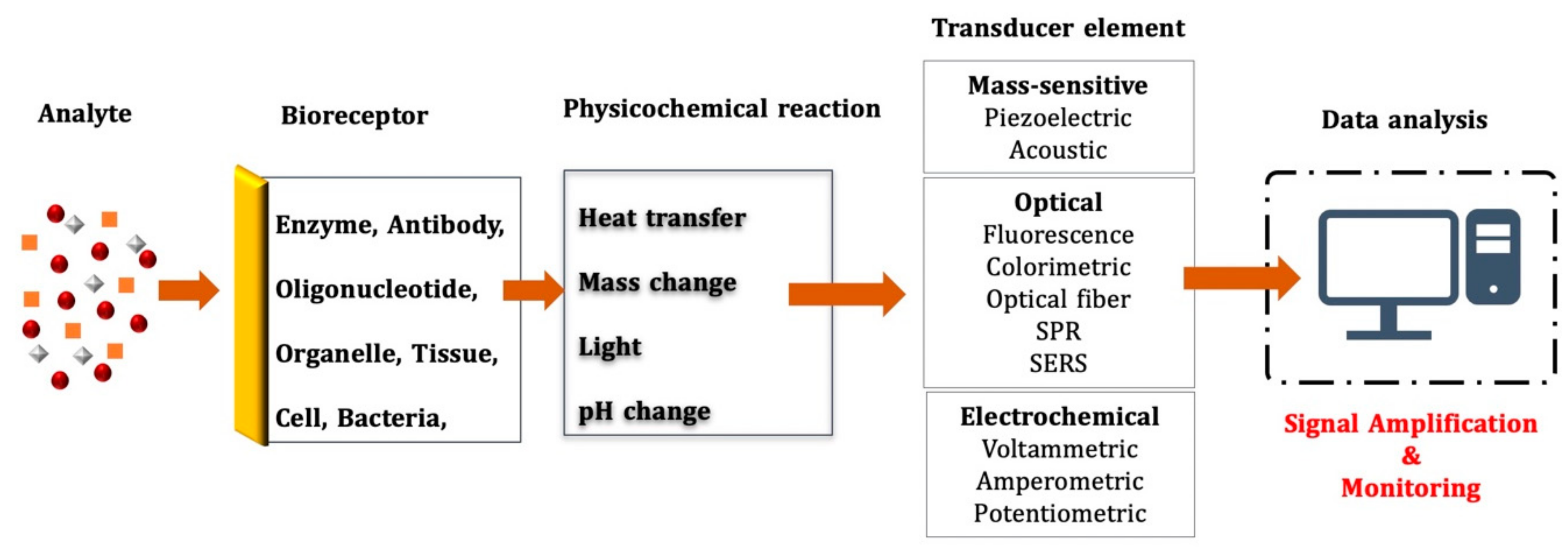
Figure 1. The classic biosensor components.
Chemosensors 10 00106 g001
Figure 2. (a) The top view of the quartz crystal electrode; (b) AT-cut quartz crystal coated with gold; (c) quartz crystal photographs. Reprinted with permission from ref. [58]. Copyright 2007 Elsevier.
Chemosensors 10 00106 g002
Figure 3. Schematic illustration for the molecular imprinting technique process; pre-complex; polymerization; and template removal/rebinding.
Chemosensors 10 00106 g003
Figure 4. (a) Design of epitope-imprinted QCM biosensor; (b) bare QCM chip and AFM measurement results. Reprinted with permission from ref. [151]. Copyright 2017 Elsevier.
Chemosensors 10 00106 g004
Figure 5. (a) QCM biosensor design; (b) schematic diagram of non-covalent recognition mechanism for amylase. Reprinted with permission from ref. [152]. Copyright 2011 ACS Publications.
Chemosensors 10 00106 g005
Figure 6. (a) QCR experiment system. Reprinted with permission from ref. [158]. Copyright 2020 Elsevier; (b) schematic representation of an LP-15-imprinted EQCM sensor. Reprinted with permission from ref. [159]. Copyright 2019 Elsevier.
Chemosensors 10 00106 g006
Figure 7. (a) QCM electrode; (b) SEM image of MIP thin film; (c) sensorgram of resonance frequency; (d) fluorescent microscope image of movement of P. aeruginosa. Reprinted with permission from ref. [161]. Copyright 2013 ACS Publications.
Chemosensors 10 00106 g007
Figure 8. (a) Schematic image of design of MIP-QCM sensors; (b) SEM image of polymer surface Reprinted with permission from ref. [165]. Copyright 2020 Elsevier.
Chemosensors 10 00106 g008
Table 1. QCM biosensors based on molecular imprinting techniques for various disease-related biomarkers.
AnalyteFunctional MonomerLinear RangeLODIF or k′Ref.
Protein
Glycoprotein4-Vinylphenylboronic acid (VPBA)1 × 10⁻⁷–1 × 10⁻⁴ g/mL-∼3.5[149]
IgMMethacryloylamidophenylboronic acid (MAPBA)0.01–0.1 mM-6.1 k′[150]
AlbuminN,N-dimethylformamide0.050–0.500 μg/mL.0.026 μg/mL6.9[151]
AmylasePoly-(ethylene-co-vinyl alcohol)0–1.0 μg/mL∼pM2.13–2.47[152]
Iron requisition protein (fbpA) 3-sulfopropyl methacrylate potassium-salt and benzyl methacrylate, N,N-methylene-bis-acrylamide5–30 ng/mL1.39 ng/mL12.27[153]
α1-Acid glycoprotein (α1-AGp)Boronate-affinity150–200 ng/mL0.25 ng/mL∼7[154]
Bacteria
E. coli and
B. subtilis
Bulk imprinting0–25 × 107 cells/mL--[156]
E. coliN-methacryloyl-l-histidine methylester0.5–3.0 McFarland3.72 × 105 CFU/mL3 k′ and 43.44 k′[157]
Mycobacterium leprae3-sulphopropyl methacrylate potassium salt, benzyl methacrylate and 4-aminothiophenol10–140 nM0.161 nM8.28[159]
E. ColiPolyurethane 102–106 CFU/mL100 CFU/mL-[160]
P. aeruginosaPolypyrrole (OPPy)1011–103 CFU/mL103 CFU/mL3.92 k′[161]
Virus
H5N1Methacrylic acid and methyl-methacrylate1–8 HAU1 HA∼4[162]
HIV type-IDopamine2–200 ng/mL2 ng/mL6[163]
Dengue virusPentadecapeptide0.5–50,000 ng/mL--[164]
Swine fever virusMulti-functional monomer/surface imprinting technique4–21 μg/mL1.7 μg/mL2 and 62[165]
PicornavirusPolyurethane MIPs 100–300 μg/mL100 μg/mL∼9[166]
Publisher’s Note: MDPI stays neutral with regard to jurisdictional claims in published maps and institutional affiliations.