Article

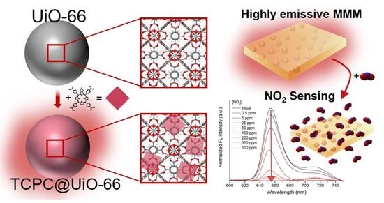
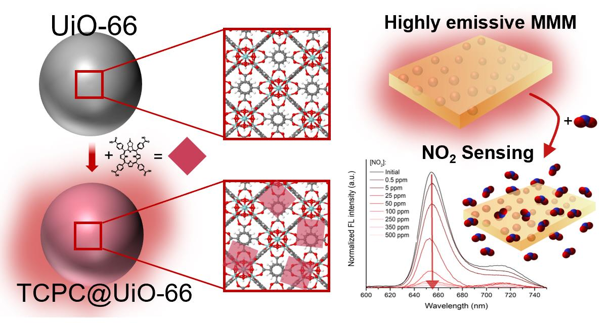
MOF-Based Materials with Sensing Potential: Pyrrolidine-Fused Chlorin at UiO-66(Hf) for Enhanced NO2 Detection

by
Carla Queirós
1,
Francisco G. Moscoso
2,
José Almeida
1,
Ana M. G. Silva
1,
Ahmad Sousaraei
3,
Juan Cabanillas-González
3,
Manuela Ribeiro Carrott
4,
Tânia Lopes-Costa
2,
José M. Pedrosa
2,* and
Luís Cunha-Silva
1,*
1
LAQV-REQUIMTE, Department of Chemistry and Biochemistry, Faculty of Sciences, University of Porto, 4169-007 Porto, Portugal
2
Department of Physical, Chemical and Natural Systems, University Pablo de Olavide, Ctra. Utrera Km. 1, 41013 Sevilla, Spain
3
Madrid Institute for Advanced Studies in Nanoscience, IMDEA Nanociencia, Universitaria de Cantoblanco, Calle Faraday 9, 28049 Madrid, Spain
4
LAQV-REQUIMTE, Institute for Research and Advanced Studies, Department of Chemistry and Biochemistry, School of Sciences and Technology, University of Évora, 7000-671 Évora, Portugal
*
Authors to whom correspondence should be addressed.
Chemosensors 2022, 10(12), 511; https://doi.org/10.3390/chemosensors10120511
Submission received: 3 November 2022 / Revised: 28 November 2022 / Accepted: 29 November 2022 / Published: 1 December 2022

Abstract

:
An efficient strategy to develop porous materials with potential for NO2 sensing was based in the preparation of a metal-organic framework (MOF), UiO-66(Hf), modified with a very small amount of meso-tetrakis(4-carboxyphenyl) N-methylpyrrolidine-fused chlorin (TCPC), TCPC@MOF. Chlorin’s incorporation into the UiO-66(Hf) framework was verified by several characterization methods and revealed that the as-synthesized TCPC@MOF brings together the chemical stability of UiO-66(Hf) and the photophysical properties of the pyrrolidine-fused chlorin which is about five times more emissive than the porphyrin counterpart. TCPC@MOF was further incorporated into polydimethylsiloxane (PDMS) and the resulting TCPC@MOF@PDMS film was tested in NO2 gas sensing. It showed notable sensitivity as well as a fast response in the range between 0.5 and 500 ppm where an emission intensity quenching is observed up to 96% for 500 ppm. This is a rare example of a chlorin-derivative used for gas-sensing applications through emission changes, and an unusual case of this type of optical-sensing composites of NO2.

Graphical Abstract

1. Introduction

Air pollution remains a major health concern worldwide. According to the 2021 Air Quality Status briefing by the European Environment Agency, many European cities still regularly exceed current Union’s emission limits for air pollutants [1]. Nitrogen oxides (NOx) belong to one category of these air pollutants, and nitrogen dioxide (NO2) is the most toxic and prevalent form of the NOx family. NO2 is a nose and throat irritant that can cause serious respiratory problems [2], reduce respiratory defense mechanisms, and increase infection rates [3], and was related to 55,000 premature deaths in 2018 in Europe alone [4]. The early detection of NO2 has also shown to be a great asset for fire detection and firefighting improvement [5]. Several colorimetric materials were shown to be quite efficient for the detection of CO and NO2 gases produced in an early fire [6]. Given this scenario, the detection of toxic gases plays an increasingly important role in keeping our environment unpolluted and ourselves safe due to their dangerous effects on both the ecosystem and human health.
Nowadays there is a wide variety of gas-sensing devices based on different materials and operation mechanisms [7]. Some of those materials are based on metal-organic frameworks (MOF), which are a category of porous coordination polymers assembled by the coordination of metal cationic centers with organic linkers. Given the wide range of available metal ions and organic linkers used in an MOF assembly, these materials have become increasingly attractive because of their wide-variety of topologies and broad-range of chemical and physical properties. Excellent literature reviews and in-depth studies have been reported regarding MOF applications as materials to capture and degrade toxic chemicals [8,9,10,11,12,13], as well as luminescent sensors [14,15,16,17,18,19,20,21], enzyme biomimetics [22,23], and catalysts [24,25]. One of the most studied and hydrothermal stable MOFs is the UiO-66(Zr) that consists of hexanuclear, octahedral zirconium(IV) oxo clusters connected through terephthalic acid linkers to form a 3D highly-porous rigid framework. This crystalline framework has shown resistance to a wide range of different solvents including strong acids and bases [26]. UiO-66(Zr) has been intensely studied as a fluorescent sensor in the past decade although terephthalic acid and consequently UiO-66(Zr) both possess low fluorescence intensities. Zirconium (Zr) MOFs are widely used; however, some characteristics of hafnium (Hf) MOFs, namely their acidic nature, mechanical and higher chemical stability, also make them promising materials for several applications [27,28]. Due to their same d0 electronic configuration and identical ionic radius (78 pm), substituting Zr(IV) with Hf(IV) in the UiO-66 synthesis results in an MOF with the same topology of UiO-66(Zr) but with stronger acid sites. This property of UiO-66(Hf) was originally shown to have applications as a radiation scavenger [26] and contrast agent for computed tomography [29]. Nevertheless, there are few examples of UiO-66(Hf) as a sensor to detect free chlorine in waters [30], uric acid in biological fluids [31], and peroxynitrite in living cells [32].
A common strategy to endow the MOF with fluorescent properties is to dope it with a lanthanide ion or to integrate (impregnate or incorporate) a fluorophore (such as rhodamine or porphyrin derivatives) into the MOF structure [33,34,35,36,37,38], creating a luminescent MOF. This strategy allows an optimization of several properties and broadens their application [39,40]. Luminescent MOF sensor characteristics/properties make them stand out in comparison with other luminescent sensors [40,41,42], namely: (i) their porosity allows the adsorption and pre-concentration of the analytes, increasing the host-guest interactions; (ii) they can be easily functionalized and therefore more tunable to sense specific analytes; (iii) they produce guest-dependent optical signals usually translated into vaporchromic or solvatochromic responses; (iv) they make it possible to study the sensing mechanisms at the molecular level due to their crystalline nature; (v) they possess stability in water even at the basic range; and (vi) they can be used to prepare composites/hybrids that combine their advantages with those of other compounds/materials.
One of the major advantages of incorporating fluorescent ligands into MOFs is to prevent their aggregation and consequently their self-quenching. This can be attained since MOFs open structures allow the separation of the fluorescent unit by relatively large distances (>5 Å), while maintaining or even amplifying the sensing signals upon interaction with analytes. The signal amplification can occur due to the existence of an efficient energy migration (exciton hoping) along the composite, similar to that of conjugated polymers, even when only a few analyte molecules interact with the composite [40]. Nevertheless, working with powder materials may be challenging and their immobilization in solid matrices like TiO2, PDMS (polydimethylsiloxane) or PMMA (poly(methyl methacrylate)) can improve the molecular recognition of analytes. These composites are also, in general, property-tunable and possess better resistance to degradation and better flexibility, among other advantageous properties [43]. This type of composite is immensely appealing when the analytes are gases or volatile organic compounds (VOC) [42].
Porphyrins are very interesting fluorophores that can be incorporated into MOF frameworks, either via chemical bonds or non-covalently [44,45]. The integration of porphyrins in MOFs was shown to be advantageous to obtain multifunctional drug carriers [46], as well as biomaterials [47]. A tremendous number of MOFs have been used as a host for the porphyrin’s incorporation, providing efficient platforms to encapsulate and deliver pharmaceuticals, imaging agents, and enzymes [45,48]. Reduced porphyrins (7,8-dihydroporphyrins, also known as chlorins; Figure 1a–d,f) are much more interesting chromo- and fluorophores for integrating into MOFs due to the enhanced absorbance of the QI (0-0) band centered at 650 nm and high fluorescence quantum yield at the same region. The incorporation of chlorins into MOF materials has been predominantly studied in the area of photodynamic therapy (PDT) of cancer, where chlorin e6 (Figure 1a) stands out as the photosensitizer of choice [49,50,51,52]. On the other hand, N-methylpyrrolidine-fused chlorins are more stable than the hydrogenated chlorins and have been studied mostly as photosensitizers in the photodynamic inactivation of bacteria [53,54,55] and PDT of cancer [56,57,58,59], as well as reaction [60], biomimetic [61], and oxidation [62] catalysts.
In the last years, our research group has been developing optical sensors for NO2 including: (i) rosamine-based composite film sensors, immobilized on SiO2 [63] and TiO2 [64]; and (ii) a Tb(BTC) MOF material (BTC: benzene-1,3,5-tricarboxylate) embedded into transparent polymeric matrices to produce mixed-matrix membranes [65,66,67,68]. In 2021, the first use of an N-methylpyrrolidine-fused chlorin in an MOF was reported by Sakamaki et al. for PDT application [69]. Meanwhile, we recently reported the first sensing application of N-methylpyrrolidine-fused 5,10,15,20-tetrakis(4-carboxyphenyl)chlorin (TCPC, Figure 1f), anchored to a nanostructured porous TiO2 substrate, as a fluorescent sensor of the explosive triacetone triperoxide (TATP) in the gas phase [70]. Following our interest in the study and application of MOF-based materials for sensing [65,66,67,68], the pyrrolidine-fused chlorin (TCPC) was incorporated into UiO-66(Hf) by a one-pot solvothermal synthesis (TCPC@MOF) and this material was characterized in the solid state, using several techniques. For comparison purposes, a porphyrinic material (TCPP@MOF) was further prepared and characterized. Then, TCPC@MOF was embedded into a polydimethylsiloxane (PDMS) matrix and used in the optical detection of NO2 gas, revealing good sensitivity. This work is one of the few examples of a chlorin@MOF composite used in optical gas sensing, particularly using fluorescence spectroscopy. In general, porphyrin derivatives are the selected sensing probes [71,72,73] and the detection “mechanism” is based on the chemiresistive or semiconducting nature of the materials/composites [74,75,76].
Figure 1. Structures (ad) are some examples of porphyrins and chlorins previously reported for their inclusion in MOF materials: (a) chlorin e6 [49,50,51,52]; (b) TAPC (meso-tetrakis(4-aminophenyl)chlorin) [77]; (c) H2DBC (5,15-bis(4-carboxylphenyl)chlorin) [78]; and (d) H2PFC (5,15-bis(4-carboxylphenyl)-10,20-bis(pentafluorophenyl)chlorin) [69]. Structures (e,f) represent those used in this work: (e) TCPP (meso-tetrakis(4-carboxyphenyl)porphyrin) and (f) TCPC (meso-tetrakis(4-carboxyphenyl) N-methylpyrrolidine-fused chlorin) [70].
Chemosensors 10 00511 g001

2. Materials and Methods

2.1. Chemicals and Instruments

Benzene-1,4-dicarboxylic acid (terephthalic acid or H2BDC) 98%, sodium hydroxide (NaOH) >98%, meso-tetrakis(4-carboxyphenyl)porphyrin (TCPP) dye content 75%, and anhydrous N,N-dimethylformamide (DMF) 99.8% were purchased from Sigma-Aldrich. Hafnium (IV) chloride (HfCl4, metal basis) Zr < 0.5%, 99.9%, was obtained from Alfa Aesar. Formic acid 98/100% (analytical reagent grade), ethanol (analytical reagent grade) ≥99.9%, methanol (analytical reagent grade) ≥99.9%, dichloromethane (analytical reagent grade) ≥99.8%, and tetrahydrofuran (analytical reagent grade) were supplied by Fisher. Chloridric acid (HCl) 37% and sodium chloride (NaCl) were purchased from VWR Chemicals. Polydimethylsiloxane (Sylgard 184) was supplied by Dow Corning. All these chemicals were purchased as solvent or reagent-grade and used without further purification, unless otherwise stated. Flash chromatography was carried out using silica gel (Merck, 230–400 mesh). The amount of TCPP and TCPC present in UiO-66(Hf) was assessed by UV-Vis (at the corresponding λabs max), using calibration curves determined for TCPP and TCPC in DMF. Room temperature PXRD patterns were collected at The Institute of Physics for Advanced Materials, Nanotechnology and Photonics (IFIMUP) using a Rigaku SmartLab diffractometer (Cu Kα1,2 radiation, λ1 = 1.540593 Å and λ2 = 1.544414 Å) using: zero-background sample holder; 200 mA and 45 kV in a Bragg−Brentano para-focusing optics configuration; a D/teX Ultra 250 silicon strip detector; step counting method (step: 0.01°) in continuous mode; 3.0° ≤ 2θ ≤ 50° range. FT-IR spectra were obtained neat with a FT-IR Perkin Elmer Spectrum BX with an attenuated total reflectance (ATR) used for acquiring FT-IR spectra (range: 400−4000 cm−1). Scanning electron microscope (SEM) images were acquired at the Materials Centre of the University of Porto (CEMUP) using a FEI Quanta 400FEG ESEM/EDAX Genesis X4M (15 keV). Thermogravimetric Analysis (TGA) was performed at FCUP|DQB Lab&Services using a Hitachi (STA7200RV) thermal analyzer (Hitachi, Japan) with a ramp of 5 °C/min under nitrogen (200 mL/min) from 30 to 600 °C. Shimadzu-UV 3600 UV-Vis-NIR spectrophotometer was used to record electronic absorption spectra, and emission spectra were recorded with a Varian Cary Eclipse spectrofluorometer (Agilent Technologies, Sta Clara, CA, USA), using 5 nm slits width. All measurements were performed at 25 °C and the absorbance’s sample values were kept below 0.1. Stock solutions of TCPP, TCPC, and suspensions of the MOFs (UiO-66(Hf), TCPP@MOF and TCPC@MOF) were prepared in DMSO. The DMSO stock solution/suspension was diluted to the required final concentration in the different solvents. Zeta potentials were obtained on a Malvern Zetasizer Nano using suspensions of the MOFs in milli-Q water and ethanol, and solutions of the ligands in ethanol, at 25 °C. Approximately 1 mg of the compounds (ligands and materials) was dispersed in 3 mL of ethanol or milli-Q water and sonicated 5 min prior to the measurement. The measurements were performed in triplicate, at 25 °C. N2 adsorption-desorption isotherms were determined at −196 °C on a Quantachrome Quadrasorb equipped with a turbomolecular pump, using helium and nitrogen of 99.999% purity. Prior to the measurements, the samples were outgassed, under turbomolecular pump vacuum, for 8 h at 80 °C using a heating rate of 1 °C min−1, and afterwards for further 12 h at 120 °C, with a heating rate of 2 °C min−1 to achieve this temperature. Photoluminescence (PL) and excitation spectra of TCPC/TCPP@MOF@PDMS films were recorded with a Hitachi F-7000 fluorescence spectrophotometer (Hitachi, Japan). Additionally, the absorption spectra of the membranes were obtained after each FL measurement using an Agilent Cary 100 UV-Vis spectrophotometer (Agilent Technologies, Sta Clara, CA, USA). Scanning electron microscopy (SEM) images of the films were obtained at 5 kV using an FEI Teneo microscope. X-Ray diffraction (XRD) pattern of the membranes were collected using a Discover D8 (Bruker) diffractometer in a with Cu Kα radiation (1.5406 Å, 50 kV, 1000 mA) in the range from 5° to 35° 2θ with a step of 0.02° per 0.5 s.

2.2. Synthesis

TCPC was synthesized from the 1,3-dipolar cycloaddition of meso-tetrakis(4-methoxycarbonylphenyl)porphyrin with azomethine ylide, and subsequent hydrolysis of the methyl ester, following the procedure described in literature [70]. The MOFs were prepared according to the literature with slight variations [79,80].
UiO-66(Hf). The solvothermal synthesis of UiO-66(Hf) was done based on one-pot solvothermal reaction reported in the literature [79]. A mixture of H2BDC (42.3 mg, 0.25 mmol, 1.00 equiv.) and HfCl4 (82.1 mg, 0.26 mmol, 1.01 equiv.) was dissolved in a solvent mixture (9:1, v/v, 10 mL) of N,N-dimethylformamide (DMF)/formic acid. Then the mixture was transferred into a Teflon-lined autoclave and heated at 123 °C for 40 h. The product was centrifuged (12.000 rpm, 3 min) and soaked in methanol for 2 h at room temperature under stirring. After this time, the material was centrifuged and soaked in dichloromethane for 2 h. The solid was recovered by centrifugation and dried under vacuum (50 mbar) at 120 °C for 24 h (99 mg).
TCPC@MOF. This material was prepared by adapting the protocol existent in the literature [80]. Briefly, we mixed 50 mg of H2BDC (0.30 mmol, 1.0 equiv.), 117 mg of HfCl4 (0.37 mmol, 1.2 equiv.), and 8.4 mg of TCPC (10 µmol, 0.03 equiv.) in a 13 mL glass vial containing 10 mL of anhydrous DMF. After 5 min of magnetic stirring, the mixture was heated at 120 °C for 24 h under solvothermal conditions (Teflon-lined reactor sealed in a stainless-steel vessel). A reddish solid was collected by centrifugation (12.000 rpm, 2 min), washed several times using DMF, methanol and THF, and ultimately dried under vacuum (50 mbar) at 120 °C for 24 h; 134 mg.
TCPP@MOF. This material was prepared in the same manner as TCPC@MOF, with slight variations. The material was prepared in a glass vial (13 mL) using a mixture of H2BDC (51 mg, 0.31 mmol, 1.0 equiv.), HfCl4 (114 mg, 0.36 mmol, 1.2 equiv.), and TCPP (8.8 mg, 11.1 µmol, 0.04 equiv.) in anhydrous DMF (10 mL). After 5 min of stirring the mixture was heated under solvothermal conditions at 120 °C for 24 h. The brownish solid was collected by centrifugation, washed several times using DMF and methanol, and dried under vacuum at 120 °C for 24 h; 133 mg.

2.3. Film Preparation

Sylgard® 184 base and curing agent were combined in a 10:1 weight ratio (1 g and 0.1 g, respectively) and were vigorously stirred for 30 min. Then, 15 mg of TCPC@MOF material was poured into the mixture under magnetic stirring until a complete dispersion of the powder was reached. Next, the mixture was spin-coated on a petri dish at 1000 rpm for 60 s and then placed in an oven at 60 °C overnight. Finally, the TCPC@UiO-66@PDMS were cut and peeled off on demand.

2.4. Time-Resolved Photoluminescence (PL) Measurements

Photoluminescence lifetime measurements were carried out with the technique of time-correlated single photon counting. The PL was excited with a PDL 828 Picoquant Sepia diode laser delivering 50 ps pulses at 405 nm at a 10 MHz repetition rate. The photoluminescence was collected in free-space and focused onto the slit of a SP2500, Acton Research ½ m spectrometer. A Picoquant PMA Hybrid-Photomultiplier Assembly with a transit time spread of less than 50 ps was employed as detector. Nanosecond PL decays were recorded with a Picoquant Hydraharp time-correlated single-photon counting (TCSPC) electronics. PL decay curves, deconvoluted from the instrumental response function, were fitted to multi-exponential laws using Picoquant Fluofit analysis software which provided individual decay components with their corresponding statistical weight as well as amplitude-averaged PL decay values.

2.5. NO2 Sensing Studies

For the sensing experiments, TCPC@MOF@PDMS films were placed into a purpose-built gas chamber made with a 3-D printer, using polylactic acid (PLA) filaments. A gas inlet and outlet, as well as two quartz windows compatible with the spectrophotometer, were also mounted on the chamber. Two Bronkhorst® F-201FV mass flow controllers were used to regulate the gas flow rates. A steady flow of dry N2 was kept during the sample handling to avoid contamination. Different NO2 concentrations were obtained from 500, 50 and 5 ppm cylinders and their subsequent dilution with N2. The gas flow rate entering the chamber was fixed at 1 L·min−1 in all cases. Once the toxic gas was introduced into the chamber, the fluorescence (FL) was recorded until the complete saturation of the sample was reached.

3. Results and Discussion

3.1. MOF-Based Materials

The incorporation of the chlorin and porphyrin molecules into the UiO-66(Hf) framework was performed by using a one-pot solvothermal reaction, with a proportion in mass of TCPC and TCPP relative to the total reactants mass of only 0.03 and 0.04, respectively. The TCPC@MOF and TCPP@MOF materials were afforded in similar yield and the content of TCPC and TCPP in the MOFs was determined as being 1.8% of TCPC and 2.0% of TCPP, for TCPC@MOF and TCPP@MOF, respectively (more details about the content determination in Supplementary Information, Section S1). An initial indication of the successful material preparation was the color change from white, correspondent to UiO-66(Hf) (MOF), to the brownish and reddish coloration of TCPC@MOF and TCPP@MOF, respectively (Figure 2a). This color was retained after several washings with DMF and methanol (plus THF for TCPC@MOF). The extent of the porphyrin derivatives incorporation (coordination) in the MOFs’ matrix, or impregnation at the pores of UiO-66(Hf) framework, is a complex process that demands an extended characterization of materials, including PXRD, SEM-EDS, FT-IR, DLS, and zeta potential.
The successful preparation of crystalline UiO-66(Hf) and corresponding TCPP and TCPC MOFs was initially verified by PXRD analysis. The similarity in the Bragg diffraction peaks validates that the UiO-66-typed topology is retained after the one-pot reaction with TCPC and TCPP (Figure 2b), in particular by the unequivocal observation of the two main peaks associated with the (111) reflection [26]. Nevertheless, the diffraction patterns reveal some loss of crystallinity for TCPC@MOF and TCPP@MOF, most probably suggesting some interaction in the UiO-66(Hf) structure with TCPC and TCPP. In terms of morphology and composition, the materials were further studied by SEM-EDS (Figure 2c–e), revealing the expected regular polyhedral particles for UiO-66(Hf) [81] and more irregular particle agglomerates for TCPC@MOF and TCPP@MOF (SEM of TCPC and TCPP and EDS information are presented in Supplementary Information, Section S2, Figures S1 and S2). From these analyses it can be inferred that the use of TCPC and TCPP affects the morphology of the particles, comparatively to UiO-66(Hf).
The FT-IR spectra (Figure S3, in Supplementary Information) of the MOFs, TCPC@MOF and TCPP@MOF revealed peak profiles much more like those of UiO-66(Hf) than of the corresponding chlorin (TCPC) and porphyrin (TCPP). The most significant peaks occur at 1700–1650 cm−1 (ν(C=O)), ≈1580 cm−1as(COO)), ≈1400 cm−1s(COO)), ≈1020 cm−1 (δ(C−H)ar) and ≈750 cm−1 (δ(C=C)). In particular, the weaker peaks corresponding to ν(C=O), observed in TCPC@MOF and TCPP@MOF in comparison with UiO-66(Hf), could be evidence of the coordination between the hexa-hafnium nodes and the porphyrin derivatives.
Figure 2. (a) Photographs of the prepared ligands (TCPC and TCPP) and MOFs (UiO-66(Hf), TCPP@MOF and TCPC@MOF), in the powder form; (b) PXRD patterns for MOF (UiO-66(Hf)) simulated structure [82] and for the prepared MOFs: UiO-66(Hf), TCPC@MOF and TCPP@MOF prepared by one-pot solvothermal reaction; (ce) SEM images of UiO-66(Hf), TCPC@MOF and TCPP@MOF, respectively (50,000 × ampliation); (f) TGA of UiO-66(Hf) (black), TCPC@MOF (blue) and TCPP@MOF (red) from 30 to 600 °C performed with a heating rate of 5 °C/min in a 200 cm3/min N2 flow stream; and (g) N2 adsorption-desorption isotherms determined at −196 °C for UiO-66(Hf), TCPC@MOF and TCPP@MOF (empty and filled symbols correspond to adsorption and desorption, respectively).
Chemosensors 10 00511 g002
The ζ (zeta potential) of the MOFs can give us some relevant information relative to their surface functionalization. The differences observed in the ζ values, in milli-Q water and ethanol, between UiO-66(Hf), TCPC@MOF and TCPP@MOF, show that UiO-66(Hf) surface is clearly changed in the presence of the TCPC and TCPP, particularly in ethanol (Table S1 in Supplementary Information).
A simple way to confirm the presence of TCPC and TCPP in the UiO-66(Hf) materials is through the study of their optical properties (the results in ethanol are summarized in Table S2 and Figure S4 in Supplementary Information). TCPC and TCPP reveal the expected Soret band and the four Q bands. The main difference between these two ligands occurs at the latter band region (639 < λ < 649 nm), where TCPC absorbs about 25 times more light (ε~1.14 × 10⁵) than TCPP (ε~4.55 × 10³). The UV-Vis spectra of UiO-66(Hf) dispersed in ethanol, show the typical continuous rise of dispersions as the wavelength decreases where an absorption band at approximately 265 nm can also be inferred as corresponding to the absorption of the benzene ring of the BDC ligand where no emission band is observed. On the other hand, TCPC@MOF and TCPP@MOF present the corresponding Soret and Q bands associated to TCPC and TCPP (Figure S4 in Supplementary Information), although they are red-shifted in comparison to the bands of the corresponding ligands (Table S2 in Supplementary Information). The main emission peak of the pyrrolidine-fused chlorin and porphyrin compounds and corresponding MOFs is around 649–660 nm followed by a lesser intense peak centered around 715–719 nm. Noteworthily, the PL spectra of TCPC and TCPP are similar to the corresponding loaded MOFs. The emission intensity of TCPC in ethanol is five-fold higher than the one corresponding to TCPP; while in the MOFs (TCPC@MOF and TCPP@MOF), the difference diminishes to less than two-fold higher.
The emission spectra comparison between TCPC/TCPP and the corresponding MOFs (TCPC@MOF and TCPP@MOF) shows: (i) a shift in the λem max; and (ii) an increase in the intensity of the longest wavelength emission band, particularly for TCPC@MOF. These changes can be correlated with a certain degree of aggregation of the porphyrinic-derivatives, a phenomenon observed both in solution and in solid matrices [83,84,85]. Nevertheless, these spectral features are typical of porphyrin-incorporated MOFs [86] and do not influence the materials’ emission properties as fluorescent sensors. The results clearly show that the MOFs present the interesting photophysical characteristics of the chlorin and porphyrin derivatives used.
The thermogravimetric analysis (TGA) of the MOFs revealed mass change profiles similar to those previously reported for UiO-66(Hf) [79,80] (Figure 2f; the TGA related to TCPC and TCPP is presented at Figure S5 in Supplementary Information). A decomposition temperature of approximately 470 °C can be determined for all materials, a value close to other reports on UiO-66(Hf) [80,87], suggesting that the materials thermal properties are highly dependent of the UiO-66(Hf) framework.
The N2 adsorption-desorption isotherms determined at −196 °C are illustrated in Figure 2g, and the values of volume adsorbed at 0.95 po and apparent specific surface area resulting from the analysis of the adsorption by the BET method, using criteria recommended by Rouquerol et al. [88] and subsequently endorsed by IUPAC [89], are shown in Table 1. All the isotherms are type I of the IUPAC classification, which is characteristic of microporous UiO-66 architectures. Nevertheless, the adsorption isotherms of TCPC@MOF and TCPP@MOF are type I(a) of the IUPAC classification indicating that the materials have mainly narrow micropores, while UiO-66(Hf) has narrow and wider micropores as inferred from the type I(b) presented in Figure 2g. The BET area obtained for UiO-66(Hf) is in close proximity to reported values [87,90], while the incorporation of either TCPC or TCPP led to reduction in the BET area and microporosity. These observations suggest that part of the microporosity is closed, probably due to the incorporation of the chlorin or porphyrin, and/or that the bulky TCPC and TCPP ligands are partially occupying the space inside the pores. Consequently, micropores become narrower and less space is available for nitrogen adsorption. It can be noted that the differences between TCPC@MOF and TCPP@MOF are not very significant, but the reduction of the BET area and adsorbed N2 volume is slightly larger in the first, possibly relating to the higher bulkiness of TCPC.

3.2. MOF-Based Membranes

Considering our interest in MOF-based films toward the development of optical sensor devices [65,66,67,68] and the various promising applications of thin porous MOF films [91,92], we proceeded to the incorporation of TCPC@MOF and TCPP@MOF materials in PDMS polymeric membranes. The SEM images of a cross-section of the TCPC@MOF@PDMS based films (Figure 3a) reveal a constant thickness of 60 µm for the obtained membranes. Figure 3b also shows the flexibility of the polymer-based films and confirms that the MOF particles are totally embedded into the porous PDMS. Similar results were obtained for the TCPP@MOF@PDMS.
The crystallinity of the TCPC@MOF and TCPP@MOF, once embedded in the PDMS membrane, was confirmed by XRD as shown in Figure 4. As can be observed, the diffractograms of both TCPC@MOF@PDMS and TCPP@MOF@PDMS reveals that the MOF particles are still crystalline after being processed into the PDMS membranes. In particular, the two main peaks of UiO-66(Hf) at 7.3° and 8.5° (2θ) can be clearly distinguished (highlighted in pale green color) from the noise signal produced by our diffractometer when analysing the mainly amorphous PDMS-based films. Other less intense peaks of the UiO-66 material can also be recognized at higher angles along with an amorphous phase around 12° (highlighted in pale pink color) corresponding to the polymeric matrix. It is noted that the membranes were not measured with a grazing incidence X-ray beam (GIXRD) because the signal coming from the amorphous phase (PDMS matrix) would be enhanced, thus masking the peaks from the crystalline MOF.
The fluorescence amplitude-average lifetime values from TCPC, TCPP, and TCPC(TCPP)@MOFs dispersed in the membrane are shown in Table 2, (PL decay curves and fits are shown in supporting information, Figure S6). The increase in PL lifetime, upon inclusion of TCPC and TCPP in UiO-66(Hf), is remarkable: a five-fold (nine-fold) lifetime increase is found when comparing TCPC@MOF (TCPP@MOF) with TCPC (TCPP). This observation underpins how porphyrin inclusion in UiO66(Hf) contributes to boosting the PL efficiency of the included chlorin and porphyrin compounds.
Figure 5 shows the absorption (black solid lines) and emission (λexc = 415 nm, red solid lines) spectra of TCPP@MOF@PDMS (a) and TCPC@MOF@PDMS (b) films. The corresponding spectra for pristine TCPC and TCPP in ethanol solution (dashed lines) are also shown for comparison purposes. As can be seen, the absorption spectra of the films show significant differences with respect to those in solution for TCPC and TCPP. Although the typical Soret and Q bands of the porphyrinic-based molecules are clearly distinguished in the spectra of the films, the large offsets seen in the spectra are ascribed to scattering produced by the MOF particles embedded into the PDMS films. Also, as stated previously, the main bands of the TCPC and TCPP molecules in solution are red-shifted when incorporated into the UiO-66 material and subsequently processed into the PDMS films. This phenomenon can be attributed to π-π stacking of the conjugated rings of TCPC and TCPP after their incorporation in the MOF. On the other hand, the fluorescence spectra of both films are similar to those of pure TCPC and TCPP in solution, demonstrating that the emission profile of the macrocyclic molecules remains almost unaltered after being incorporated into the non-emissive MOF particle embedded into the PDMS films. Moreover, a comparison of the emission intensities of the different films (Figure S7 in Supplementary Information) reveals that TCPC@MOF@PDMS exhibit 2.5 times more fluorescence than TCPP@MOF@PDMS. This higher fluorescence comes from the more emissive TCPC molecules compared to TCPP, as stated previously (Figure S4 in Supplementary Information). We then took advantage of these improved emission properties of the chlorin molecules for their application as more sensitive optical sensors using TCPC@MOF@PDMS films.

3.3. NO2 Sensing Assays

TCPC@MOF@PDMS films were exposed to different concentrations of NO2 gas, i.e., 500, 350, 250, 100, 50, 5, and 0.5 ppm, and their absorption and emission spectra were recorded. As shown in Figure 6a, exposure to increasing concentrations of the toxic gas resulted in a progressive red shift of the TCPC Soret band while a change in the number and position of the Q bands could also be inferred in the absorption spectra. These changes are compatible with a charge transfer between the electron-rich system of the chlorin and the strongly oxidizing NO2, as previously reported for several free-base porphyrins [93,94,95,96,97,98]. However, a more sensitive sensing signal can be obtained in this case by following the emission changes as depicted in Figure 6b. As can be seen, a strong and progressive fluorescence quenching is observed when the TCPC@MOF@PDMS films were exposed to relatively low and increasing concentrations of NO2. In particular, 500 ppm of the toxic gas produced a 96% quenching of the original emission of the complex film. Despite these high spectral changes, the recovery of the system was not possible after heating the sample or exposing it to a dry N2 flow (data not shown), probably due to a covalent interaction between the NO2 molecules and the product from the TCPC oxidation [99]. Other recovery strategies are in progress and deserve further research.
The fluorescent decay (quenching) in the presence of different concentrations of NO2 was quantified as Φ = 1 − I/I0, where I0 and I are the FL intensity at the wavelength of maximum emission (650 nm) before and after NO2 exposure, respectively [93,100,101]. Figure 7 shows the fraction of the fluorescence quenching Φ as a function of the NO2 concentration. The sensor exhibits a high response change at low concentrations of NO2 (0–50 ppm). However, a saturation is observed when it is exposed to concentrations above 50 ppm, reaching a maximum response (Φ × 100) of >96%. Quenching-based fluorescent sensors in solution usually follow the Stern-Volmer model, where I0/I follow a linear dependence with the analyte concentration [102]. However, a multi-site model is a better approximation for fluorescence-based gas sensors that do not follow the expected linear dependence [65,67,103,104]. In the two-site Stern-Volmer model, it is assumed that the sensor possesses at least two different binding sites, with their respective Stern-Volmer constants [65,105]. The equation of this model as a function of Φ can be written as:
Φ = 1 f 1 1 + K 1 1 s v [ Q ] f 2 1 + K 1 2 s v [ Q ]  
where f1 and f2 are the fractions of sites 1 and 2, respectively, where f1 + f2 = 1, K 1 1 s v and K 1 2 s v are the Stern-Volmer constants for each site, and [Q] is the quencher concentration; in our case, NO2. Using this model, the experimental results showed a good fitting (R2 = 0.9837) as can be seen in Figure 7. This good agreement with the experimental data could be explained in terms of the structural heterogeneity of the film where the TCPC sensing molecules must be occluded into the MOF cavities. This means that, in contrast to solution, the accessibility of the NO2 molecules to the binding sites depends on many factors like their specific distribution, or the crystal shape and size. More particularly, the numerical fitting values of the proposed model for f1 and f2 were 0.9 and 0.1 respectively, and their corresponding Stern-Volmer constants were 0.098 and 24 ppm−1. This means that around 90% of the available sites are less accessible, resulting in a lower binding energy, and therefore only occupied at high NO2 concentrations.
Finally, the speed of the response was analysed by monitoring the temporal evolution of the FL emission intensity changes (Φ × 100) at different NO2 concentrations as shown in Figure 8a. The corresponding spectra are shown in Figure S8 in Supplementary Information. As can be seen, the kinetic curves are characterized by a fast increasing of the quenching, followed by a stabilization of the curve until a complete saturation is reached. To evaluate the speed of response we used the t50 parameter, calculated as the time taken for the FL decay to reach 50% of its maximum change. Figure 8b shows the dependence of the t50 values with the gas concentration. As expected, the t50 decreases with the [NO2], showing that a few minutes are enough to obtain a measurable signal even for low analyte concentrations.

4. Conclusions

In summary, a pyrrolidine-fused chlorin derivative (TCPC) was successfully incorporated into an MOF framework (UiO-66(Hf)) using a one-pot solvothermal synthesis. The material characterization indicates that the interesting photophysical properties of the chlorin derivative, even with a very small amount incorporated. were impressed in the framework and that the structural and stability features of the framework are maintained in the final material (TCPC@MOF). The composite was later embedded into a PDMS matrix and used in NO2 sensing. Notably, despite the very low amount of chlorin incorporated in the MOF framework, the sensing experiments reveal that the composite responds to NO2 in a range between 0 and 500 ppm, observing a 96% reduction of the original emission of the complex film for 500 ppm (t50 = 5 min). This work uncovers a new series of potential applications for chlorin derivatives usually associated to PDT applications, namely as optical gas sensors based on fluorescence properties.

Supplementary Materials

The following supporting information can be downloaded at: https://www.mdpi.com/article/10.3390/chemosensors10120511/s1, Figure S1: Scanning electron microscopy (SEM) images of TCPC (a), TCPP (b), TCPC@MOF (c) and TCPP@MOF (d); Figure S2: EDS graphical representations of TCPC (a), TCPP (b), UiO-66(Hf) (c), TCPC@MOF (d) and TCPP@MOF (e); Figure S3: FTIR-ATR spectra of (a): TCPC, MOF (UiO-66(Hf)) and TCPC@MOF and (b): TCPP, MOF (UiO-66(Hf)) and TCPP@MOF in the range 2500–500 cm−1; Table S1: Zeta potential (ζ) values for TCPC, TCPP and MOFs (UiO-66(Hf), TCPC@MOF and TCPP@MOF) in milli-Q water/ethanol at 25 °C; Table S2: Absorption and emission properties of TCPC, TCPP, UiO-66(Hf), TCPC@MOF, TCPP@MOF in ethanol, at 25 °C; Figure S4: Normalized absorption and emission spectra in ethanol (25 °C) of: (a) TCPC (violet solid and dashed lines), MOF (UiO-66(Hf), black line) and TCPC@MOF (blue solid and dashed lines) and (b) TCPP (green solid and dashed lines), MOF (UiO-66(Hf), black line) and TCPP@MOF (red solid and dashed lines); Figure S5: TGA of TCPC (purple line) and TCPP (green line) from 30 to 600 °C performed with a heating rate of 5 °C/min in a 200 cm3/min N2 flow stream; Figure S6: PL decay curves (blue) of the different samples. (ab): PL decay of TCPC; (cd): PL decay of TCPP; (ef): PL decay of UiO66(Hf); (gh): PL decay of TCPC@MOF; (ij): PL decay of TCPP@MOF. Multi-exponential fits are shown as black lines with the individual decay components and fractional amplitudes displayed on the right. The red lines stand for the instrumental response function which was employed deconvoluted from each PL decay trace; Figure S7: Comparison of PL emission spectra between MOF@PDMS (black line), TCPC@MOF@PDMS (red line) and TCPP@MOF@PDMS (blue line) films; Figure S8: Time evolution (interval 5 min) of the PL emission spectra of TCPC@MOF@PDMS at different concentrations of NO2 gas. Reference [85] is cited in the supplementary materials.

Author Contributions

Conceptualization, A.M.G.S., M.R.C., J.M.P. and L.C.-S.; Data curation, C.Q., F.G.M. and M.R.C.; Formal analysis, C.Q., J.A., A.S., J.C.-G., T.L.-C. and L.C.-S.; Funding acquisition, F.G.M., A.M.G.S., J.C.-G., J.M.P. and L.C.-S.; Investigation, C.Q., F.G.M., J.A., A.S., T.L.-C. and J.M.P.; Methodology, F.G.M. and M.R.C.; Project administration, A.M.G.S.; Supervision, J.M.P. and L.C.-S.; Writing—original draft, C.Q. and F.G.M.; Writing—review & editing, A.M.G.S., J.M.P. and L.C.-S. All authors have read and agreed to the published version of the manuscript.

Funding

This work received financial support from PT national funds (FCT/MCTES, Fundação para a Ciência e a Tecnologia and Ministério da Ciência, Tecnologia e Ensino Superior) through the projects UIDB/50006/2020|UIDP/50006/2020, PTDC/QUI-QOR/29426/2017 (X-Sensors) and EXPL/QUI-OUT/1554/2021 (Plas2Nano). The authors are also grateful to the Ministry of Science and Innovation, the State Research Agency of Spain (AEI) (10.13039/501100011033) and the NextGenerationEU funds (PRTR), through projects PID2019-110430 GB-C22 (ADLIGHT) and PCI2020-112241(SALMOS). The European Regional Development Fund and the Consejería de Transformación Económica, Industria, Conocimiento y Universidades de la Junta de Andalucía, in the framework of the Operative Programme FEDER-Andalucia 2014–2020 (objective 01) through projects P20_01258, P20_01234 and UPO-1381028 also contributed to the present research. UPO group also thanks the laboratory of materials characterization INMALAB of University Pablo de Olavide for experiments and technical support. J.C.G received support from the Spanish Ministry of Science and Innovation (RTI2018-097508–B-I00, PID2021-128313OB-I00), the Regional Government of Madrid through projects NMAT2D-CM (S2018/NMT-4511), Proyectos Sinérgicos de I + D (Grant Y2018/NMT-5028 FULMATEN-CM) and NANOCOV-CM (REACT-UE). IMDEA Nanociencia acknowledges support from the Severo Ochoa Programme for Centres of Excellence in R&D (MINECO, grant CEX2020-001039-S).

Data Availability Statement

Not applicable.

Acknowledgments

J. Almeida acknowledges FCT and ESF (European Social Fund) through POCH (Programa Operacional Capital Humano) for his PhD grant (PD/BD/142868/2018). A. M. G. Silva and L. Cunha-Silva thanks FCT for funding through programs DL 57/2016—Norma transitória and Individual Call to Scientific Employment Stimulus (Ref. CEECIND/00793/2018), respectively.

Conflicts of Interest

The authors declare no conflict of interest.

References

  1. Briefing: Europe’s Air Quality Status. 2021. Available online: https://www.eea.europa.eu/publications/air-quality-status-2021 (accessed on 12 October 2021).
  2. Meulenbelt, J. Nitrogen and nitrogen oxides. Medicine 2007, 35, 638. [Google Scholar] [CrossRef]
  3. Curtis, L. PM2.5, NO2, wildfires, and other environmental exposures are linked to higher Covid 19 incidence, severity, and death rates. Environ. Sci. Pollut. Res. 2021, 28, 54429–54447. [Google Scholar] [CrossRef] [PubMed]
  4. Agency, E.E. Air Quality in Europe—2020 Report. Available online: https://www.eea.europa.eu/publications/air-quality-in-europe-2020-report (accessed on 12 October 2021).
  5. Gutmacher, D.; Hoefer, U.; Wöllenstein, J. Gas sensor technologies for fire detection. Sens. Actuators B Chem. 2012, 175, 40–45. [Google Scholar] [CrossRef]
  6. Schmitt, K.; Tarantik, K.; Pannek, C.; Wöllenstein, J. Colorimetric Materials for Fire Gas Detection—A Review. Chemosensors 2018, 6, 14. [Google Scholar] [CrossRef] [Green Version]
  7. Korotcenkov, G. Properties, Advantages and Shortcomings for Applications Volume 1: Conventional Approaches. In Handbook of Gas Sensor Materials; Korotcenkov, G., Ed.; Springer-Verlag New York: New York, NY, USA, 2013; Volume 1. [Google Scholar]
  8. Ma, Y.; Han, X.; Xu, S.; Wang, Z.; Li, W.; da Silva, I.; Chansai, S.; Lee, D.; Zou, Y.; Nikiel, M.; et al. Atomically Dispersed Copper Sites in a Metal–Organic Framework for Reduction of Nitrogen Dioxide. J. Am. Chem. Soc. 2021, 143, 10977–10985. [Google Scholar] [CrossRef]
  9. Martínez-Ahumada, E.; Díaz-Ramírez, M.L.; Velásquez-Hernández, M.d.J.; Jancik, V.; Ibarra, I.A. Capture of toxic gases in MOFs: SO2, H2S, NH3 and NOx. Chem. Sci. 2021, 12, 6772–6799. [Google Scholar] [CrossRef]
  10. Islamoglu, T.; Chen, Z.; Wasson, M.C.; Buru, C.T.; Kirlikovali, K.O.; Afrin, U.; Mian, M.R.; Farha, O.K. Metal–Organic Frameworks against Toxic Chemicals. Chem. Rev. 2020, 120, 8130–8160. [Google Scholar] [CrossRef]
  11. Liu, Y.; Howarth, A.J.; Hupp, J.T.; Farha, O.K. Selective Photooxidation of a Mustard-Gas Simulant Catalyzed by a Porphyrinic Metal–Organic Framework. Angew. Chem. Int. Ed. 2015, 54, 9001–9005. [Google Scholar] [CrossRef]
  12. DeCoste, J.B.; Peterson, G.W. Metal–Organic Frameworks for Air Purification of Toxic Chemicals. Chem. Rev. 2014, 114, 5695–5727. [Google Scholar] [CrossRef]
  13. Barea, E.; Montoro, C.; Navarro, J.A.R. Toxic gas removal—Metal–organic frameworks for the capture and degradation of toxic gases and vapours. Chem. Soc. Rev. 2014, 43, 5419–5430. [Google Scholar] [CrossRef]
  14. Feng, Y.; Wang, Y.; Ying, Y. Structural design of metal–organic frameworks with tunable colorimetric responses for visual sensing applications. Coord. Chem. Rev. 2021, 446, 214102. [Google Scholar] [CrossRef]
  15. Pamei, M.; Puzari, A. Luminescent transition metal–organic frameworks: An emerging sensor for detecting biologically essential metal ions. Nano-Struct. Nano-Objects 2019, 19, 100364. [Google Scholar] [CrossRef]
  16. Dolgopolova, E.A.; Rice, A.M.; Martin, C.R.; Shustova, N.B. Photochemistry and photophysics of MOFs: Steps towards MOF-based sensing enhancements. Chem. Soc. Rev. 2018, 47, 4710–4728. [Google Scholar] [CrossRef] [PubMed]
  17. Lustig, W.P.; Mukherjee, S.; Rudd, N.D.; Desai, A.V.; Li, J.; Ghosh, S.K. Metal–organic frameworks: Functional luminescent and photonic materials for sensing applications. Chem. Soc. Rev. 2017, 46, 3242–3285. [Google Scholar] [CrossRef]
  18. Cui, Y.; Li, B.; He, H.; Zhou, W.; Chen, B.; Qian, G. Metal–Organic Frameworks as Platforms for Functional Materials. Acc. Chem. Res. 2016, 49, 483–493. [Google Scholar] [CrossRef]
  19. Hu, Z.; Deibert, B.J.; Li, J. Luminescent metal–organic frameworks for chemical sensing and explosive detection. Chem. Soc. Rev. 2014, 43, 5815–5840. [Google Scholar] [CrossRef] [Green Version]
  20. Cui, Y.; Yue, Y.; Qian, G.; Chen, B. Luminescent Functional Metal–Organic Frameworks. Chem. Rev. 2012, 112, 1126–1162. [Google Scholar] [CrossRef]
  21. Gamonal, A.; Sun, C.; Mariano, A.L.; Fernandez-Bartolome, E.; Guerrero-SanVicente, E.; Vlaisavljevich, B.; Castells-Gil, J.; Marti-Gastaldo, C.; Poloni, R.; Wannemacher, R.; et al. Divergent Adsorption-Dependent Luminescence of Amino-Functionalized Lanthanide Metal–Organic Frameworks for Highly Sensitive NO2 Sensors. J. Phys. Chem. Lett. 2020, 11, 3362–3368. [Google Scholar] [CrossRef]
  22. Lian, X.; Fang, Y.; Joseph, E.; Wang, Q.; Li, J.; Banerjee, S.; Lollar, C.; Wang, X.; Zhou, H.-C. Enzyme–MOF (metal–organic framework) composites. Chem. Soc. Rev. 2017, 46, 3386–3401. [Google Scholar] [CrossRef]
  23. Nath, I.; Chakraborty, J.; Verpoort, F. Metal organic frameworks mimicking natural enzymes: A structural and functional analogy. Chem. Soc. Rev. 2016, 45, 4127–4170. [Google Scholar] [CrossRef]
  24. Wang, Q.; Astruc, D. State of the Art and Prospects in Metal–Organic Framework (MOF)-Based and MOF-Derived Nanocatalysis. Chem. Rev. 2020, 120, 1438–1511. [Google Scholar] [CrossRef]
  25. Lee, J.; Farha, O.K.; Roberts, J.; Scheidt, K.A.; Nguyen, S.T.; Hupp, J.T. Metal–organic framework materials as catalysts. Chem. Soc. Rev. 2009, 38, 1450–1459. [Google Scholar] [CrossRef]
  26. Jakobsen, S.; Gianolio, D.; Wragg, D.S.; Nilsen, M.H.; Emerich, H.; Bordiga, S.; Lamberti, C.; Olsbye, U.; Tilset, M.; Lillerud, K.P. Structural determination of a highly stable metal-organic framework with possible application to interim radioactive waste scavenging: Hf-UiO-66. Phys. Rev. B 2012, 86, 125429. [Google Scholar] [CrossRef]
  27. Hu, Z.; Wang, Y.; Zhao, D. The chemistry and applications of hafnium and cerium(iv) metal–organic frameworks. Chem. Soc. Rev. 2021, 50, 4629–4683. [Google Scholar] [CrossRef] [PubMed]
  28. Pal, S.; Yu, S.-S.; Kung, C.-W. Group 4 Metal-Based Metal—Organic Frameworks for Chemical Sensors. Chemosensors 2021, 9, 306. [Google Scholar] [CrossRef]
  29. deKrafft, K.E.; Boyle, W.S.; Burk, L.M.; Zhou, O.Z.; Lin, W. Zr- and Hf-based nanoscale metal–organic frameworks as contrast agents for computed tomography. J. Mater. Chem. 2012, 22, 18139–18144. [Google Scholar] [CrossRef] [Green Version]
  30. Nandi, S.; Biswas, S. A diamino functionalized metal-organic framework for fluorometric recognition of free chlorine in environmental water samples. Microporous Mesoporous Mater. 2020, 299, 110116. [Google Scholar] [CrossRef]
  31. Dalapati, R.; Biswas, S. A Pyrene-Functionalized Metal–Organic Framework for Nonenzymatic and Ratiometric Detection of Uric Acid in Biological Fluid via Conformational Change. Inorg. Chem. 2019, 58, 5654–5663. [Google Scholar] [CrossRef]
  32. Sk, M.; Nandi, S.; Singh, R.K.; Trivedi, V.; Biswas, S. Selective Sensing of Peroxynitrite by Hf-Based UiO-66-B(OH)2 Metal–Organic Framework: Applicability to Cell Imaging. Inorg. Chem. 2018, 57, 10128–10136. [Google Scholar] [CrossRef]
  33. Shahat, A.; Hassan, H.M.A.; Azzazy, H.M.E. Optical metal-organic framework sensor for selective discrimination of some toxic metal ions in water. Anal. Chim. Acta 2013, 793, 90–98. [Google Scholar] [CrossRef]
  34. Xu, X.-Y.; Yan, B. Eu(III) functionalized Zr-based metal-organic framework as excellent fluorescent probe for Cd2+ detection in aqueous environment. Sens. Actuators B Chem. 2016, 222, 347–353. [Google Scholar] [CrossRef]
  35. Yang, Y.; Liu, X.; Yan, D.; Deng, P.; Guo, Z.; Zhan, H. Europium ion post-functionalized zirconium metal–organic frameworks as luminescent probes for effectively sensing hydrazine hydrate. RSC Adv. 2018, 8, 17471–17476. [Google Scholar] [CrossRef] [PubMed] [Green Version]
  36. Xiaoxiong, Z.; Wenjun, Z.; Cuiliu, L.; Xiaohong, Q.; Chengyu, Z. Eu3+-Postdoped UIO-66-Type Metal–Organic Framework as a Luminescent Sensor for Hg2+ Detection in Aqueous Media. Inorg. Chem. 2019, 58, 3910–3915. [Google Scholar] [CrossRef] [PubMed]
  37. Zhao, X.; Wang, Y.; Hao, X.; Liu, W. Fluorescent molecule incorporated metal-organic framework for fluoride sensing in aqueous solution. Appl. Surf. Sci. 2017, 402, 129–135. [Google Scholar] [CrossRef] [Green Version]
  38. Govindaraju, S.; Puthiaraj, P.; Lee, M.-H.; Yun, K. Photoluminescent AuNCs@UiO-66 for Ultrasensitive Detection of Mercury in Water Samples. ACS Omega 2018, 3, 12052–12059. [Google Scholar] [CrossRef] [Green Version]
  39. Liu, L.; Zhou, Y.; Liu, S.; Xu, M. The Applications of Metal−Organic Frameworks in Electrochemical Sensors. ChemElectroChem 2018, 5, 6–19. [Google Scholar] [CrossRef] [Green Version]
  40. Diamantis, S.A.; Margariti, A.; Pournara, A.D.; Papaefstathiou, G.S.; Manos, M.J.; Lazarides, T. Luminescent metal–organic frameworks as chemical sensors: Common pitfalls and proposed best practices. Inorg. Chem. Front. 2018, 5, 1493–1511. [Google Scholar] [CrossRef]
  41. Zhang, Y.; Yuan, S.; Day, G.; Wang, X.; Yang, X.; Zhou, H.-C. Luminescent sensors based on metal-organic frameworks. Coord. Chem. Rev. 2018, 354, 28–45. [Google Scholar] [CrossRef]
  42. Li, H.-Y.; Zhao, S.-N.; Zang, S.-Q.; Li, J. Functional metal–organic frameworks as effective sensors of gases and volatile compounds. Chem. Soc. Rev. 2020, 49, 6364–6401. [Google Scholar] [CrossRef]
  43. Melnikov, P.; Bobrov, A.; Marfin, Y. On the Use of Polymer-Based Composites for the Creation of Optical Sensors: A Review. Polymers 2022, 14, 4448. [Google Scholar] [CrossRef] [PubMed]
  44. Huh, S.; Kim, S.-J.; Kim, Y. Porphyrinic metal–organic frameworks from custom-designed porphyrins. CrystEngComm 2016, 18, 345–368. [Google Scholar] [CrossRef]
  45. Yu, W.; Zhen, W.; Zhang, Q.; Li, Y.; Luo, H.; He, J.; Liu, Y. Porphyrin-Based Metal−Organic Framework Compounds as Promising Nanomedicines in Photodynamic Therapy. ChemMedChem 2020, 15, 1766–1775. [Google Scholar] [CrossRef]
  46. Horcajada, P.; Chalati, T.; Serre, C.; Gillet, B.; Sebrie, C.; Baati, T.; Eubank, J.F.; Heurtaux, D.; Clayette, P.; Kreuz, C.; et al. Porous metal–organic-framework nanoscale carriers as a potential platform for drug delivery and imaging. Nat. Mater. 2010, 9, 172–178. [Google Scholar] [CrossRef]
  47. Cai, H.; Huang, Y.-L.; Li, D. Biological metal–organic frameworks: Structures, host–guest chemistry and bio-applications. Coord. Chem. Rev. 2019, 378, 207–221. [Google Scholar] [CrossRef]
  48. Morozova, S.M.; Sharsheeva, A.; Morozov, M.I.; Vinogradov, A.V.; Hey-Hawkins, E. Bioresponsive metal–organic frameworks: Rational design and function. Coord. Chem. Rev. 2021, 431, 213682. [Google Scholar] [CrossRef]
  49. Fu, X.; Yang, Z.; Deng, T.; Chen, J.; Wen, Y.; Fu, X.; Zhou, L.; Zhu, Z.; Yu, C. A natural polysaccharide mediated MOF-based Ce6 delivery system with improved biological properties for photodynamic therapy. J. Mater. Chem. B 2020, 8, 1481–1488. [Google Scholar] [CrossRef] [PubMed]
  50. Wang, H.; Chen, Y.; Wang, H.; Liu, X.; Zhou, X.; Wang, F. DNAzyme-Loaded Metal–Organic Frameworks (MOFs) for Self-Sufficient Gene Therapy. Angew. Chem. Int. Ed. 2019, 58, 7380–7384. [Google Scholar] [CrossRef] [PubMed]
  51. Ding, L.; Lin, X.; Lin, Z.; Wu, Y.; Liu, X.; Liu, J.; Wu, M.; Zhang, X.; Zeng, Y. Cancer Cell-Targeted Photosensitizer and Therapeutic Protein Co-Delivery Nanoplatform Based on a Metal–Organic Framework for Enhanced Synergistic Photodynamic and Protein Therapy. ACS Appl. Mater. Interfaces 2020, 12, 36906–36916. [Google Scholar] [CrossRef]
  52. Sheng, S.; Liu, F.; Lin, L.; Yan, N.; Wang, Y.; Xu, C.; Tian, H.; Chen, X. Nanozyme-mediated cascade reaction based on metal-organic framework for synergetic chemo-photodynamic tumor therapy. J. Control. Release 2020, 328, 631–639. [Google Scholar] [CrossRef]
  53. Calmeiro, J.M.D.; Dias, C.J.; Ramos, C.I.V.; Almeida, A.; Tomé, J.P.C.; Faustino, M.A.F.; Lourenço, L.M.O. Comparative photodynamic inactivation of bioluminescent E. coli by pyridinium and inverted pyridinium chlorins. Dye. Pigment. 2020, 173, 107410. [Google Scholar] [CrossRef]
  54. Sobotta, L.; Sniechowska, J.; Ziental, D.; Dlugaszewska, J.; Potrzebowski, M.J. Chlorins with (trifluoromethyl)phenyl substituents—Synthesis, lipid formulation and photodynamic activity against bacteria. Dye. Pigment. 2019, 160, 292–300. [Google Scholar] [CrossRef]
  55. Heredia, D.A.; Durantini, A.M.; Sarotti, A.M.; Gsponer, N.S.; Ferreyra, D.D.; Bertolotti, S.G.; Milanesio, M.E.; Durantini, E.N. Proton-Dependent Switching of a Novel Amino Chlorin Derivative as a Fluorescent Probe and Photosensitizer for Acidic Media. Chem. A Eur. J. 2018, 24, 5950–5961. [Google Scholar] [CrossRef]
  56. Almeida, J.; Zhang, G.; Wang, M.; Queirós, C.; Cerqueira, A.F.R.; Tomé, A.C.; Barone, G.; Vicente, M.G.H.; Hey-Hawkins, E.; Silva, A.M.G.; et al. Synthesis, characterization, and cellular investigations of porphyrin– and chlorin–indomethacin conjugates for photodynamic therapy of cancer. Org. Biomol. Chem. 2021, 19, 6501–6512. [Google Scholar] [CrossRef] [PubMed]
  57. Thompson, S.A.; Aggarwal, A.; Singh, S.; Adam, A.P.; Tome, J.P.C.; Drain, C.M. Compromising the plasma membrane as a secondary target in photodynamic therapy-induced necrosis. Bioorganic Med. Chem. 2018, 26, 5224–5228. [Google Scholar] [CrossRef]
  58. Hirohara, S.; Oka, C.; Totani, M.; Obata, M.; Yuasa, J.; Ito, H.; Tamura, M.; Matsui, H.; Kakiuchi, K.; Kawai, T.; et al. Synthesis, Photophysical Properties, and Biological Evaluation of trans-Bisthioglycosylated Tetrakis(fluorophenyl)chlorin for Photodynamic Therapy. J. Med. Chem. 2015, 58, 8658–8670. [Google Scholar] [CrossRef] [PubMed]
  59. Hao, E.; Friso, E.; Miotto, G.; Jori, G.; Soncin, M.; Fabris, C.; Sibrian-Vazquez, M.; Vicente, M.G.H. Synthesis and biological investigations of tetrakis(p-carboranylthio-tetrafluorophenyl)chlorin (TPFC). Org. Biomol. Chem. 2008, 6, 3732–3740. [Google Scholar] [CrossRef]
  60. Castro, K.A.D.F.; Pires, S.M.G.; Ribeiro, M.A.; Simões, M.M.Q.; Neves, M.G.P.M.S.; Schreiner, W.H.; Wypych, F.; Cavaleiro, J.A.S.; Nakagaki, S. Manganese chlorins immobilized on silica as oxidation reaction catalysts. J. Colloid Interface Sci. 2015, 450, 339–352. [Google Scholar] [CrossRef]
  61. Pires, S.M.G.; Paula, R.D.; Simões, M.M.Q.; Neves, M.G.P.M.S.; Santos, I.C.M.S.; Tomé, A.C.; Cavaleiro, J.A.S. A new silica-supported manganese chlorin as a biomimetic oxidation catalyst. Catal. Commun. 2009, 11, 24–28. [Google Scholar] [CrossRef]
  62. Marui, K.; Nomoto, A.; Akashi, H.; Ogawa, A. Green Oxidation of Amines to Imines Based on the Development of Novel Catalytic Systems Using Molecular Oxygen or Hydrogen Peroxide. Synthesis 2016, 48, 31–42. [Google Scholar]
  63. Guillén, M.G.; Gámez, F.; Suárez, B.; Queirós, C.; Silva, A.M.G.; Barranco, Á.; Sánchez-Valencia, J.R.; Pedrosa, J.M.; Lopes-Costa, T. Preparation and Optimization of Fluorescent Thin Films of Rosamine-SiO2/TiO2 Composites for NO2 Sensing. Materials 2017, 10, 124. [Google Scholar] [CrossRef] [Green Version]
  64. Guillén, M.G.; Suárez, B.; Roales, J.; Gámez, F.; Vargas, A.P.; Moscoso, F.G.; Lopes-Costa, T.; Queirós, C.; Silva, A.M.G.; Pedrosa, J.M. Fluorescent Rosamine/TiO2 Composite Films for the Optical Detection of NO2. J. Sens. 2018, 2018, 7954839. [Google Scholar] [CrossRef]
  65. Moscoso, F.G.; Almeida, J.; Sousaraei, A.; Lopes-Costa, T.; Silva, A.M.G.; Cabanillas-Gonzalez, J.; Cunha-Silva, L.; Pedrosa, J.M. Luminescent MOF crystals embedded in PMMA/PDMS transparent films as effective NO2 gas sensors. Mol. Syst. Des. Eng. 2020, 5, 1048–1056. [Google Scholar] [CrossRef]
  66. Sousaraei, A.; Queirós, C.; Moscoso, F.G.; Silva, A.M.G.; Lopes-Costa, T.; Pedrosa, J.M.; Cunha-Silva, L.; Cabanillas-Gonzalez, J. Reversible Protonation of Porphyrinic Metal-Organic Frameworks Embedded in Nanoporous Polydimethylsiloxane for Colorimetric Sensing. Adv. Mater. Interfaces 2021, 8, 2001759. [Google Scholar] [CrossRef]
  67. Moscoso, F.G.; Almeida, J.; Sousaraei, A.; Lopes-Costa, T.; Silva, A.M.G.; Cabanillas-Gonzalez, J.; Cunha-Silva, L.; Pedrosa, J.M. A lanthanide MOF immobilized in PMMA transparent films as a selective fluorescence sensor for nitroaromatic explosive vapours. J. Mater. Chem. C 2020, 8, 3626–3630. [Google Scholar] [CrossRef]
  68. Sousaraei, A.; Queirós, C.; Moscoso, F.G.; Lopes-Costa, T.; Pedrosa, J.M.; Silva, A.M.G.; Cunha-Silva, L.; Cabanillas-Gonzalez, J. Subppm Amine Detection via Absorption and Luminescence Turn-On Caused by Ligand Exchange in Metal Organic Frameworks. Anal. Chem. 2019, 91, 15853–15859. [Google Scholar] [CrossRef] [PubMed]
  69. Sakamaki, Y.; Ozdemir, J.; Heidrick, Z.; Azzun, A.; Watson, O.; Tsuji, M.; Salmon, C.; Sinha, A.; Batta-Mpouma, J.; McConnell, Z.; et al. A Bioconjugated Chlorin-Based Metal–Organic Framework for Targeted Photodynamic Therapy of Triple Negative Breast and Pancreatic Cancers. ACS Appl. Bio Mater. 2021, 4, 1432–1440. [Google Scholar] [CrossRef]
  70. Vargas, A.P.; Almeida, J.; Gámez, F.; Roales, J.; Queirós, C.; Rangel, M.; Lopes-Costa, T.; Silva, A.M.G.; Pedrosa, J.M. Synthesis of a highly emissive carboxylated pyrrolidine-fused chlorin for optical sensing of TATP vapours. Dye. Pigment. 2021, 195, 109721. [Google Scholar] [CrossRef]
  71. Wang, Y.; Liu, D.; Yin, J.; Shang, Y.; Du, J.; Kang, Z.; Wang, R.; Chen, Y.; Sun, D.; Jiang, J. An ultrafast responsive NO2 gas sensor based on a hydrogen-bonded organic framework material. Chem. Commun. 2020, 56, 703–706. [Google Scholar] [CrossRef] [PubMed]
  72. Tuerdi, G.; Yan, Y.; Yimit, A. Optical and electrochemical gas sensing applications of Cu Tetraphenylporphyrin thin films. Optik 2019, 183, 114–123. [Google Scholar] [CrossRef]
  73. Zhu, P.; Song, F.; Ma, P.; Wang, Y.; Chen, C.; Feng, J. Morphology-controlled self-assembly of a ferrocene–porphyrin based NO2 gas sensor: Tuning the semiconducting nature via solvent–solute interaction. J. Mater. Chem. C 2016, 4, 10471–10478. [Google Scholar] [CrossRef]
  74. Su, H.C.; Tran, T.-T.; Bosze, W.; Myung, N.V. Chemiresistive sensor arrays for detection of air pollutants based on carbon nanotubes functionalized with porphyrin and phthalocyanine derivatives. Sens. Actuators Rep. 2020, 2, 100011. [Google Scholar] [CrossRef]
  75. Wang, Y.; Ma, P.; Song, F.; Yao, S.; Chen, C.; Zhu, P. Comparative NO2-sensing in cobalt and metal-free porphyrin nanotubes. J. Colloid Interface Sci. 2017, 490, 129–136. [Google Scholar] [CrossRef]
  76. Lee, J.I.; Kim, M.; Park, K.C.; Lee, C.Y.; Park, Y.D. Polythiophene hybrid film with zirconium–porphyrin metal–organic framework for improved charge carrier transport and NO2 gas sensing. Mater. Chem. Phys. 2022, 278, 125661. [Google Scholar] [CrossRef]
  77. Zheng, X.; Wang, L.; Guan, Y.; Pei, Q.; Jiang, J.; Xie, Z. Integration of metal-organic framework with a photoactive porous-organic polymer for interface enhanced phototherapy. Biomaterials 2020, 235, 119792. [Google Scholar] [CrossRef]
  78. Lu, K.; He, C.; Lin, W. A Chlorin-Based Nanoscale Metal-Organic Framework for Photodynamic Therapy of Colon Cancers. J. Am. Chem. Soc. 2015, 137, 7600–7603. [Google Scholar] [CrossRef] [Green Version]
  79. Hu, Z.; Nalaparaju, A.; Peng, Y.; Jiang, J.; Zhao, D. Modulated Hydrothermal Synthesis of UiO-66(Hf)-Type Metal–Organic Frameworks for Optimal Carbon Dioxide Separation. Inorg. Chem. 2016, 55, 1134–1141. [Google Scholar] [CrossRef]
  80. Zheng, X.; Wang, L.; Liu, M.; Lei, P.; Liu, F.; Xie, Z. Nanoscale Mixed-Component Metal–Organic Frameworks with Photosensitizer Spatial-Arrangement-Dependent Photochemistry for Multimodal-Imaging-Guided Photothermal Therapy. Chem. Mater. 2018, 30, 6867–6876. [Google Scholar] [CrossRef]
  81. Liao, X.; Wang, X.; Wang, F.; Yao, Y.; Lu, S. Ligand Modified Metal Organic Framework UiO-66: A Highly Efficient and Stable Catalyst for Oxidative Desulfurization. J. Inorg. Organomet. Polym. Mater. 2021, 31, 756–762. [Google Scholar] [CrossRef]
  82. Øien, S.; Wragg, D.; Reinsch, H.; Svelle, S.; Bordiga, S.; Lamberti, C.; Lillerud, K.P. Detailed Structure Analysis of Atomic Positions and Defects in Zirconium Metal–Organic Frameworks. Cryst. Growth Des. 2014, 14, 5370–5372. [Google Scholar] [CrossRef] [Green Version]
  83. Melnikov, P.V.; Naumova, A.O.; Alexandrovskaya, A.Y.; Zaitsev, N.K. Optimizing Production Conditions for a Composite Optical Oxygen Sensor Using Mesoporous SiO2. Nanotechnologies Russ. 2018, 13, 602–608. [Google Scholar] [CrossRef]
  84. Ghosh, M.; Nath, S.; Hajra, A.; Sinha, S. Fluorescence self-quenching of tetraphenylporphyrin in liquid medium. J. Lumin. 2013, 141, 87–92. [Google Scholar] [CrossRef]
  85. Ni, M.; Gong, M.; Li, X.; Gu, J.; Li, B.; Chen, Y. Dimensions of fluorescence kinetic concentration of doped morphology homologs synthesized by TCPP and UiO-66 MOF. Appl. Mater. Today 2021, 23, 100982. [Google Scholar] [CrossRef]
  86. Simon-Yarza, T.; Mielcarek, A.; Couvreur, P.; Serre, C. Nanoparticles of Metal-Organic Frameworks: On the Road to In Vivo Efficacy in Biomedicine. Adv. Mater. 2018, 30, 1707365. [Google Scholar] [CrossRef]
  87. Dang, Y.T.; Hoang, H.T.; Dong, H.C.; Bui, K.-B.T.; Nguyen, L.H.T.; Phan, T.B.; Kawazoe, Y.; Doan, T.L.H. Microwave-assisted synthesis of nano Hf- and Zr-based metal-organic frameworks for enhancement of curcumin adsorption. Microporous Mesoporous Mater. 2020, 298, 110064. [Google Scholar] [CrossRef]
  88. Rouquerol, J.; Rouquerol, F.; Sing, K. Adsorption by Powders and Porous Solids; Academic Press: London, UK, 1999. [Google Scholar]
  89. Thommes, M.; Kaneko, K.; Neimark, A.V.; Olivier, J.P.; Rodriguez-Reinoso, F.; Rouquerol, J.; Sing, K.S.W. Physisorption of gases, with special reference to the evaluation of surface area and pore size distribution (IUPAC Technical Report). Pure Appl. Chem. 2015, 87, 1051–1069. [Google Scholar] [CrossRef] [Green Version]
  90. Bakuru, V.R.; Churipard, S.R.; Maradur, S.P.; Kalidindi, S.B. Exploring the Brønsted acidity of UiO-66 (Zr, Ce, Hf) metal–organic frameworks for efficient solketal synthesis from glycerol acetalization. Dalton Trans. 2019, 48, 843–847. [Google Scholar] [CrossRef] [PubMed]
  91. Crivello, C.; Sevim, S.; Graniel, O.; Franco, C.; Pané, S.; Puigmartí-Luis, J.; Muñoz-Rojas, D. Advanced technologies for the fabrication of MOF thin films. Mater. Horiz. 2021, 8, 168–178. [Google Scholar] [CrossRef]
  92. Shekhah, O.; Liu, J.; Fischer, R.A.; Wöll, C. MOF thin films: Existing and future applications. Chem. Soc. Rev. 2011, 40, 1081–1106. [Google Scholar] [CrossRef]
  93. Roales, J.; Pedrosa, J.M.; Guillén, M.G.; Lopes-Costa, T.; Castillero, P.; Barranco, A.; González-Elipe, A.R. Free-Base Carboxyphenyl Porphyrin Films Using a TiO2 Columnar Matrix: Characterization and Application as NO2 Sensors. Sensors 2015, 15, 11118–11132. [Google Scholar] [CrossRef] [Green Version]
  94. Richardson, T.H.; Dooling, C.M.; Jones, L.T.; Brook, R.A. Development and optimization of porphyrin gas sensing LB films. Adv. Colloid Interface Sci. 2005, 116, 81–96. [Google Scholar] [CrossRef]
  95. Pedrosa, J.M.; Dooling, C.M.; Richardson, T.H.; Hyde, R.K.; Hunter, C.A.; Martín, M.T.; Camacho, L. Influence of Molecular Organization of Asymmetrically Substituted Porphyrins on Their Response to NO2 Gas. Langmuir 2002, 18, 7594–7601. [Google Scholar] [CrossRef]
  96. Gulino, A.; Mineo, P.; Scamporrino, E.; Vitalini, D.; Fragalà, I. Molecularly Engineered Silica Surfaces with an Assembled Porphyrin Monolayer as Optical NO2 Molecular Recognizers. Chem. Mater. 2004, 16, 1838–1840. [Google Scholar] [CrossRef]
  97. Dooling, C.M.; Worsfold, O.; Richardson, T.H.; Tregonning, R.; Vysotsky, M.O.; Hunter, C.A.; Kato, K.; Shinbo, K.; Kaneko, F. Fast, reversible optical sensing of NO2 using 5,10,15,20-tetrakis[3,4-bis(2-ethylhexyloxy)phenyl]-21H,23H-porphine assemblies. J. Mater. Chem. 2001, 11, 392–398. [Google Scholar] [CrossRef]
  98. Gulino, A.; Bazzano, S.; Mineo, P.; Scamporrino, E.; Vitalini, D.; Fragalà, I. Characterization, Optical Recognition Behavior, Sensitivity, and Selectivity of Silica Surfaces Functionalized with a Porphyrin Monolayer. Chem. Mater. 2005, 17, 521–526. [Google Scholar] [CrossRef]
  99. Abudukeremu, H.; Kari, N.; Zhang, Y.; Wang, J.; Nizamidin, P.; Abliz, S.; Yimit, A. Highly sensitive free-base-porphyrin-based thin-film optical waveguide sensor for detection of low concentration NO2 gas at ambient temperature. J. Mater. Sci. 2018, 53, 10822–10834. [Google Scholar] [CrossRef]
  100. Zakavi, S.; Omidyan, R.; Ebrahimi, L.; Heidarizadi, F. Substitution effects on the UV–vis and 1H NMR spectra of the dications of meso and/or β substituted porphyrins with trifluoroacetic acid: Electron-deficient porphyrins compared to the electron-rich ones. Inorg. Chem. Commun. 2011, 14, 1827–1832. [Google Scholar] [CrossRef]
  101. Shen, Y.; Zhong, X.; Zhang, J.; Li, T.; Zhao, S.; Cui, B.; Wei, D.; Zhang, Y.; Wei, K. In-situ growth of mesoporous In2O3 nanorod arrays on a porous ceramic substrate for ppb-level NO2 detection at room temperature. Appl. Surf. Sci. 2019, 498, 143873. [Google Scholar] [CrossRef]
  102. Lakowicz, J.R. Principles of Fluorescence Spectroscopy, 3rd ed.; Springer: Boston, MA, USA, 2006. [Google Scholar]
  103. Guillén, M.G.; Gámez, F.; Lopes-Costa, T.; Cabanillas-González, J.; Pedrosa, J.M. A fluorescence gas sensor based on Förster Resonance Energy Transfer between polyfluorene and bromocresol green assembled in thin films. Sens. Actuators B Chem. 2016, 236, 136–143. [Google Scholar] [CrossRef]
  104. Guillén, M.G.; Gámez, F.; Lopes-Costa, T.; Castro-Smirnov, J.R.; Wannemacher, R.; Cabanillas-González, J.; Pedrosa, J.M. Amplified spontaneous emission in action: Sub-ppm optical detection of acid vapors in poly[2-methoxy-5-(2-ethylhexyloxy)-1,4-phenylenevinylene] thin films. Sens. Actuators B Chem. 2018, 255, 1354–1361. [Google Scholar] [CrossRef]
  105. Demas, J.N.; DeGraff, B.A.; Xu, W. Modeling of Luminescence Quenching-Based Sensors: Comparison of Multisite and Nonlinear Gas Solubility Models. Anal. Chem. 1995, 67, 1377–1380. [Google Scholar] [CrossRef]
Figure 3. SEM images of (a) cross-section and (b) top view of TCPC@MOF@PDMS films.
Chemosensors 10 00511 g003
Figure 4. XRD diffractograms of UiO-66 simulated, TCPC@MOF@PDMS and TCPP@MOF@PDMS.
Chemosensors 10 00511 g004
Figure 5. Absorbance (black lines) and FL emission (red lines) spectra of (a) TCPC@MOF@PDMS and (b) TCPP@MOF@PDMS films (solid lines) and the corresponding spectra of (a) TCPC and (b) TCPP in ethanol solution (dashed lines).
Chemosensors 10 00511 g005
Figure 6. (a) Absorbance and (b) fluorescence (λexc = 415 nm) spectra of TCPC@MOF@PDMS films exposed to different NO2 concentrations.
Chemosensors 10 00511 g006
Figure 7. Quenching values Φ of the TCPC@MOF@PDMS film emission vs. NO2 concentration and their fitting to the two-site Stern-Volmer model (see text for details).
Chemosensors 10 00511 g007
Figure 8. (a) Kinetic response of TCPC@MOF@PDMS under exposure to 0.5–500 ppm NO2 gas and (b) the corresponding t50 values for this analyte concentration range.
Chemosensors 10 00511 g008
Table 1. BET area (ABET) and adsorbed N2 volume for UiO-66(Hf), TCPC@MOF and TCPP@MOF.
MaterialABET/m2 g−1Vads (0.95 po)/cm3 g−1
UiO-66(Hf)9400.43
TCPC@MOF3610.20
TCPP@MOF4140.24
Table 2. PL lifetime and values obtained for TCPC, TCPP, UiO-66(Hf), TCPC@MOF and TCPP@MOF using the materials-embedded membranes.
Materialτav/nsχ2
TCPC0.111.388
TCPP0.121.066
UiO-66(Hf)2.000.872
TCPC@MOF0.551.111
TCPP@MOF1.030.884
Publisher’s Note: MDPI stays neutral with regard to jurisdictional claims in published maps and institutional affiliations.

