Article

Age-Stratified Classification of Common Middle Ear Pathologies Using Pressure-Less Acoustic Immittance (PLAI™) and Machine Learning

by
Aleksandar Miladinović
1,*,†,
Francesco Bassi
2,†,
Miloš Ajčević
2 and
Agostino Accardo
2
1
Institute for Maternal and Child Health-IRCCS “Burlo Garofolo”, 34137 Trieste, Italy
2
Department of Engineering and Architecture, University of Trieste, via A. Valerio 10, 34127 Trieste, Italy
*
Author to whom correspondence should be addressed.
These authors contributed equally to this work.
Healthcare 2025, 13(15), 1921; https://doi.org/10.3390/healthcare13151921
Submission received: 12 July 2025 / Revised: 1 August 2025 / Accepted: 1 August 2025 / Published: 6 August 2025

Abstract

Background/Objective: This study explores a novel approach for diagnosing common middle ear pathologies using Pressure-Less Acoustic Immittance (PLAI™), a non-invasive alternative to conventional tympanometry. Methods: A total of 516 ear measurements were collected and stratified into three age groups: 0–3, 3–12, and 12+ years, reflecting key developmental stages. PLAI™-derived acoustic parameters, including resonant frequency, peak admittance, canal volume, and resonance peak frequency boundaries, were analyzed using Random Forest classifiers, with SMOTE addressing class imbalance and SHAP values assessing feature importance. Results: Age-specific models demonstrated superior diagnostic accuracy compared to non-stratified approaches, with macro F1-scores of 0.79, 0.84, and 0.78, respectively. Resonant frequency, ear canal volume, and peak admittance consistently emerged as the most informative features. Notably, age-based stratification significantly reduced false negative rates for conditions such as Otitis Media with Effusion and tympanic membrane retractions, enhancing clinical reliability. These results underscore the relevance of age-aware modeling in pediatric audiology and validate PLAI™ as a promising tool for early, pressure-free middle ear diagnostics. Conclusions: While further validation on larger, balanced cohorts is recommended, this study supports the integration of machine learning and acoustic immittance into more accurate, developmentally informed screening frameworks.

1. Introduction

The current landscape of general population otologic screening highlights a critical need for early detection and intervention strategies for hearing loss and related otologic conditions [1]. Early assessments of children’s hearing and periodic screenings throughout life are essential for identifying otologic diseases, thereby mitigating the health and social consequences associated with hearing impairment [2,3,4]. The World Health Organization (WHO) estimates that over 360 million individuals globally experience disabling hearing loss, emphasizing the urgency for effective screening strategies, particularly in low- and middle-income countries where healthcare access is often limited [5]. In pediatric populations, otologic screening is increasingly recognized as vital due to the high incidence of conditions such as acute otitis media, which affects over 80% of children by age three [6].
The American Academy of Audiology’s Clinical Practice Guidelines for Childhood Hearing Screening recommend initial assessments using pure tone audiometry, followed by tympanometry [7]. If pure tone assessment is not feasible, otoacoustic emission measurements are employed [8]. This structured approach underscores the necessity for comprehensive screening protocols that address both hearing and developmental concerns, particularly since untreated hearing loss can lead to significant cognitive and educational deficits [9,10,11,12].
In contrast, otologic screening for adults, especially those aged 50 and older, remains inconsistent. The U.S. Preventive Services Task Force (USPSTF) has historically stated that there is insufficient evidence to recommend routine screening for hearing loss in asymptomatic adults, which has resulted in a lack of standardized practices in primary care settings [13,14,15]. However, recent studies advocate for the integration of hearing screening into routine health assessments, emphasizing that early detection can significantly improve quality of life and cognitive outcomes [16,17]. Hearing loss has been shown to severely affect social function and mental health, contributing to cognitive decline and isolation in both pediatric and adult populations [8,18,19].
While screening programs are well established among children and industrial workers exposed to occupational hazards, broader population-level implementation remains limited. Disparities in screening rates, particularly among older adults and socioeconomically disadvantaged groups, underline the need for scalable, low-cost, and effective diagnostic technologies.
A key limitation in current screening practice lies in the diagnostic methods themselves. Tympanometry remains a gold standard due to its low cost and ease of use, and it is supported by robust clinical literature. It measures ear function via changes in acoustic impedance at a single frequency under variable air pressure [20]. However, the method is not without drawbacks: it can induce discomfort or even risk damage in sensitive patients [21], and may produce similar readings across diverse pathologies [22]. Efforts to improve diagnostic resolution through wideband acoustic immittance (WAI), which extends the frequency range of analysis, have shown promise, but the complexity of data interpretation limits its widespread use [23,24,25,26,27].
To overcome these limitations, the Pressure-Less Acoustic Immittance (PLAI™) system has been introduced as a truly non-invasive alternative. Based on microelectromechanical systems (MEMS), PLAI™ eliminates the need for ear canal pressurization while enabling multi-parameter measurements of middle ear function [28]. Although promising, PLAI™ relies on a novel paradigm and must therefore be validated against gold standard methods through rigorous clinical evaluation [29].
Initial investigations into PLAI™ suggest that its derived parameters exhibit strong physiological correlation and are sensitive to both pathology and patient age [30,31]. Therefore, the aim of this study is to determine whether age-dependent stratification improves diagnostic performance and whether non-invasive machine-learning based identification of common otologic pathologies can be achieved using PLAI™-derived features without reliance on conventional tympanometry.

2. Materials and Methods

2.1. Study Population

Outpatients aged from 0 to 80 years, both healthy and affected by various ear pathologies, were enrolled in the study from six Italian hospitals: Gemelli University Hospital IRCCS (Rome), “Federico II” University Hospital (Naples), Fondazione IRCCS Policlinico “San Matteo” Hospital (Pavia), “Giovanni XXIII” Children’s Hospital (Bari), “Guglielmo da Saliceto” Hospital (Piacenza), and IRCCS Maternal and Child Health Institute “Burlo Garofolo” (Trieste). Each patient underwent a clinical otologic examination, which included otoscopy and, where feasible, tympanometry to confirm the diagnosis. Following diagnosis, PLAI measurements were acquired to assess the middle ear’s acoustic properties under pressure-less conditions. The study was approved by the Ethics Committees of all participating institutions (Protocol No. 5389, 19 January 2023, IRCCS “A. Gemelli” Hospital, Rome), and by the Italian Ministry of Health (MEDWAVE-2, IT-23-03-042692, 9 May 2023). Written informed consent was obtained from adult participants; for minors, consent was provided by a parent or legal guardian.
Only patients with ears evaluated as healthy or as being affected by a single pathology were enrolled in the study, also excluding patients with a history of otologic surgery, significant comorbidities, and/or cognitive impairments that could affect measurements. All subjects underwent otoscopy and tympanometry to confirm the status of their auditory organs. Table 1 reports the distribution of individual ears by age group and diagnosis.

2.2. PLAI™ Device Description

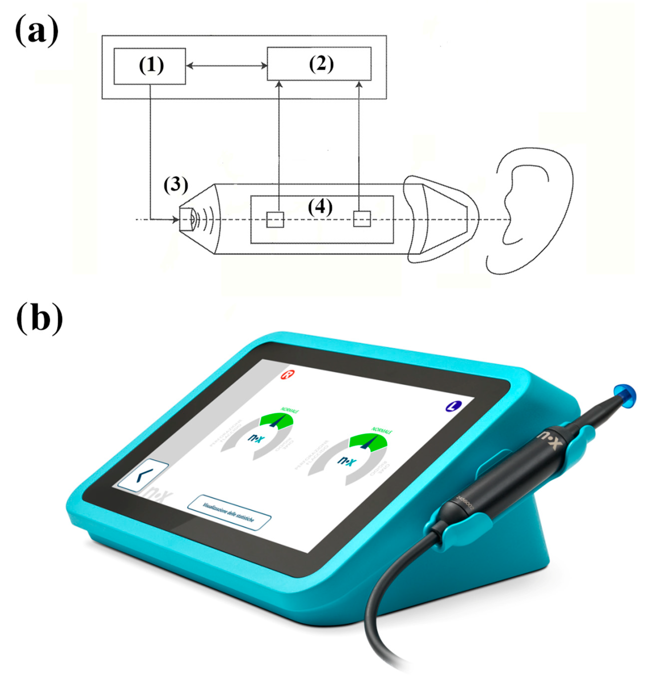
The Pressure-Less Acoustic Immittance (PLAI™; Neuranix Srl, Naples, Italy) system is a MEMS-based diagnostic tool designed for middle ear assessment without the need for external pressurization. Unlike conventional tympanometry, PLAI operates under ambient pressure, using a dual-sensor setup capable of measuring both acoustic pressure and particle velocity at the same location (Figure 1). The device emits a wideband acoustic signal spanning 100 to 2000 Hz, which interacts with the ear canal and tympanic membrane. The reflected signal, containing key information about the mechanical and acoustic properties of the middle and outer ear, is captured by two microphones. These microphones measure the pressure and axial velocity of the air particles in the ear canal.
From the acquired signals, PLAI™ computes an acoustic admittance curve, from which it extracts numerous parameters, including the following:
  • Resonance frequency (Fres);
  • Peak admittance (Peak);
  • Minimum and maximum frequency bounds (minBnd, maxBnd);
  • Bandwidth amplitude (AltBnd);
  • Equivalent ear canal volume (Vol).
The full feature set also includes age and labels used for classification.

2.3. Statistical Analysis

To assess whether PLAI™-derived parameters differ significantly between healthy and pathological ears, each acoustic feature was compared across diagnosis groups within each age category using Wilcoxon rank-sum test (Bonferroni correction was applied for multiple comparisons). A significance threshold of p < 0.05 was set.

2.4. Classification Pipeline

Following statistical analysis, a classification pipeline was implemented to evaluate the ability of PLAI™ features to distinguish between healthy and pathological ears.
The dataset was split into training (80%) and testing (20%) subsets within each age group. Feature selection was performed using a Random Forest classifier’s intrinsic feature importance score, retaining only the most relevant features per group. SMOTE (Synthetic Minority Over-sampling Technique) was applied to the training set to mitigate class imbalance by generating synthetic samples for underrepresented diagnostic categories.
Random Forest was selected as the classifier due to its robustness to overfitting, ability to handle nonlinear relationships, and compatibility with feature importance analysis. Hyperparameters were tuned based on performance on the validation split. Model evaluation included accuracy, precision, recall, F1-score, and specificity, computed both per class and as micro- and macro-averages. ROC curves were plotted, and confusion matrices were generated in grayscale for each age category.
To support model interpretability, SHAP (SHapley Additive exPlanations) values were computed, revealing the impact of individual features on classification decisions [32]. Feature importance visualizations are included in the results section for each age group.
All analyses and visualizations were performed in Python 3.8 using Scikit-learn, Imbalanced-learn, and SHAP libraries.

3. Results

3.1. Descriptive Statistics

3.1.1. Age 0–3

In Table 2, we report the descriptive statistics and comparative analysis of key PLAI™ parameters between healthy children and those diagnosed with Otitis Media with Effusion (OME) in the 0–3 year age group. Statistically significant differences were found in multiple parameters.
These findings are visually represented in Figure 2 through comparative boxplots, highlighting the dispersion and central tendency of each parameter.

3.1.2. Age 3–12

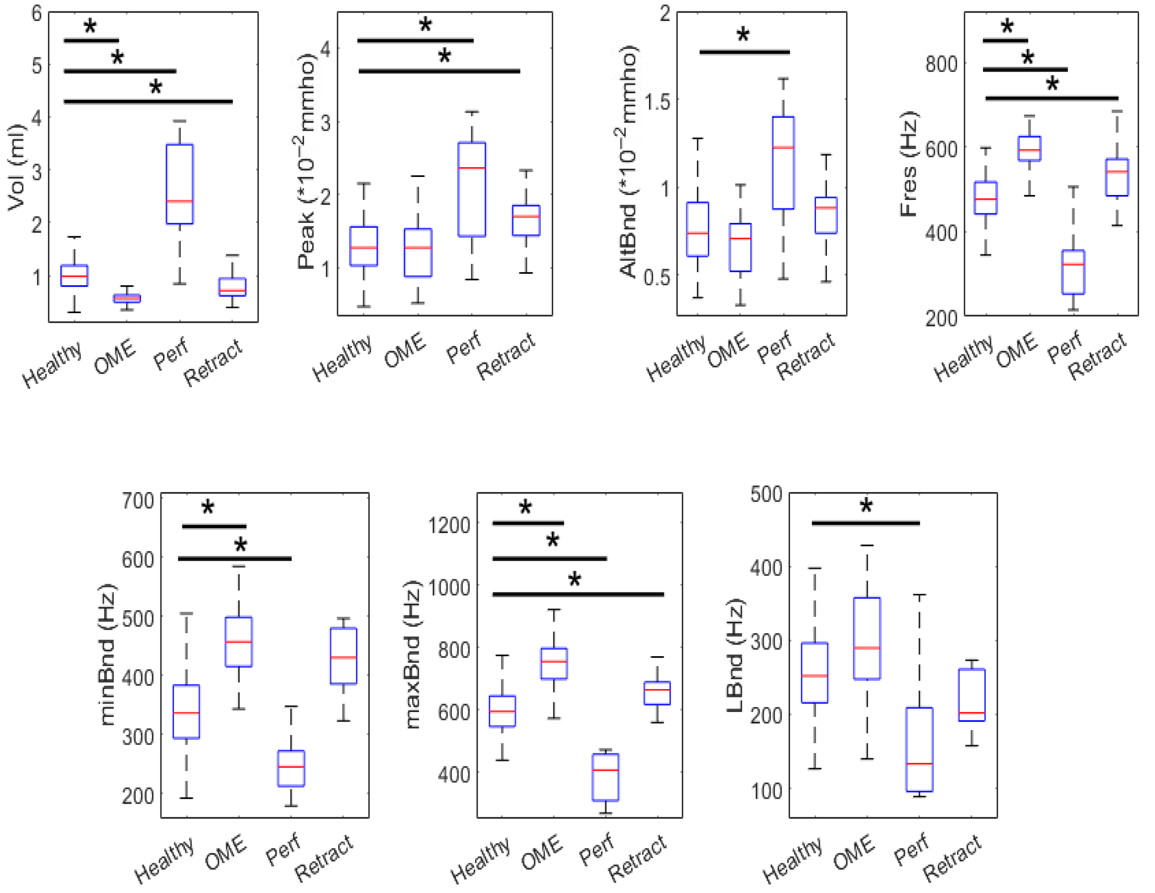
In Table 3, we report the descriptive statistics and comparative analysis for the main PLAI™-derived parameters in children aged 3 to 12 years, divided into healthy individuals, those diagnosed with Otitis Media with Effusion (OME), and individuals with tympanic membrane retraction. The comparisons were performed using appropriate statistical tests, with a Bonferroni-corrected significance threshold of p < 0.0167. These findings can be observed in boxplot form in Figure 3.

3.1.3. Age Greater than 12

In Table 4 we present the descriptive statistics and group comparisons for each parameter in the age group older than 12 years. This table includes the mean, median, standard deviation (SD), first (25th percentile) and third (75th percentile) quartiles, along with p-values comparing the healthy group with each pathological condition. The comparison threshold is set to p < 0.0125 to account for multiple comparisons. The corresponding boxplots are shown in Figure 4. Notably, there are no statistically significant differences in any of the parameters for otosclerosis in this age group, suggesting that the parameters analyzed may have limited discriminative value for this condition within the current feature set.

3.2. Feature Relevance via SHAP Analysis

To interpret the relevance of different features in the classification process, SHAP (SHapley Additive exPlanations) analysis was performed for each age group. Table 5 presents the mean SHAP values for the top-ranked features selected by the Random Forest classifier across the three age strata. These values quantify the average contribution of each feature to the model’s output.
For the 0–3 age group, the most influential feature was Fres, followed by maxBnd, minBnd, and Vol, suggesting that the frequency-related parameters played a central role in the classification of middle ear conditions. In the 3–12 age group, Vol and Fres again emerged as prominent contributors, alongside minBnd and LBnd, pointing to consistent diagnostic value across developmental stages. In adolescents and adults (12+), Vol, Fres, and AltBnd were key contributors, indicating that the volumetric and resonance features maintain importance in older patients as well.
These findings underscore the physiological plausibility of the selected features, as tympanic volume and resonance boundaries are commonly affected by pathological middle ear conditions such as effusions, retractions, or perforations.

3.3. Random Forest Classification Performance

Random Forest classifiers were trained and evaluated for each age group using a 5-class diagnostic classification task. Synthetic oversampling Via SMOTE [33] was employed to mitigate class imbalance. Performance was assessed using macro-averaged and micro-averaged metrics for precision, recall, F1-score, and specificity. Additionally, class-wise accuracy was extracted from the classification reports.
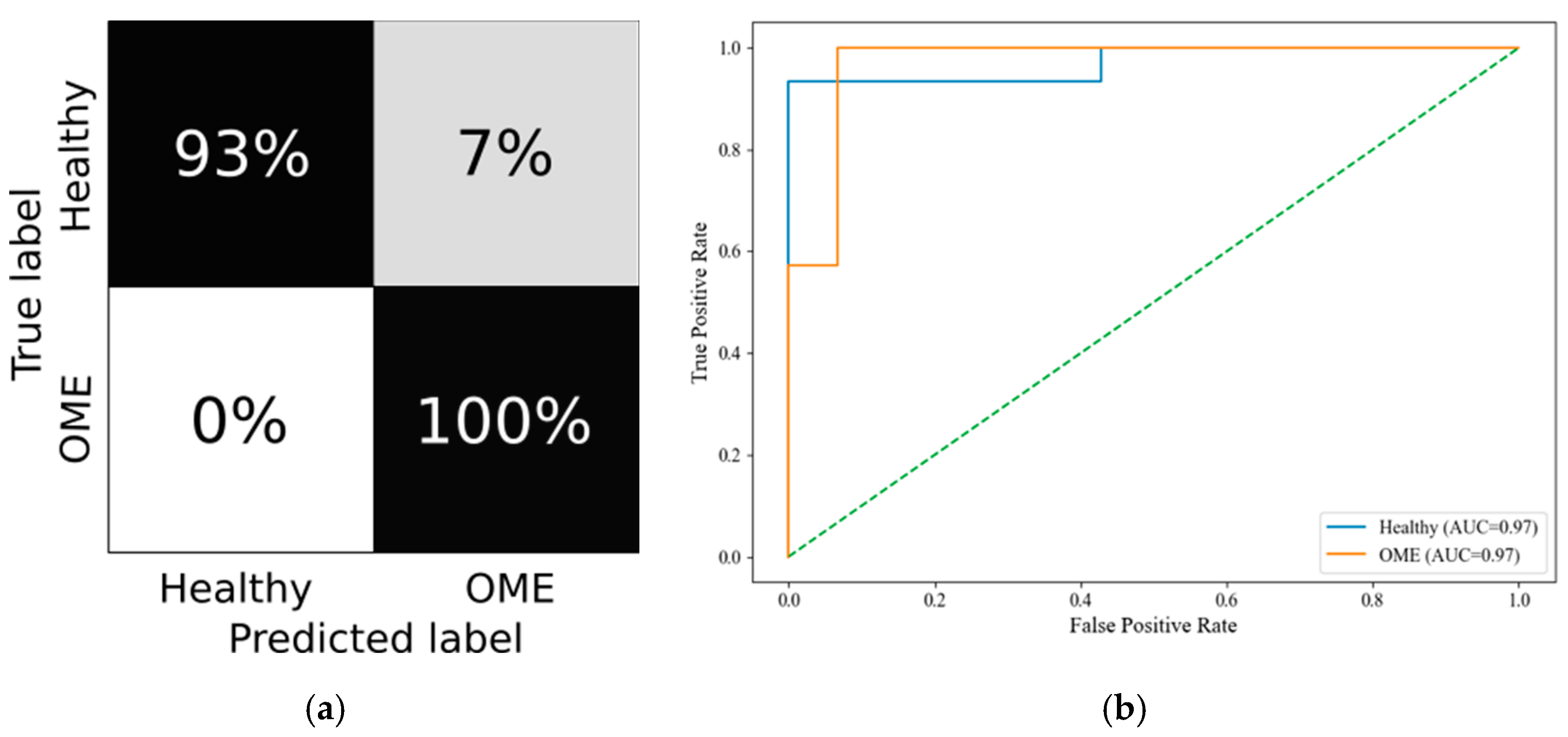
The 0–3 year group yielded a macro F1-score of 0.79 and a micro F1-score of 0.80, indicating good overall performance despite developmental anatomical variability. The model maintained a macro specificity of 0.95, emphasizing its reliability in distinguishing between pathological and non-pathological cases. The confusion matrix and ROC curves are depicted in Figure 5a and Figure 5b, respectively.
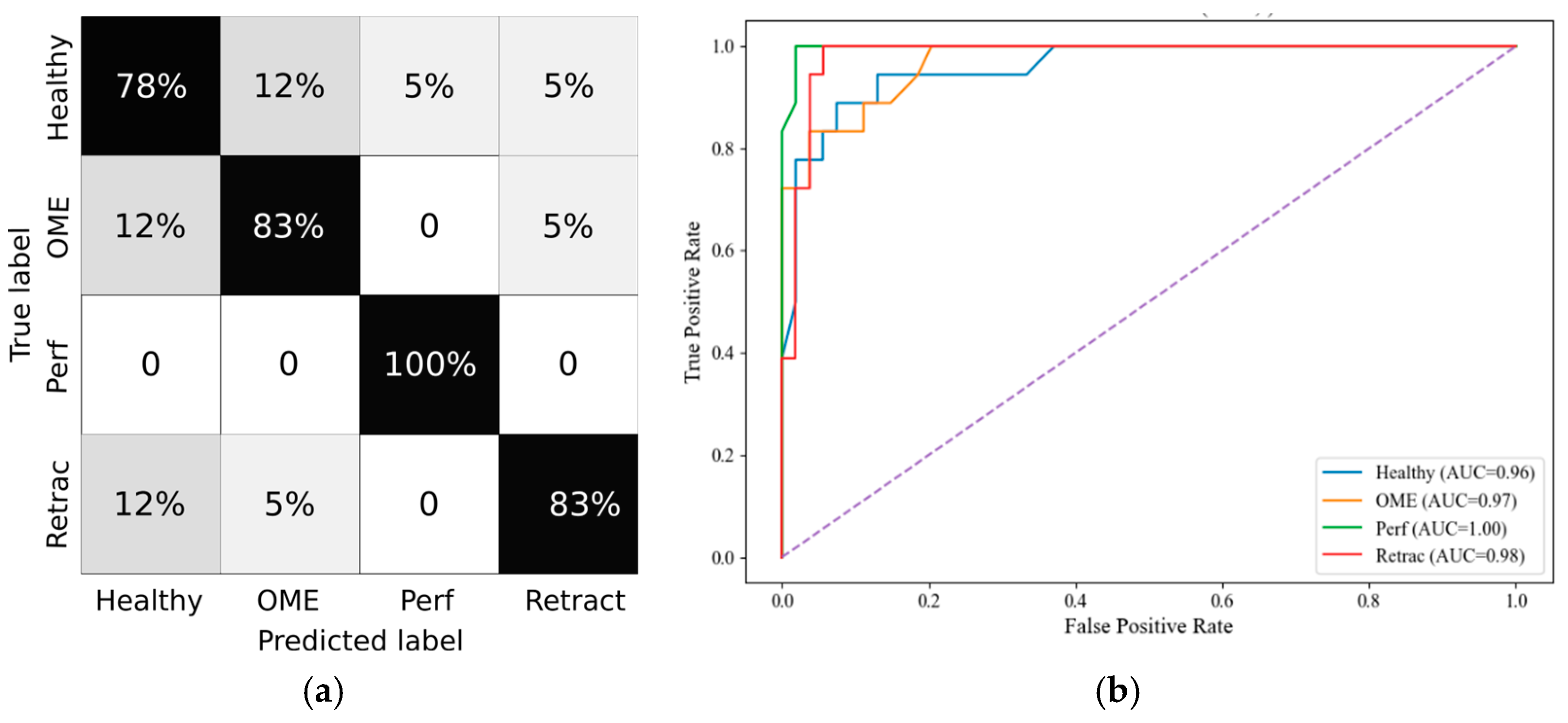
In the 3–12 group, the classifier demonstrated the highest performance, with a macro F1-score of 0.84 and a micro F1-score of 0.86. Notably, the macro precision (0.85) and specificity (0.96) affirm strong discriminatory capacity in this age range, which typically presents frequent but well-characterized middle ear pathologies. The confusion matrix and ROC curves are depicted in Figure 6a and Figure 6b, respectively.
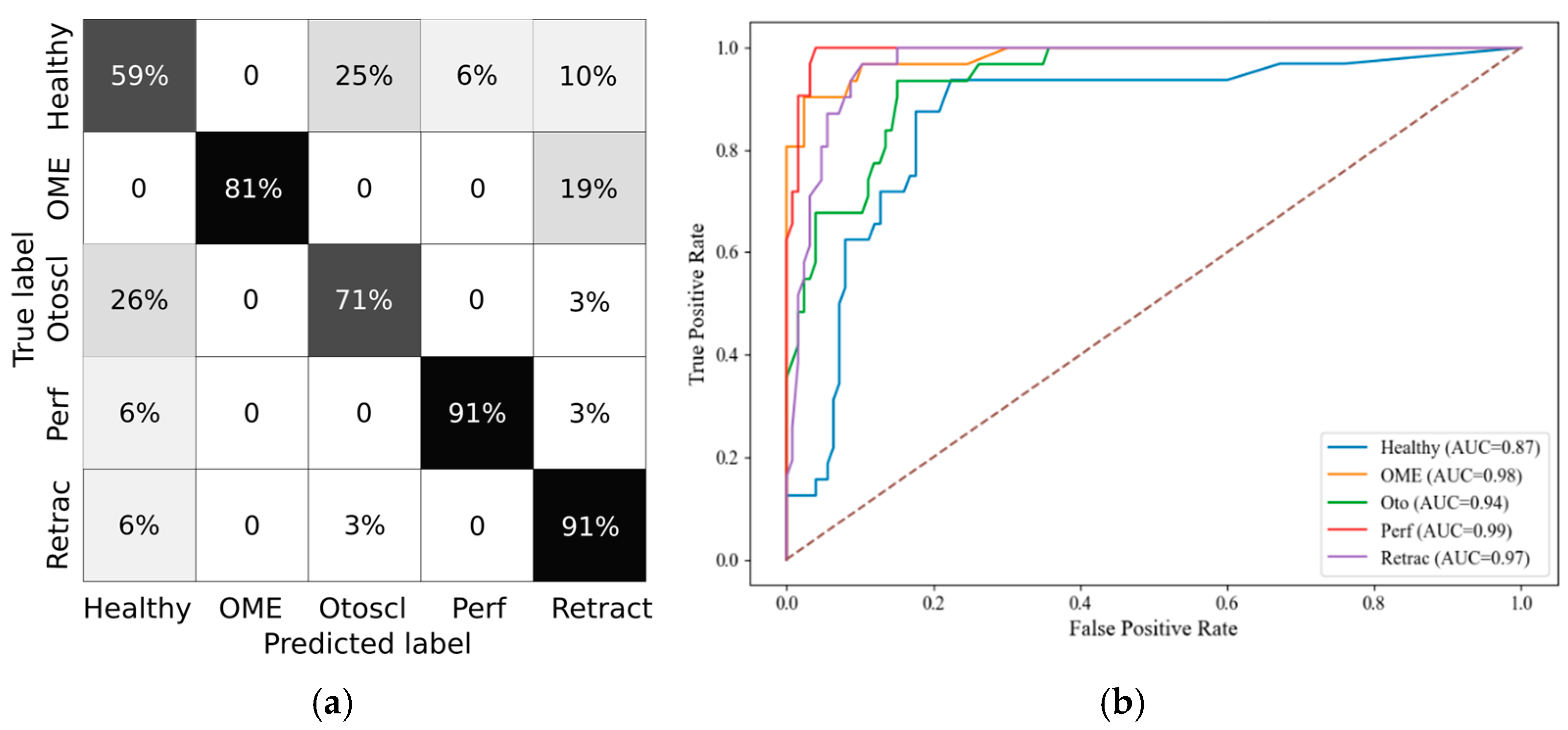
In the 12+ group, performance remained robust, albeit slightly reduced, with a macro F1-score of 0.78 and micro F1-score of 0.80. The confusion matrix and ROC curves are depicted in Figure 7a and Figure 7b, respectively. This may reflect increased inter-individual anatomical variability and the presence of more diverse pathophysiological mechanisms in adolescents and adults.
The overall classification performance is summarized in Table 6, while the per-class classification accuracy is reported in Table 7.

4. Discussion

This study investigated whether stratifying pediatric subjects by age groups improves the diagnostic modeling of middle ear conditions using quantitative PLAI™ features. Motivated by the known developmental changes in the auditory system during infancy and childhood [34,35], we hypothesized that age-related physiological differences significantly influence tympanometric measurements, thus impacting classification accuracy when predicting pathologies such as otitis media with effusion (OME), tympanic membrane perforation (Perf), otosclerosis (Otoscl), and retraction (Retract).
The primary aim of this study was to determine whether stratifying by age reveals statistically significant differences between diagnostic groups and to evaluate the performance of machine learning classifiers trained within these age-defined strata for detecting ear pathologies without relying on conventional, and potentially invasive, tympanometry.
Statistical comparisons of wideband tympanometry parameters showed that age stratification enhances the discrimination between healthy and pathological ears, particularly in the youngest (0–3 years) and oldest (12+ years) age groups. These results justified the creation of age-specific models, which subsequently demonstrated robust classification performance. Specifically, random forest classifiers, trained with SMOTE-balanced datasets, achieved macro F1-scores above 80% in both the 0–3 and 12+ groups. The SHAP analysis further revealed consistent feature relevance patterns, with Peak, Fres, and Vol emerging as the most discriminative parameters across all age groups.
Importantly, stratifying the analysis by age groups led to a marked reduction in false negative rates for conditions like OME and tympanic membrane retractions. This improvement strengthens diagnostic reliability and highlights the model’s potential utility as a non-invasive screening tool in clinical settings. In particular, the PLAI™ system could be integrated into routine examinations by general practitioners and pediatricians, enabling them to assess ear health rapidly and with minimal training. By flagging abnormal patterns suggestive of middle ear pathology, the system could support early identification and prompt referral of at-risk children for further evaluation by an otolaryngologist. This triage function may be especially valuable in primary care and community health settings where access to specialist equipment and expertise is limited.
Physiologically, these observations align with known developmental changes in the ear canal and middle ear structures. In infants and toddlers (0–3 years), the ear canal is shorter and more compliant, while the ossicular chain and tympanic membrane undergo maturation that can dramatically affect acoustic transfer functions [36]. This likely contributes to the distinct parameter distributions observed in this group and underscores the need for specialized diagnostic thresholds in early life. Conversely, adolescents (12+ years) display more adult-like anatomy and auditory responses, which could explain the enhanced classification performance and clearer parameter separation in this group.
Among the tympanometric features, Peak admittance, representing energy flow at the resonant frequency, was repeatedly selected as a key predictor. Fres, the resonant frequency of the middle ear system, also showed high discriminative power, likely reflecting changes in ossicular ant tympanic stiffness. Volume (Vol), while highly informative in distinguishing perforations, showed some limitations in overlapping pathologies such as OME and retractions, particularly in the middle group (3–12 years), where developmental heterogeneity may blur diagnostic distinctions.
Despite promising results, this study has limitations. The dataset, while substantial, exhibited class imbalance, particularly in less frequent pathologies such as otosclerosis and retraction, which may affect classifier generalizability. While SMOTE was employed to address class imbalance, particularly for underrepresented diagnoses such as otosclerosis and tympanic membrane retraction, several limitations should be considered. SMOTE synthesizes new examples by interpolating between existing minority class samples, which may not fully capture the variability inherent in complex or rare pathologies. Moreover, age bins were defined in three broad categories; finer stratification might yield further insights but would require a larger cohort to maintain statistical power. In this study, the small samples available for these rare conditions were assumed to be representative of their respective classes. However, this assumption carries inherent risks: if the original samples do not adequately reflect the broader clinical variability of the condition, the synthetic data may reinforce sampling biases rather than mitigate them. Future work should prioritize collecting more real-world data for the minority classes to improve generalizability and reduce reliance on synthetic augmentation.
Lastly, although the models demonstrated strong diagnostic potential, especially in early and late childhood, further validation on external datasets is necessary to confirm generalizability. Expanding the dataset, especially with more balanced class representation and longitudinal follow-ups, would be beneficial.
Nonetheless, the study underscores the importance of age-aware diagnostic modeling in pediatric audiology and highlights PLAI™ as a promising non-invasive tool for early and reliable identification of middle ear conditions.

5. Conclusions

This study demonstrated that age-stratified analysis enables the classification of common otologic pathologies using a non-invasive screening methodology. By dividing subjects into physiologically meaningful age groups (0–3, 3–12, and 12+ years), we observed enhanced parameter discrimination and superior classification performance, particularly in the youngest and oldest cohorts. The application of random forest classifiers, combined with SHAP-based feature relevance analysis, identified Peak admittance, Fres, and Volume as key contributors to diagnostic accuracy.
These findings highlight the critical role of age-dependent anatomical and physiological changes in shaping acoustic immittance responses, reinforcing the necessity of age-specific diagnostic frameworks. Despite limitations related to class imbalance and sample size, our results indicate strong potential for integrating machine learning and tympanometric features into future clinical tools for early detection of middle ear pathologies.
Further research is warranted to validate these models in larger, more diverse populations and to refine age-specific diagnostic thresholds. Future work will also aim to establish diagnostic criteria for specific ear pathologies based on PLAI™ measurements and to validate these criteria through comparison with established clinical gold standards, such as otoscopic evaluation and audiological testing. Ultimately, this approach holds promise for improving diagnostic precision and guiding early interventions in pediatric audiology.
Author Contributions

Conceptualization, F.B. and A.A.; formal analysis, A.M. and F.B.; funding acquisition, A.A.; methodology, A.M., M.A. and A.A.; project administration, A.A.; resources, F.B., M.A. and A.A.; software, A.M.; supervision, A.M., M.A. and A.A.; validation, F.B. and M.A.; visualization, F.B.; writing—original draft, A.M. and F.B.; writing—review and editing, A.M., M.A. and A.A. All authors have read and agreed to the published version of the manuscript.

Funding

This study is based on data collected during the clinical trial “MEDWAVE-2” (Study ID: IT-23-03-042692), funded by Neuranix Srl as part of the certification process for their medical device under CE marking regulations. No additional external funding was received for the preparation of this manuscript.

Institutional Review Board Statement

The study was approved by the Ethics Committees of all participating institutions (Protocol No. 5389, 19 January 2023, IRCCS “A. Gemelli” Hospital, Rome), and by the Italian Ministry of Health (MEDWAVE-2, IT-23-03-042692, 9 May 2023).

Informed Consent Statement

Written informed consent was obtained from adult participants; for minors, consent was provided by a parent or legal guardian.

Data Availability Statement

The datasets presented in this article are not readily available because the data are part of an ongoing study. Requests to access the datasets should be directed to Neuranix Srl.

Acknowledgments

The authors thank the six centers where the data collection was conducted: Gemelli University Hospital IRCCS-Rome, “Guglielmo da Saliceto” Hospital-Piacenza, “Federico II” University Hospital—Napoli, “Giovanni XXIII” Children Hospital—Bari, Fondazione IRCCS Policlinico “S. Matteo” Hospital—Pavia, IRCCS Maternal and Child Health Institute “Burlo Garofolo”-Trieste.

Conflicts of Interest

Agostino Accardo and Aleksandar Miladinović report a relationship with Neuranix Srl, Napoli, Italy that includes consulting or advisory. Francesco Bassi and Miloš Ajčević declare no known competing financial interests or personal relationships that could have appeared to influence the work reported in this paper.

Abbreviations

The following abbreviations are used in this manuscript:
OMEOtitis Media with Effusion
PLAIPressure-Less Acoustic Immittance
SHAPSHapley Additive exPlanations
RFRandom Forest

References

  1. Graydon, K.; Waterworth, C.; Miller, H.; Gunasekera, H. Global Burden of Hearing Impairment and Ear Disease. J. Laryngol. Otol. 2019, 133, 18–25. [Google Scholar] [CrossRef] [PubMed]
  2. Deshmukh, B.G. Accuracy of the Novel ENTraview Mobile Device to Increase Access to Screening and Provisional Diagnosis of Ear Disease in Underserved Communities Across India. Epidemiol. Int. 2022, 07, 21–24. [Google Scholar] [CrossRef]
  3. Godse, N.R.; Lu, N.; Shaffer, A.D.; Stapleton, A.L. Long Term Incidence and Outcomes of Sinonasal and Otologic Disease in Patients With Pyriform Aperture Stenosis and Choanal Atresia. J. Craniofac. Surg. 2022, 33, 1409–1412. [Google Scholar] [CrossRef]
  4. James, O.; Otoghile, B.; Adamson, O.O.; Egbunah, U.P.; Sabo, V.Y.; Adekunle, A.A.; Adeyemo, W.L.; Ladeinde, A.L.; Ogunlewe, O.M. Otological Challenges in Patients With Non-Syndromic Cleft Lip and Palate: A Systematic Review. FACE 2023, 4, 281–293. [Google Scholar] [CrossRef]
  5. Feltner, C.; Wallace, I.F.; Kistler, C.E.; Coker-Schwimmer, M.; Jonas, D.E. Screening for Hearing Loss in Older Adults: Updated Evidence Report and Systematic Review for the US Preventive Services Task Force. JAMA 2021, 325, 1202. [Google Scholar] [CrossRef] [PubMed]
  6. Smith, S.L.; Francis, H.W.; Witsell, D.L.; Dubno, J.R.; Dolor, R.J.; Bettger, J.P.; Silberberg, M.; Pieper, C.F.; Schulz, K.A.; Majumder, P.; et al. A Pragmatic Clinical Trial of Hearing Screening in Primary Care Clinics: Effect of Setting and Provider Encouragement. Ear Hear. 2024, 45, 23–34. [Google Scholar] [CrossRef] [PubMed]
  7. Clinical Practice Guidelines: Childhood Hearing Screening. 2011. Available online: https://www.audiology.org/practice-guideline/clinical-practice-guidelines-childhood-hearing-screening/ (accessed on 31 July 2025).
  8. Mertens, G.; Andries, E.; Claes, A.J.; Topsakal, V.; Van De Heyning, P.; Van Rompaey, V.; Calvino, M.; Sanchez Cuadrado, I.; Muñoz, E.; Gavilán, J.; et al. Cognitive Improvement After Cochlear Implantation in Older Adults With Severe or Profound Hearing Impairment: A Prospective, Longitudinal, Controlled, Multicenter Study. Ear Hear. 2021, 42, 606–614. [Google Scholar] [CrossRef]
  9. Fu, X.; Eikelboom, R.H.; Liu, B.; Wang, S.; Jayakody, D.M.P. The Longitudinal Relationship between Hearing Loss and Cognitive Decline in Tonal Language-Speaking Older Adults in China. Front. Aging Neurosci. 2023, 15, 1122607. [Google Scholar] [CrossRef]
  10. Kiely, K.; Anstey, K. Putting Age-Related Hearing Loss on the Public Health Agenda in Australia. Public Health Res. Pract. 2021, 31, e3152125. [Google Scholar] [CrossRef]
  11. Stevenson, J.; McCann, D.; Watkin, P.; Worsfold, S.; Kennedy, C.; on behalf of the Hearing Outcomes Study Team. The Relationship between Language Development and Behaviour Problems in Children with Hearing Loss. J. Child Psychol. Psychiatry 2010, 51, 77–83. [Google Scholar] [CrossRef]
  12. Williams, C.J.; Jacobs, A.M. The Impact of Otitis Media on Cognitive and Educational Outcomes. Med. J. Aust. 2009, 191, S69–S72. [Google Scholar] [CrossRef]
  13. Siggaard, L.D.; Jacobsen, H.; Hougaard, D.D.; Høgsbro, M. Digital vs. Physical Ear-Nose-and-Throat Specialist Assessment Screening for Complicated Hearing Loss and Serious Ear Disorders in Hearing-Impaired Adults Prior to Hearing Aid Treatment: A Randomized Controlled Trial. Front. Digit. Health 2023, 5, 1182421. [Google Scholar] [CrossRef]
  14. Paglialonga, A.; Cleveland Nielsen, A.; Ingo, E.; Barr, C.; Laplante-Lévesque, A. eHealth and the Hearing Aid Adult Patient Journey: A State-of-the-Art Review. Biomed. Eng. OnLine 2018, 17, 101. [Google Scholar] [CrossRef] [PubMed]
  15. Moyer, V.A.; on behalf of the U.S. Preventive Services Task Force*. Screening for Hearing Loss in Older Adults: U.S. Preventive Services Task Force Recommendation Statement. Ann. Intern. Med. 2012, 157, 655–661. [Google Scholar] [CrossRef] [PubMed]
  16. Chadha, S.; Dillard, L.K.; Mariotti, S.P.; Keel, S. Monitoring Hearing and Vision Functions in Older Adults: Rationale and Process. Age Ageing 2023, 52, iv158–iv161. [Google Scholar] [CrossRef] [PubMed]
  17. Bettger, J.P.; Dolor, R.J.; Witsell, D.L.; Dubno, J.R.; Pieper, C.F.; Walker, A.R.; Silberberg, M.; Schulz, K.A.; Majumder, P.; Juhlin, E.; et al. Comparative Implementation-Effectiveness of Three Strategies to Perform Hearing Screening among Older Adults in Primary Care Clinics: Study Design and Protocol. BMC Geriatr. 2020, 20, 170. [Google Scholar] [CrossRef] [PubMed]
  18. Everett, A.; Wong, A.; Piper, R.; Cone, B.; Marrone, N. Sensitivity and Specificity of Pure-Tone and Subjective Hearing Screenings Using Spanish-Language Questions. Am. J. Audiol. 2020, 29, 35–49. [Google Scholar] [CrossRef]
  19. Tseng, Y.C.; Liu, S.H.Y.; Lou, M.F.; Huang, G.S. Quality of Life in Older Adults with Sensory Impairments: A Systematic Review. Qual. Life Res. 2018, 27, 1957–1971. [Google Scholar] [CrossRef]
  20. Onusko, E. Tympanometry. Am. Fam. Physician 2004, 70, 1713–1720. [Google Scholar]
  21. Himelfarb, M.Z.; Popelka, G.R.; Shanon, E. Tympanometry in Normal Neonates. J. Speech Lang. Hear. Res. 1979, 22, 179–191. [Google Scholar] [CrossRef]
  22. Nakayama, J.R. Tympanometry. In Encyclopedia of Otolaryngology, Head and Neck Surgery; Springer: Berlin/Heidelberg, Germany, 2013; ISBN 978-3-642-23498-9. [Google Scholar]
  23. Margolis, R.H.; Saly, G.L.; Keefe, D.H. Wideband Reflectance Tympanometry in Normal Adults. J. Acoust. Soc. Am. 1999, 106, 265–280. [Google Scholar] [CrossRef]
  24. Al-Salim, S.; Skretta, D.; Merchant, G.R. Survey of Wideband Acoustic Immittance Use by Clinical Audiologists. Am. J. Audiol. 2024, 33, 254–268. [Google Scholar] [CrossRef]
  25. Stuppert, L.; Nospes, S.; Bohnert, A.; Läßig, A.K.; Limberger, A.; Rader, T. Clinical Benefit of Wideband-Tympanometry: A Pediatric Audiology Clinical Study. Eur. Arch. Otorhinolaryngol. 2019, 276, 2433–2439. [Google Scholar] [CrossRef] [PubMed]
  26. Holte, L.; Margolish, R.H.; Cavanaugh, R.M. Developmental Changes in Multifrequency Tympanograms. Int. J. Audiol. 1991, 30, 1–24. [Google Scholar] [CrossRef]
  27. Jiang, W.; Mu, Y.; Zhao, F.; Wang, P. Advancements in Pediatric Audiological Assessments Using Wideband Acoustic Immittance: A Review. Audiol. Res. 2024, 14, 684–700. [Google Scholar] [CrossRef] [PubMed]
  28. Graffigna, C.E.; Stanzial, D. Apparatus and Method for Measuring Clinical-Audiometric Parameters. U.S. Patent 12150757, 26 November 2024. [Google Scholar]
  29. Bassi, F.; Accardo, A. Estimation of Middle Ear Characteristics by an Innovative Pressure-Less Acoustic Immittance (PLAITM) Device. In 9th European Medical and Biological Engineering Conference; Jarm, T., Šmerc, R., Mahnič-Kalamiza, S., Eds.; IFMBE Proceedings; Springer Nature Switzerland: Cham, Switzerland, 2024; Volume 113, pp. 36–42. ISBN 978-3-031-61627-3. [Google Scholar]
  30. Just, T. Comparison of Conventional Single-Frequency Tympanometry with Pressure-Less Acoustic Immittance Measurements of Pathological and Normal Middle Ears. Online J. Otolaryngol. Rhinol. 2024, 7, 1–11. [Google Scholar] [CrossRef]
  31. Bassi, F.; Miladinovic, A.; Accardo, A. Relations between Age, Tympanometric Parameters and Pressureless Measurements. In Proceedings of the 9th National Bioengineering Group (GNB) Congress, Palermo, Italy, 16–18 June 2025. [Google Scholar]
  32. Lundberg, S.M.; Lee, S.I. A Unified Approach to Interpreting Model Predictions. In Advances in Neural Information Processing Systems; Guyon, I., Luxburg, U.V., Bengio, S., Wallach, H., Fergus, R., Vishwanathan, S., Garnett, R., Eds.; Curran Associates, Inc.: Sydney, Australian, 2017; Volume 30. [Google Scholar]
  33. Chawla, N.V.; Bowyer, K.W.; Hall, L.O.; Kegelmeyer, W.P. SMOTE: Synthetic Minority over-Sampling Technique. J. Artif. Int. Res. 2002, 16, 321–357. [Google Scholar] [CrossRef]
  34. Farkas, L.G.; Posnick, J.C.; Hreczko, T.M. Anthropometric Growth Study of the Ear. Cleft Palate-Craniofacial J. 1992, 29, 324–329. [Google Scholar] [CrossRef] [PubMed]
  35. Kalcioglu, M.T.; Miman, M.C.; Toplu, Y.; Yakinci, C.; Ozturan, O. Anthropometric Growth Study of Normal Human Auricle. Int. J. Pediatr. Otorhinolaryngol. 2003, 67, 1169–1177. [Google Scholar] [CrossRef]
  36. Keefe, D.H.; Bulen, J.C.; Arehart, K.H.; Burns, E.M. Ear-Canal Impedance and Reflection Coefficient in Human Infants and Adults. J. Acoust. Soc. Am. 1993, 94, 2617–2638. [Google Scholar] [CrossRef]
Figure 1. (a) Schematic of the Pressure-Less Acoustic Immittance (PLAITM) device, showing key components: (1) Sound source, (3) Speaker, (4) Sensors to capture reflected sound, and (2) Processing unit for data analysis. (b) Image of the PLAITM device.
Healthcare 13 01921 g001
Figure 2. Box plots comparing PLAI parameters between Healthy and OME subjects in the 0–3 age group. Statistically significant differences are indicated by asterisks.
Healthcare 13 01921 g002
Figure 3. Box plots comparing PLAI parameters between Healthy, OME, Perforated, and Retracted ears in the 3–12 age group. Statistically significant differences are indicated by asterisks.
Healthcare 13 01921 g003
Figure 4. Box plots comparing PLAI parameters between Healthy, OME, Perforated, Otosclerotic, and Retracted ears in the 12+ age group. Statistically significant differences are indicated by asterisks.
Healthcare 13 01921 g004
Figure 5. The 0–3 group RF metrics. (a) Confusion Matrix; (b) ROC Curve.
Healthcare 13 01921 g005
Figure 6. The 3–12 group RF metrics (a) Confusion Matrix; (b) ROC Curve.
Healthcare 13 01921 g006
Figure 7. The 12+ group RF metrics. (a) Confusion Matrix; (b) ROC Curve.
Healthcare 13 01921 g007
Table 1. Distribution of individual ears by age and pathology.
Age GroupHealthyOMEPerforationOtosclerosisRetractionTotal
0–3 years7122---93
3–12 years90287-18143
12+ years15718186317273
Total31868256335509
Table 2. Descriptive statistics and p-values for PLAI™ parameters in the 0–3 year age group (p-values in bold are statistically significant).
ParameterGroupMeanSDp-Value
Volume (mL)Healthy0.780.333<0.001
OME0.620.666
Peak (×10−2 mmho)Healthy1.020.3530.732
OME1.010.398
AltBnd (×10−2 mmho)Healthy0.610.190.184
OME0.580.19
Fres (Hz)Healthy54894<0.001
OME638132
minBnd (Hz)Healthy357118<0.001
OME47090
maxBnd (Hz)Healthy711151<0.001
OME827154
LBnd (Hz)Healthy3541770.193
OME356123
Table 3. Descriptive statistics and p-values for PLAI™ parameters in the 3–12 year age group (p-values in bold are statistically significant).
ParameterGroupMeanSDp-Value
OMEPerforationRetraction
Volume (mL)Healthy1.060.40<0.001<0.0010.002
OME0.590.17
Perforation2.581.07
Retraction0.810.29
Peak (×10−2 mmho)Healthy1.290.370.7400.01030.005
OME1.270.44
Perforation2.120.83
Retraction1.590.45
AltBnd (×10−2 mmho)Healthy0.760.200.1280.0100.12
OME0.690.21
Perforation1.130.39
Retraction0.830.21
Fres (Hz)Healthy47765<0.001<0.0010.002
OME59668
Perforation32396
Retraction53169
minBnd (Hz)Healthy33677<0.0010.0050.105
OME45371
Perforation24854
Retraction42758
maxBnd (Hz)Healthy59582<0.0010.0020.003
OME75496
Perforation414148
Retraction65683
LBnd (Hz)Healthy259740.0220.0060.058
OME30190
Perforation16798
Retraction22969
Table 4. Descriptive statistics and p-values for PLAI™ parameters in the 12+ year age group (p-values in bold are statistically significant).
ParameterGroupMeanSDp-Value
OMEPerf+Retract
Volume (mL)Healthy1.760.55<0.0010.1010.929<0.001
OME1.040.44
Perforation2.060.97
Otosclerosis1.780.66
Retraction1.150.54
Peak (×10−2 mmho)Healthy1.570.390.2450.0950.0940.100
OME1.530.69
Perforation1.961.06
Otosclerosis1.660.38
Retraction1.830.81
AltBnd (×10−2 mmho)Healthy0.900.210.1040.0520.0140.091
OME0.850.35
Perforation1.120.55
Otosclerosis0.960.18
Retraction1.040.44
Fres (Hz)Healthy38458<0.0010.1010.929<0.001
OME48475
Perforation388161
Otosclerosis38667
Retraction489152
minBnd (Hz)Healthy28456<0.0010.5020.761<0.001
OME36681
Perforation302146
Otosclerosis28669
Retraction380143
maxBnd (Hz)Healthy47773<0.0010.0390.6700.003
OME61270
Perforation463177
Otosclerosis47370
Retraction573172
LBnd (Hz)Healthy193560.0130.0020.3080.389
OME24692
Perforation16187
Otosclerosis18748
Retraction19381
Table 5. Mean SHAP values for PLAI™ derived features.
Feature0–3 Years3–12 Years12+ Years
Vol0.14610.09080.1573
Peak0.03060.03740.0343
AltBnd0.05720.04840.0739
Fres0.16030.08580.1242
minBnd0.11100.07390.0516
maxBnd0.14610.03800.0529
LBnd0.09670.06960.0384
Table 6. Classification performance metrics of Random Forest classifiers for each age group.
Metric0–3 Years3–12 Years12+ Years
Macro Precision0.800.850.77
Micro Precision0.810.860.80
Macro Recall0.780.840.79
Micro Recall0.810.860.80
Macro F1-score0.790.840.78
Micro F1-score0.810.860.80
Macro Specificity0.950.960.93
Table 7. Per class accuracy of obtained classifiers.
Per Class Accuracy0–3 Years3–12 Years12+ Years
Healthy0.930.780.59
OME1.00.830.81
Otosclerosis--0.71
Perforation-1.00.90
Retraction-0.830.90
Disclaimer/Publisher’s Note: The statements, opinions and data contained in all publications are solely those of the individual author(s) and contributor(s) and not of MDPI and/or the editor(s). MDPI and/or the editor(s) disclaim responsibility for any injury to people or property resulting from any ideas, methods, instructions or products referred to in the content.