
Pain and Disability Reduction Following Rib Manipulation in a Patient Recovering from Osteomyelitis of the Thoracic Spine
 , , , , and
Abstract
1. Introduction
2. Detailed Case Presentation
2.1. Differential Diagnosis
2.2. Treatment
2.3. Outcomes and Follow-Up
3. Discussion
4. Conclusions
Author Contributions
Funding
Institutional Review Board Statement
Informed Consent Statement
Data Availability Statement
Conflicts of Interest
References
- IsIssa, K.; Boylan, M.R.; Faloon, M.J.; Pourtaheri, S.; Naziri, Q.; Sahai, N.K.; Paulino, C.; Emami, A. The incident trends, Epidemiology, mortality and economic evaluation of vertebral osteomyelitis in the United States: A nationwide inpatient database study of 283,022 cases from 1998 to 2010. Spine J. 2016, 16, S168. [Google Scholar] [CrossRef]
- Issa, K.; Diebo, B.G.; Faloon, M.; Naziri, Q.; Pourtaheri, S.; Paulino, C.B.; Emami, A. The epidemiology of vertebral osteomyelitis in the United States from 1998 to 2013. Clin. Spine Surg. A Spine Publ. 2018, 31, E102–E108. [Google Scholar] [CrossRef]
- Zimmerli, W. Vertebral Osteomyelitis. N. Engl. J. Med. 2010, 362, 1022–1029. [Google Scholar] [CrossRef]
- Berbari, E.F.; Kanj, S.S.; Kowalski, T.J.; Darouiche, R.O.; Widmer, A.F.; Schmitt, S.K.; Hendershot, E.F.; Holtom, P.D.; Huddleston, P.M., 3rd; Petermann, G.W.; et al. 2015 Infectious Diseases Society of America (IDSA) clinical practice guidelines for the diagnosis and treatment of native vertebral osteomyelitis in adults. Clin. Infect. Dis. 2015, 61, e26–e46. [Google Scholar] [CrossRef]
- Gonzalez, G.A.; Porto, G.; Tecce, E.; Oghli, Y.S.; Miao, J.; O’Leary, M.; Chadid, D.P.; Vo, M.; Harrop, J. Advances in diagnosis and management of atypical spinal infections: A comprehensive review. North Am. Spine Soc. J. (NASSJ) 2023, 16, 100282. [Google Scholar] [CrossRef]
- Jha, Y.; Chaudhary, K. Diagnosis and Treatment Modalities for Osteomyelitis. Cureus 2022, 14, e30713. [Google Scholar] [CrossRef] [PubMed]
- Gupta, A.; Kowalski, T.J.; Osmon, D.R.; Enzler, M.; Steckelberg, J.M.; Huddleston, P.M.; Nassr, A.; Mandrekar, J.M.; Berbari, E.F. Long-term outcome of pyogenic vertebral osteomyelitis: A cohort study of 260 patients. Open Forum Infect. Dis. 2014, 1, ofu107. [Google Scholar] [CrossRef]
- Zuluaga, A.F.; Galvis, W.; Saldarriaga, J.G.; Agudelo, M.; Salazar, B.E.; Vesga, O. Etiologic diagnosis of chronic osteomyelitis. Arch. Intern. Med. 2006, 166, 95–100. [Google Scholar] [CrossRef] [PubMed]
- Dunn, R. Management of spinal osteomyelitis-associated deformity. Orthop. Trauma 2021, 35, 328–335. [Google Scholar] [CrossRef]
- Herman, G.; Zehr, S. Osteomyelitis of the Glenohumeral Joint. J. Orthop. Sports Phys. Ther. 2019, 49, 865. [Google Scholar] [CrossRef]
- Kavanagh, N.; Ryan, E.J.; Widaa, A.; Sexton, G.; Fennell, J.; O’Rourke, S.; Cahill, K.C.; Kearney, C.J.; O’Brien, F.J.; Kerrigan, S.W. Staphylococcal osteomyelitis: Disease progression, treatment challenges, and future directions. Clin. Microbiol. Rev. 2018, 31, e00084–17. [Google Scholar] [CrossRef] [PubMed]
- Park, K.-H.; Cho, O.-H.; Lee, Y.-M.; Moon, C.; Park, S.Y.; Moon, S.M.; Lee, J.H.; Park, J.S.; Ryu, K.N.; Kim, S.-H.; et al. Therapeutic outcomes of hematogenous vertebral osteomyelitis with instrumented surgery. Clin. Infect. Dis. 2015, 60, 1330–1338. [Google Scholar] [CrossRef] [PubMed]
- Priest, D.H.; Peacock, J.E. Hematogenous vertebral osteomyelitis due to Staphylococcus aureus in the adult: Clinical features and therapeutic outcomes. South. Med. J. 2005, 98, 854–862. [Google Scholar] [CrossRef] [PubMed]
- Segreto, F.A.; Beyer, G.A.; Grieco, P.; Horn, S.R.; Bortz, C.A.; Jalai, C.M.; Passias, P.G.; Paulino, C.B.; Diebo, B.G. Vertebral osteomyelitis: A comparison of associated outcomes in early versus delayed surgical treatment. Int. J. Spine Surg. 2018, 12, 703–712. [Google Scholar] [CrossRef]
- Yagdiran, A.; Otto-Lambertz, C.; Lingscheid, K.M.; Sircar, K.; Samel, C.; Scheyerer, M.J.; Zarghooni, K.; Eysel, P.; Sobottke, R.; Jung, N.; et al. Quality of life and mortality after surgical treatment for vertebral osteomyelitis (VO): A prospective study. Eur. Spine J. 2020, 30, 1721–1731. [Google Scholar] [CrossRef]
- Zadran, S.; Pedersen, P.H.; Eiskjær, S. Vertebral osteomyelitis: A mortality analysis comparing surgical and conservative management. Glob. Spine J. 2019, 10, 456–463. [Google Scholar] [CrossRef]
- Chu, J.; Allen, D.D.; Pawlowsky, S.; Smoot, B. Peripheral response to cervical or thoracic spinal manual therapy: An evidence-based review with meta-analysis. J. Man. Manip. Ther. 2014, 22, 220–229. [Google Scholar] [CrossRef]
- Heneghan, N.R.; Lokhaug, S.M.; Tyros, I.; Longvastøl, S.; Rushton, A. Clinical reasoning framework for thoracic spine exercise prescription in sport: A systematic review and narrative synthesis. BMJ Open Sport Amp; Exerc. Medicine 2020, 6, e000713. [Google Scholar] [CrossRef]
- Heneghan, N.R.; Lokhaug, S.; Tyros, I.; Rushton, A. Thoracic spine exercise prescription in sport: A narrative review. Physiotherapy 2019, 105, e4. [Google Scholar] [CrossRef]
- Lena, F.; Pappaccogli, M.; Santilli, M.; Torre, M.; Modugno, N.; Perrotta, A. How does semantic pain and words condition pain perception? A short communication. Neurol. Sci. 2022, 43, 691–696. [Google Scholar] [CrossRef] [PubMed]
- McDevitt, A.; Young, J.; Mintken, P.; Cleland, J. Regional interdependence and manual therapy directed at the thoracic spine. J. Man. Manip. Ther. 2015, 23, 139–146. [Google Scholar] [CrossRef] [PubMed]
- Schenk, R.; Donaldson, M.; Parent-Nichols, J.; Wilhelm, M.; Wright, A.; Cleland, J.A. Effectiveness of cervicothoracic and thoracic manual physical therapy in managing upper quarter disorders—A systematic review. J. Man. Manip. Ther. 2021, 30, 46–55. [Google Scholar] [CrossRef] [PubMed]
- Walser, R.F.; Meserve, B.B.; Boucher, T.R. The effectiveness of thoracic spine manipulation for the management of musculoskeletal conditions: A systematic review and meta-analysis of randomized clinical trials. J. Man. Manip. Ther. 2009, 17, 237–246. [Google Scholar] [CrossRef] [PubMed]
- Funabashi, M.; Son, J.; Pecora, C.G.; Tran, S.; Lee, J.; Howarth, S.J.; Kawchuk, G.; de Luca, K. Characterization of thoracic spinal manipulation and mobilization forces in older adults. Clin. Biomech. 2021, 89, 105450. [Google Scholar] [CrossRef]
- Horn, K.K.; Jennings, S.; Richardson, G.; van Vliet, D.; Hefford, C.; Abbott, J.H. The patient-specific functional scale: Psychometrics, clinimetrics, and application as a clinical outcome measure. J. Orthop. Sports Phys. Ther. 2012, 42, 30–42. [Google Scholar] [CrossRef]
- Michener, L.A.; Snyder, A.R.; Leggin, B.G. Responsiveness of the numeric pain rating scale in patients with shoulder pain and the effect of surgical status. J. Sport Rehabil. 2011, 20, 115–128. [Google Scholar] [CrossRef]
- Stratford, P. Assessing disability and change on individual patients: A report of a patient specific measure. Physiother. Can. 1995, 47, 258–263. [Google Scholar] [CrossRef]
- Stratford, P.W.; Kennedy, D.M.; Wainwright, A.V. Assessing the patient-specific functional scale’s ability to detect early recovery following total knee arthroplasty. Phys. Ther. 2014, 94, 838–844. [Google Scholar] [CrossRef]
- Magee, D.J.; Manske, R.C. Orthopedic Physical Assessment, 7th ed.; Elsevier: Amsterdam, The Netherlands, 2021. [Google Scholar]
- López-de-Uralde-Villanueva, I.; Acuyo-Osorio, M.; Prieto-Aldana, M.; La Touche, R. Reliability and minimal detectable change of a modified passive neck flexion test in patients with chronic nonspecific neck pain and asymptomatic subjects. Musculoskelet. Sci. Pract. 2017, 28, 10–17. [Google Scholar] [CrossRef]
- Lonstein, J.E. Point of view: A study of the diagnostic accuracy and reliability of the scoliometer and Adam’s Forward Bend Test. Spine 1998, 23, 803. [Google Scholar] [CrossRef]
- Gavin, D. The effect of joint manipulation techniques on active range of motion in the mid-thoracic spine of asymptomatic subjects. J. Man. Manip. Ther. 1999, 7, 114–122. [Google Scholar] [CrossRef]
- Kovanur Sampath, K.; Treffel, L.; PThomson, O.; Rodi, J.D.; Fleischmann, M.; Tumilty, S. Changes in biochemical markers following a spinal manipulation—A systematic review update. J. Man. Manip. Ther. 2023, 32, 28–50. [Google Scholar] [CrossRef] [PubMed]
- Pasquier, M.; Young, J.J.; Lardon, A.; Descarreaux, M. Factors associated with clinical responses to spinal manipulation in patients with non-specific thoracic back pain: A prospective cohort study. Front. Pain Res. 2022, 2, 742119. [Google Scholar] [CrossRef]
- Puentedura, E.J.; O’Grady, W.H. Safety of thrust joint manipulation in the thoracic spine: A systematic review. J. Man. Manip. Ther. 2015, 23, 154–161. [Google Scholar] [CrossRef]
- Sillevis, R.; Cleland, J.; Hellman, M.; Beekhuizen, K. Immediate effects of a thoracic spine thrust manipulation on the autonomic nervous system: A randomized clinical trial. J. Man. Manip. Ther. 2010, 18, 181–190. [Google Scholar] [CrossRef] [PubMed]
- Blanpied, P.R.; Gross, A.R.; Elliott, J.M.; Devaney, L.L.; Clewley, D.; Walton, D.M.; Sparks, C.; Robertson, E.K.; Altman, R.D.; Beattie, P.; et al. Neck pain: Revision 2017. J. Orthop. Sports Phys. Ther. 2017, 47, A1–A83. [Google Scholar] [CrossRef]
- George, S.Z.; Fritz, J.M.; Silfies, S.P.; Schneider, M.J.; Beneciuk, J.M.; Lentz, T.A.; Gilliam, J.R.; Hendren, S.; Norman, K.S.; Beattie, P.F.; et al. Interventions for the Management of Acute and Chronic Low Back Pain: Revision 2021. J Orthop Sports Phys Ther. 2021, 51, CPG1–CPG60. [Google Scholar] [CrossRef] [PubMed]
- Aspegren, D.; Hyde, T.; Miller, M. Conservative treatment of a female collegiate volleyball player with Costochondritis. J. Manip. Physiol. Ther. 2007, 30, 321–325. [Google Scholar] [CrossRef]
- Rabey, I.M. Costochondritis: Are the symptoms and signs due to neurogenic inflammation. Two cases that responded to manual therapy directed towards posterior spinal structures. Man. Ther. 2008, 13, 82–86. [Google Scholar] [CrossRef]
- Young, B.A.; Walker, M.J.; Strunce, J.B.; Boyles, R.E.; Whitman, J.M.; Childs, J.D. Responsiveness of the neck disability index in patients with mechanical neck disorders. Spine J. 2009, 9, 802–808. [Google Scholar] [CrossRef]
- Hotta, G.H.; Alaiti, R.K.; Ribeiro, D.C.; McQuade, K.J.; de Oliveira, A.S. Causal mechanisms of a scapular stabilization intervention for patients with subacromial pain syndrome: A secondary analysis of a randomized controlled trial. Arch. Physiother. 2022, 12, 12. [Google Scholar] [CrossRef]
- Ravichandran, H.; Janakiraman, B.; Gelaw, A.Y.; Fisseha, B.; Sundaram, S.; Sharma, H.R. Effect of scapular stabilization exercise program in patients with subacromial impingement syndrome: A systematic review. J. Exerc. Rehabil. 2020, 16, 216–226. [Google Scholar] [CrossRef] [PubMed]
- Yuksel, E.; Yesilyaprak, S.S. Scapular Stabilization exercise training improves treatment effectiveness on shoulder pain, scapular dyskinesis, muscle strength, and function in patients with subacromial pain syndrome: A randomized controlled trial. J. Bodyw. Mov. Ther. 2024, 37, 101–108. [Google Scholar] [CrossRef] [PubMed]
- Fernandes, J.B.; Ferreira, N.; Domingos, J.; Ferreira, R.; Amador, C.; Pardal, N.; Castro, C.; Simões, A.; Fernandes, S.; Bernardes, C.; et al. Health professionals’ motivational strategies to enhance adherence in the rehabilitation of people with lower limb fractures: Scoping review. Int. J. Environ. Res. Public Health 2023, 20, 7050. [Google Scholar] [CrossRef] [PubMed]
- Hurst, M.J.; Kedzie, M.; Hughes, H.J.; Burns, P.R.; Manway, J.M. Osteomyelitis and Fracture Susceptibility: A Review of the Literature and Case Series of Surgical Treatment of Pathologic Calcaneal Fractures. Foot Ankle Surg. Tech. Rep. Cases 2023, 3, 100329. [Google Scholar] [CrossRef]
- Pickar, J.G. Neurophysiological effects of spinal manipulation. Spine J. 2002, 2, 357–371. [Google Scholar] [CrossRef]


| Description | |
|---|---|
| Medical history | Osteomyelitis Osteoarthritis Sacroiliac joint pain Hypertension |
| Surgical history | Multiple root canals |
| Medications | OxyContin (20 mg) prn for pain Norvasc (5 mg) 1 time daily Ceftriaxone (1–2 g) IV once daily Daptomycin (4 mg/kg) IV every 24 h for 7 days |
| Symptom Location | Pain Description | Aggravating Factor | Easing Factor |
|---|---|---|---|
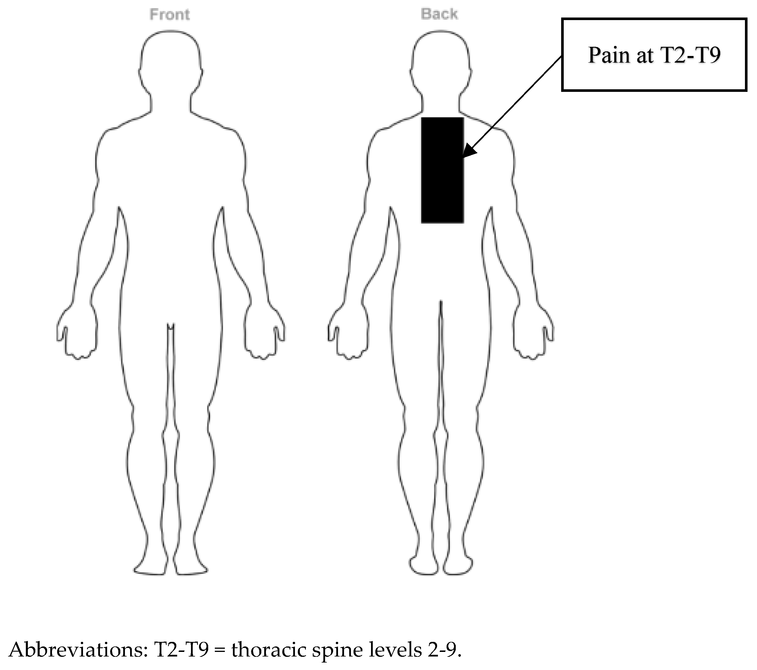
| Upper and middle thoracic spine (T2–T9) | NPRS severity: 8 Irritability: mild Stage: sub-acute Stability: worsening | Standing > 15 min Sleeping in supine Walking > 30 min Donning and doffing clothing | Sitting with exaggerated thoracic flexion Side-lying position |
| Cervical Spine | AROM | PROM | Strength |
|---|---|---|---|
| Flexion | 80° | 86° | 4/5 |
| Extension | 72° | 75° | 4/5 |
| Right side-bending | 40° | 44° | 4/5 |
| Left side-bending | 42° | 46° | 4/5 |
| Right rotation | 85° | 88° | 4/5 |
| Left rotation | 80° | 84° | 4/5 |
| Thoracic Spine | AROM | PROM | Strength |
| Flexion | 60° | 66° | 2/5 |
| Extension | 5° | 7° | 2/5 |
| Right side-bending | 18° | 22° | 2/5 |
| Left side-bending | 17° | 20° | 2/5 |
| Right rotation | 12° | 15° | 2/5 |
| Left rotation | 16° | 20° | 2/5 |
| Test | Description | Positive Test Result |
|---|---|---|
| Slump test [29] | The patient flexes the spine and shoulders while the examiner holds the chin and head erect. The patient is asked whether any symptoms are produced. If no symptoms are produced, the examiner flexes the patient’s neck and holds the head down with shoulders slumped to see whether symptoms are produced. If no symptoms are produced, the examiner passively extends one of the patient’s knees to see whether symptoms are produced. If no symptoms are produced, the examiner then passively dorsiflexes the foot of the same leg to see if symptoms are produced. | Reproduction of the patient’s symptoms indicates a positive test, implicating neural tension |
| Passive neck flexion test [30] | The patient actively performs an upper cervical nod. The examiner passively flexes the lower cervical spine. | A reproduction of pain or other neural symptoms in the thoracic spine is a positive test result |
| Adam’s forward bending test [31] | The patient needs to bend forward, starting at the waist until the back comes in the horizontal plane, with the feet together, arms hanging, and the knees in extension. The palms are held together. | An asymmetry is observed on one side |
| Reflex hammer test [29] | The patient is seated, and the examiner taps over each spinous process to see whether pain or muscle spasm is provoked. | If pain or muscle spasm is provoked, a fracture is possible |
| Thoracic Movement | AROM | Pain Location | NPRS |
|---|---|---|---|
| Flexion | 60° | Central, T2 | 2/10 |
| Extension | 5° | Central, T5–T9 | 8/10 |
| Right side-bending | 18° | Right unilateral, T5–T9 | 6/10 |
| Left side-bending | 17° | Left unilateral, T5–T9 | 6/10 |
| Right rotation | 12° | Right unilateral, T2–T9 | 4/10 |
| Left rotation | 16° | Left unilateral, T2–T9 | 4/10 |
| Visit | Treatment Plan |
|---|---|
| 1–4 | Mobility: Thoracic extension, side-bending, and rotation AROM in sitting position, side-lying rotational AROM, doorway stretch Motor control: Scapular squeezes in sitting position Manual therapy: Grades 1–4 central and unilateral PA mobilizations to reduce pain at levels T2–T9; STM to thoracic paraspinals to reduce muscle tightness; pectoralis minor STM to reduce muscle tightness |
| 5–8 | Mobility: Continued mobility from visits 1–4 with progressions of ½ foam roll extensions over chair with arms crossed; begin quadruped unilateral rotation exercises; downward dog; tabletop stress with physioball Motor control: Continued exercises from visits 1–4; progressed to serratus anterior strengthening, and resistance exercises targeting scapular stabilizers Manual therapy: Grades 3–4 central and unilateral PA mobilizations at levels T2–T9 to improve thoracic mobility; mobilization with movement for thoracic rotation, side-bending, and rotation; thoracic manipulation to T5–T7 |
| 9–12 | Mobility: AROM exercises from sessions 1–4 discharged to HEP; progressed to quadruped rotation AROM Motor control: Scapular stabilization including resistance bands and dumbbells Manual therapy: Continued techniques from visits 5–8; initiation of costotransverse joint manipulation bilaterally from T2 to T7 |
| 13–16 | Mobility: Discharged to patient’s HEP Motor control: Scapular stabilization including resistance bands and dumbbells until failure; functional activities including farmers carries, crate carries, and sled pushing and pulling Manual therapy: Costotransverse joint manipulation bilaterally from T2 to T7 |
| Assessment | NPRS | NDI | PSFS |
|---|---|---|---|
| Initial evaluation | 8/10 | 46/50 | 3/30 |
| Re-evaluation (Week 4) | 6/10 | 34/50 | 9/30 |
| Discharge (Week 8) | 1/10 | 22/50 | 21/30 |
| Cervical Spine (Re-Evaluation) | AROM | PROM | Strength |
|---|---|---|---|
| Flexion | 88° | 90° | 4/5 |
| Extension | 74° | 81° | 4/5 |
| Right side-bending | 45° | 49° | 4/5 |
| Left side-bending | 46° | 50° | 4/5 |
| Right rotation | 86° | 88° | 4-/5 |
| Left rotation | 85° | 86° | 4-/5 |
| Thoracic Spine (Re-Evaluation) | AROM | PROM | Strength |
| Flexion | WFL | WFL | 2+/5 |
| Extension | 11° | 15° | 2+/5 |
| Right side-bending | 22° | 25° | 2+/5 |
| Left side-bending | 25° | 27° | 2+/5 |
| Right rotation | 20° | 23° | 2+/5 |
| Left rotation | 20° | 22° | 2+/5 |
| Cervical Spine (Discharge) | |||
| Flexion | WFL | WFL | 4+/5 |
| Extension | WFL | WFL | 4+/5 |
| Right side-bending | WFL | WFL | 4+/5 |
| Left side-bending | WFL | WFL | 4+/5 |
| Right rotation | WFL | WFL | 4/5 |
| Left rotation | WFL | WFL | 4/5 |
| Thoracic Spine (Discharge) | |||
| Flexion | WFL | WFL | 4/5 |
| Extension | 35° | 40° | 3+/5 |
| Right side-bending | 40° | 46° | 3+/5 |
| Left side-bending | 35° | 39° | 3+/5 |
| Right rotation | 50° | 55° | 3+/5 |
| Left rotation | 45° | 50° | 3+/5 |
Disclaimer/Publisher’s Note: The statements, opinions and data contained in all publications are solely those of the individual author(s) and contributor(s) and not of MDPI and/or the editor(s). MDPI and/or the editor(s) disclaim responsibility for any injury to people or property resulting from any ideas, methods, instructions or products referred to in the content. |
© 2025 by the authors. Licensee MDPI, Basel, Switzerland. This article is an open access article distributed under the terms and conditions of the Creative Commons Attribution (CC BY) license (https://creativecommons.org/licenses/by/4.0/).
Share and Cite
Prall, J.; Dunning, J.; Young, I.; Ross, M.; Escaloni, J.; Bliton, P. Pain and Disability Reduction Following Rib Manipulation in a Patient Recovering from Osteomyelitis of the Thoracic Spine. Healthcare 2025, 13, 1355. https://doi.org/10.3390/healthcare13121355
Prall J, Dunning J, Young I, Ross M, Escaloni J, Bliton P. Pain and Disability Reduction Following Rib Manipulation in a Patient Recovering from Osteomyelitis of the Thoracic Spine. Healthcare. 2025; 13(12):1355. https://doi.org/10.3390/healthcare13121355
Chicago/Turabian StylePrall, Joshua, James Dunning, Ian Young, Michael Ross, James Escaloni, and Paul Bliton. 2025. "Pain and Disability Reduction Following Rib Manipulation in a Patient Recovering from Osteomyelitis of the Thoracic Spine" Healthcare 13, no. 12: 1355. https://doi.org/10.3390/healthcare13121355
APA StylePrall, J., Dunning, J., Young, I., Ross, M., Escaloni, J., & Bliton, P. (2025). Pain and Disability Reduction Following Rib Manipulation in a Patient Recovering from Osteomyelitis of the Thoracic Spine. Healthcare, 13(12), 1355. https://doi.org/10.3390/healthcare13121355