
Prevalence of Chlamydia trachomatis, Neisseria gonorrhoeae, and Mycoplasma genitalium among Patients with Urogenital Symptoms in Istanbul

 , , ,
, , ,  ,
,
Abstract
1. Introduction
2. Materials and Methods
2.1. Subjects and Sample Collections
2.2. DNA Extraction and Multiplex Real-Time PCR
2.3. Statistical Analysis
3. Results
4. Discussion
5. Conclusions
Author Contributions
Funding
Institutional Review Board Statement
Informed Consent Statement
Data Availability Statement
Conflicts of Interest
References
- Ahmad, N.; Alspaugh, J.A.; Lawrence Drew, W.; Lagunoff, M.; Pottinger, P.; Barth Reller, L.; Reller, M.E.; Sterling, C.R.; Weisman, S. Infectious Diseases; Syndromes and Etiologies. In Sherris Medical Microbiology 7th Press; Ryan, K.J., Ed.; Mc Graw-Hill Education: New York, NY, USA, 2018; p. 982. [Google Scholar]
- Igietseme, J.U.; Omosun, Y.; Black, C.M. Bacterial Sexually Transmitted Infections (STIs): A Clinical Overview. Mol. Med. Microbiol. 2015, 3, 1403–1420. [Google Scholar]
- McCormack, D.; Koons, K. Sexually Transmitted Infections. Emerg. Med. Clin. N. Am. 2019, 37, 725–738. [Google Scholar] [CrossRef] [PubMed]
- Serradji, N.; Vu, T.H.; Kim, H.; Panyam, J.; Verbeke, P. Inhibition of Chlamydia trachomatis growth during the last decade: A mini-review. Mini Rev. Med. Chem. 2018, 18, 1363–1372. [Google Scholar] [CrossRef]
- Rondeau, P.; Valin, N.; Decré, D.; Girard, P.M.; Lacombe, K.; Surgers, L. Chlamydia trachomatis screening in urine among asymptomatic men attending an STI clinic in Paris: A cross-sectional study. BMC Infect. Dis. 2019, 19, 31. [Google Scholar] [CrossRef] [PubMed]
- Nijhuis, R.H.T.; Duinsbergen, R.G.; Pol, A.; Godschalk, P.C.T. Prevalence of Chlamydia trachomatis, Neisseria gonorrhoeae, Mycoplasma genitalium and Trichomonas vaginalis including relevant resistance-associated mutations in a single center in the Netherlands. Eur. J. Clin. Microbiol. Infect. Dis. 2021, 40, 591–595. [Google Scholar] [CrossRef]
- Workowski, K.A.; Bachmann, L.H.; Chan, P.A.; Johnston, C.M.; Muzny, C.A.; Park, I.; Reno, H.; Zenilman, J.M.; Bolan, G.A. Sexually Transmitted Infections Treatment Guidelines, 2021. MMWR Recomm. Rep. 2021, 70, 1–187. [Google Scholar] [CrossRef] [PubMed]
- Gnanadurai, R.; Fifer, H. Mycoplasma genitalium: A Review. Microbiology 2020, 166, 21–29. [Google Scholar] [CrossRef]
- Manhart, L.E.; Broad, J.M.; Golden, M.R. Mycoplasma genitalium: Should we treat and how? Clin. Infect. Dis. 2011, 53 (Suppl. S3), 129–142. [Google Scholar] [CrossRef]
- Taylor-Robinson, D.; Jensen, J.S. Mycoplasma genitalium: From Chrysalis to multicolored butterfly. Clin. Microbiol. Rev. 2011, 24, 498–514. [Google Scholar] [CrossRef]
- Foschi, C.; Zagarrigo, M.; Belletti, M.; Marangoni, A.; Re, M.C.; Gaspari, V. Genital and extra-genital Chlamydia trachomatis and Neisseria gonorrhoeae infections in young women attending a Sexually Transmitted Infections (STI) clinic. New Microbiol. 2020, 43, 115–120. [Google Scholar]
- Kersh, E.N.; Geisler, W.M. A Commentary on current diagnostic challenges and research needs for evaluating reproductive sequelae of sexually transmitted infections. J. Infect. Dis. 2021, 224 (Suppl. S2), S72–S74. [Google Scholar] [CrossRef] [PubMed]
- Tsevat, D.G.; Wiesenfeld, H.C.; Parks, C.; Peipert, J.F. Sexually transmitted diseases and infertility. Am. J. Obstet. Gynecol. 2017, 216, 1–9. [Google Scholar] [CrossRef] [PubMed]
- Jimoh, O.; Aliyu, S.; Ejembi, J.; Abdulaziz, M.M.; Ibrahim, M.; Ibrahim, A.; Olayinka, A.T. Changing susceptibility pattern of Neisseria gonorrhoeae: A threat to management of sexually transmitted infections—Case series. Niger. J. Clin. Pract. 2021, 24, 778–781. [Google Scholar] [PubMed]
- Gaydos, C.A.; Cartwright, C.P.; Colaninno, P.; Welsch, J.; Holden, J.; Ho, S.Y.; Webb, E.M.; Anderson, C.; Bertuzis, R.; Zhang, L.; et al. Performance of the Abbott RealTime CT/NG for detection of Chlamydia trachomatis and Neisseria gonorrhoeae. J. Clin. Microbiol. 2010, 48, 3236–3243. [Google Scholar] [CrossRef]
- Hopkins, M.J.; Ashton, L.J.; Alloba, F.; Alawattegama, A.; Hart, I.J. Validation of a laboratory-developed real-time PCR protocol for detection of Chlamydia trachomatis and Neisseria gonorrhoeae in urine. Sex. Transm. Infect. 2010, 86, 207–211. [Google Scholar] [CrossRef] [PubMed]
- Pereyre, S.; Caméléna, F.; Hénin, N.; Berçot, B.; Bébéar, C. Clinical performance of four multiplex real-time PCR kits detecting urogenital and sexually transmitted pathogens. Clin. Microbiol. Infect. 2022, 28, 733.e7–733.e13. [Google Scholar] [CrossRef]
- Gossé, M.; Lysvand, H.; Pukstad, B.; Nordbø, S.A. A Novel SimpleProbe PCR Assay for Detection of Mutations in the 23S rRNA Gene Associated with Macrolide Resistance in Mycoplasma genitalium in Clinical Samples. J. Clin. Microbiol. 2016, 54, 2563–2567. [Google Scholar] [CrossRef]
- World Health Organization. Laboratory Diagnosis of Sexually Transmitted Infections, Including Human Immunodeficiency Virus; World Health Organization: Geneva, Switzerland, 2013. [Google Scholar]
- Barrientos-Durán, A.; Salazar, A.; Fuentes-Lopez, A.; Serrano-Conde, E.; Espadafor, B.; Chueca, N.; Alvarez-Estevez, M.; Garcia, F. Comparison between Aptima® assays (Hologic) and the CoBAS® 6800 system (Roche) for the diagnosis of sexually transmitted infections caused by Chlamydia trachomatis, Neisseria gonorrhoeae, and Mycoplasma genitalium. Eur. J. Clin. Microbiol. Infect. Dis. 2021, 40, 1337–1342. [Google Scholar] [CrossRef]
- Le Roy, C.; Le Hen, I.; Clerc, M.; Arfel, V.; Normandin, F.; Bebear, C.; Barbeyrac, B. The first performance report fort he Bio-Rad Dx CT/NG/MG assay for simultaneous detection of Chlamydia trachomatis, Neisseria gonorrhoeae and Mycoplasma genitalium in urogenital samples. J. Microbiol. Methods 2012, 89, 193–197. [Google Scholar] [CrossRef]
- Roy, A.; Dadwal, R.; Yadav, R.; Singh, P.; Krishnamoorthi, S.; Dasgupta, A.; Chakraborti, A.; Sethi, S. Association of Chlamydia trachomatis, Neisseria gonorrhoeae, Mycoplasma genitalium and Ureaplasma species infection and organism load with cervicitis in north Indian population. Lett. Appl. Microbiol. 2021, 73, 506–514. [Google Scholar] [CrossRef]
- de Souza, L.S.; Sardinha, J.C.; Talhari, S.; Heibel, M.; dos Santos, M.N.; Talhari, C. Main etiological agents identified in 170 men with urethritis attended at the Fundação Alfredo da Matta, Manaus, Amazonas, Brazil. An. Bras. Dermatol. 2021, 96, 176–183. [Google Scholar] [CrossRef] [PubMed]
- Van Der Pol, B.; Daniel, G.; Kodsi, S.; Paradis, S.; Cooper, C.K. Molecular-based testing for sexually transmitted infections using samples previously collected for vaginitis diagnosis. Clin. Infect. Dis. 2019, 68, 375–381. [Google Scholar] [CrossRef] [PubMed]
- Tomusiak, A.; Heczko, P.B.; Janeczko, J.; Adamski, P.; Pilarczyk-Zurek, M.; Strus, M. Bacterial infections of the lower genital tract in fertile and infertile women from the southeastern Poland. Ginekol. Pol. 2013, 84, 352–358. [Google Scholar] [PubMed]
- den Heijer, C.D.J.; Hoebe, C.J.P.A.; Driessen, J.H.M.; Wolffs, P.; van den Broek, I.V.F.; Hoenderboom, B.M.; Williams, R.; de Vries, F.; Dukers-Muijrers, N.H.T.M. Chlamydia trachomatis and the risk of pelvic inflammatory disease, ectopic pregnancy, and female infertility: A retrospective cohort study among primary care patients. Clin. Infect. Dis. 2019, 69, 1517–1525. [Google Scholar] [CrossRef]
- Debonnet, C.; Robin, G.; Prasivoravong, J.; Vuotto, F.; Catteau-Jonard, F.; Faure, K.; Dessein, R.; Robin, C. Update of Chlamydia trachomatis infection. Gynecol. Obstet. Fertil. Senol. 2021, 49, 608–616. [Google Scholar]
- Paira, D.A.; Tissera, G.M.A.D.; Molina, C.O.R.I.; Motrich, R.D. Results from a large cross-sectional study assessing Chlamydia trachomatis, Ureaplasma spp. and Mycoplasma hominis urogenital infections in patients with primary infertility. Sci. Rep. 2021, 11, 13655. [Google Scholar] [CrossRef]
- Pottorff, A.; Duarte, P.; Chow, J.; Luque, A.; Nijhawan, A.E. Extragenital testing for Neisseria gonorrhoeae and Chlamydia trachomatis in a large HIV clinic in the US South: Implementation and epidemiology. Sex. Transm. Dis. 2021, 48, e22–e26. [Google Scholar] [CrossRef]
- Patterson-Lomba, O.; Goldstein, E.; Gómez-Liévano, A.; Castillo-Chavez, C.; Towers, S. Per-capita incidence of sexually transmitted infections increases systematically with urban population size: A cross-sectional study. Sex. Transm. Infect. 2015, 91, 610–614. [Google Scholar] [CrossRef]
- Zou, X.; Chow, E.P.F.; Zhao, P.; Xu, Y.; Ling, L.; Zhang, L. Rural-to-urban migrants are at high risk of sexually transmitted and viral hepatitis infections in China: A systematic review and meta-analysis. BMC Infect. Dis. 2014, 14, 490–497. [Google Scholar] [CrossRef]
- Sachdev, D.; Wasnik, K.; Patel, A.L.; Sonkar, S.C.; Desai, P.; Mania-Pramanik, J.; Kerkar, S.; Sethi, S.; Sharma, N.; Mittal, P.; et al. Multi-centric validation of an in-house-developed beacon-based PCR diagnostic assay kit for Chlamydia and Neisseria and portable fluorescence detector. J. Med. Microbiol. 2018, 67, 1287–1293. [Google Scholar] [CrossRef]
- Meyer, T.; Buder, S. The Laboratory Diagnosis of Neisseria gonorrhoeae: Current Testing and Future Demands. Pathogens 2020, 9, 91. [Google Scholar] [CrossRef] [PubMed]
- Harrison, M.; Harding-Esch, E.; Broad, C.; Soares, C.; Fuller, S.; Okala, S.; Saunders, J.; Barber, T.; Hay, P.; Sadiq, T.S. P169 Comparison of the FTD™ Urethritis Plus (7-Plex) detection kit with routine sexual health clinic nucleic acid amplification testing for detection of Neisseria gonorrhoeae and Chlamydia trachomatis in urine, vaginal, pharyngeal and rectal samples. Sex. Transm. Infect. 2016, 92, A76–A77. [Google Scholar] [CrossRef]
- Calas, A.; Zemali, N.; Camuset, G.; Jaubert, J.; Manaquin, R.; Saint-Pastou, C.; Koumar, Y.; Poubeau, P.; Gerardin, P.; Bertolotti, A. Prevalence of urogenital, anal, and pharyngeal infections with Chlamydia trachomatis, Neisseria gonorrhoeae, and Mycoplasma genitalium: A cross-sectional study in Reunion island. BMC Infect. Dis. 2021, 21, 95. [Google Scholar] [CrossRef] [PubMed]
- Köksal, M.O.; Beka, H.; Demirci, M.; Kadioglu, A.; Agacfidan, A.; Akgül, B. Prevalence and genotyping of Chlamydia trachomatis in symptomatic male patients from Istanbul, Turkey. Springerplus 2016, 5, 1706. [Google Scholar] [CrossRef]
- Güralp, O.; Bostancı, A.; Özerkman Başaran, E.; Schild-Suhren, M.; Kaya, B. Evaluation of the prevalence of sexually transmitted bacterial pathogens in Northern Cyprus by nucleic acid amplification tests, and investigation of the relationship between these pathogens and cervicitis. Turk. J. Obstet. Gynecol. 2019, 16, 242–248. [Google Scholar] [CrossRef]


| Total [n (%)] | CT [n (%)] | NG [n (%)] | MG [n (%)] | p1 * [p (95%CI)] ** | p2 * [p (95%CI)] ** | p3 * [p (95%CI)] ** | ||
|---|---|---|---|---|---|---|---|---|
| Overall | 360 | 40 (11.1) | 14 (3.9) | 28 (7.8) | ||||
| CP | Urethritis | 138 (38.3) | 18 (13.0) | 8 (5.8) | 14 (10.1) | 0.052 | 0.233 | 0.323 |
| Vaginitis *** | 140 (38.9) | 11 (7.9) | 3 (2.1) | 11 (7.9) | ||||
| Cervicitis *** | 42 (11.7) | 9 (21.4) | 3 (7.1) | 3 (7.1) | ||||
| Infertility | 30 (8.3) | 2 (6.7) | - | - | ||||
| Others Ø | 12 (2.8) | - | - | - | ||||
| Gender # | M | 180 (50) | 20 (11.1) | 8 (4.4) | 14 (7.8) | - | 0.586 [1.35 (0.46–3.97)] | - |
| F | 180 (50) | 20 (11.1) | 6 (3.3) | 14 (7.8) | ||||
| Age (years) | ≤20 | 8 (2.2) | - | - | - | 0.145 | 0.455 | 0.002 |
| 21–30 | 136 (37.8) | 21 (15.4) | 8 (5.9) | 19 (14.0) | ||||
| 31–40 | 110 (30.5) | 13 (11.8) | 3 (2.7) | 8 (7.2) | ||||
| 41–50 | 70 (19.4) | 4 (5.7) | 3 (4.3) | - | ||||
| ≥51 | 36 (10) | 2 (5.5) | - | - | ||||
| Location | Urban | 320 | 38 (11.9) | 14 (4.4) | 27 (8.4) | 0.192 [0.4 (0.1–0.7)] | 0.177 [0.96 (0.93–0.98)] | 0.313 [0.28 (0.04–2.1)] |
| Suburban | 40 | 2 (5) | - | 1 (2.5) | ||||
| Age | Gender | Single Infection/Co-Infection | |||||||
|---|---|---|---|---|---|---|---|---|---|
| Female | Male | CT | NG | MG | CT + MG | NG + CT | NG + MG | CT + NG + MG | |
| ≤20 | 1 | - | - | - | 1 | - | - | - | - |
| 21–30 | 18 | 19 | 12 | 3 | 12 | 5 | 3 | 1 | 1 |
| 31–40 | 10 | 12 | 11 | 2 | 7 | 1 | 1 | - | - |
| 41–50 | 3 | 3 | 3 | 2 | - | - | 1 | - | - |
| ≥51 | 1 | 1 | 2 | - | - | - | - | - | - |
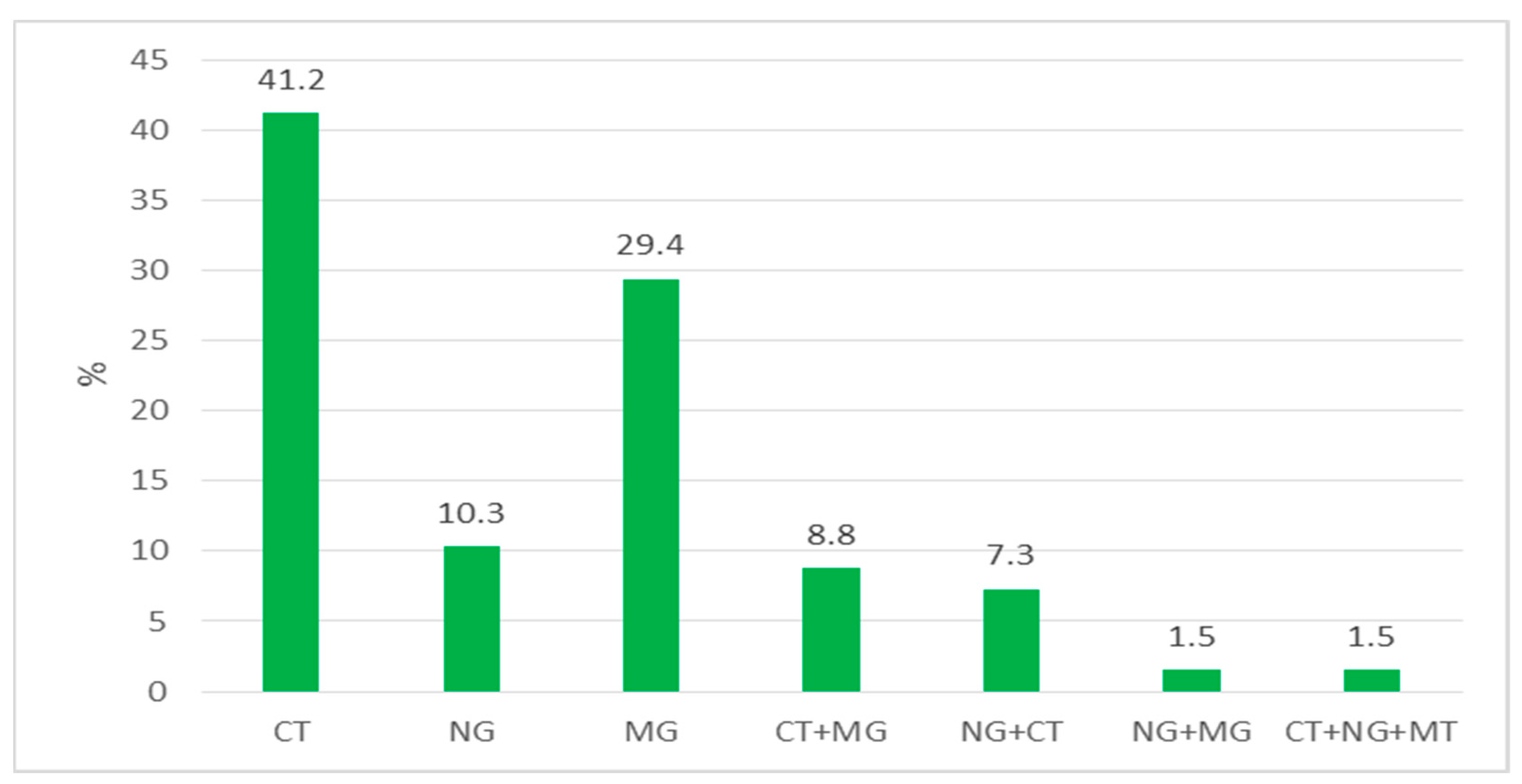
| Total | 33 | 35 | 28 | 7 | 20 | 6 | 5 | 1 | 1 |
| Agents | Gender | Single Infection/Co-Infection | |||||||
|---|---|---|---|---|---|---|---|---|---|
| Female | Male | CT | NG | MG | CT + MG | NG + CT | NG + MG | CT + NG + MG | |
| HIV (n: 3) | 1 | 2 | 1 | - | 1 | - | 1 | - | - |
| Treponema pallidum (n: 3) | 1 | 2 | 1 | 1 | - | - | 1 | - | - |
| Trichomonas vaginalis (n: 2) | 1 | - | 1 | - | - | - | - | - | - |
| HPV (n: 2) | - | 2 | 1 | 1 | - | - | - | - | - |
| HBV (n: 1) | 1 | - | - | - | 1 | - | - | - | - |
| HSV2 (n: 1) | 1 | - | 1 | - | - | - | - | - | - |
| Target | Estimate | Lower 95% CL | Upper 95% CL | |
|---|---|---|---|---|
| CT | Apparent Prevalence (WilsonCL) | 0.1111 | 0.0827 | 0.1478 |
| True Prevalence (BlakerCL) | 0.1021 | 0.0734 | 0.1392 | |
| NG | Apparent Prevalence (WilsonCL) | 0.0389 | 0.0233 | 0.0642 |
| True Prevalence (BlakerCL) | 0.0292 | 0.0134 | 0.0548 | |
| MG | Apparent Prevalence (WilsonCL) | 0.0778 | 0.0544 | 0.1101 |
| True Prevalence (BlakerCL) | 0.0685 | 0.0448 | 0.1011 |
| CT [n (%)] | NG [n (%)] | MG [n (%)] | ||
|---|---|---|---|---|
| In this study | Istanbul/Turkey | 40 (11.1) | 14 (3.9) | 28 (7.8) |
| De Souza et al. [23] | Brazil | 50 (29.4) | 102 (60.0) | 11 (6.5) |
| Van Der Pol et al. [24] | Canada | 34 (6.1) | 10 (1.8) | |
| Le Roy et al. [21] | France | (8.3) | (5.8) | |
| Calas et al. [35] | Reunion Island | (10.2) | (1.07) | (2.69) |
| Koksal et al. [36] | Istanbul/Turkey | 57 (13.6) | ||
| Guralp et al. [37] | Cyprus | (5.4) | (2.5) | (2.9) |
| In this study | Infertility | 2 (6.7) | - | - |
| Tomusiak et al. [25] | Infertility | (4) | ||
| Paira et al. [28] | Infertility | (5.3) | ||
| In this study | Cervicitis | 9 (21.4) | 3 (7.1) | 3 (7.1) |
| Roy et al. [22] | Cervicitis | 5 (3.3) | 10 (6.6) | 37 (24.6) |
| In this study | Urethritis | 18 (13.0) | 8 (5.8) | 14 (10.1) |
| De Souza et al. [23] | Urethritis | 13 (7.6) | 46 (27.1) | 5 (2.9) |
Disclaimer/Publisher’s Note: The statements, opinions and data contained in all publications are solely those of the individual author(s) and contributor(s) and not of MDPI and/or the editor(s). MDPI and/or the editor(s) disclaim responsibility for any injury to people or property resulting from any ideas, methods, instructions or products referred to in the content. |
© 2023 by the authors. Licensee MDPI, Basel, Switzerland. This article is an open access article distributed under the terms and conditions of the Creative Commons Attribution (CC BY) license (https://creativecommons.org/licenses/by/4.0/).
Share and Cite
Kirkoyun Uysal, H.; Koksal, M.O.; Sarsar, K.; Ilktac, M.; Isik, Z.; Akgun Karapinar, D.B.; Demirci, M.; Ongen, B.; Buyukoren, A.; Kadioglu, A.; et al. Prevalence of Chlamydia trachomatis, Neisseria gonorrhoeae, and Mycoplasma genitalium among Patients with Urogenital Symptoms in Istanbul. Healthcare 2023, 11, 930. https://doi.org/10.3390/healthcare11070930
Kirkoyun Uysal H, Koksal MO, Sarsar K, Ilktac M, Isik Z, Akgun Karapinar DB, Demirci M, Ongen B, Buyukoren A, Kadioglu A, et al. Prevalence of Chlamydia trachomatis, Neisseria gonorrhoeae, and Mycoplasma genitalium among Patients with Urogenital Symptoms in Istanbul. Healthcare. 2023; 11(7):930. https://doi.org/10.3390/healthcare11070930
Chicago/Turabian StyleKirkoyun Uysal, Hayriye, Muammer Osman Koksal, Kutay Sarsar, Mehmet Ilktac, Zeynep Isik, Deniz Bahar Akgun Karapinar, Mehmet Demirci, Betigul Ongen, Ahmet Buyukoren, Ates Kadioglu, and et al. 2023. "Prevalence of Chlamydia trachomatis, Neisseria gonorrhoeae, and Mycoplasma genitalium among Patients with Urogenital Symptoms in Istanbul" Healthcare 11, no. 7: 930. https://doi.org/10.3390/healthcare11070930
APA StyleKirkoyun Uysal, H., Koksal, M. O., Sarsar, K., Ilktac, M., Isik, Z., Akgun Karapinar, D. B., Demirci, M., Ongen, B., Buyukoren, A., Kadioglu, A., Yurtsever, E., & Agacfidan, A. (2023). Prevalence of Chlamydia trachomatis, Neisseria gonorrhoeae, and Mycoplasma genitalium among Patients with Urogenital Symptoms in Istanbul. Healthcare, 11(7), 930. https://doi.org/10.3390/healthcare11070930