


Abstract
This study aims to investigate the prevalence of locomotive syndrome (LS) and to examine the relationship of LS with musculoskeletal symptoms (pain, generalized joint laxity (GJL)) in young Chinese adults. Our study population (n = 157; mean age of 19.8 ± 1.2 years) comprises college student residents at Tsinghua University in Beijing, China. Three screening methods were used to evaluate LS: 25-question Geriatric Locomotive Function Scale (GLFS-25), a two-step test, and a stand-up test. Musculoskeletal pain was assessed by self-report and visual analog scale (VAS), and joint body laxity was evaluated using the GJL test. The prevalence of LS was 21.7% of all participants. Musculoskeletal pain affected 77.8% of the college students with LS and was strongly associated with LS. A total of 55.0% of college students with LS had four or more site joints that were positive for GJL, and higher scores of GJL were associated with a higher prevalence rate of LS. Young Chinese college students have a relatively high prevalence of LS, and musculoskeletal pain and GJL were significantly related to LS. The present results suggest that we need early screening of musculoskeletal symptoms and LS health education in young adults to prevent the mobility limitations of LS in the future.
1. Introduction
China has rapidly advanced into a super-aging society in recent years. By 2020, 13.5% of the Chinese population were older than 65, approximately 190 million people [1]. Since various chronic diseases are gradually trending in youths [2], health interventions from youth are urgently needed, given the rising geriatric problem. The Japanese Orthopaedic Association (JOA) proposed the term “locomotive syndrome” (LS) in 2007, which is defined as a decline in mobility or quality of life due to the dysfunction of the motor system, including muscles, bones, joints, spinal cord, and peripheral nerves [3,4]. However, LS affects not only middle and older adults, whose incidence of LS is approximately 21.1% and 49.3% [5], respectively, but also several people in their 20s meet the LS definition [6,7,8,9]. However, studies on the prevalence of LS in young people are still scarce. Since the decrease in mobility and muscle strength leading to disability may start at a young age [4,8,10,11], it is necessary to pay more attention to the early screening and prevention of LS in young people [9,12,13].
Some studies have found that the risk of LS is associated with muscle mass, lower limb muscle exertion [9,12], BMI [6], physical activity [7,14], and walking speed [5,12] in young adults. Previous studies claimed that older patients with LS frequently experience musculoskeletal pain in multiple sites [15,16,17]. However, few studies have focused on young people [7,18], but the percentage of young adults with patellofemoral and knee pain was reported to be 20.7% and 35.6%, respectively [19]. In addition, a connective tissue disorder known as generalized joint laxity (GJL) [20] causes musculoskeletal symptoms and physical limitations, particularly in adolescents [21,22]. Therefore, starting LS preventive measures in young adults requires attention to body pain and joint laxity.
There are two main purposes of this study: (1) to determine the prevalence of LS and (2) to investigate the relationship between LS and musculoskeletal symptoms (e.g., pain, GJL) in young Chinese college students.
2. Materials and Methods
2.1. Participants
The participants of this study were recruited from June to August 2022, and data were collected from September to November 2022 at Tsinghua University, Beijing, China. The recruitment methods included oral communications in physical education (PE) classes and fielding responses from posters on campus for three months. Participants were excluded if they had walking function problems and could not participate in all assessments of LS and GJL. All participants were free from chronic disease (e.g., diabetes, cardiovascular) and musculoskeletal injuries (e.g., fracture, orthopedic surgery) as assessed by their annual check-ups. There were no professional college athletes, and no one participated in long-term professional or intensive sports training prior to this study. The data analyses included 157 students (mean age, 19.8 ± 1.2 years) who met the criteria. This research was approved by the Ethics Committee at Tsinghua University, and the methods were carried out in accordance with the principles of the Declaration of Helsinki.
2.2. Measurement and Measuring Equipment
Physical Measurement
Physical measurements included height and body composition analysis, which adapted direct segmental multifrequency bioelectrical impedance analysis (BIA) (InBody720; Biospace Co, Ltd., Seoul, Korea). The BIA provided absolute values for body weight, skeletal muscle mass (SMM), appendicular skeletal muscle mass (ASM), body fat mass (PBF%), and total body water (TBW). Grip strength (kg) was measured using a handheld dynamometer (GRIP-D; Takei Ltd., Niigata, Japan). Both hands were tested, and the higher value was used to characterize the maximum muscle strength of the subject. Gait speed was assessed with the usual gait speed, and balance was measured using the 5-times sit-to-stand test (5xSST). To measure gait speed, two photocells were placed 6 m apart to record the time participants could walk at their usual speed, and the average speed of two walks was recorded. In the 5xSST, participants were instructed to sit against the back of a chair. Participants were asked to stand up and sit down as quickly as possible 5 times with their arms crossed in front of their chest. A chair with armrests and a standard height of 0.47 m was used.
2.3. Locomotive Syndrome (LS) Risk Tests
The “stand-up test,” “two-step test,” and “the 25-question geriatric locomotive function scale” (GLFS-25) were components of the LS risk test [4,5,12] recommended by the JOA, which also applies to young adults [13].
Stand-up test
The stand-up test measures lower muscle strength by having the participants stand on one or both legs from a 40, 30, 20, or 10 cm–high seat. The details of the testing method and scoring criteria were based on previous studies [8,12], and a participant‘ successful performance was given a score between 0 and 8.
Two-step test
(a) Participants stood with both feet behind the starting line; (b) participants were instructed to align their feet after taking two extremely long steps; (c) the length of the two steps between the starting line and the tips of the participant’s toes was measured. The following formula was used to determine the score for the two-step test: length of the two stages (cm) ÷ height (cm).
GLFS-25
It is a self-administered, comprehensive measure consisting of 25 items that include four questions regarding pain during the last month [23], 16 questions regarding activities of daily living during the last month, 3 questions regarding social functions, and 2 questions regarding mental health status during the last month. These 25 items were graded on a five-point scale, from no impairment (0 points) to severe impairment (4 points), and then arithmetically added to produce a total score.
We defined LS as follows based on the results of the three tests:
No-LS: If all three conditions were met—(a) two-step score ≥1.3; (b) ability to stand up on a single leg from a 40 cm–high seat with each leg; and (c) GLFS-25 score <7. LS 1: If any of the three conditions (a–c) were met—(a) stand-up test, difficulty in standing from a 40 cm high seat using one leg (either leg); (b) two-step test <1.3; or (c) 25-question score ≥7). LS 2: If any of the three conditions (a–c) were met—(a) stand-up test, difficulty in standing from a 20 cm high seat using both legs; (b) two-step test <1.1; or (c) 25-question score ≥16).
2.4. Musculoskeletal Pain
Musculoskeletal pain was defined by asking a question about nine anatomical sites (neck, shoulder, upper limb, lumbar, hip, knee, ankle, foot, and other): Have you experienced pain on most days (that continue on at least one day) in the past month in addition to the current pain? Musculoskeletal pain was deemed present in an anatomical site for participants who answered “yes.” [7,24]. Furthermore, we used the pain visual analog scale (VAS) [18] to evaluate the pain degrees.
2.5. General Joint Laxity Test
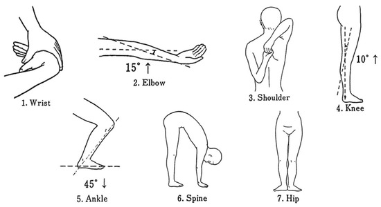
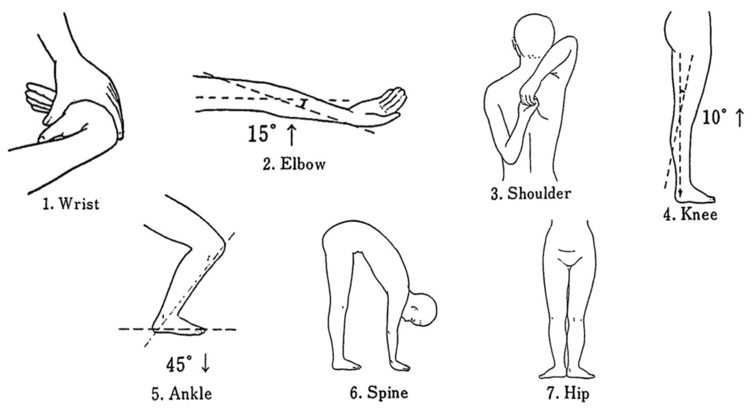
Seven maneuvers of the University of Tokyo GJL test were performed [25]. The judgment criteria of the positive joint laxity were the following: (1) wrist: passive opposition of the thumb to the flexor aspect of the forearm; (2) elbow: hyperextension >15 degrees; (3) shoulder: fingers overlap or grasping; (4) knee: hyperextension >10 degrees; (5) ankle: dorsiflexion >45 degrees; (6) spine: trunk flexion with the knee extended and both palms contacting the mat; and (7) hip: toes pointing outwards >180 degrees [26] (Figure 1). Seven positions were measured, with one point being given to each item; except for the spine, the left and right positions were each given a value of 0.5 points for the six major bilateral joints. A goniometer was used to measure the items with the joint angle as the criterion. One operator was responsible for taking and recording joint angle measurements.
Figure 1.
The University of Tokyo joint laxity test. Laxity of six major joints in the body (wrist, elbow, shoulder, knee, ankle, hip) and of the spine was examined.
Demographic variables and behavioral characteristics included age, gender, smoking (current smoker, ex-smoker, non-smoker) and drinking habits (drinks every day, drinks occasionally, ex-drinker, never drinks), and the International Physical Activity Questionnaire (IPAQ). All parts of the tests were completed by professionally trained medical staff. Part of the questionnaire test was conducted via a face-to-face interview.
2.6. Statistical Analysis
The anthropometric characteristics and background data with continuous variables were expressed as mean ± SD and categorical variables as absolute numbers and percentages (%) of the total. Differences between variables were examined using ANOVA with Bonferroni correction (continuous variables) or using the chi-square test (categorical variables). Logistic regression analyses were used to assess the association between the LS group, the No-LS group and pain sites, pain VAS and GJL sites, and GJL score. Model 1 was adjusted for age and sex. Model 2 was adjusted for Model 1 covariates plus BMI, grip strength/weight, ASMI, PBF%, GJL score (or total pain VAS), smoking, drinking, and IPAQ. The odds ratio (OR) value and 95% CI were calculated. All statistical analyses were performed using SPSS v 26.0 (SPSS Inc., Chicago, IL, USA), and p-values of <0.05 were considered statistically significant.
3. Results
Anthropometric characteristics and background data of all subjects, the No-LS group, and the LS group are shown in Table 1. The mean age of the 157 participants was 19.8 ± 1.2 years, and 57 participants (36.3%) were female. The prevalence of LS was 21.7% of all participants (16.0% of males (LS1: 13.0%, LS2: 3.0%) and 31.6% of females (LS1: 28.1%, LS2: 3.5%)). The prevalence of a two-step test score <1.3, the inability to stand with one leg from a 40 cm high seat, and a GLFS-25 score ≥7 were 0.6%, 2.5%, and 18.5%, respectively. The total number of pain sites, pain VAS score, GJL score, PBF%, and GLFS-25 score were significantly higher in the LS group than in the No-LS group. The LS group had a significantly lower stand-up test score, ASMI, and adjusted grip strength.
Table 1.
Anthropometric characteristics and background data of each group.
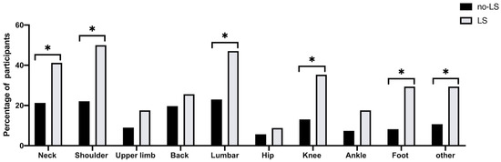
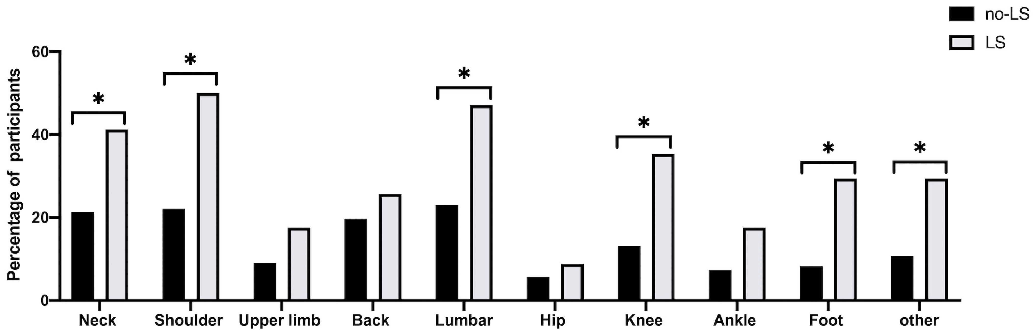
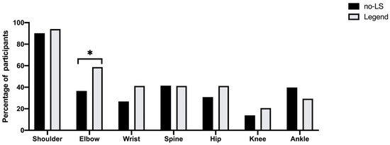
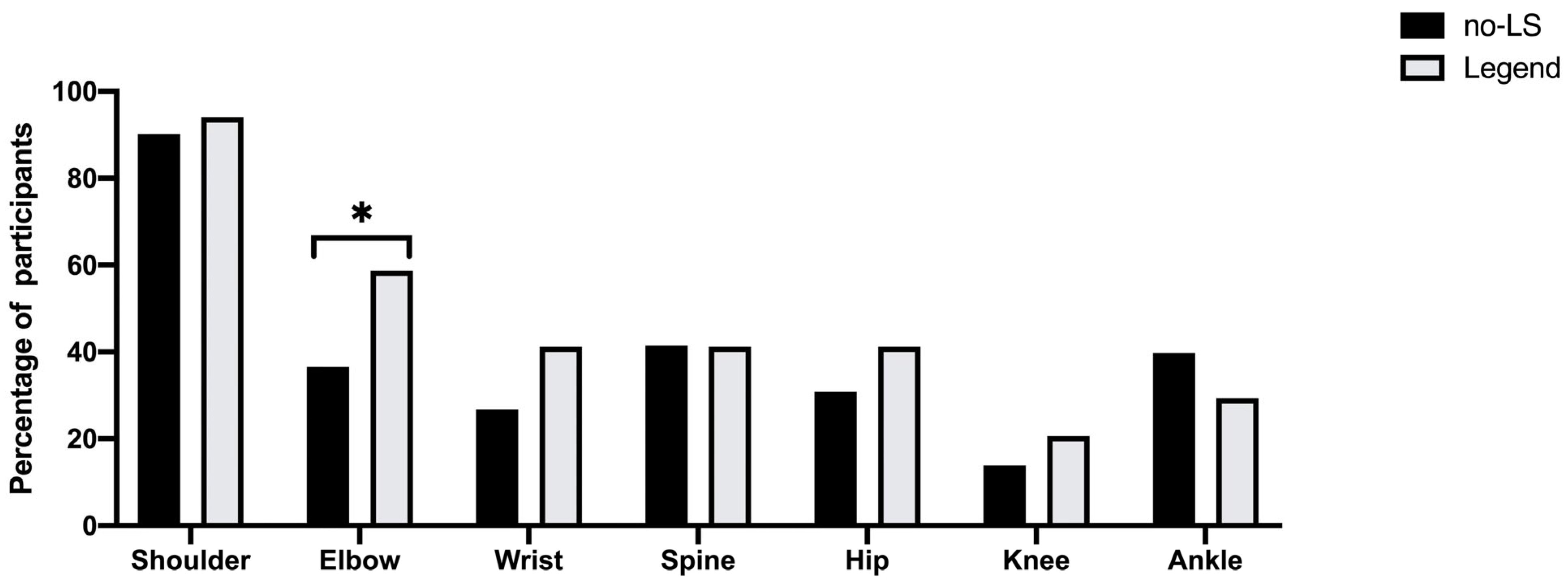
Figure 2 shows the prevalence of musculoskeletal pain in the nine anatomical areas, and 57.3% of persons had experienced musculoskeletal pain somewhere in their bodies. The typical pain sites in the LS group were the neck (p = 0.021), shoulder (p = 0.002), lumbar (p = 0.007), knee (p = 0.004), foot (p = 0.002), and other sites (p = 0.009). After the multivariate analysis (Table 2), the prevalence of pain in shoulder (OR = 2.99, 95% CI = 1.18–7.59, p = 0.021), lumbar (OR = 2.93, 95% CI = 1.19–7.21, p = 0.019), knee (OR = 3.19, 95% CI = 1.16–8.73, p = 0.024), and foot (OR = 6.18, 95% CI = 1.95–19.6, p = 0.002) were significantly higher in the LS group than in the No-LS group. Furthermore, the VAS of the total pain (OR = 1.13, 95% CI = 1.05–1.22, p = 0.002), neck pain (OR = 1.57, 95% CI = 1.13–2.19, p = 0.008), shoulder pain (OR = 1.67, 95% CI = 1.15–2.42, p = 0.008), ankle pain (OR = 1.80, 95% CI = 1.04–3.11, p = 0.034), and foot pain (OR = 3.55, 95% CI = 1.47–8.56, p = 0.005) were significantly higher in the LS group than in the No-LS group after the multivariate analysis (Table 3). There were no differences in GJL sites between the LS group and the No-LS group, except for the elbow in unadjusted crude (OR = 2.48, 95% CI = 1.14–5.38, p = 0.022) (Table 4 and Figure 3). However, after the multivariate analysis, the GJL score was significantly higher in the LS group than in the No-LS group, and the risk of LS increased with the increase of the GJL score (OR = 1.42, 95% CI = 1.06–1.91, p = 0.019) (Table 5).
Figure 2.
Prevalence of musculoskeletal pain in each anatomical area in No-LS and LS groups. * p < 0.05.
Table 2.
Logistic regression analysis of locomotive syndrome and pain sites.
Table 3.
Logistic regression analysis of locomotive syndrome and pain VAS.
Table 4.
Logistic regression analysis of locomotive syndrome and joint laxity risk sites.
Figure 3.
Prevalence of joint laxity risk sites in each anatomical area in No-LS and LS groups. * p < 0.05.
Table 5.
Logistic regression analysis of locomotive syndrome and general joint laxity score.
4. Discussion
The main findings of this study are as follows: First, we investigated the prevalence of LS in young college students (mean age of 19.8 ± 1.2 years). The overall prevalence of LS was 21.7% (16.0% of males (LS1: 13.0%, LS2: 3.0%) and 31.6% (LS1: 28.1%, LS2: 3.5%) of females). Second, musculoskeletal pain affected 77.8% of the college students with LS. Musculoskeletal pain was more common in the LS group than in the No-LS group and was strongly associated with LS. Third, 55.0% of college students with LS had four or more site joints that were positive for GJL (No-LS group: 29.3%), and higher scores of GJL were associated with a higher prevalence rate of LS.
4.1. Prevalence of LS in Young Adults
A few previous studies from Japan have investigated the incidence and risk propensity of LS in young people [6,8,9,12,13]. Akinobu et al. [13] researched the prevalence of LS among young workers under 29 years of age to be 19.6% in men and 5.3% in women. In addition, the prevalence was higher in men <29 years old than in those 30–39 and 40–49 years old. Another study [9] found that the prevalence of LS among young female students was 18.5%, while the prevalence in males was 0% (mean age of 18.6 years), which was lower than that determined by the present study. The main reason for the difference was that our participants had relatively high GLFS-25 scores, with approximately 18.5% of them ≥7 [5]. Another possible reason was that the participants (especially males) in the present study had higher BMIs (21.9 kg/m2 vs. 20.4 kg/m2) [6] and PBF% (17.3% vs. 13.2%) [12] and lower muscle strength (35.2 kg vs. 42.1 kg) [9], which are significant risk factors for LS in young adults. Moreover, because of the increase in online classes and outdoor activity limitations during the novel coronavirus disease 2019 (COVID-19), the participants in this study had more sedentary time compared to previous studies [27,28], which might also contribute to a relatively high prevalence in our study [7,29].
In addition, one study found that 65.0% of female college students (20.0 ± 1.5 years) were classified in the high LS risk group [12] based on the JOA-recommended cut-off values (one leg stand of 30 cm or less, two-step test <1.55) for those in their 20s [8]. In this study, 56.0% of female students were assigned to the LS risk group after using the same cut-off values. A study found that the stand-up and two-step tests might start to deteriorate in those in their 30s and younger, which might support the high rate of LS risk in young people [5]. However, the majority of the studies mentioned above were from Japan, and the differences in ethnicity and lifestyle [30] might also contribute to different prevalence.
4.2. Musculoskeletal Pain and LS
Several previous studies clarified that musculoskeletal disorders such as lower extremity pain [16,17], multiple sites pain [31], osteoporosis [32], and lumbar spinal stenosis [16] are associated with LS in older people. However, few studies focused on young adults, especially those under 30 [7,33]. In the present study, 77.8% of the college students with LS had experienced musculoskeletal pain. The incidence of pain in the shoulders (22.1% of No-LS and 50.0% of LS), lumbar (23.0% of No-LS and 47.1% of LS), knee (13.1% of No-LS and 35.3% of LS), and foot (8.2% of No-LS and 29.4% of LS) were significantly higher in the LS group than in the No-LS group (Table 2), consistent with previous research in young and middle-aged adults [7]. Furthermore, Hironori et al. [17] revealed that 85.8% of patients over 40 with chronic pain were diagnosed with LS2, indicating that chronic pain patients had an earlier risk of developing LS.
Moreover, the students with LS had more pain sites in the lower extremities than upper extremities, indicating the pain origin of LS was induced by the degeneration of the lumbar disk [34] and mainly affected mobility functions, such as gait and climbing ability [4,5]. In addition, because the spine and knee joint are primarily supporting the person’s upright body and trunk, lower extremity pain is the primary cause of locomotive disability [35]. Multivariate analysis revealed the pain degree (total pain of VAS, OR = 1.13, p = 0.002) was significantly worse in the LS group than in the No-LS group in our study. This result was similar to Takeshi et al.’s [18] finding that pain VAS was the risk factor for LS in patients with rheumatoid arthritis (OR = 1.04, p < 0.001). In addition, as an evaluation of locomotive organ deterioration, GLFS-25 was found to be associated with chronic pain [17,32]. In contrast, our results showed a relatively higher score of GLFS-25 (mean value: 4.0 ± 4.9) in young adults [5]; in particular, questions about pain review scored higher. According to the current findings, LS was associated with musculoskeletal pain even in college students, as in older adults with LS.
4.3. GJL and LS
GJL has a high prevalence among adolescents (especially in women) and declines with age [25,36]. Previous studies have shown that GJL is a risk factor for musculoskeletal pain, lower extremity joint injuries [37], increased plantar loading [20], and proprioception deficits [21,38]. In the present study, we found the GJL score was significantly higher in the LS group than in the No-LS group (OR = 1.42, p = 0.019); 55.0% of college students with LS had four or more site joints that were positive for GJL. Two possible mechanisms explain this result. One is that LS is mainly associated with lower extremity function, and the range of motion is greater than normal in GJL, which may affect dynamic balance and alignment of the lower extremities [20]. Second, knee hyperextension results in impaired proprioception [39] and lower muscle strength [22], which can induce knee injury and inadequate control of lower motor function [38]. For instance, 20.6% of college students with LS had knee hyperextension >10 degrees in our study. Hence, a lack of awareness about the relationship between GJL and LS may lead to unnecessary motor system dysfunction in young college adults.
4.4. Strength and Limitations
This study was the first cross-sectional study to identify the prevalence of LS in young Chinese adults; we clarified that musculoskeletal pain and GJL at a young age are associated with LS.
Several limitations must be considered in this study. Firstly, the current findings were only representative of some Chinese college students because they were from one university, and most were first-year students. Secondly, there was a small sample size; additional studies with large sample sizes and robust experimental designs should be carried out, including verifying the reliability and validity of the GLFS-25 in young adults. Thirdly, our data did not include the medical examinations for bone mineral density, scoliosis, etc., and a more standard GJL assessment method, such as the Beighton test, should be supplemented in future research.
5. Conclusions
In conclusion, this cross-sectional study aimed to identify the prevalence of LS and its association with musculoskeletal pain and GJL in young Chinese college students. The overall prevalence of LS was 21.7% (16.0% of males and 31.6% of females). Musculoskeletal pain was more common in the LS group than in the No-LS group, and higher scores of pain VAS and GJL were associated with a higher prevalence rate of LS. Therefore, we need to develop LS interventions for young adults, including the early screening of musculoskeletal symptoms and LS health education to prevent the mobility limitations of LS in the future.
Author Contributions
Conceptualization, B.Z. and X.Z.; methodology, Y.Q., M.S. and Y.L.; formal analysis, Y.M. and X.W.; investigation, Y.Q., M.S. and Y.L.; data curation, Y.Q., M.S. and Y.L.; writing—original draft preparation, Y.M. and X.W.; writing—review and editing, Y.M., W.H., S.S. and X.Z. All authors have read and agreed to the published version of the manuscript.
Funding
This research received no external funding.
Institutional Review Board Statement
The study was conducted in accordance with the Declaration of Helsinki and approved by the Ethics Committee at Tsinghua University (protocol code: 20220233; approval date: 20230104).
Informed Consent Statement
Informed consent was obtained from all subjects involved in the study.
Data Availability Statement
Data is unavailable due to privacy or ethical restrictions.
Acknowledgments
We thank the Tsinghua University Student Physical Fitness Test Center for supporting infrastructure and planning.
Conflicts of Interest
The authors declare no conflict of interest.
References
- Bao, J.; Zhou, L.; Liu, G.; Tang, J.; Lu, X.; Cheng, C.; Jin, Y.; Bai, J. Current state of care for the elderly in China in the context of an aging population. Biosci. Trends 2022, 16, 107–118. [Google Scholar] [CrossRef] [Scilit]
- Ramos, G.F.; Ribeiro, V.P.; Mercadante, M.P.; Ribeiro, M.P.; Delgado, A.F.; Farhat, S.C.L.; Leal, M.M.; Marques, H.H.; Odone-Filho, V.; Tannuri, U.; et al. Mortality in adolescents and young adults with chronic diseases during 16 years: A study in a Latin American tertiary hospital. J. Pediatr. 2019, 95, 667–673. [Google Scholar] [CrossRef] [Scilit]
- Nakamura, K. A “super-aged” society and the “locomotive syndrome”. J. Orthop. Sci. 2008, 13, 1–2. [Google Scholar] [CrossRef] [Scilit] [PubMed]
- Nakamura, K.; Ogata, T. Locomotive Syndrome: Definition and Management. Clin. Rev. Bone Miner Metab. 2016, 14, 56–67. [Google Scholar] [CrossRef] [Scilit]
- Yoshimura, N.M.S.; Oka, H.; Tanaka, S.; Ogata, T.; Kawaguchi, H.; Akune, T.; Nakamura, K. Association between new indices in the locomotive syndrome risk test and decline in mobility: Third survey of the ROAD study. J. Orthop. Sci. 2015, 20, 896–905. [Google Scholar] [CrossRef] [Scilit]
- Yoshimura, Y.; Ishijima, M.; Ishibashi, M.; Liu, L.; Arikawa-Hirasawa, E.; Machida, S.; Naito, H.; Hamada, C.; Kominami, E. A nationwide observational study of locomotive syndrome in Japan using the ResearchKit: The Locomonitor study. J. Orthop. Sci. 2019, 24, 1094–1104. [Google Scholar] [CrossRef] [Scilit]
- Kato, T.; Nishimura, A.; Ohtsuki, M.; Wakasugi, Y.; Nagao-Nishiwaki, R.; Fukuda, A.; Kato, K.; Sudo, A. Is musculoskeletal pain related to locomotive syndrome even in young and middle-aged adults? Mod. Rheumatol. 2022, 32, 213–220. [Google Scholar] [CrossRef] [Scilit] [PubMed]
- Yamada, K.; Ito, Y.M.; Akagi, M.; Chosa, E.; Fuji, T.; Hirano, K.; Ikeda, S.; Ishibashi, H.; Ishibashi, Y.; Ishijima, M.; et al. Reference values for the locomotive syndrome risk test quantifying mobility of 8681 adults aged 20-89 years: A cross-sectional nationwide study in Japan. J. Orthop. Sci. 2020, 25, 1084–1092. [Google Scholar] [CrossRef] [Scilit]
- Yasuda, T. Identifying preventative measures against frailty, locomotive syndrome, and sarcopenia in young adults: A pilot study. J. Phys. Ther. Sci. 2021, 33, 823–827. [Google Scholar] [CrossRef] [Scilit] [PubMed]
- Suetta, C.; Haddock, B.; Alcazar, J.; Noerst, T.; Hansen, O.M.; Ludvig, H.; Kamper, R.S.; Schnohr, P.; Prescott, E.; Andersen, L.L.; et al. The Copenhagen Sarcopenia Study: Lean mass, strength, power, and physical function in a Danish cohort aged 20–93 years. J. Cachexia Sarcopenia Muscle 2019, 10, 1316–1329. [Google Scholar] [CrossRef] [Scilit]
- Nakamura, K. Locomotive syndrome: Disability-free life expectancy and locomotive organ health in a “super-aged” society. J. Orthop. Sci. 2009, 14, 1–2. [Google Scholar] [CrossRef] [Scilit] [PubMed]
- Uesugi, Y.; Kanaya, S.; Nakanishi, H.; Naito, Y. The Relationship between Locomotive Syndrome Risk, Gait Pattern, and Standing Posture in Young Japanese Women: A Cross-Sectional Study. Healthcare 2020, 8, 565. [Google Scholar] [CrossRef] [Scilit]
- Nishimura, A.; Ohtsuki, M.; Kato, T.; Nagao, R.; Ito, N.; Kato, K.; Ogura, T.; Sudo, A. Locomotive syndrome testing in young and middle adulthood. Mod. Rheumatol. 2020, 30, 178–183. [Google Scholar] [CrossRef] [Scilit]
- Shen, S.; Suzuki, K.; Kohmura, Y.; Fuku, N.; Someya, Y.; Naito, H. Association of physical fitness and motor ability at young age with locomotive syndrome risk in middle-aged and older men: J-Fit(+) Study. BMC Geriatr. 2021, 21, 89. [Google Scholar] [CrossRef] [Scilit]
- Arbex, M.; Okazaki, J.E.F.; Tavares, D.R.B.; Figueiredo Bersani, A.L.; Santos, F.C. Locomotive Syndrome is associated with chronic pain and poor quality of life in Brazilian oldest old: LOCOMOV Project. J. Orthop. Sci. 2021, 26, 162–166. [Google Scholar] [CrossRef] [Scilit] [PubMed]
- Chiba, D.; Tsuda, E.; Wada, K.; Kumagai, G.; Sasaki, E.; Nawata, A.; Nakagomi, S.; Takahashi, I.; Nakaji, S.; Ishibashi, Y. Lumbar spondylosis, lumbar spinal stenosis, knee pain, back muscle strength are associated with the locomotive syndrome: Rural population study in Japan. J. Orthop. Sci. 2016, 21, 366–372. [Google Scholar] [CrossRef] [Scilit]
- Tsuji, H.; Tetsunaga, T.; Tetsunaga, T.; Misawa, H.; Nishida, K.; Ozaki, T. Cognitive factors associated with locomotive syndrome in chronic pain patients: A retrospective study. J. Orthop. Sci. 2021, 26, 896–901. [Google Scholar] [CrossRef] [Scilit] [PubMed]
- Mochizuki, T.; Ikari, K.; Yano, K.; Okazaki, K. Evaluation of factors associated with locomotive syndrome in Japanese elderly and younger patients with rheumatoid arthritis. Mod. Rheumatol. 2019, 29, 733–736. [Google Scholar] [CrossRef] [Scilit] [PubMed]
- Xu, X.; Yao, C.; Wu, R.; Yan, W.; Yao, Y.; Song, K.; Jiang, Q.; Shi, D. Prevalence of patellofemoral pain and knee pain in the general population of Chinese young adults: A community-based questionnaire survey. BMC Musculoskelet Disord. 2018, 19, 165. [Google Scholar] [CrossRef] [Scilit]
- Aydin, E.; Metin Tellioglu, A.; Kurt Omurlu, I.; Turan, Y. Impact of Generalized Joint Laxity on Plantar Loading Patterns in Young Females. Foot Ankle Int. 2017, 38, 909–915. [Google Scholar] [CrossRef] [Scilit]
- Wolf, J.M.; Cameron, K.L.; Owens, B.D. Impact of joint laxity and hypermobility on the musculoskeletal system. J. Am. Acad. Orthop. Surg. 2011, 19, 463–471. [Google Scholar] [CrossRef] [Scilit]
- Sahin, N.; Baskent, A.; Ugurlu, H.; Berker, E. Isokinetic evaluation of knee extensor/flexor muscle strength in patients with hypermobility syndrome. Rheumatol. Int. 2008, 28, 643–648. [Google Scholar] [CrossRef] [Scilit] [PubMed]
- Seichi, A.; Hoshino, Y.; Doi, T.; Akai, M.; Tobimatsu, Y.; Iwaya, T. Development of a screening tool for risk of locomotive syndrome in the elderly: The 25-question Geriatric Locomotive Function Scale. J. Orthop. Sci. 2012, 17, 163–172. [Google Scholar] [CrossRef] [Scilit]
- Muraki, S.; Akune, T.; Oka, H.; En-yo, Y.; Yoshida, M.; Saika, A.; Suzuki, T.; Yoshida, H.; Ishibashi, H.; Tokimura, F.; et al. Association of radiographic and symptomatic knee osteoarthritis with health-related quality of life in a population-based cohort study in Japan: The ROAD study. Osteoarthr. Cartil. 2010, 18, 1227–1234. [Google Scholar] [CrossRef] [Scilit]
- Yamazaki, T.; Maruyama, S.; Sato, Y.; Suzuki, Y.; Shimizu, S.; Kaneko, F.; Ikezu, M.; Matsuzawa, K.; Edama, M. A preliminary study exploring the change in ankle joint laxity and general joint laxity during the menstrual cycle in cis women. J. Foot Ankle Res. 2021, 14, 21. [Google Scholar] [CrossRef] [Scilit]
- Motohashi, M. Profile of bilateral anterior cruciate ligament injuries: A retrospective follow-up study. J. Orthop. Surg. 2004, 12, 210–215. [Google Scholar] [CrossRef] [Scilit] [PubMed]
- Shi, M.; Zhai, X.; Li, S.; Shi, Y.; Fan, X. The Relationship between Physical Activity, Mobile Phone Addiction, and Irrational Procrastination in Chinese College Students. Int. J. Environ. Res. Public Health 2021, 18, 5325. [Google Scholar] [CrossRef] [Scilit]
- Zhang, Y.; Zhang, H.; Ma, X.; Di, Q. Mental Health Problems during the COVID-19 Pandemics and the Mitigation Effects of Exercise: A Longitudinal Study of College Students in China. Int. J. Environ. Res. Public Health 2020, 17, 3722. [Google Scholar] [CrossRef] [Scilit] [PubMed]
- Nishimura, A.; Ito, N.; Asanuma, K.; Akeda, K.; Ogura, T.; Sudo, A. Do exercise habits during middle age affect locomotive syndrome in old age? Mod. Rheumatol. 2018, 28, 334–338. [Google Scholar] [CrossRef] [Scilit]
- Akahane, M.; Yoshihara, S.; Maeyashiki, A.; Tanaka, Y.; Imamura, T. Lifestyle factors are significantly associated with the locomotive syndrome: A cross-sectional study. BMC Geriatr. 2017, 17, 241. [Google Scholar] [CrossRef] [Scilit]
- Nakatoh, S. Relationships between chronic pain with locomotive syndrome and somatic symptom disorder in general community-dwelling population: A cross-sectional evaluation of individuals aged 50 years or older undergoing primary specific health screening. Mod. Rheumatol. 2020, 30, 1067–1073. [Google Scholar] [CrossRef] [Scilit]
- Iizuka, Y.; Iizuka, H.; Mieda, T.; Tajika, T.; Yamamoto, A.; Takagishi, K. Population-based study of the association of osteoporosis and chronic musculoskeletal pain and locomotive syndrome: The Katashina study. J. Orthop. Sci. 2015, 20, 1085–1089. [Google Scholar] [CrossRef] [Scilit]
- Akahane, M.; Maeyashiki, A.; Tanaka, Y.; Imamura, T. The impact of musculoskeletal diseases on the presence of locomotive syndrome. Mod. Rheumatol. 2019, 29, 151–156. [Google Scholar] [CrossRef] [Scilit]
- de Schepper, E.I.; Damen, J.; van Meurs, J.B.; Ginai, A.Z.; Popham, M.; Hofman, A.; Koes, B.W.; Bierma-Zeinstra, S.M. The association between lumbar disc degeneration and low back pain: The influence of age, gender, and individual radiographic features. Spine 2010, 35, 531–536. [Google Scholar] [CrossRef] [Scilit] [PubMed]
- Muraki, S.; Akune, T.; Oka, H.; En-Yo, Y.; Yoshida, M.; Saika, A.; Suzuki, T.; Yoshida, H.; Ishibashi, H.; Tokimura, F.; et al. Health-related quality of life in subjects with low back pain and knee pain in a population-based cohort study of Japanese men: The Research on Osteoarthritis Against Disability study. Spine 2011, 36, 1312–1319. [Google Scholar] [CrossRef] [Scilit] [PubMed]
- Decoster, L.C.; Bernier, J.N.; Lindsay, R.H.; Vailas, J.C. Generalized Joint Hypermobility and Its Relationship to Injury Patterns Among NCAA Lacrosse Players. J. Athl. Train. 1999, 34, 99–105. [Google Scholar]
- Grahame, R.; Hakim, A.J. Hypermobility. Curr. Opin. Rheumatol. 2008, 20, 106–110. [Google Scholar] [CrossRef] [Scilit]
- Roberts, D.; Ageberg, E.; Andersson, G.; Friden, T. Clinical measurements of proprioception, muscle strength and laxity in relation to function in the ACL-injured knee. Knee Surg. Sport. Traumatol. Arthrosc. 2007, 15, 9–16. [Google Scholar] [CrossRef] [Scilit]
- Griffin, L.Y.; Agel, J.; Albohm, M.J.; Arendt, E.A.; Dick, R.W.; Garrett, W.E.; Garrick, J.G.; Hewett, T.E.; Huston, L.; Ireland, M.L.; et al. Noncontact anterior cruciate ligament injuries: Risk factors and prevention strategies. J. Am. Acad. Orthop. Surg. 2000, 8, 141–150. [Google Scholar] [CrossRef] [Scilit] [PubMed]

Disclaimer/Publisher’s Note: The statements, opinions and data contained in all publications are solely those of the individual author(s) and contributor(s) and not of MDPI and/or the editor(s). MDPI and/or the editor(s) disclaim responsibility for any injury to people or property resulting from any ideas, methods, instructions or products referred to in the content. |
© 2023 by the authors. Licensee MDPI, Basel, Switzerland. This article is an open access article distributed under the terms and conditions of the Creative Commons Attribution (CC BY) license (https://creativecommons.org/licenses/by/4.0/).