Article

Intraocular Pressure Fluctuation during Aerobic Exercise at Different Exercise Intensities

1
Department of Physical Therapy, Makuhari Human Care Faculty, Tohto University, Hibino 1-1, Mihama-ku, Chiba City 261-0021, Chiba, Japan
2
Department of Rehabilitation Sciences, Kansai University of Welfare Sciences, Kashiwara City 582-0026, Osaka, Japan
3
Division of Rehabilitation, Department of Clinical Practice and Support, Hiroshima University Hospital, Kasumi 1-2-3, Minami-ku, Hiroshima City 734-8551, Hiroshima, Japan
4
Department of Physical Therapy, Faculty of Health Sciences, Okayama Healthcare Professional University, Daiku 3-2-18, Kitaku, Okayama City 700-0913, Okayama, Japan
5
Department of Rehabilitation, Hiroshima University Hospital, Kasumi 1-2-3, Minami-ku, Hiroshima City 734-8551, Hiroshima, Japan
*
Author to whom correspondence should be addressed.
Healthcare 2022, 10(7), 1196; https://doi.org/10.3390/healthcare10071196
Submission received: 9 May 2022 / Revised: 17 June 2022 / Accepted: 23 June 2022 / Published: 26 June 2022

Abstract

:
Few studies have examined the effects of different aerobic-exercise intensities on intraocular-pressure (IOP) changes. This may be important for eye diseases that are impacted by IOP or its fluctuation, including glaucoma, and diabetes that is complicated by diabetic retinopathy. We investigated the effects of low-, moderate-, and high-intensity exercise on IOP in healthy subjects. A submaximal cardiopulmonary exercise test was performed in 18 healthy male subjects, and the maximal oxygen uptake was calculated. The subjects then exercised for 20 min at 30%, 50%, and 70% ·VO2 of maximal oxygen uptake, and their IOP was measured at rest and every 5 min during exercise. Oxygen uptake was monitored using an expiratory gas analyzer during exercise to maintain accurate exercise intensity and adjust exercise load. Oxygen uptake during exercise was significantly higher at all intensities from 5 to 20 min than at rest. IOP was significantly lower at 70% exercise intensity from 5 to 20 min than at rest. A negative correlation existed between IOP and ·VO2. IOP remained unchanged during low- and moderate-intensity exercise but significantly declined during high-intensity exercise compared with that at rest. Although various factors, such as β-blockers, are involved in IOP decline at rest, a different mechanism is involved in IOP decline during exercise.

1. Introduction

The number of people who have diabetes worldwide was estimated to be 537 million in 2021, corresponding to approximately one in 10 people. The prevalence of diabetes is predicted to increase to 643 million by 2030 and 783 million by 2045 [1]. Therefore, there is an urgent need to prevent the onset and severity of diabetes worldwide. Exercise is a well-established treatment for type 2 diabetes, as it improves glycemic control [2], obesity [3], and the accumulation of visceral fat [4]. Aerobic and resistance exercises are the recommended types of exercise [5]. Complications of diabetes mellitus include neuropathy, nephropathy, and retinopathy. In a recent study of 121 patients with diabetic nephropathy, an intervention that included dietary guidance and brisk walking for at least 300 min per week showed no improvement in HbA1c, a measure of glycemic control, compared with that in control subjects. However, body mass index (BMI), albumin-to-creatinine ratio, serum and urinary 8-OHdG, and plasma TGF-β1 were significantly lower in the intervention group [6]. Robinson et al. investigated 256 patients with chronic kidney disease to determine the association between physical activity and estimated glomerular filtration rate (eGFR) [7]. Their study revealed that each additional 60 min of physical activity per week showed an annual 0.5% decline in eGFR, suggesting that interventions including exercise therapy may delay kidney damage in patients with diabetes.
In one study, a 10-week intervention including aerobic and resistance exercise in 20 patients with diabetic neuropathy resulted in reduced adiposity and altered resting tissue properties in the lower leg compared with those in the control group. This study also reported that continuous exercise therapy improved muscle quality in patients with diabetic neuropathy [8]. Furthermore, after 16 weeks of aerobic exercise, perceived pain interference was reduced among patients with painful diabetic polyneuropathy [9]. On the contrary, exercise has been reported to improve the severity of symptoms in patients with diabetic nephropathy and neuropathy, but there have been no studies on its effect in patients with retinopathy [10,11]. The reason for this finding is that neovascularization is a problem in retinopathy, and the vulnerability of these vessels is thought to increase the risk of hemorrhage with an exercise-induced increase in blood pressure [5]. A point to consider during exercise in retinopathy is the association of proliferative diabetic retinopathy with neovascular glaucoma (NVG) [12,13]. NVG is an eye disease in which ischemia of the retina causes the formation of new blood vessels in the eye, resulting in an increased IOP. Therefore, the clarification of exercise-induced IOP fluctuation may be important in glaucoma and other eye diseases.
On the other hand, few studies have been conducted on IOP changes during exercise. In a previous study that evaluated IOP before and after 60–80 watts of moderate-intensity aerobic exercise, both normal subjects and patients with glaucoma showed a significant reduction in IOP after exercise [14]. In another study on resistance exercise, Vieira et al. investigated changes in IOP in healthy subjects during weightlifting. The exercise was performed at 80% of the one repetition maximum for four repetitions, and the IOP was measured. The results showed an increase of 2.2 ± 3.0 mmHg [15]. However, there are no detailed reports on the effects of different intensities of aerobic exercise on changes in IOP. Therefore, this study aimed to clarify the effects of low-, moderate-, and high-intensity exercise on IOP in healthy subjects.

2. Materials and Methods

2.1. Participants

This study included 18 healthy male subjects who agreed to participate in the study and were diagnosed as healthy by a rehabilitation doctor before the study (age, 24.6 ± 2.7 years; height, 172.0 ± 5.0 cm; bodyweight, 62.9 ± 5.9 kg; and BMI, 21.2 ± 1.1 kg/m2). The exclusion criteria were as follows: past and present smoking habits, musculoskeletal diseases that made exercise difficult, cardiovascular diseases, eye diseases under treatment, and a resting IOP < 10 mmHg or >21 mmHg. Written consent from each participant was obtained before the study. The study was approved by the Epidemiological Ethics Review of Hiroshima University (E-529).

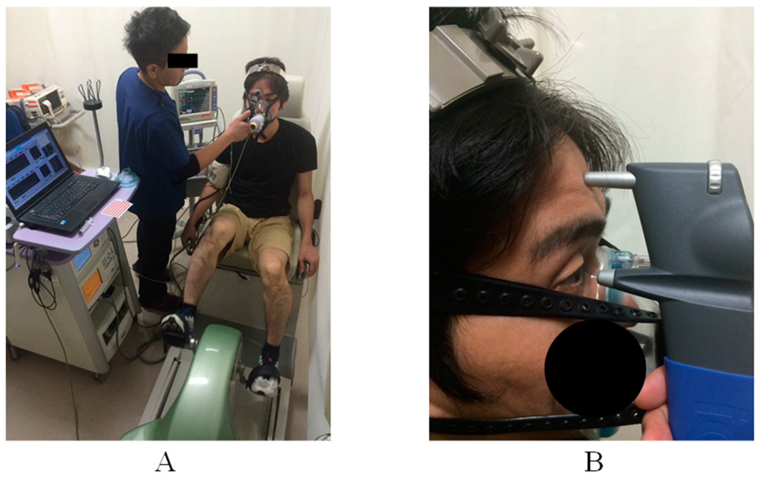
2.2. Measurement Protocol

On the first day, the submaximal cardiopulmonary exercise test was performed. An expiratory gas analyzer (aero monitor: AE-310s, Minato Medical Science Co., Ltd., Osaka, Japan) and a bicycle ergometer (strength ergometer: BK-ERG-003, Mitsubishi Engineering Co. Ltd., Tokyo, Japan) were used to assess the oxygen uptake at each stage and calculate the maximal oxygen uptake by extrapolation. At intervals of at least 1 day, aerobic exercise on a bicycle ergometer was performed for 20 min at 30% ·VO2, 50% ·VO2, and 70% ·VO2, and IOP was assessed at rest and during exercise. Each exercise session was performed at intervals of at least 1 day. All measurements were performed from 19:00, as IOP has diurnal fluctuations.

2.3. Maximal Cardiopulmonary Exercise Test

The submaximal exercise stress test, performed using an expiratory gas analyzer and a bicycle ergometer, was used to measure the maximal oxygen uptake. After 3 min of rest, the subjects underwent a four-step exercise load test, with a single step of 50 W for 3 min and a recovery step of 3 min. From rest to the end of the exercise, oxygen uptake was measured by an expiratory gas analysis, and an electrocardiogram (Bedside monitor: BSM-2301, Nihon Kohden Co., Ltd., Tokyo, Japan) and automatic blood pressure monitor (EBP-300, Minato Medical Science Co., Ltd., Osaka, Japan) were used to monitor the circulatory dynamics.

2.4. Constant Aerobics Exercise Protocol

Aerobic exercise trials of three different intensities were performed at least 1 day apart from the submaximal constant exercise test. Constant exercise comprised 3 min of rest, 20 min of exercise, and 3 min of recovery. The exercise intensities were 30%, 50%, and 70% ·VO2 of the maximal oxygen uptake that was calculated from the submaximal exercise test, and these were performed randomly. Exercise intensity was monitored using an expiratory gas analyzer. Blood pressure and IOP measurements were taken at rest, every 5 min of exercise, and during recovery (Figure 1A).

2.5. Oxygen Consumption

The maximal oxygen uptake was calculated from the oxygen uptake (x) at 3 min and the heart rate (y) at each stage of the multistage exercise test. A linear regression line was constructed, and the oxygen uptake when the maximum heart rate (220—age) was substituted into the equation was used as the maximal oxygen uptake. The oxygen uptake was calculated for each minute of quantitative exercise from rest to the end of the exercise.

2.6. Intraocular Pressure

IOP was measured in the right eye using a handheld tonometer (iCare: TA01i, M.E. Technica, Tokyo, Japan). The tonometer was fixed on the subject’s forehead and the probe was placed perpendicular to the apex of the cornea. Two measurements were taken, and the average of the two measurements was used. Measurements were taken at rest, every 5 min of exercise, and during recovery (Figure 1B).

2.7. Statistical Analysis
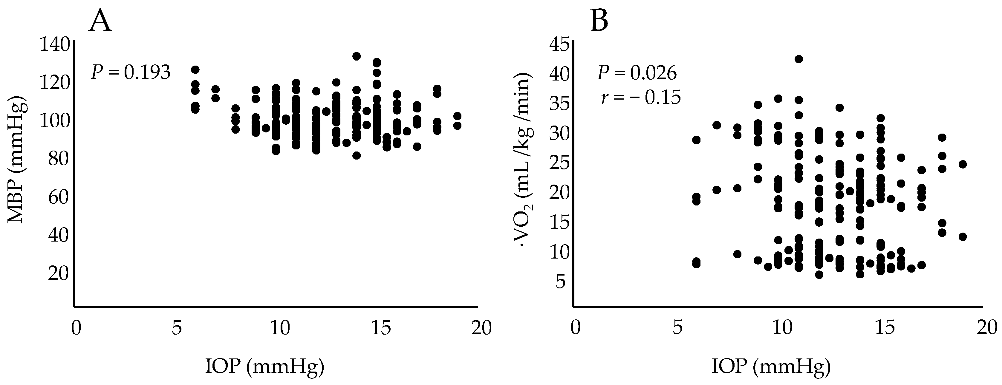
SPSS® version 25 (SPSS, Chicago, IL, USA) was used for all statistical analyses. Dunnett’s test was used to compare the dynamic changes in IOP, mean blood pressure (MBP), and ·VO2 during rest at each stage of exercise. Pearson’s correlation coefficient was used to correlate IOP with MBP and ·VO2. The statistical significance was set at P < 0.05.

3. Results

All subjects were able to perform the multistage exercise test with a ·VO2max of 53.4 ± 8.0 mL/kg/min. The oxygen uptake during exercise was significantly higher at all intensities from 5 to 20 min of exercise than that at rest. MBP was significantly higher at 70% intensity from 5 to 20 min of exercise than that at rest. IOP was significantly lower at 70% intensity from 5 to 20 min of exercise than that at rest (Table 1).
There was no correlation between IOP and MBP (P = 0.193), but there was a negative correlation between IOP and ·VO2 (r = −0.15, P = 0.026) (Figure 2).

4. Discussion

In the present study, we investigated the effects of low-, moderate-, and high-intensity exercise on IOP in healthy subjects. The results showed that there was no change in IOP during low- and moderate-intensity exercise compared with that at rest. However, during high-intensity exercise (70% ·VO2), IOP was significantly lower than that at rest until 20 min after the start of exercise, and IOP was correlated with ·VO2, an index of exercise intensity. Shapiro et al. investigated IOP changes before and after exercise using a bicycle ergometer at different intensities using absolute exercise intensity in watts and reported that load amount did not correlate with changes in IOP [16]. When prescribing exercise at absolute exercise intensity, the load is constant, but oxygen uptake and the circulatory response during exercise for each individual is different. Therefore, it is likely that the effective limit, which is important when prescribing exercise, will be lowered, or that the safe limit will be exceeded. Therefore, it is necessary to investigate the relationship between IOP and relative exercise intensity more accurately. In the present study, exercise intensity was observed by adjusting the load so that oxygen uptake was constant during exercise to demonstrate the relationship more accurately between exercise intensity and IOP. The results of this study may also apply to general clinical practice, as exercise prescriptions are generally based on relative exercise intensity using the Karvonen or other methods [17].
IOP is generated by the aqueous humor and describes the pressure that is experienced by the entire closed system. Aqueous humor is produced by the ciliary epithelium and drains through two independent outflow pathways: the trabecular meshwork and the uveal tract. Recently, it was reported that uveal lymphatics are also involved in IOP regulation [18]. The decrease in aqueous humor, which is related to IOP, is due to the inhibition of NO synthesis [19], and β-blockers, which have long been used to reduce IOP in glaucoma [20]. As observed in this study, exercise is not a factor in lowering IOP due to NO production and increased sympathetic nerve activity, suggesting that other mechanisms may be involved in aerobic exercise. Hariss et al. evaluated the relationship among IOP, PCO2, and plasma osmolality after 7 min of exercise at 30 W and 90 W on a bicycle ergometer, followed by gradual increases in exercise intensity. The results showed that PCO2 and plasma osmolality were not related to the decrease in IOP; however, there was a negative correlation between IOP and lactate levels [21]. In the present study, IOP decreased with high-intensity exercise. There was no relationship between IOP and MBP but a negative correlation between ·VO2 and IOP, and this might have been due to the rapid increase in lactate levels in high-intensity exercise.
In previous studies of transient exercise and training in healthy volunteers, optical coherence tomography angiography revealed a reduction in the blood flow in the optic papilla and macula [22,23]. The effects of exercise intensity on optic nerve and macular fundus perfusion in patients with retinopathy, glaucoma, and other ocular diseases are not clear. Therefore, fundus blood flow, including IOP, should be investigated to determine the safe level of exercise for patients with various ocular diseases.
The first limitation of the present study is that the data were derived exclusively from healthy men. However, IOP reportedly varies according to sex and age [24]. Therefore, future studies should take these differences into account by including a more diverse cohort of individuals. A second limitation is that we did not measure the lactate level, which is an IOP-lowering factor. Therefore, future studies should include this parameter.
In healthy participants, IOP did not change during low- and moderate-intensity exercise, but significantly decreased during high-intensity exercise compared with that at rest. The results suggest that different mechanisms are involved in the lowering of IOP at rest and during exercise.

Author Contributions

Conceptualization, T.K. and T.N.; methodology, T.K.; software, T.K.; validation, T.N., H.K. (Hiroaki Kataoka) and Y.N.; formal analysis, Y.N.; investigation, K.F.; resources, D.I.; data curation, D.I.; writing—original draft preparation, T.K.; writing—review and editing, T.I.; visualization, D.I.; supervision, H.K. (Hiroaki Kimura); project administration, H.K. (Hiroaki Kimura); funding acquisition, T.K. and T.N. All authors have read and agreed to the published version of the manuscript.

Funding

This research was funded by the JSPS KAKENHI (grant numbers: 15K01440 and 18K10674).

Institutional Review Board Statement

The study was conducted in accordance with the Declaration of Helsinki and approved by the Institutional Review Board of Hiroshima University (E-529).

Informed Consent Statement

Written informed consent has been obtained from the patients for the publication of this paper.

Data Availability Statement

Data are not available to the public.

Conflicts of Interest

The authors declare no conflict of interest. The funders had no role in the design of the study; in the collection, analyses, or interpretation of data; in the writing of the manuscript; or in the decision to publish the results.

References

  1. International Diabetes Federation. IDF Diabetes Atlas: Diabetes around the World in 2021. Available online: https://diabetesatlas.org/ (accessed on 28 September 2021).
  2. Boulé, N.G.; Haddad, E.; Kenny, G.P.; Wells, G.A.; Sigal, R.J. Effects of exercise on glycemic control and body mass in type 2 diabetes mellitus: A meta-analysis of controlled clinical trials. JAMA 2001, 286, 1218–1227. [Google Scholar] [CrossRef] [PubMed]
  3. Qiu, S.; Cai, X.; Schumann, U.; Velders, M.; Sun, Z.; Steinacker, J.M. Impact of walking on glycemic control and other cardiovascular risk factors in type 2 diabetes: A meta-analysis. PLoS ONE 2014, 9, e109767. [Google Scholar] [CrossRef] [PubMed] [Green Version]
  4. Sabag, A.; Way, K.L.; Keating, S.E.; Sultana, R.N.; O’Connor, H.T.; Baker, M.K.; Chuter, V.H.; George, J.; Johnson, N.A. Exercise and ectopic fat in type 2 diabetes: A systematic review and meta-analysis. Diabetes Metab. 2017, 43, 195–210. [Google Scholar] [CrossRef] [PubMed]
  5. Araki, E.; Goto, A.; Kondo, T.; Noda, M.; Noto, H.; Origasa, H.; Osawa, H.; Taguchi, A.; Tanizawa, Y.; Tobe, K.; et al. Japanese Clinical Practice Guideline for Diabetes 2019. J. Diabetes Investig. 2020, 11, 1020–1076. [Google Scholar] [CrossRef] [PubMed]
  6. Dong, L.; Li, J.; Lian, Y.; Tang, Z.X.; Zen, Z.; Yu, P.; Li, Y. Long-term intensive lifestyle intervention promotes improvement of stage III diabetic nephropathy. Med. Sci. Monit. 2019, 25, 3061–3068. [Google Scholar] [CrossRef] [PubMed]
  7. Robinson-Cohen, C.; Littman, A.J.; Duncan, G.E.; Weiss, N.S.; Sachs, M.C.; Ruzinski, J.; Kundzins, J.; Rock, D.; de Boer, I.H.; Ikizler, T.A.; et al. Physical activity and change in estimated GFR among persons with CKD. J. Am. Soc. Nephrol. 2014, 25, 399–406. [Google Scholar] [CrossRef] [Green Version]
  8. Yoo, M.; D’Silva, L.J.; Martin, K.; Sharma, N.K.; Pasnoor, M.; LeMaster, J.W.; Kluding, P.M. Pilot study of exercise therapy on painful diabetic peripheral neuropathy. Pain Med. 2015, 16, 1482–1489. [Google Scholar] [CrossRef] [Green Version]
  9. Brown, R.; Sharafi, A.; Slade, J.M.; Convit, A.; Davis, N.; Baete, S.; Milton, H.; Mroczek, K.J.; Kluding, P.M.; Regatte, R.R.; et al. Lower extremity MRI following 10-week supervised exercise intervention in patients with diabetic peripheral neuropathy. BMJ Open Diabetes Res. Care 2021, 9, e002312. [Google Scholar] [CrossRef]
  10. Aro, A.; Kauppinen, A.; Kivinen, N.; Selander, T.; Kinnunen, K.; Tuomilehto, J.; Keinänen-Kiukaanniemi, S.; Lindström, J.; Uusitupa, M.; Kaarniranta, K. Life style intervention improves retinopathy status-the Finnish Diabetes Prevention Study. Nutrients 2019, 11, 1691. [Google Scholar] [CrossRef] [Green Version]
  11. Kuwata, H.; Okamura, S.; Hayashino, Y.; Tsujii, S.; Ishii, H.; Diabetes Distress and Care Registry at Tenri Study Group. Higher levels of physical activity are independently associated with a lower incidence of diabetic retinopathy in Japanese patients with type 2 diabetes: A prospective cohort study, Diabetes Distress and Care Registry at Tenri (DDCRT15). PLoS ONE 2017, 12, e0172890. [Google Scholar] [CrossRef]
  12. Woodcock, M.G.L.; Richards, J.C.; Murray, A.D.N. The last 11 years of Molteno implantation at the University of Cape Town. Refining our indications and surgical technique. Eye 2008, 22, 18–25. [Google Scholar] [CrossRef]
  13. Al-Shamsi, H.N.; Dueker, D.K.; Nowilaty, S.R.; Al-Shahwan, S.A. Neovascular glaucoma at King Khaled Eye Specialist Hospital—Etiologic consideration. Middle East Afr. J. Ophthalmol. 2009, 16, 15–19. [Google Scholar] [CrossRef] [PubMed]
  14. Natsis, K.; Asouhidou, I.; Nousios, G.; Chatzibalis, T.; Vlasis, K.; Karabatakis, V. Aerobic exercise and intraocular pressure in normotensive and glaucoma patients. BMC Ophthalmol. 2009, 9, 6. [Google Scholar] [CrossRef] [PubMed]
  15. Vieira, G.M.; Oliveira, H.B.; de Andrade, D.T.; Bottaro, M.; Ritch, R. Intraocular pressure variation during weight lifting. Arch. Ophthalmol. 2006, 124, 1251–1254. [Google Scholar] [CrossRef] [Green Version]
  16. Shapiro, A.; Shoenfeld, Y.; Shapiro, Y. The effect of standardised submaximal work load on intraocular pressure. Br. J. Ophthalmol. 1978, 62, 679–681. [Google Scholar] [CrossRef] [Green Version]
  17. Karvonen, M.J.; Kentala, E.; Mustala, O. The effects of training on heart rate; a longitudinal study. Ann. Med. Exp. Biol. Fenn. 1957, 35, 307–315. [Google Scholar] [PubMed]
  18. Wu, F.; Zhao, Y.; Zhang, H. Ocular autonomic nervous system: An update from anatomy to physiological functions. Vision 2022, 6, 6. [Google Scholar] [CrossRef]
  19. Kiel, J.W.; Reitsamer, H.A.; Walker, J.S.; Kiel, F.W. Effects of nitric oxide synthase inhibition on ciliary blood flow, aqueous production and intraocular pressure. Exp. Eye Res. 2001, 73, 355–364. [Google Scholar] [CrossRef]
  20. Phillips, C.I.; Howitt, G.; Rowlands, D.J. Propranolol as ocular hypotensive agent. Br. J. Ophthalmol. 1967, 51, 222–226. [Google Scholar] [CrossRef] [Green Version]
  21. Harris, A.; Malinovsky, V.; Martin, B. Correlates of acute exercise-induced ocular hypotension. Investig. Ophthalmol. Vis. Sci. 1994, 35, 3852–3857. [Google Scholar]
  22. Alnawaiseh, M.; Lahme, L.; Treder, M.; Rosentreter, A.; Eter, N. Short-term effects of exercise on optic nerve and macular perfusion measured by optical coherence tomography angiography. Retina 2017, 37, 1642–1646. [Google Scholar] [CrossRef] [PubMed]
  23. Schmitz, B.; Nelis, P.; Rolfes, F.; Alnawaiseh, M.; Klose, A.; Krüger, M.; Eter, N.; Brand, S.-M.; Alten, F. Effects of high-intensity interval training on optic nerve head and macular perfusion using optical coherence tomography angiography in healthy adults. Atherosclerosis 2018, 247, 8–15. [Google Scholar] [CrossRef] [PubMed]
  24. Shiose, Y. The aging effect on intraocular pressure in an apparently normal population. Arch. Ophthalmol. 1984, 102, 883–887. [Google Scholar] [CrossRef] [PubMed]
Figure 1. (A) Measurement of intraocular pressure (IOP) during exercise. (B) Probe position at rest and during exercise measuring IOP.
Healthcare 10 01196 g001
Figure 2. (A) Relationship between intraocular pressure and mean arterial pressure (MBP). (B) Relationship between intraocular pressure and oxygen consumption during exercise (VO2).
Healthcare 10 01196 g002
Table 1. Differences in intraocular pressure, blood pressure, and oxygen uptake at each exercise intensity.
30%-intensity aerobic exercise
Rest5 min10 min15 min20 minRecovery
IOP (mmHg)13.7 ± 2.213.0 ± 2.212.9 ± 2.212.4 ± 2.412.1 ± 2.312.5 ± 2.9
MBP (mmHg)93.2 ± 9.597.6 ± 6.395.2 ± 8.795.5 ± 7.195.0 ± 8.593.6 ± 7.1
·VO2 (mL/kg/min)5.5 ± 1.58.7 ± 2.7 *8.4 ± 2.7 *8.6 ± 2.9 *8.6 ± 2.7 *5.7 ± 1.5 *
50%-intensity aerobic exercise
Rest5 min10 min15 min20 minRecovery
IOP (mmHg)14.3 ± 2.713.9 ± 2.513.1 ± 2.713.4 ± 2.913.1 ± 3.313.0 ± 2.9
MBP (mmHg)93.7 ± 11.297.5 ± 7.195.7 ± 7.997.3 ± 8.196.8 ± 9.993.6 ± 8.3
·VO2 (mL/kg/min)5.7 ± 1.617.7 ± 4.3 *17.6 ± 4.4 *16.5 ± 3.8 *15.8 ± 4.3 *7.8 ± 2.6
70%-intensity aerobic exercise
Rest5 min10 min15 min20 minRecovery
IOP (mmHg)14.2 ± 2.612.4 ± 2.8 *11.5 ± 2.6 *11.5 ± 2.5 *11.6 ± 2.8 *13.1 ± 2.3
MAP (mmHg)94.3 ± 10.4110.0 ± 12.4 *103.3 ± 9.9 *102.2 ± 7.5 *100.3 ± 7.1 *94.1 ± 11.2
·VO2 (mL/kg/min)5.8 ± 1.728.7 ± 5.2 *25.7 ± 4.7 *24.6 ± 3.9 *24.6 ± 4.3 *7.0 ± 2.0
Mean ± standard deviation. * Statistically significant (P < 0.05). IOP, intraocular pressure; MBP, mean blood pressure; ·VO2, oxygen consumption during exercise.
Publisher’s Note: MDPI stays neutral with regard to jurisdictional claims in published maps and institutional affiliations.

