Article

Subsiding of Periodontitis in the Permanent Dentition in Individuals with Papillon-Lefèvre Syndrome through Specific Periodontal Treatment: A Systematic Review

by
Dagmar Schnabl
1,
Felix Maximilian Thumm
1,
Ines Kapferer-Seebacher
2,* and
Peter Eickholz
3
1
Department of Prosthetic Dentistry, Medical University Innsbruck, 6020 Innsbruck, Austria
2
Department of Conservative Dentistry and Periodontology, Medical University Innsbruck, Anichstraße 35, 6020 Innsbruck, Austria
3
Department of Periodontology, Center for Dentistry and Oral Medicine (Carolinum), Johann Wolfgang Goethe-University Frankfurt/Main, 60596 Frankfurt, Germany
*
Author to whom correspondence should be addressed.
Healthcare 2022, 10(12), 2505; https://doi.org/10.3390/healthcare10122505
Submission received: 13 October 2022 / Revised: 2 December 2022 / Accepted: 5 December 2022 / Published: 10 December 2022

Abstract

:
Papillon–Lefèvre syndrome (PLS) is a rare hereditary disease characterized by palmoplantar hyperkeratosis (PPK) and periodontitis in the primary and permanent dentition, usually resulting in edentulism in youth. Subsiding of PLS-associated periodontitis through specific therapy has occasionally been reported. We aimed to systematically assess periodontal treatment strategies that may decelerate disease progression. A systematic literature search was conducted at PubMed/LIVIVO/Ovid (Prospero registration number CRD42021223253). Clinical studies describing periodontal treatment success—defined as loss of ≤four permanent teeth because of periodontitis and the arrest of periodontitis or probing depths ≤ 5 mm—in individuals with PLS followed up for ≥24 months. Out of the 444 primarily identified studies, 12 studies reporting nine individuals were included. The timely extraction of affected or, alternatively, all primary teeth, compliance with oral hygiene instructions, supra- and subgingival debridement within frequent supportive periodontal care intervals, and—in eight patients—adjunctive systemic antibiotic therapy (mostly amoxicillin/metronidazole) effected a halt in disease progression. The suppression of Aggregatibacter actinomycetemcomitans below the detection limit was correlated with the subsiding of periodontitis. Successful controlling of PLS-associated periodontitis may be achieved if high effort and patient compliance are provided.

1. Introduction

Papillon–Lefèvre syndrome (PLS) is a rare autosomal recessive disease with a prevalence of 1 in 4,000,000 at birth [1]. Haim–Munk syndrome (synonyms: Cochin Jewish disorder and Congenital Keratosis) is an extremely rare variant of PLS that has only been seen in descendants of an inbred Jewish family originally from Cochin, India, who migrated to Israel [1]. Cardinal features are palmoplantar hyperkeratosis (PPK) and severe periodontal destruction of the primary and permanent dentition. The first dermatological and periodontal manifestations commonly emerge in toddlerhood [2]. After a proper eruption of the primary teeth, gingivitis occurs around the age of two to three years, and subsequent periodontitis leads to alveolar bone resorption, attachment loss, and premature loss of the primary dentition [2,3]. In permanent dentition, the destructive process recurs, usually leading to complete tooth loss in youth.
Periodontitis is a chronic inflammatory disease that is triggered by dysbiotic bacterial deposits on teeth (dental biofilm) and it destroys the tooth supporting tissues (connective tissue attachment and bone) [4,5]. The interface of the tooth surface and oral mucosa (i.e., gingiva) is a weak spot of the body’s integrity. Dental biofilm accumulating at the gingival margin may enter between the tooth surface and gingiva into the connective tissue or even bone. As a respective defense mechanism, the body gathers inflammatory cells in the affected gingiva to prevent the microbiota from entering. Some asaccharolytic and iron-dependent bacteria (e.g., Porphyromonas gingivalis) may, on the one hand, trigger inflammation to thrive on proteins of the inflammatory gingival fluid, but, on the other hand, evade the respective immune reaction (dysbiosis) maintaining chronic inflammation [5]. Certain periodontal bacteria, such as Aggregatibacter actinomycetemcomitans (A. actinomycetemcomitans), are strongly associated with destructive inflammatory responses [6]. Inflammatory and immunological defense may be disturbed by genetic factors (e.g., PLS) that deteriorate the function of cells and mediators. In most people, highly prevalent periodontitis progresses slowly and starts to manifest at a middle age [7]. In rare monogenic disorders such as PLS, genetic factors severely affect immunity, and rapidly progressing periodontitis is likely to appear early in life. Thus, the treatment of periodontitis aims to prevent biofilm accumulation on tooth surfaces (effective individual oral hygiene) and to remove the biofilm from periodontal pockets (subgingival instrumentation) [8].
Homozygous or compound heterozygous loss-of-function mutations of the cathepsin C gene, encoding dipeptidyl-aminopeptidase cathepsin C (CTSC), are associated with PLS [1,9,10]. CTSC, a lysosomal cysteine protease, impacts epidermal differentiation and scaling and is expressed in epithelial regions typically affected in PLS patients. Furthermore, the cysteine protease—through the removal of N-terminal inhibitory peptides—activates certain serine proteases (e.g., proteinase 3 (PR3)) in immunologic cells, such as neutrophil granulocytes, cytotoxic lymphocytes, natural killer cells, alveolar macrophages, and mast cells, which participate in cell-mediated phagocytosis, cytotoxicity, and activation of various inflammatory mediators [1,9,10]. PR3 activates cathelicidin LL-37 (LL-37), known as a potent antimicrobial to most strains of A. actinomycetemcomitans. Consequently, loss-of-function mutation of CTSC in PLS resulting in a lack of PR3 leads to the deficit of antimicrobial and immunomodulatory functions of LL-37 in the gingiva, allowing for infection with A. actinomycetemcomitans and contributing to the development of severe periodontal disease [11]. A. actinomycetemcomitans cannot reliably be suppressed below levels of detection using a subgingival instrumentation alone [12,13,14]. Thus, the adjunctive use of systemic antibiotics is recommended. The combination of amoxicillin and metronidazole has been shown to be effective at suppressing A. actinomycetemcomitans below the limits of detection [15]. A loss of CTSC function increases susceptibility to infections frequently seen in PLS patients, e.g., furunculosis, pyodermia, liver abscess, pneumonia, and others [3,10,16,17]. In some PLS patients, intracranial calcifications and mental retardation were reported [3]. The likely pathomechanism of periodontal destruction with PLS is a reduced immune response due to inadequate chemotaxis, phagocytosis, and bactericidal activity of the granulocytes, a diminished number of T-helper and regulatory cells, and a decreased proliferation of lymphocytes [18,19,20,21]. While disease progression seems default in most affected individuals, occasional cases with loss-of function mutations in CTSC displaying PPK, but no periodontitis, have been reported. Furthermore, PLS cases with successful periodontal treatment have been documented. To the best of the authors’ knowledge, up until now, interventional studies on periodontal treatment of PLS-associated periodontitis do not exist.
The aim of this systematic review was a compilation of reported PLS cases with successfully arrested periodontitis to identify effective periodontal treatment strategies.

2. Materials and Methods

2.1. Protocol and Registration

The systematic search was performed according to the Preferred Reporting Items for Systematic Review and Meta-Analysis (PRISMA) guidelines [22] and was registered at Prospero under CRD42021223253.
A structured approach was used to formulate the research question for this systematic review using five components, commonly known by the acronym “PICOS”: the patient population (P), the interventions (I), the comparison group (C), the outcome of interest (O), and the study design (S). The respective PICOS components for this review are:
P: Subjects with PLS who have completed at least one cycle of non-surgical periodontal therapy rendering clinical success;
I: Non-surgical/surgical periodontal treatment;
C: PLS patients without periodontal treatment;
O: Loss of less than four permanent teeth because of periodontitis and arrest of clinical attachment loss with probing depths ≤ 5 mm at the end of a determined follow-up interval of ≥24 months;
S: Clinical trials, case-control studies, cross-sectional studies, cohort studies, case series, and case reports published in peer-reviewed scientific journals.

2.2. Literature Search Strategy

The systematic search was accomplished in the electronic databases PubMed, LIVIVO, and Ovid through thr use of the following keywords: (Papillon–Lefèvre OR “papillon lefevre” OR Haim–Munk OR “Haim Munk” OR palmoplantar kerato*) AND (Cathepsin C OR CTSC). The publication date was open to the past. The determined cut-off date was 31 March 2021. Additionally, a manual search was conducted, including browsing of the reference lists of the selected studies.

2.3. Screening and Selection

Clinical studies of successful periodontal treatment of adult individuals clinically and/or genetically diagnosed with PLS were included. Children were included if the follow-up interval extended after the eruption of all permanent incisors and first molars. Treatment success was evaluated at a follow-up-interval of ≥ 24 months, and was defined as loss of less than four permanent teeth because of periodontitis, corresponding to a remainder of at least 24 permanent teeth in the absence of dental aplasia (third molars excluded), ensuring a proper chewing function and posterior tooth support. According to the S3 clinical practice guideline of the European Federation of Periodontology, endpoint of periodontal therapy was defined when probing depths ≤ 5 mm were achieved [8]. (Moderately deep pockets are considered manageable by subgingival instrumentation during supportive periodontal care.) Studies in English or German, available in full text, were included.
The following types of studies were considered: clinical trials, case-control studies, cross-sectional studies, cohort studies, case series, and case reports published in peer-reviewed scientific journals. Excluded studies were cell culture laboratory studies, animal studies, narrative reviews, and questionnaire studies. Titles and abstracts were checked regarding the pre-defined eligibility criteria. Abstracts with unclear methodology were included in full-text assessment to avoid the exclusion of potentially relevant articles.

2.3.1. Assessment of Heterogeneity

The heterogeneity of the included studies was evaluated based on following factors: (1) study design and (2) subjects’ characteristics.

2.3.2. Quality Assessment

The Joanna Briggs Institute (Adelaide, Australia) quality assessment tool [23] was applied. Each study was classified into the following groups: low risk of bias if all quality criteria were judged as “present”, moderate risk of bias if one or more key domains were “unclear”, and high risk of bias if one or more key domains were “absent”. Only low risk of bias studies were eligible.

2.4. Data Extraction

The extracted main outcomes in the studies reporting successful periodontal treatment in PLS were as follows: (1) clinical and genetic diagnosis of PLS; (2) age at baseline; (3) initial dental, periodontal parameters, and microbiological assessment, if available; (4) description of disease progression and applied therapies; and (5) outcome and follow-up.

3. Results

3.1. Study Selection

The literature search flowchart is presented in Figure 1. Out of 266 identified studies (after removal of the duplicates), 81 were excluded based on the title and abstract. Of these, 26 studies were laboratory studies, 16 reported the treatment of other symptoms but periodontitis, 15 reported periodontitis that was not (successfully) treated, 11 were written in another language besides English or German, 6 described the periodontal treatment of children with a follow-up interval ending before the eruption of incisors/first molars, 3 presented genetic analyses in individuals diagnosed with periodontitis and severe tooth loss, 2 described other pathologies than PLS, 1 was an animal study, and 1 was an erratum.
Of the remaining 185 studies, eight were not available in full text, seven of which published between 1965 and 1997 and one abstract of an oral presentation delivered in the year 2005. One hundred and seventy-seven articles were screened by full text. Eighty eight studies reported individuals diagnosed with periodontitis who received no or unsuccessful periodontal treatment, twenty seven were genetic analyses/laboratory studies, 26 twenty six with other aspects of PLS than the periodontal condition, ten documented a follow-up period of <24 months after periodontal treatment, ten reported on young children with permanent incisors and first molars not yet erupted, three studies reported PLS cases (two alleged and one genetically confirmed) without signs of periodontitis [24,25,26], and one reported a CTSC mutation not effecting PLS (neither PPK nor PD) [27].
Finally, 12 studies were included [2,28,29,30,31,32,33,34,35,36,37,38]. Eight of these were case series [2,28,29,30,33,34,35,38] and four were case reports [31,32,36,37].

3.2. Subjects, Diagnosis, and Therapy

Twelve of the included studies reported on a total of nine individuals (six males and three females) meeting the inclusion criteria. Multiple reports existed of four individuals (identification numbers (IDs) 4, 5, 6, and 7). In these cases, previous reports of early treatment were included in synopsis, with subsequent reports of treatment success after an overall follow-up of ≥24 months.
Table 1 presents an overview of the subjects’ characteristics, periodontal treatment measures, and follow-up period of nine included individuals [2,28,29,30,31,32,33,34,35,36,37,38]. In four individuals, PLS was confirmed by genetic examination (ID 6: homozygous variants in c.755 A>T, p.Q252L; ID 7: compound heterozygous variants in c.947 T>G, p.L316R and c.1268 G>C, p.W423S [29]; ID 8: homozygous variants in c.854 C>T; p.P285L [29]; ID 9: compound heterozygous variants in c.322A>T, p.Lys108Ter and c.504C>G, p.Tyr168Ter). In the remaining subjects, PLS diagnosis was based on clinical symptoms and positive family history.
The following treatment steps were unanimously considered essential in the reduction of the probing depths and periodontal inflammation by authors reporting successful periodontal therapy: (I) strict biofilm management including repeated oral hygiene instructions and chlorhexidine mouth rinses; (II) professional supra- and subgingival debridement within a stringent supportive periodontal therapy with intervals ranging from one week up to three months; and the (III) extraction of severely affected teeth. Only the dentist of patient ID 9 chose to not extract severely affected primary teeth [38]. In this patient, primary teeth exfoliated at the usual age and, intriguingly, the permanent dentition was not affected by periodontitis by the age of 18 years, despite poor biofilm management at the time.
In seven patients, initial microbiological testing was performed and was positive for A. actinomycetemcomitans and/or other periopathogenic bacteria (Table 2). In seven patients, antibiotic courses were conducted, and in six the suppression of A. actinomycetemcomitans below the level of detection after treatment was documented and related with the arrest of periodontitis [28,29,33,34,35,36,37]. In three individuals, all of the primary teeth were extracted, inducing an edentulous period of nine months to two years until the eruption of the permanent teeth [2,28,31,32]. The authors emphasized the benefit of a complete (temporary) extinction of microbiological niches with regard to the elimination of periopathogens that did not (re-) occur in the permanent dentition. In one individual (ID 6), treatment success was accomplished by the extraction of only severely affected primary teeth, together with mechanical cleaning and antibiotic therapy [33,34].

4. Discussion

Numerous case reports have recounted the default progression of periodontal destruction and the failure of periodontal treatment in individuals afflicted with PLS. The aim of this systematic review was to identify promising therapeutic strategies by detecting and analyzing case reports considering the successful treatment of PLS-associated periodontitis.
Regarding treatment success, we deliberately set tight inclusion criteria (requiring an arrest of periodontitis implying steadily reduced probing depths of maximally 5 mm for 24 months and a maximum loss of four permanent teeth, third molars excluded, due to periodontitis) with respect to favorable esthetics and proper masticatory function. In all of the included reports, conformingly, periodontal treatment comprised classical mechanical debridement (when necessary, under general anesthesia), repeated oral hygiene instructions, chlorhexidine mouth rinses, the extraction of severely affected primary and/or permanent teeth, and a strict and frequent recall interval. In the majority of studies, microbiological testing for A. actinomycetemcomitans and/or other periopathogenic bacteria was performed before and after treatment. The suppression of A. actinomycetemcomitans below the level of detection through mechanical treatment with adjunctive systemic antibiotic courses was correlated with the subsiding of periodontitis [28,29,33,34,35,36,37].
For antibiotic therapy, a combination of amoxicillin and metronidazole, as used in individuals ID 4, 6, 7, 8, and 9, has been shown to be a commendable strategy [29,33,34,35,36,37,38]. In individual ID 3, after unsuccessful courses of minocycline and erythromycin, according to antibiogram, ofloxacin proved to be effective in the suppression of A. actinomycetemcomitans below the level of detection [28]. Individual ID 1, after an edentulous interval through the extraction of all primary teeth, was administered repeated courses of tetracycline to prevent the onset of periodontitis in permanent teeth [2]. The administration of antibiotics (mostly initially and at recurrence of periodontitis or A. actinomycetemcomitans) varied from one week to two years [2,28,29,31,32,33,34,35,36,37,38].
In three cases, the complete extraction of the primary dentition effecting an edentulous period of several months (with or without the delivery of intermediate full dentures) was suspected to have eliminated all periopathogenic bacteria and facilitated the eruption of permanent teeth in a steadily physiological microbiological environment [2,28,31,32]. In individual ID 6 (as well as his younger brother, who was excluded from this review because of the incomplete eruption of incisors at the end of the study period), the extraction of only severely affected primary teeth, together with mechanical cleaning and antibiotic therapy, effected treatment success [33,34]. Whenever permanent teeth were already erupted in the presence of severe periodontitis of the primary teeth, they were usually also afflicted with periodontitis and in need of periodontal therapy or, eventually, extraction [28,35,36,37].
Intensive maintenance therapy and compliance with homecare seem to constitute key success factors [35]. Lux et al. (2005), in the description of the course of disease of individual ID 6, demonstrated a deterioration of periodontal parameters after 60 months of successfully controlled periodontitis when, the patient’s compliance with home care decreased, which emphasized the necessity for a continuous motivation and recall of PLS patients [36]. In individual ID 6, fixed orthodontic therapy was required and included the extraction of two central mandibular incisors with severe alveolar bone loss. Originally, all four mandibular incisors were scheduled for extraction. However, the patient’s mother insisted on the retention of these teeth. The result of the orthodontic treatment was satisfactory, but the long-term prognosis of the remaining mandibular incisors was rated as critical due to severe clinical attachment loss of up to 6 mm, and isolated probing depths of 4 mm with bleeding on probing remaining after 79 months of continuous monitoring and treatment [36]. Seventeen years after active treatment, both incisors still were present [35]. Nickles et al. (2013) also reported orthodontic treatment in individual ID 7 [35], and Wiebe et al. (2001) considered orthodontics following the successful periodontal therapy of individual ID 4 [31]. This is in accordance with the recently published periodontal practice guidelines, which state that orthodontic therapy has no detrimental effects on periodontal conditions in periodontitis patients with a healthy but reduced periodontium, provided the results of the periodontal therapy are maintained during the active orthodontic treatment phase [39].
More than 75 different mutations in the CTSC gene may cause PLS, Haim–Munk syndrome, or prepubertal periodontitis, which are subsumed to the same phenotypical spectrum [40,41]. PLS manifestation seems to range in the severity and progression of tissue destruction and age of disease onset. Atypical phenotypes of CTSC mutations with palmoplantar hyperkeratosis but lack of periodontal involvement and vice versa have been described [24,25,26,38]. Atypical phenotypes also may occur within one molecular family. Soskolne et al. (1996) reported on an individual without periodontal involvement who had two (out of five) siblings that suffered from both PPK and periodontitis with complete tooth loss in teenage years, and a second molecular family with three siblings exhibiting PPK plus periodontitis, but one brother displaying periodontitis only [24]. The basis of phenotypic variation is genetic variability between individuals in conjunction with acquired risk factors. The authors suspect the influence of certain genetic or epigenetic mechanisms to cause a variable or discordant expression of cardinal features of PLS, such as the co-expression of linked loci or non-linked modifying genes, compound heterozygosity, or genomic imprinting (gene silencing) in homozygous cases with absent consanguinity, or also occasional manifestations of heterozygosity [24,26]. Genetic variability probably also influences the periodontal treatment success of PLS.
The identified treatment strategies and the proposed treatment goal of the suppression of A. actinomycetemcomitans must be investigated in larger cohorts of PLS patients. The European research project “Solving the unsolved Rare Diseases (RD)”, which was funded under the European Union’s Horizon 2020 research and innovation program, aims to solve large numbers of RDs and to improve the diagnostics and therapy of RD patients. The resolution of the remaining open questions with PLS will primarily rely on the standardization of the diagnostic criteria. Clinical centers that specialize in this rare pathology need to apply congruent approaches for the exact characterization of clinical features in conjunction with genetic validation, which should be reached without exception in all patients and relevant family members [42].
In conclusion, only a few case reports on the successful treatment of PLS associated periodontitis exist. The early diagnosis of PLS and the suppression of A. actinomycetemcomitans below the detection level might be critical factors for treatment success. Suppression of A. actinomycetemcomitans may be achieved through extraction of (affected or all) primary teeth, preferably before the eruption of permanent teeth and/or supra- and subgingival debridement with adjunctive systemic antibiotic treatment with amoxicillin and metronidazole. The suppression of A. actinomycetemcomitans in conjunction with frequent supportive periodontal care, preferably carried out by a specialized dentist or team, seems to enable a delay or halt of the usual default disease progression. Successful controlling of PLS-associated periodontitis implies a high effort and compliance within a stringent treatment concept.

Author Contributions

Conceptualization, D.S., I.K-S., and F.M.T.; literature search, F.M.T. and I.K.-S.; screening and assessment, F.M.T. and D.S.; writing—original draft preparation, D.S.; review and editing, I.K.-S. and P.E. All authors have read and agreed to the published version of the manuscript.

Funding

This research received no external funding.

Institutional Review Board Statement

This manuscript is a systematic review (SR). Thus, all data reported and analyzed in this SR was retrieved from articles already published and therefore represent publicly accessible information. Thus, an institutional review board approval is not applicable.

Informed Consent Statement

The authors of the respective publications that we used for this SR report informed consent given by patients they report on.

Data Availability Statement

All data analyzed for this SR is publicly available in the respective articles that are referred to.

Conflicts of Interest

The authors declare no conflict of interest.

References

  1. Bloch-Zupan, A.; Sedano, H.O.; Scully, C. Anomalies of Teeth Eruption and/or Resorption. In Dento/Oro/Craniofacial Anomalies and Genetics, 1st ed.; Elsevier: Amsterdam, The Netherlands, 2012; pp. 170–174. [Google Scholar]
  2. Preus, H.; Gjermo, P. Clinical management of prepubertal periodontitis in 2 siblings with Papillon-Lefèvre syndrome. J. Clin. Periodontol. 1987, 14, 156–160. [Google Scholar] [CrossRef] [PubMed]
  3. Haneke, E. The Papillon-Lefèvre syndrome: Keratosis palmoplantaris with periodontopathy. Report of a case and review of the cases in the literature. Hum Genet. 1979, 51, 1–35. [Google Scholar] [CrossRef] [PubMed]
  4. Papapanou, P.N.; Sanz, M.; Buduneli, N.; Dietrich, T.; Feres, M.; Fine, D.H.; Flemmig, T.F.; Garcia, R.; Giannobile, W.V.; Graziani, F.; et al. Periodontitis: Consensus report of workgroup 2 of the 2017 World Workshop on the Classification of Periodontal and Peri-Implant Diseases and Conditions. Clin. Periodontol. 2018, 45 (Suppl. 20), S162–S170. [Google Scholar] [CrossRef] [PubMed]
  5. Hajishengallis, G.; Lamont, R.J. Beyond the red complex and into more complexity: The polymicrobial synergy and dysbiosis (PSD) model of periodontal disease etiology. Mol. Oral Microbiol. 2012, 27, 409–419. [Google Scholar] [CrossRef] [Green Version]
  6. Hajishengallis, G. Immunomicrobial pathogenesis of periodontitis: Keystones, pathobionts, and host response. Trends Immunol. 2014, 35, 3–11. [Google Scholar] [CrossRef] [Green Version]
  7. Löe, H.; Anerud, A.; Boysen, H.; Morrison, E. Natural history of periodontal disease in man. Rapid, moderate and no loss of attachment in Sri Lankan laborers 14 to 46 years of age. J. Clin. Periodontol. 1986, 13, 431–445. [Google Scholar] [CrossRef]
  8. Sanz, M.; Herrera, D.; Kebschull, M.; Chapple, I.; Jepsen, S.; Beglundh, T.; Sculean, A.; Tonetti, M.S.; EFP Workshop Participants and Methodological Consultants. Treatment of stage I-III periodontitis-The EFP S3 level clinical practice guideline. J. Clin. Periodontol. 2020, 47 (Suppl. 22), 4–60. [Google Scholar] [CrossRef]
  9. Janjua, S.A.; Khachemoune, A. Papillon-Lefèvre syndrome: Case report and review of the literature. Dermatology 2004, 10, 13. [Google Scholar] [CrossRef]
  10. Hart, T.C.; Hart, P.S.; Bowden, D.W.; Michalec, M.D.; Callison, S.A.; Walker, S.J.; Zang, Y.; Firatli, E. Mutations of the cathepsin C gene are responsible for Papillon-Lefèvre syndrome. J. Med. Genet. 1999, 36, 881–887. [Google Scholar] [CrossRef]
  11. Eick, S.; Puklo, M.; Adamowicz, K.; Kantyka, T.; Hiemstra, P.; Stennicke, H.; Guentsch, A.; Schacher, B.; Eickholz, P.; Potempa, J. Lack of cathelicidin processing in Papillon-Lefèvre syndrome patients reveals essential role of LL-37 in periodontal homeostasis. Orphanet. J. Rare Dis. 2014, 9, 148. [Google Scholar] [CrossRef]
  12. Christersson, L.A.; Slots, J.; Rosling, B.G.; Genco, R.J. Microbiological and clinical effects of surgical treatment of localized juvenile periodontitis. J. Clin. Periodontol. 1985, 12, 465–476. [Google Scholar] [CrossRef] [PubMed]
  13. Kornman, K.S.; Robertson, P.B. Clinical and microbiological evaluation of therapy for juvenile periodontitis. J. Periodontol. 1985, 56, 443–446. [Google Scholar] [CrossRef] [PubMed]
  14. Mombelli, A.; Gmür, R.; Gobbi, C.; Lang, N.P. Actinobacillus actinomycetemcomitans in adult periodontitis. I. Topographic distribution before and after treatment. J. Periodontol. 1994, 65, 820–826. [Google Scholar] [CrossRef]
  15. van Winkelhoff. A.J.; Rodenburg, J.P.; Goené, R.J.; Abbas, F.; Winkel, E.G.; de Graaff, J. Metronidazole plus amoxycillin in the treatment of Actinobacillus actinomycetemcomitans associated periodontitis. J. Clin. Periodontol. 1989, 16, 128–131. [Google Scholar] [CrossRef] [PubMed]
  16. Guay, D.; Beaulieu, C.; Percival, M.D. Therapeutic utility and medicinal chemistry of cathepsin C inhibitors. Curr. Top. Med. Chem. 2010, 10, 708–716. [Google Scholar] [CrossRef] [PubMed]
  17. Toomes, C.; James, J.; Wood, A.J.; Wu, C.L.; McCormick, D.; Lench, N.; Hewitt, C.; Moynihan, L.; Roberts, E.; Woods, C.G.; et al. Loss-of-function mutations in the cathepsin C gene result in periodontal disease and palmoplantar keratosis. Nat. Genet. 1999, 23, 421–424. [Google Scholar] [CrossRef] [PubMed]
  18. Nakano, A.; Nomura, K.; Nakano, H.; Ono, Y.; LaForgia, S.; Pulkkinen, L.; Hashimoto, I.; Uitto, J. Papillon-Lefèvre syndrome: Mutations and polymorphisms in the cathepsin C gene. J. Investig. Dermatol. 2001, 116, 339–343. [Google Scholar] [CrossRef] [Green Version]
  19. Czauderna, P.; Sznurkowska, K.; Korzon, M.; Roszkiewicz, A.; Stoba, C. Association of inflammatory pseudotumor of the liver and Papillon-Lefevre syndrome—Case report. Eur. J. Pediatr. Surg. 1999, 9, 343–346. [Google Scholar] [CrossRef]
  20. Van Dyke, T.E.; Taubman, M.A.; Ebersole, J.L.; Haffajee, A.D.; Socransky, S.S.; Smith, D.J.; Genko, R.J. The Papillon-Lefèvre syndrome: Neutrophil dysfunction with severe periodontal disease. Clin. Immunol. Immunopathol. 1984, 31, 419–429. [Google Scholar] [CrossRef]
  21. Borroni, G.; Pagani, A.; Carcaterra, A.; Pericoli, R.; Gabba, P.; Marconi, M. Immunological alterations in a case of Papillon-Lefèvre syndrome with recurrent cutaneous infections. Dermatologica 1985, 170, 27–30. [Google Scholar] [CrossRef]
  22. Moher, D.; Liberati, A.; Tetzlaff, J.; Altman, D.G.; PRISMA Group. Preferred reporting items for systematic reviews and meta-analyses: The PRISMA statement. BMJ 2009, 339, b2535. [Google Scholar] [CrossRef] [PubMed] [Green Version]
  23. The Joanna Briggs Institute Critical Appraisal Tools for Use in JBI Systematic Reviews. Checklist for Case Reports. Available online: https://jbi.global/sites/default/files/201905/JBI_Critical_Appraisal-Checklist_for_Case_Reports2017_0.pdf (accessed on 15 July 2022).
  24. Soskolne, W.A.; Stabholz, A.; van Dyke, T.E.; Hart, T.C.; Meyle, J. Partial expression of the Papillon-Lefèvre syndrome in 2 unrelated families. J. Clin. Periodontol. 1999, 23, 764–769. [Google Scholar] [CrossRef] [PubMed]
  25. Fardal, O.; Drangsholt, E.; Olsen, I. Palmar plantar keratosis and unusual periodontal findings. Observations from a family of 4 members. J. Clin. Periodontol. 1998, 25, 181–184. [Google Scholar] [CrossRef] [PubMed]
  26. Kobayashi, T.; Sugiura, K.; Takeichi, T.; Akiyama, M. The novel CTSC homozygous nonsense mutation p.Lys106X in a patient with Papillon-Lefèvre syndrome with all permanent teeth remaining at over 40 years of age. Br. J. Dermatol. 2016, 16, 948–950. [Google Scholar] [CrossRef]
  27. Allende, L.M.; García-Pérez, M.A.; Moreno, A.; Corell, A.; Carasol, M.; Martínez-Canut, P.; Arnaiz-Villena, A. Cathepsin C gene: First compound heterozygous patient with Papillon-Lefèvre syndrome and a novel symptomless mutation. Hum. Mutat. 2001, 17, 152–153. [Google Scholar] [CrossRef]
  28. Ishikawa, I.; Umeda, M.; Laosrisin, N. Clinical, bacteriological, and immunological examinations and the treatment process of two Papillon-Lefèvre syndrome patients. J. Periodontol. 1994, 65, 364–371. [Google Scholar] [CrossRef]
  29. Rüdiger, S.; Petersilka, G.; Flemmig, T.F. Combined systemic and local antimicrobial therapy of periodontal disease in Papillon-Lefèvre syndrome. A report of 4 cases. J. Clin. Periodontol. 1999, 2, 847–854. [Google Scholar]
  30. Kressin, S.; Herforth, A.; Preis, S.; Wahn, V.; Lenard, H.G. Papillon-Lefèvre syndrome--successful treatment with a combination of retinoid and concurrent systematic periodontal therapy: Case reports. Quintessence Int. 1995, 26, 795–803. [Google Scholar]
  31. Wiebe, C.B.; Häkkinen, L.; Putnins, E.E.; Walsh, P.; Larjava, H.S. Successful periodontal maintenance of a case with Papillon-Lefèvre syndrome: 12-year follow-up and review of the literature. J. Periodontol. 2001, 72, 824–830. [Google Scholar] [CrossRef]
  32. French, D.; Scott, H.; Overall, C.M. Papillon-Lefèvre syndrome associated early onset periodontitis: A review and case study. J. Can. Dent. Assoc. 1995, 61, 432–438. [Google Scholar]
  33. Nickles, K.; Schacher, B.; Schuster, G.; Valesky, E.; Eickholz, P. Evaluation of two siblings with Papillon-Lefèvre syndrome 5 years after treatment of periodontitis in primary and mixed dentition. J. Periodontol. 2011, 82, 1536–1547. [Google Scholar] [CrossRef] [PubMed]
  34. Schacher, B.; Baron, F.; Ludwig, B.; Valesky, E.; Noack, B.; Eickholz, P. Periodontal therapy in siblings with Papillon-Lefèvre syndrome and tinea capitis: A report of two cases. J. Clin. Periodontol. 2006, 33, 829–836. [Google Scholar] [CrossRef] [PubMed]
  35. Nickles, K.; Schacher, B.; Ratka-Krüger, P.; Krebs, M.; Eickholz, P. Long-term results after treatment of periodontitis in patients with Papillon-Lefèvre syndrome: Success and failure. J. Clin. Periodontol. 2013, 40, 789–798. [Google Scholar] [CrossRef] [PubMed]
  36. Lux, C.J.; Kugel, B.; Komposch, G.; Pohl, S.; Eickholz, P. Orthodontic treatment in a patient with Papillon-Lefèvre syndrome. J. Periodontol. 2005, 76, 642–650. [Google Scholar] [CrossRef] [PubMed]
  37. Eickholz, P.; Kugel, B.; Pohl, S.; Näher, H.; Staehle, H.J. Combined mechanical and antibiotic periodontal therapy in a case of Papillon-Lefèvre syndrome. J. Periodontol. 2001, 72, 542–549. [Google Scholar] [CrossRef]
  38. Umlauft, J.; Schnabl, D.; Blunder, S.; Moosbrugger-Martinz, V.; Kapferer-Seebacher, I.; Zschocke, J.; Schmuth, M.; Gruber, R. Two patients with Papillon-Lefèvre syndrome without periodontal involvement of the permanent dentition. J. Dermatol. 2021, 48, 537–541. [Google Scholar] [CrossRef]
  39. Herrera, D.; Sanz, M.; Kebschull, M.; Jepsen, S.; Sculean, A.; Berglundh, T.; Papapanou, P.N.; Chapple, I.; Tonetti, M.S. EFP Workshop Participants and Methodological Consultant. Treatment of stage IV periodontitis: The EFP S3 level clinical practice guideline. J. Clin. Periodontol. 2022, 49 (Suppl. 24), 4–71. [Google Scholar] [CrossRef]
  40. Nagy, N.; Vályi, P.; Csoma, Z.; Sulák, A.; Tripolszki, K.; Farkas, K.; Paschali, E.; Papp, F.; Tóth, L.; Fábos, B.; et al. CTSC and Papillon-Lefèvre syndrome: Detection of recurrent mutations in Hungarian patients, a review of published variants and database update. Mol. Genet. Genom. Med. 2014, 2, 217–228. [Google Scholar] [CrossRef] [Green Version]
  41. Selvaraju, V.; Markandaya, M.; Prasad, P.V.; Sathyan, P.; Sethuraman, G.; Srivastava, S.C.; Thakker, N.; Kumar, A. Mutation analysis of the cathepsin C gene in Indian families with Papillon-Lefèvre syndrome. BMC Med. Genet. 2003, 4, 5. [Google Scholar] [CrossRef] [Green Version]
  42. Atalaia, A.; Thompson, R.; Corvo, A.; Carmody, L.; Piscia, D.; Matalonga, L.; Macaya, A.; Lochmuller, A.; Fontaine, B.; Zurek, B.; et al. A guide to writing systematic reviews of rare disease treatments to generate FAIR-compliant datasets: Building a Treatabolome. Orphanet. J. Rare Dis. 2020, 15, 206. [Google Scholar] [CrossRef]
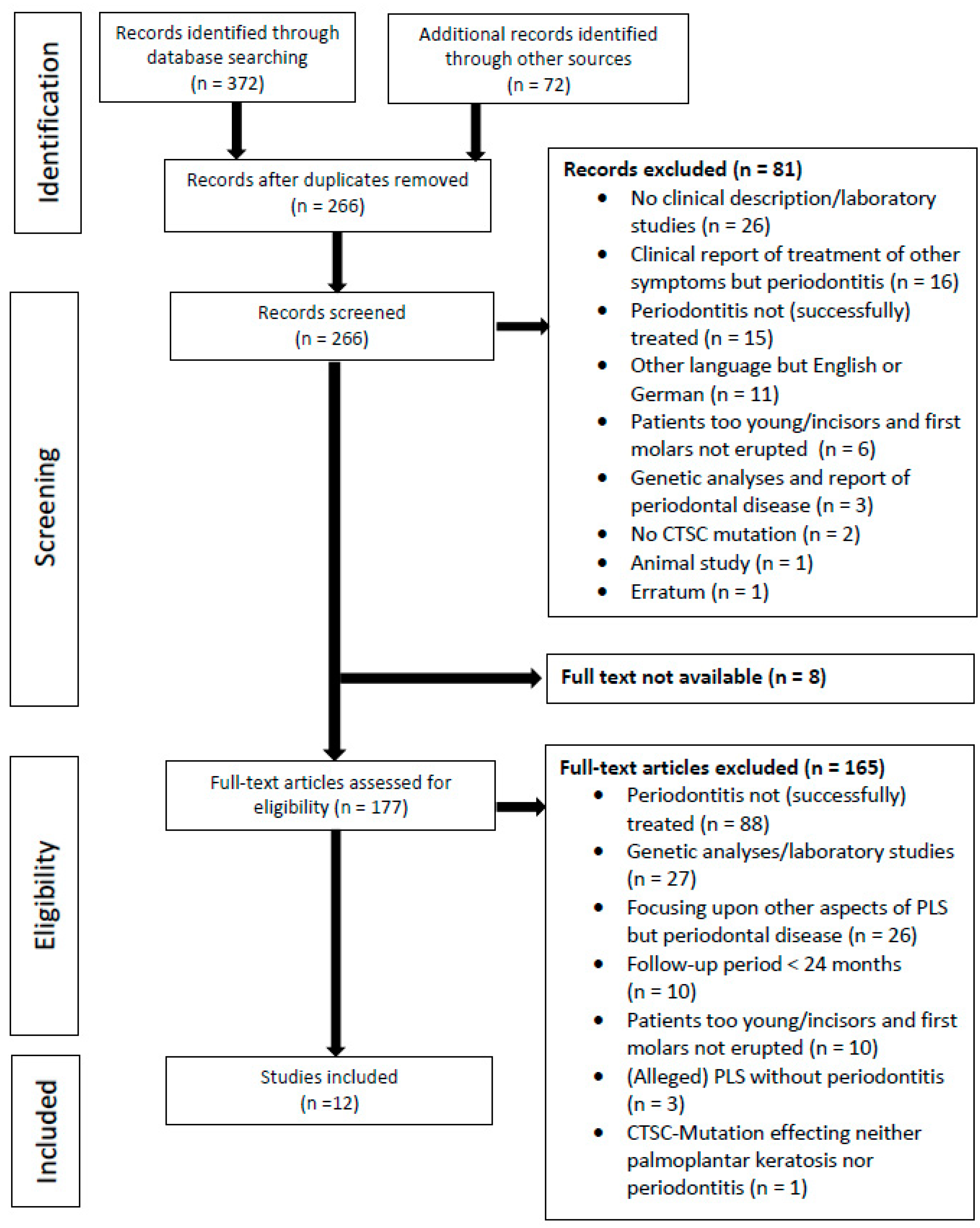
Figure 1. PRISMA flow diagram illustrating the literature search and selection process.
Healthcare 10 02505 g001
Table 1. Subjects’ characteristics including gender, age at the end of follow-up, periodontal therapy, and follow-up interval of included cases with successful periodontal treatment. Clinical diagnosis of Papillon–Lefèvre syndrome was based on severe periodontitis in early childhood and palmoplantar hyperkeratosis in all of the included individuals. Successful periodontal treatment was defined as the loss of less than four permanent teeth because of periodontitis and arrest of periodontitis with steady probing depths ≤ 5 mm for ≥24 months. Multiple reports existed from individuals with IDs 4, 5, 6, and 7.
ReferenceIDAge (y)GenderPeriodontal TherapyAdjunctive Antibiotic TreatmentAdditional Therapeutic InterventionsFollow-Up (y)
Preus et al. (1987) [2]19malesupra- and subgingival debridement, OHI,
0.2% chlorhexidine mouthrinse
extraction of all primary teeth
tetracycline 250 mg/d for 2–4 weeks intermittently in case of suspected gingival abnormality
and 0.25 mg/day continuously for 2 y
none5
Ishikawa et al. (1994) [28]29femaleextraction of all primary teethnonefull dentures5
Ishikawa et al. (1994) [28]312femalesupra- and subgingival debridement, OHI, flap surgery minocycline 200 mg/d for 2 weeks
erythromycin 1 g/d for 1 month
ofloxacin 300 mg/day for 1 month
none5
Rüdiger et al. (1999) [29]
Kressin et al. (1995) [30]
411femalesupra- and subgingival debridement, OHI
Meridol® mouth rinse
extraction of two mandibular incisors
0.06% chlorhexidine jet irrigation
amoxicillin 750 mg/d
plus clavulanic acid
and metronidazole 375 mg/d for 1 week
removable partial denture6
Wiebe et al. (2001) [31]
French et al. (1995) [32]
517malesupra- and subgingival debridement, OHI
0.12% chlorhexidine mouthrinse
extraction of all primary teeth
2 rounds of metronidazolefull dentures12
Nickles et al. (2011) [33]
Schacher et al. (2006) [34]
610malesupra- and subgingival debridement, OHI
extraction of severely affected primary teeth
1% chlorhexidine gel
0.12% chlorhexidine mouthrinse
amoxicillin 750 mg/d and metronidazole 750 mg/d for 1 weeknone5
Nickles et al.
(2013) [35]
Lux et al. (2005) [36]
Eickholz et al. (2001) [37]
724malesupra- and subgingival debridement, OHI
0.1% chlorhexidine mouthrinse
e 1% chlorhexidine gel
30% chlorhexidine chips in 4 teeth
amoxicillin 750 mg/d and metronidazole 500 mg/d for 1 weekorthodontics17
Nickles et al. (2013) [35]
817malesupra- and subgingival debridement, OHI
extraction of severely affected teeth
amoxicillin 750 mg/d plus clavulanic acid
and metronidazole 600 mg/d for 1 week
repeated in case of recurrence
orthodontics13
Umlauft et al. (2021) [38]918malesupra- and subgingival debridement, OHI
0.2% chlorhexidine mouthrinse
amoxicillin
and metronidazole
none15
ID, identification number; y, years; OHI, repeated oral hygiene instructions; et al., et al. teri; d, day(s).
Table 2. Microbiolocigal findings in patients with Papillon–Lefèvre syndrome. The presence of Aggregatibacter actinomycetemcomitans, Porphyromonas gingivalis, Prevotella intermedia, Fusobacterium nucleatum, Campylobacter rectus, and Eikenella corrodens before and/or after periodontal therapy was evaluated.
ReferenceIDMicrobiological Testing BaselineAntibioticsExtraction of All Primary TeethPresence of A. actinomycetemcomitans
after Therapy
Preus et al. (1987) [2]1not evaluatedTetracyclineyesnot evaluated
Ishikawa et al. (1994) [28]2A. actinomycetemcomitans
P. gingivalis
P. intermedia
F. nucleatum
C. rectus
E. corrodens
Noneyesnegative after extraction of all primary teeth
Ishikawa et al. (1994) [28]3A. actinomycetemcomitans
P. gingivalis
P. intermedia
F. nucleatum
C. rectus
E. corrodens
minocycline
erythromycin
ofloxacin
nopositive after minocycline
positive after erythromycin
negative after ofloxacine
Rüdiger et al. (1999) [29]
Kressin et al. (1995) [30]
4A. actinomycetemcomitans
P. gingivalis
amoxicillin and metronidazolenonegative after 3 y
Wiebe et al. (2001) [31],
French et al. (1995) [32]
5P. gingivalis
T. forsythia
Noneyesnot evaluated
Nickles et al. (2011) [33]
Schacher et al. (2006) [34]
6A. actinomycetemcomitans
P. gingivalis
T. forsythia
T. denticola
amoxicillin and metronidazolenonegative 1 and 5 y
Nickles et al.
(2013) [35]
Lux et al. (2005) [36]
Eickholz et al. (2001) [37]
7A. actinomycetemcomitans
P. gingivalis
P. intermedia
F. nucleatum
C. rectus
E. corrodens
amoxicillin and metronidazolenonegative after 7 months, 2, 3, and 4 y
positive after 7 y
negative after 8 and 12 y
Nickles et al.
(2013) [35]
8A. actinomycetemcomitans
amoxicillin/clavulanic acid and metronidazole initially and at recurrences after 3 and 8 ynopositive after 3 and 8 y negative after 9 y
Umlauft et al. (2021) [38]9not evaluatedamoxicillin and metronidazolenonot evaluated
ID, identification number; et al., et al.; y, years.
Publisher’s Note: MDPI stays neutral with regard to jurisdictional claims in published maps and institutional affiliations.