

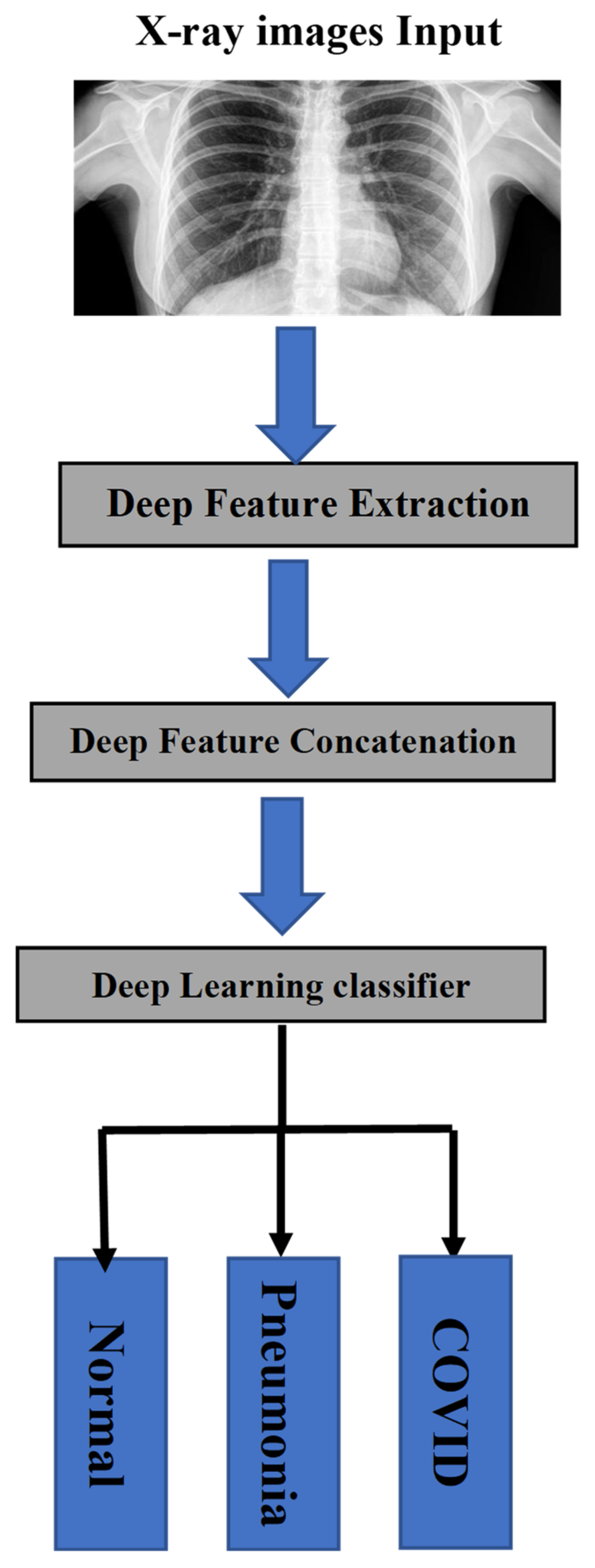
COVID-AleXception: A Deep Learning Model Based on a Deep Feature Concatenation Approach for the Detection of COVID-19 from Chest X-ray Images


Abstract
:1. Introduction
- ❖
- The collection of a medical X-ray image dataset includes three main classes (normal, pneumonia, and COVID-19) for the training and testing of the proposed system.
- ❖
- To perform a DFC technique in order to benefit from combining deep features which were extracted from AlexNet and Xception models.
- ❖
- We propose COVID-AlexCeption: a deep learning model with concatenation of AlexNet and Xception models to detect COVID-19 from X-ray images.
- ❖
- The performance of COVID-AlexCeption has been tested.
- ❖
- The comparison of COVID-AlexCeption to competitive methods in terms of different performance metrics has been performed, mainly in terms of f-measure, accuracy, precision, and recall.
2. State-of-the-Art Methods

| Ref | CNN Model | Data Sources | Accuracy (%) | Limitations |
|---|---|---|---|---|
| [15] | nCOVnet | Cohen et al. [25] | 97.97 | High execution time |
| [16] | ResNet50 | Radiography Database [26] | 99.17 | Imbalanced data |
| [17] | ResNet18 | CT scan images [27] | 99.60 | Overfitting issue |
| ResNet50 | 99.20 | |||
| ResNet101 | 99.30 | |||
| SqueezeNet | 99.50 | |||
| [18] | VGG19 +CNN | GitHub+ cancer X-ray and CT images [28] | 98.05 | Imbalanced data |
| ResNet152V2 | 95.31 | |||
| ResNet152V2 + GRU | 96.09 | |||
| ResNet152V2+ Bi-GRU | 93.36 | |||
| [19] | ResNet50 | Cohen [25] Kaggle [29] | 93.01 | Imbalanced data Overfitting issue |
| ResNet101 | 97.22 | |||
| [20] | VGG-16 | Khan et al. [22] Ozturk [20] | 79.58 | Overfitting issue |
| [21] | FocusCOVID | Kaggle-1 [30] Kaggle-2 [31] | 95.20 | Cannot provide the optimal accuracy. |
| [22] | CoroNet | Chest X-ray Images [32] | 89.6 | This method is slow. |
| [23] | CheXImageNet | Cohen [25] Kaggle [29] | 100 | Overfitting issue |
| [24] | ResNet50 | Kaggle chest X-ray [33] RSNA pneumonia [34] | 91.13 | Cannot provide the optimal accuracy. |
| MobileNet | 93.73 | |||
| Hybrid model | 94.43 |
3. Deep Learning Architectures
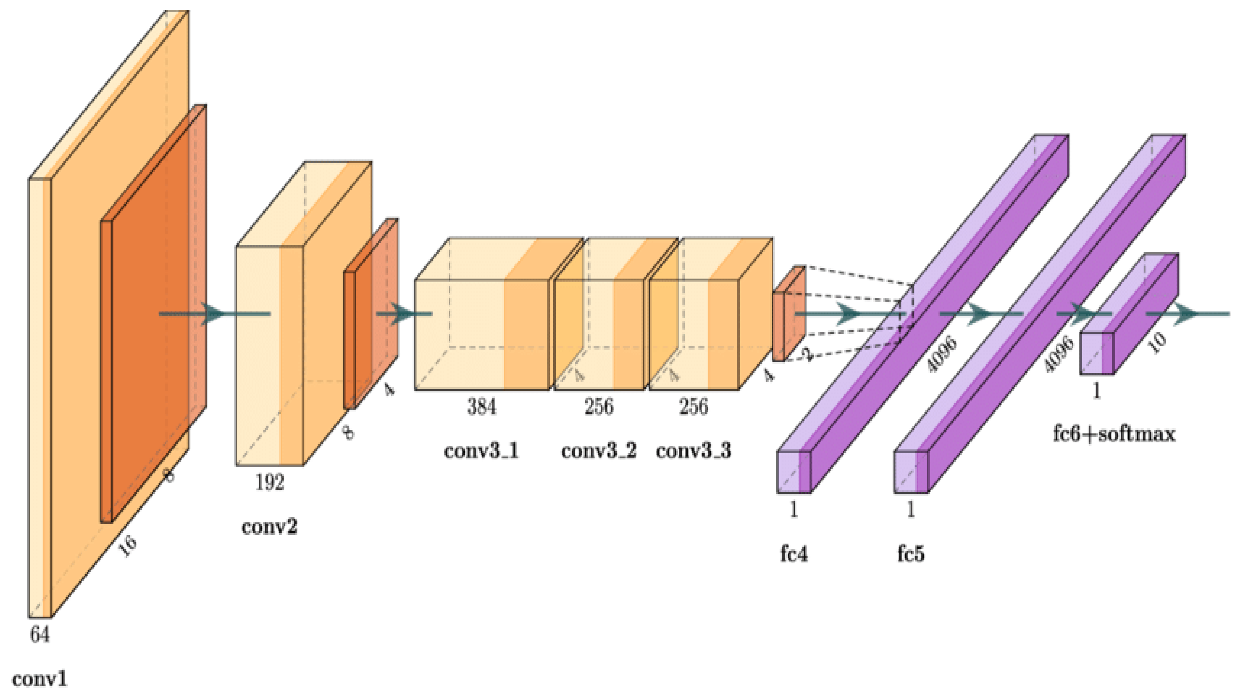
3.1. AlexNet Model Architecture
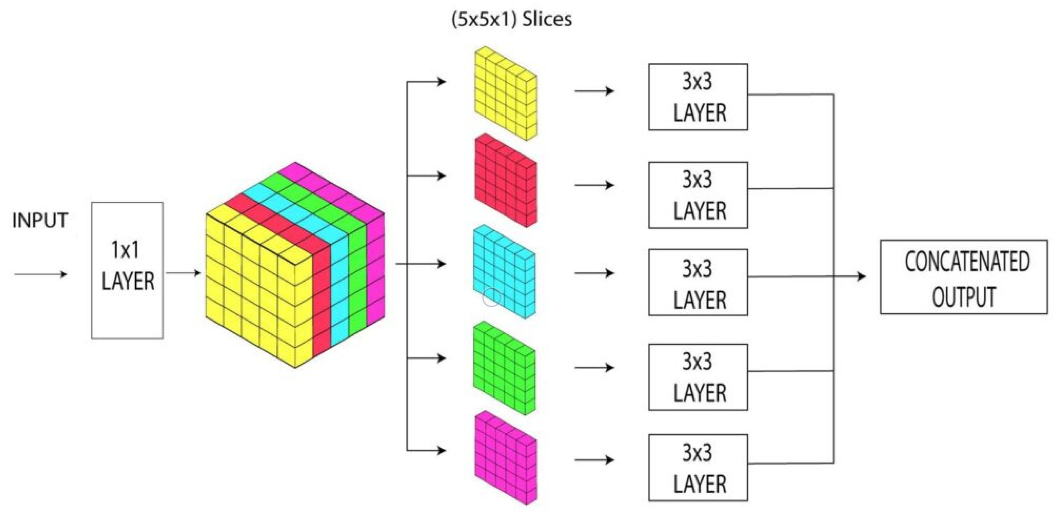
3.2. Xception Model Architecture
4. Materials and Methods
4.1. Dataset Description
4.2. Data Preparation and Preprocessing
- Image resizing: The images in this dataset have a wide range of dimensions. All of the images that were originally obtained were examined to see if they met the dataset’s minimum height and width requirements. All of the images in the dataset were scaled to this minimum dimension after it was determined. A minimum dimension as obtained in our work is 224 × 224. So, to fit to the input image size in AlexNet and Xception pretrained models, all images in the dataset were resized to the dimension of 224 × 224.
- Image normalization: In this work, contrast enhancement and image normalization methods were utilized to adjust pixel intensity values in order to provide a better-enhanced image in this study. Hidden information that occurs inside the low range is revealed by changing the pixel intensity. As a result, we normalized all image intensity values to a range of [–1, 1]. Given the fact that this method helps remove bias and achieve uniform distribution, it helps in accelerating the convergence of the model.
4.3. Data Augmentation
4.4. Deep Feature Concatenation (DFC)
5. Implementation and Testbed
- ❖
- Accuracy: The accuracy is the percentage of properly predicted images out of the total number of predictions [24]. The accuracy is calculated as:
- ❖
- Precision: The ratio of correctly predicted positive results (TP) to the total number of positive results (TP + FP) forecasted by the model is the accuracy metric. The vast number of FPs results in a lower precision [30]. The precision range is between 0 and 1 and is calculated as follows:
- ❖
- Recall: The recall is utilized to measure the right positive forecasts by calculating the ratio between the number of true positive results (TP) to the total number of samples (TP + FN) [30]. The recall is calculated using the following equation:
- ❖
- F1 score: The F1 score is one of the metrics used to measure and evaluate the model’s performance. The weighted harmonic between the accuracy and recall is used to compute the F1 score. Its definition is as follows:
5.1. Performance Analysis and Analysis
5.2. Comparative Analysis and Discussion
6. Conclusions
Author Contributions
Funding
Institutional Review Board Statement
Informed Consent Statement
Data Availability Statement
Conflicts of Interest
References
- Luz, E.; Silva, P.; Silva, R.; Silva, L.; Guimarães, J.; Miozzo, G.; Moreira, G.; Menotti, D. Towards an effective and efficient deep learning model for COVID-19 patterns detection in X-ray images. Res. Biomed. Eng. 2022, 38, 149–162. [Google Scholar] [CrossRef]
- Senan, E.M.; Alzahrani, A.; Alzahrani, M.Y.; Alsharif, N.; Aldhyani, T.H.H. Automated Diagnosis of Chest X-Ray for Early Detection of COVID-19 Disease. Comput. Math. Methods Med. 2021, 2021, 6919483. [Google Scholar] [CrossRef] [PubMed]
- Iwendi, C.; Huescas, C.G.Y.; Chakraborty, C.; Mohan, S. COVID-19 health analysis and prediction using machine learning algorithms for Mexico and Brazil patients. J. Exp. Theor. Artif. Intell. 2022, 34, 725–733. [Google Scholar] [CrossRef]
- Sitaula, C.; Shahi, T.B. Monkeypox Virus Detection Using Pre-Trained Deep Learning-based Approaches. J. Med. Syst. 2022, 46, 78. [Google Scholar] [CrossRef] [PubMed]
- Krishnaraj, C.; Chinmay, C.; Srikanth, P.; Shashikiran, U.; KVivekananda, B.; Niranjana, S. Clinical and Laboratory approach to Diagnose COVID-19 using Machine Learning. Interdiscip. Sci. Comput. Life Sci. 2022, 14, 452–470. [Google Scholar] [CrossRef]
- Wu, C.; Khishe, M.; Mohammadi, M.; Taher Karim, S.H.; Rashid, T.A. Evolving deep convolutional neutral network by hybrid sine–cosine and extreme learning machine for real-time COVID19 diagnosis from X-ray images. Soft Comput. 2021, 5, 9663–9676. [Google Scholar] [CrossRef] [PubMed]
- Nasiri, H.; Hasani, S. Automated detection of COVID-19 cases from chest X-ray images using deep neural network and XGBoost. Radiography 2022, 28, 732–738. [Google Scholar] [CrossRef] [PubMed]
- Chakraborty, C.; Kishor, A.; Rodrigues, J.J. Novel Enhanced-Grey Wolf Optimization hybrid machine learning technique for biomedical data computation. Comput. Electr. Eng. 2022, 99, 107778. [Google Scholar] [CrossRef]
- Hassan, H.; Ren, Z.; Zhou, C.; Khan, M.A.; Pan, Y.; Zhao, J.; Huang, B. Supervised and weakly supervised deep learning models for COVID-19 CT diagnosis: A systematic review. Comput. Methods Progr. Biomed. 2022, 218, 106731. [Google Scholar] [CrossRef]
- Rahman, A.; Chakraborty, C.; Anwar, A.; Karim, M.; Islam, M.; Kundu, D.; Rahman, Z.; Band, S.S. SDN-IoT Empowered Intelligent Framework for Industry 4.0 Applications during COVID-19 Pandemic. Clust. Comput. 2021, 25, 2351–2368. [Google Scholar] [CrossRef]
- Sujata, D.; Chinmay, C.; Sourav, K.G.; Subhendu, K.P.; Jaroslav, F. BIFM: Big-data Driven Intelligent Forecasting Model for COVID-19. IEEE Access 2021, 9, 97505–97517. [Google Scholar] [CrossRef]
- Muhammad, S.; Javeria, A.; Nadia, G.; Seifedine, K.; Chinmay, C. Quantum Machine Learning Architecture for COVID-19 Classification based on Synthetic Data Generation using Conditional Adversarial Neural Network (CGAN). Cogn. Comput. 2021, 14, 1677–1688. [Google Scholar] [CrossRef]
- Kumar, A.; Tripathi, A.R.; Satapathy, S.C.; Zhang, Y.D. SARS-Net: COVID-19 detection from chest x-rays by combining graph convolutional network and convolutional neural network. Pattern Recognit. 2022, 122, 108255. [Google Scholar] [CrossRef] [PubMed]
- Zouch, W.; Sagga, D.; Echtioui, A.; Khemakhem, R.; Ghorbel, M.; Mhiri, C.; Hamida, A.B. Detection of COVID-19 from CT and Chest X-ray Images Using Deep Learning Models. Ann. Biomed. Eng. 2022, 50, 825–835. [Google Scholar] [CrossRef]
- Panwar, H.; Gupta, P.K.; Siddiqui, M.K.; Morales-Menendez, R.; Singh, V. Application of deep learning for fast detection of COVID-19 in X-Rays using nCOVnet. Chaos Solitons Fractals 2020, 138, 109944. [Google Scholar] [CrossRef]
- Hossain, M.B.; Iqbal, S.H.S.; Islam, M.M.; Akhtar, M.N.; Sarker, I.H. Transfer learning with fine-tuned deep CNN ResNet50 model for classifying COVID-19 from chest X-ray images. Inform. Med. Unlocked 2022, 30, 100916. [Google Scholar] [CrossRef]
- Ahuja, S.; Panigrahi, B.K.; Dey, N.; Rajinikanth, V.; Gandhi, T.K. Deep transfer learning-based automated detection of COVID-19 from lung CT scan slices. Appl. Intell. 2020, 51, 571–585. [Google Scholar] [CrossRef] [PubMed]
- Ibrahim, D.M.; Elshennawy, N.M.; Sarhan, A.M. Deep-chest: Multi-classification deep learning model for diagnosing COVID-19, pneumonia, and lung cancer chest diseases. Comput. Biol. Med. 2021, 132, 104348. [Google Scholar] [CrossRef] [PubMed]
- Jain, G.; Mittal, D.; Thakur, D.; Mittal, M.K. A deep learning approach to detect COVID-19 coronavirus with X-ray images. Biocybern. Biomed. Eng. 2020, 40, 1391–1405. [Google Scholar] [CrossRef]
- Sitaula, C.; Hossain, M.B. Attention-based VGG-16 model for COVID-19 chest X-ray image classification. Appl. Intell. 2021, 51, 2850–2863. [Google Scholar] [CrossRef] [PubMed]
- Li, L.; Qin, L.; Xu, Z.; Yin, Y.; Wang, X.; Kong, B.; Bai, J.; Lu, Y.; Fang, Z.; Song, Q.; et al. Using Artificial Intelligence to Detect COVID-19 and Community-acquired Pneumonia Based on Pulmonary CT: Evaluation of the Diagnostic Accuracy. Radiology 2020, 296, E65–E71. [Google Scholar] [CrossRef] [PubMed]
- Khan, A.I.; Shah, J.L.; Bhat, M.M. CoroNet: A deep neural network for detection and diagnosis of COVID-19 from chest X-ray images. Comput. Methods Progr. Biomed. 2020, 196, 105581. [Google Scholar] [CrossRef]
- Shastri, S.; Kansal, I.; Kumar, S.; Singh, K.; Popli, R.; Mansotra, V. CheXImageNet: A novel architecture for accurate classification of COVID-19 with chest x-ray digital images using deep convolutional neural networks. Health Technol. 2022, 12, 193–204. [Google Scholar] [CrossRef]
- Nandi, R.; Mulimani, M. Detection of COVID-19 from X-rays using hybrid deep learning models. Res. Biomed. Eng. 2021, 37, 687–695. [Google Scholar] [CrossRef]
- Cohen, J.P.; Morrison, P.; Dao, L. COVID-19 image data collection. arXiv 2003, arXiv:2003.11597. [Google Scholar]
- Rahman, T.; Khandakar, A.; Qiblawey, Y.; Tahir, A.; Kiranyaz, S.; Kashem, S.B.A.; Islam, M.T.; Al Maadeed, S.; Zughaier, S.M.; Khan, M.S.; et al. Exploring the effect of image enhancement techniques on COVID-19 detection using chest X-ray images. Comput. Biol. Med. 2021, 132, 104319. [Google Scholar] [CrossRef] [PubMed]
- Zhao, J.; Zhang, Y.; He, X.; Xie, P. COVID-ct-dataset: A ct scan dataset about COVID-19. arXiv 2020, arXiv:2003.13865. [Google Scholar]
- Bhandary, A.; Prabhu, G.A.; Rajinikanth, V.; Thanaraj, K.P.; Satapathy, S.C.; Robbins, D.E.; Shasky, C.; Zhang, Y.D.; Tavares, J.M.R.; Raja, N.S.M. Deep-learning framework to detect lung abnormality—A study with chest X-ray and lung CT scan images. Pattern Recognit. Lett. 2020, 129, 271–278. [Google Scholar] [CrossRef]
- Mooney, P. Chest X-ray Images (Pneumonia). Available online: https://www.kaggle.com/datasets/paultimothymooney/chest-xray-pneumonia (accessed on 5 April 2022).
- Chowdhury, M.E.H.; Rahman, T.; Khandakar, A.; Mazhar, R.; Kadir, M.A.; Mahbub, Z.B.; Islam, K.R.; Khan, M.S.; Iqbal, A.; Emadi, N.A.; et al. Can ai help in screening viral and COVID- 19 pneumonia? IEEE Access 2020, 8, 132665–132676. [Google Scholar] [CrossRef]
- Asraf, A. COVID Dataset. 2020. Available online: https://www.kaggle.com/amanullahasraf/COVID19-pneumonia-normal-chest-xraypa-dataset (accessed on 2 January 2022).
- Chest X-ray Images (Pneumonia). Available online: https://www.kaggle.com/datasets/prashant268/chest-xray-COVID19-pneumonia (accessed on 1 April 2022).
- Kermany, D.S.; Zhang, K.; Goldbaum, M.H. Labeled optical coherence tomography (oct) and chest x-ray images for classification. Mendeley Data 2018, 2. [Google Scholar] [CrossRef]
- Rsna Pneumonia Detection Challenge. 2018. Available online: https://www.kaggle.com/c/rsna-pneumonia-detection-challenge/data (accessed on 25 June 2022).
- Alzubaidi, L.; Zhang, J.; Humaidi, A.J.; Al-Dujaili, A.; Duan, Y.; Al-Shamma, O.; Santamaría, J.; Fadhel, M.A.; Al-Amidie, M.; Farhan, L. Review of deep learning: Concepts, CNN architectures, challenges, applications, future directions. J. Big Data 2021, 8, 53. [Google Scholar] [CrossRef] [PubMed]
- Turkoglu, M. COVIDetectioNet: COVID-19 diagnosis system based on X-ray images using features selected from pre-learned deep features ensemble. Appl. Intell. 2021, 51, 1213–1226. [Google Scholar] [CrossRef] [PubMed]
- Ozturk, T.; Talo, M.; Yildirim, E.A.; Baloglu, U.B.; Yildirim, O. Automated detection of COVID-19 cases using deep neural networks with X-ray images. Comput. Biol. Med. 2020, 121, 103792. [Google Scholar] [CrossRef] [PubMed]


| Name of Class | Normal | Pneumonia | COVID-19 |
|---|---|---|---|
| No. of images before augmentation | 10,192 | 1345 | 3616 |
| No. of images after augmentation | 10,192 | 8087 | 8107 |
| Classes | Training (80%) | Validation (20%) | Total |
|---|---|---|---|
| Normal | 8154 | 2038 | 10,192 |
| Pneumonia | 6470 | 1617 | 8087 |
| COVID-19 | 6486 | 1621 | 8107 |
| Model | Accuracy (%) | Precision (%) | Recall (%) | F1-Score (%) | Training Time (s) | Testing Time (s) |
|---|---|---|---|---|---|---|
| AlexNet | 94.86 | 94.75 | 94.08 | 94.78 | 690 | 3.29 |
| Xception | 95.63 | 94.26 | 94.12 | 95.16 | 740 | 2.49 |
| COVID-AleXception | 98.68 | 99.11 | 98.77 | 98.46 | 938 | 4.23 |
| Model | Category | Accuracy (%) | Precision (%) | Recall (%) | F1-Score (%) |
|---|---|---|---|---|---|
| AlexNet | Normal | 96.25 | 95.25 | 95.55 | 95.36 |
| Pneumonia | 95.25 | 95.45 | 95.85 | 95.57 | |
| COVID-19 | 93.55 | 93.75 | 93.35 | 93.03 | |
| Xception | Normal | 97.15 | 97.52 | 97.25 | 97.55 |
| Pneumonia | 95.52 | 95.63 | 95.25 | 95.85 | |
| COVID-19 | 95.01 | 95.21 | 95.15 | 95.05 | |
| COVID-AleXception | Normal | 99.05 | 99.35 | 99.27 | 99.78 |
| Pneumonia | 98.75 | 99.85 | 99.48 | 99.05 | |
| COVID-19 | 98.55 | 98.45 | 98.72 | 98.75 |
| Ref | CNN Model | Data Sources | Accuracy (%) | Precision (%) | Recall (%) | F1-Score (%) |
|---|---|---|---|---|---|---|
| [15] | nCOVnet | Cohen et al. [25] | 97.97 | 97.61 | 97.78 | 97.67 |
| [16] | ResNet50 | Radiography Database [26] | 99.17 | 99.45 | 98.97 | 99.25 |
| [17] | ResNet18 | CT scan images [27] | 99.60 | 99.52 | 99.71 | 99.75 |
| ResNet50 | 99.20 | 99.05 | 99.35 | 99.25 | ||
| ResNet101 | 99.30 | 99.35 | 99.25 | 99.18 | ||
| SqueezeNet | 99.50 | 99.45 | 99.40 | 99.55 | ||
| [18] | VGG19 +CNN | GitHub+ cancer X-ray and CT images [28] | 98.05 | 97.95 | 97.87 | 97.90 |
| ResNet152V2 | 95.31 | 95.23 | 95.17 | 95.25 | ||
| ResNet152V2 + GRU | 96.09 | 95.85 | 95.95 | 95.80 | ||
| ResNet152V2+ Bi-GRU | 93.36 | 93.15 | 93.22 | 93.25 | ||
| [19] | ResNet50 | Cohen [25] Kaggle [29] | 93.01 | 92.90 | 92.95 | 92.85 |
| ResNet101 | 97.22 | 97.05 | 97.15 | 97.03 | ||
| [20] | VGG-16 | Khan et al. [22] Ozturk [20] | 79.58 | 79.58 | 85.43 | 87.49 |
| [21] | FocusCOVID | Kaggle-1 [30] Kaggle-2 [31] | 95.20 | 95.36 | 95.16 | 95.26 |
| [22] | CoroNet | Chest X-ray Images [32] | 89.6 | 89.45 | 89.36 | 89.50 |
| [23] | CheXImageNet | Cohen [25] Kaggle [29] | 100 | 100 | 100 | 100 |
| [24] | ResNet50 | Kaggle chest X-ray [33] RSNA pneumonia [34] | 91.13 | 92.96 | 92.85 | 92.73 |
| MobileNet | 93.73 | 93.66 | 93.55 | 93.48 | ||
| Hybrid model | 94.43 | 94.26 | 94.35 | 94.15 | ||
| COVID-AleXception | Xception | Ref: [25,26,30,31,32] | 94.86 | 94.75 | 94.08 | 94.78 |
| AlexNet | 95.63 | 94.26 | 94.12 | 95.16 | ||
| COVID-AleXception | 98.68 | 99.11 | 98.77 | 98.46 |
Publisher’s Note: MDPI stays neutral with regard to jurisdictional claims in published maps and institutional affiliations. |
© 2022 by the authors. Licensee MDPI, Basel, Switzerland. This article is an open access article distributed under the terms and conditions of the Creative Commons Attribution (CC BY) license (https://creativecommons.org/licenses/by/4.0/).
Share and Cite
Ayadi, M.; Ksibi, A.; Al-Rasheed, A.; Soufiene, B.O. COVID-AleXception: A Deep Learning Model Based on a Deep Feature Concatenation Approach for the Detection of COVID-19 from Chest X-ray Images. Healthcare 2022, 10, 2072. https://doi.org/10.3390/healthcare10102072
Ayadi M, Ksibi A, Al-Rasheed A, Soufiene BO. COVID-AleXception: A Deep Learning Model Based on a Deep Feature Concatenation Approach for the Detection of COVID-19 from Chest X-ray Images. Healthcare. 2022; 10(10):2072. https://doi.org/10.3390/healthcare10102072
Chicago/Turabian StyleAyadi, Manel, Amel Ksibi, Amal Al-Rasheed, and Ben Othman Soufiene. 2022. "COVID-AleXception: A Deep Learning Model Based on a Deep Feature Concatenation Approach for the Detection of COVID-19 from Chest X-ray Images" Healthcare 10, no. 10: 2072. https://doi.org/10.3390/healthcare10102072
APA StyleAyadi, M., Ksibi, A., Al-Rasheed, A., & Soufiene, B. O. (2022). COVID-AleXception: A Deep Learning Model Based on a Deep Feature Concatenation Approach for the Detection of COVID-19 from Chest X-ray Images. Healthcare, 10(10), 2072. https://doi.org/10.3390/healthcare10102072