Communication

Establishment of a Genetic Transformation and Gene Editing Method by Floral Dipping in Descurainia sophia

Lushan Botanical Garden, Jiangxi Province and Chinese Academy of Sciences, NO.9 Zhiqing Road, Jiujiang 332900, China
*
Author to whom correspondence should be addressed.
These authors contributed equally to this work.
Plants 2024, 13(20), 2833; https://doi.org/10.3390/plants13202833
Submission received: 8 September 2024 / Revised: 30 September 2024 / Accepted: 1 October 2024 / Published: 10 October 2024
(This article belongs to the Special Issue Advances and Applications of Genome Editing in Plants)

Abstract

:
Descurainia sophia L. Webb ex Prantl is used in traditional medicine globally. However, the lack of an efficient and reliable genetic transformation system has seriously limited the investigation of gene function and further utilization of D. sophia. In this study, a highly efficient, time-saving, and cost-effective Agrobacterium tumefaciens-mediated genetic transformation system has been developed in D. sophia. In this method, the transformation was accomplished by simply dipping developing D. sophia inflorescences for 45 s into an Agrobacterium suspension (OD600 = 0.6) containing 5% sucrose and 0.03% (v/v) Silwet L-77. Treated plants were allowed to set seeds which were then plated on a selective medium with hygromycin B (HygB) to screen transformants. Additionally, the CRISPR/Cas9 genomic editing system was validated by targeting phytoene desaturase (PDS) gene using this floral dip method, and mutant plants with the expected albino phenotype could be obtained in 2.5 months. This genetic transformation and targeted editing system will be a valuable tool for routine investigation of gene function and further exploitation in D. sophia.

1. Introduction

Descurainia sophia L. Webb ex Prantl, an annual dicot weed belonging to Brassicaceae family, has been used in traditional medicine globally because of its important pharmaceutical values [1,2]. D. sophia is a diploid, self-pollinating plant with a height ranging from 60 to 80 cm. The flowering process of D. sophia starts approximately 1.5 months after germination, and the period of seed to seed takes around 2 months in our laboratory conditions. It exhibits remarkable reproductive and growth potential in diverse environments, yielding about 4000–10,000 seeds per plant. To date, the genome sequence of D. sophia remains unreported. Various compounds have been isolated from this plant, such as phenolic compounds (flavonoids and coumarins), fatty acids, lipids, and steroids, with diverse biological effects, which include anti-inflammatory, anti-oxidative, antipyretic, anthelmintic activities, etc., [3,4,5,6,7]. Additionally, many studies have proven its effects on cancer, respiratory, gastrointestinal, and cardiac systems, without any significant adverse effects [2,3,8,9]. Moreover, D. sophia also has potential use as an oil crop because of its high productivity, high seed oil content up to 44.1%, and unique fatty acid composition, which mainly consists of linolenic acid and palmitic acid having biological effects within the organism [8,10]. D. sophia has successfully attracted the attention of researchers from different countries, as a valuable medicinal plant and oil crop. In the past decades, substantial progress of D. sophia has been experienced on chemical composition analysis and pharmacological mechanism [3,4,5,6,7,11,12,13]. However, the lack of an efficient transformation protocol for D. sophia severely hinders the fundamental research and further utilization.
Plant genetic transformation provides a range of opportunities for basic research and genetic modification of valuable traits in plants of economic importance. Agrobacterium-mediated transformation remains the most frequently used approach for the introduction of transgenes into Brassicaceae [14]. As a classic Agrobacterium-mediated transformation method, the floral dip method is simple, convenient, stable, and inexpensive. This method was first established in Arabidopsis thaliana and has been successfully utilized in other Brassicaceae plants, such as Brassica rapa, B. napus, and B. carinata [15,16,17]. Possibly, it could be utilized in D. sophia, which belongs to the Brassicaceae family. CRISPR/Cas9 is a powerful tool for precise genome editing and broadly used to modify metabolic pathways and develop plants with optimized secondary metabolite profiles in many medicine plants, such as Dioscorea zingiberensis, Salvia miltiorrhiza, and Symphytum officinale [18,19,20,21,22].
In this study, we established an Agrobacterium-mediated genetic transformation system for D. sophia via the floral dip method. Additionally, the targeted genome editing was also successfully accomplished in D. sophia, using the transformation method developed in this study. The genetic transformation and targeted editing system presented here will significantly advance basic research and utilization of D. sophia in the near future.

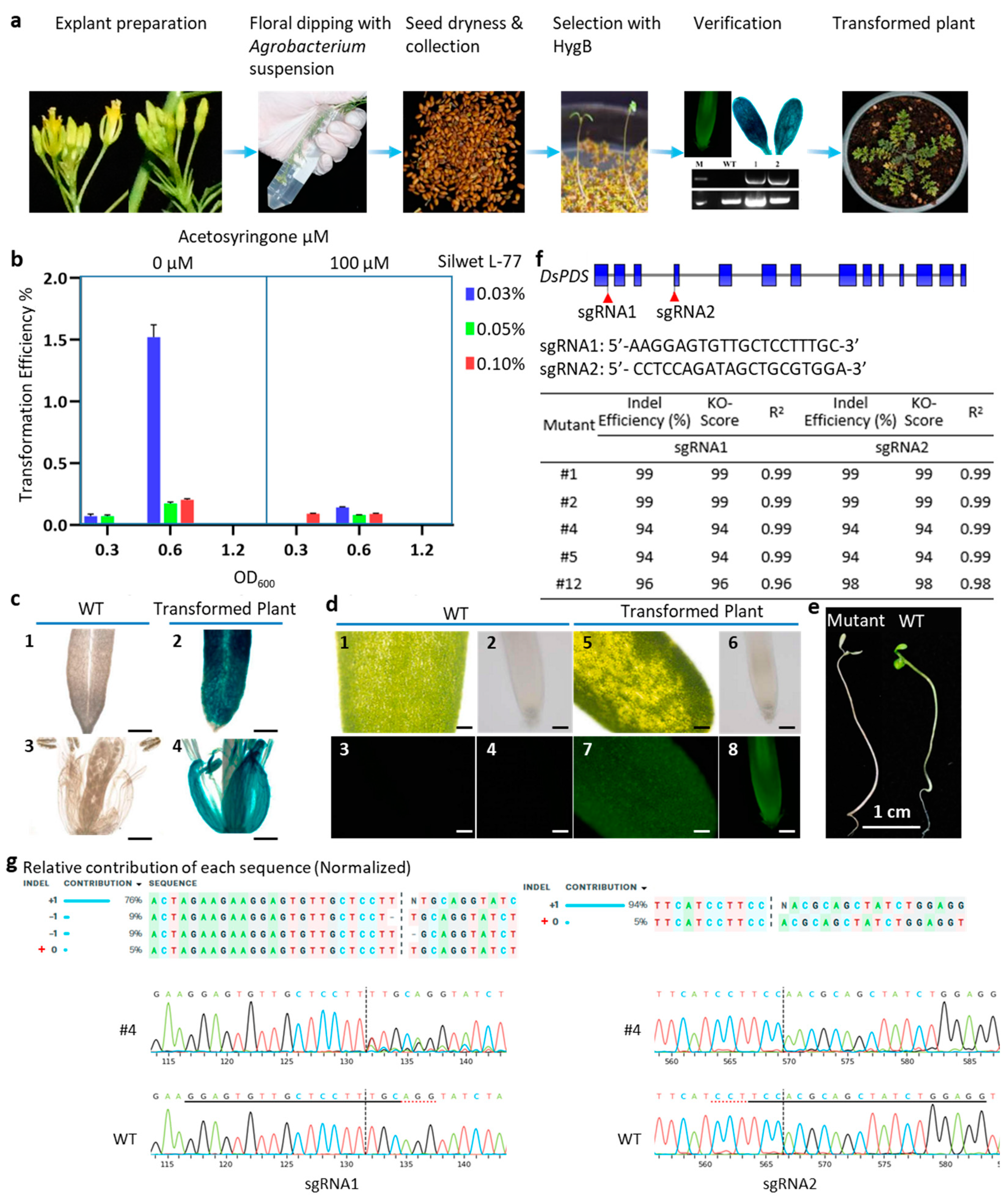
2. Results and Discussion

In this study, the transformation in D. sophia was accomplished by dipping its buds for 45 s into Agrobacterium suspension, and the collected seeds were screened by selectable makers or molecular verification (Figure 1a and Figure S1). However, in planta transformation efficiency is influenced by several factors, such as the density of bacteria evaluated by optical density at 600 nm (OD600), application and concentration of acetosyringone (AS), concentration of surfactants, etc. [23]. To develop an effective and efficient floral dip transformation method for D. sophia, A. tumefaciens strain GV3101 containing pCAMBIA1301 was used to optimize the workflow with 18 treatments consisting of different variables (Figure 1b, Table S1). The result showed that a high Agrobacterium density (OD600 = 1.2) in the inoculation culture caused D. sophia flower wilting and death at any concentration of AS and Silwet L-77. It was consistent with the reports that high Agrobacterium density usually damaged the plant cells and resulted in lower cell recovery that ultimately reduced the stable transformation efficiency in Pinus tabuliformis, Camellia sinensis, spring bread wheat, etc. [23,24,25,26]. In addition, the transformation efficiency of OD600 = 0.6 was higher than that of OD600 = 0.3, illustrating that the optimal Agrobacterium density was OD600 of 0.6 (Figure 1b, Table S1).
Acetosyringone is usually employed to increase transformation efficiency by inducing vir genes expression to promote transfer and integration of Agrobacterium T-DNA into plant genomes [27,28,29]. Here, we evaluated the effect of AS at two concentrations (0 and 100 μM) on D. sophia transformation efficiency, showing that the inoculation solution without addition of AS resulted in a higher transformation rate than that of the culture with 100 μM AS (Figure 1b, Table S1). It was notable that with 100 μM AS application, the transformation efficiency dramatically reduced from 1.521% to 0.143% at the condition of OD600 = 0.6 and 0.03% Silwet L-77 (Figure 1b, Table S1). However, it was reported that in Tamarix hispida, 120 μM AS is the most suitable concentration for genetic transformation, as this treatment could significantly induce the expression of all vir genes in A. tumefaciens strain EHA105 with higher expression level except virE2, compared with other AS concentrations (0, 100, 150, 200 μM) [29]. It suggested that the optimal concentration of AS for Agrobacterium-mediated transformation could be affected by many factors, such as explant species and type, Agrobacterium strain. It may be worth confirming whether low concentration (<100 μM) of AS in A. tumefaciens GV3101 inoculation solution could improve D. sophia transformation efficiency in the future. The result in this study illustrated that no AS application in the A. tumefaciens inoculation solution may produce more transgenic seedlings.
Silwet L-77 is a low toxic surfactant that can reduce surface tension, which can improve the penetration of Agrobacterium to affect the transformation efficiency [15,30]. Many studies have found that appropriate dosage of Silwet L-77 can significantly increase the transformation rate in plant, such as Linum usitatissimum, Cosmos sulphureus Cav, Geng/Japonica and Xian/Indica rice [31,32,33]. To detect the effect of Silwet L-77 in D. sophia transformation, three different concentrations (0.03%, 0.05%, and 0.10% v/v) were assessed in this study. The result illustrated that the transformation efficiency (1.521%) with 0.03% Silwet L-77 was highest with OD600 = 0.6 and without AS (Figure 1b, Table S1). However, the efficiency dramatically reduced to 0.174% and 0.204% with the Silwet L-77concentration of 0.05% and 0.10%, separately (Figure 1b, Table S1). Thus, 0.03% Silwet L-77 was most effective in the floral dipping of D. sophia, and excessive Silwet L-77 could cause deleterious effects for D. sophia (Figure 1b, Table S1).
Although the floral dip method is a common technique of plant transformation, it has been mainly developed for some species belonging to the Brassicaceae family. Many factors more than those mentioned above have been suggested to impact the efficiency and application of the floral dip method in other plant species, such as physical barriers associated with flower morphology, necrotic reaction to the presence of Agrobacterium causing abortions in the flowers, low seed set, and big size of the plant and/or flower structures [34].
In summary, the floral dip method for D. sophia was most efficient when the flower buds were dipped into Agrobacterium suspension with OD600 = 0.6 and containing 0.03% Silwet L-77 for 45 s.
Various genetically encodable reporters have been widely used in transformation studies to monitor gene expression, protein localization, hormonal signaling, and other cellular activities [35,36,37]. In this study, Green fluorescent protein (GFP) and β-Glucuronidase (GUS) were utilized to confirm whether GFP and GUS could be acted as reporters in D. sophia. The HygB-resistant seedlings transformed with pCAMBIA1301 were verified by GUS staining assay. The result showed that strong GUS signals were observed in transgenic plants, using the WT as the control (Figure 1c). In addition, HygB-resistant seedlings infected with Agrobacteria carrying pCAMBIA1301-GFP were constructed to detect GFP in this study. The fluorescence microscope analysis demonstrated that transgenic seedlings showed strong green fluorescence, compared with WT (Figure 1d). In this study, it was validated that both GFP and GUS worked well in D. sophia and could be applied as reporters of D. sophia in the future studies.
CRISPR/Cas9 system-mediated gene editing is a powerful tool for gene function analysis and generation of elite germplasm. Additionally, phytoene desaturase (PDS) is involved in the carotenoid biosynthesis pathway, and the loss of function mutant of PDS genes shows obvious albino phenotype, prompting it to be widely used as an indicator for genome editing [38,39,40,41]. To detect whether the CRISPR/Cas9 system works well in D. sophia, the binary vector pHSE401-sgDsPDS carrying two sgRNAs (sg1 and sg2) targeting DsPDS gene was transformed into D. sophia by the floral dip method developed in this study. After antibiotic selection, a total of 36 HygB-resistant seedlings were obtained, and five of them were albino (Figure 1e).
Aiming to detect mutations generated by CRISPR in D. sophia, partial sequences of DsPDS gene, covering two sgRNA sites, were amplified from genomic DNA of albino seedlings. Sequencing results were further analyzed by ICE (ice.synthego.com (accessed on 7 September 2024)) to quantify the efficiency of CRISPR/Cas9 gene editing. Analysis of ICE showed that the editing efficiencies by CRISPR/Cas9 were 94–99% in both sgRNA1 and sgRNA2 of DsPDS, mainly contributed by the insertion event (Figure 1f and Figure S2). This insertion was mostly observed as a single-nucleotide insertion at each 3-bp upstream of the two PAMs in albino seedlings (T or A at sgRNA1 and T at sgRNA2), compared with WT (Figure 1g and Figure S2). For example, the mutant #4 showed 94 knockout score with indel of 94% in both sgRNA1 and sgRNA2 sites (Figure 1f,g). In sgRNA1 site, there were very clear deletions (18%) and insertions (76%) with a p value of <0.001 (Figure 1g). Whereas, only insertion (94%) was observed in sgRNA2 site with a p value of <0.001 (Figure 1g). In addition, substitutions were not detected in the genotyped plants, which might be caused by the low number of mutants used for sequencing. Finally, the targeted genome editing mediated by CRISPR/Cas9 system was successfully established in D. sophia.

3. Materials and Methods

3.1. Plant Materials and Growth Conditions

D. sophia used in this study was kept in our laboratory. The seeds were surface-sterilized with 70% ethanol for 30 s and 10% NaClO for 10 min, then rinsed three times in sterile water and dried on sterilized filter paper. The treated seeds were sown on half-strength Murashige and Skoog (1/2 MS) plates containing 0.8% agar and 2% sucrose and then kept in the artificial climate chamber (22 °C, under a 16 h day/8 h night photoperiod) for germinating. After one week, the young seedlings were transplanted to 15 cm diameter plastic pots with a 3:1 mixture of sterilized soil and vermiculite, at 28 °C, light/dark, 16/8 h with light intensity of 15,000 Lux in greenhouse conditions.
In about 30 days after sowing, D. sophia started to bolt and were ready for floral dipping (Figure 1a). The bolting plants carried plenty of young unopened flower buds and only a few open flowers. Notably, the buds for transformation should be at a relatively early stage of inflorescence development, when the oldest flower buds still have their petals mostly enclosed by sepals. These open flowers were excised before floral dipping. In this study, each individual plant of D. sophia represented a repeat in each treatment.

3.2. Vector Construction

A total of three vectors were used in this study. The T-DNA region of pCAMBIA1301 (Abcam, Cambridge, UK, Cat# ab275753) vector contains the Cauliflower Mosaic Virus 35S (CaMV 35S) promoter-driven hygromycin phosphotransferase II (HptII) gene, and the GUS gene as plant selection and reporter markers, respectively (Figure S3). In this vector, the GUS gene has a 5′ extension with a catalase intron to ensure expression in plants but not bacteria (Figure S3).
The pCAMBIA1301-GFP vector was constructed by replacing the GUS gene of pCAMBIA1301 with the GFP gene (Figure S3). In brief, the sequence of GFP was amplified with Nco-LP (5′-acgggggactcttgaccatggATGAGTAAAGGAGAAGAACTTTTCACTG-3′) Pml-RP (5′-gtcacctgtaattcacacgtgCTATTTGTATAGTTCATCCATGCCATG-3′) and inserted in the backbone of pCAMBIA1301 cut with NcoI and PmlI using a recombinant cloning kit (Vazyme, Wuhan, China, Cat#c-112).
The pHSE401 vector contains gRNA, Cas9, and HptII, a selectable marker [42]. Cas9 is optimized by maize codon under the control of CaMV 35S promoter, and the gRNA is controlled by Arabidopsis U6 promoter. The pHSE401-sgDsPDS plasmid was constructed based on pHSE401, which harbored two expression cassettes. The sgRNAs of DsPDS were designed using the online tool CRISPR-P (http://crispr.hzau.edu.cn/CRISPR2/ (accessed on 7 September 2024)) (listed in Figure 1f), and synthesized as oligos (Sangon, Shanghai, China), annealed, and cloned into pHSE401 vector.

3.3. A. tumefaciens-Mediated Delivery System of D. sophia

The GV3101 strain utilized in this study harbors the C58 chromosomal backbone containing rifampicin resistance and the Ti plasmid pMP90 (pTiC58DT-DNA) harboring the gentamicin resistance. The reporter and gene-editing vectors were transferred into A. tumefaciens strain GV3101 (pMP90) by the freeze/thaw method (keeping the mixture of vectors and Agrobacterium competent cells on ice for 30 min, liquid nitrogen for 5 min, 37 °C water bath for 5 min, back on ice for 5 min, separately). Colonies were selected with LB solid medium containing antibiotics (50 mg/L of rifampicin/Rif, 50 mg/L of kanamycin/Kan), and these positive colonies were dispersed into 50 mL LB liquid medium with 50 mg/L of Rif and 50 mg/L of Kan and cultured overnight (28 °C, 220 RPM). The overnight culture was pelleted by centrifuge and resuspended in 5% sucrose solution to a certain OD600 value. The transformation efficiency is influenced by several factors, such as the density of bacteria, concentration of AS and surfactants, etc. To establish the floral dip transformation method in D. sophia, we utilized three different OD600 of Agrobacterium culture, i.e., 0.3, 0.6, and 1.2. The suspension was added with varied concentrations of Silwet L-77 (0.03%, 0.05%, and 0.1% v/v) and AS (0 and 100 μM). A total of 18 treatments had been applied, and three plants were used in each treatment, each plant being a repeat. AS was added to Agrobacterium cell suspension 2 h before the floral dip, while the Silwet L-77 was added just prior to dipping. The D. sophia flower buds were dipped in Agrobacterium cell suspension for 45 s with a 50 mL falcon tube, and the dipped plants were wrapped with a plastic cover to maintain dark and high humidity for 12 h. Then, the treated plants were sent back to the normal growth conditions until the seeds were harvested. Collected seeds were surface-sterilized as mentioned above and germinated on 1/2 MS medium with 50 mg/L of HygB (Thermo-Fisher Scientific, Lenexa, USA, Cat#10687010) at 28 °C in the dark conditions for 5–7 days, to screen out the HygB-resistant seedlings. Then, these seedlings were transferred to wet soil and moved to the greenhouse at normal growth conditions for further verification.

3.4. GFP Detection and GUS Staining

The leaf and root of HygB-resistant seedlings transferred with pCAMBIA1301-GFP were applied to detect the fluorescence GFP gene, with WT as a control. The samples were visualized and imaged using an ultraviolet (UV) transilluminator (Olympus, Tokyo, Japan, BX53). HygB-resistant seedlings transferred with pCAMBIA1301 were stained according to the preparation protocol of GUS staining kit (Biosharp, Hefei, China, Cat#BL622A), with WT as a control. These samples were photographed under a microscope.

3.5. Evaluation of the Editing Efficiency in D. sophia

Genomic DNA was extracted from plant tissues with the CTAB method. To determine gene-editing type and efficiency, T1 transgenic plants transferred with pHSE401-sgDsPDS were analyzed by PCR amplification using the primers of PDS-F (5′-ATGGTTGTGTTTGGGAATG-3′) and PDS-R (5′-ATCATTGATCCCAAGTTCTC-3′), with WT as a control. The PCR products were separated by 1.0% agarose gel electrophoresis and sequenced (Sangon, Shanghai, China). The data obtained from Sanger sequencing were analyzed to determine the indel percentage and knockout score in genetically edited D. sophia through online software ICE (ice.synthego.com).

3.6. Statistical Analysis

Transformation efficiency = number of positive transgenic seedlings/total number of seeds × 100%. The data in Figure 1b are means ± standard deviations (SD), and the SDs were derived from three biological replicates.

4. Conclusions

In conclusion, a simple and efficient Agrobacterium-mediated genetic transformation system of medicinal and oilseed plant D. sophia was established via floral dip method. This method was accomplished by dipping the flower buds into Agrobacterium suspension with OD600 = 0.6 and containing 0.03% Silwet L-77 for 45 s. Moreover, the DsPDS gene was successfully edited by a CRISPR/Cas9 system that was developed in this study by Agrobacterium-mediated floral dip method. This genetic transformation and targeted editing system will be a valuable tool for routine investigation of gene function and further exploitation in D. sophia.

Supplementary Materials

The following supporting information can be downloaded at: https://www.mdpi.com/article/10.3390/plants13202833/s1, Figure S1: Preliminary screening of T1 D. sophia plants on 1/2 MS media containing 50 mg/L HygB; Figure S2: Sequencing results of representative DsPDS knockout albino seedlings using CRISPR/Cas9 system, analyzed by ICE; Figure S3: The structure of the T-DNA region of pCAMBIA1301 and pCAMBIA1301-GFP. Table S1: Transformation efficiencies for D. sophia under different OD600 values of Agrobacterium, As concentrations and Silwet L-77 concentrations.

Author Contributions

Conceptualization, Y.L.; methodology, T.J. and H.Y.; validation, D.Z. and S.Z.; formal analysis, T.J. and H.Y.; writing—original draft preparation, T.J., H.Y., and Y.L.; writing—review and editing, J.W., T.Z., D.K., and M.H.; funding acquisition, Y.L. All authors have read and agreed to the published version of the manuscript.

Funding

This work is supported by the Key Program of the Chinese Academy of Sciences (KJZD-SW-L13), the Talents Program of Jiangxi Province (jxsq2020101020), and the Key Research Projects of Jiangxi Province (20223BBG71003).

Data Availability Statement

The genomic sequence of the DsPDS genes have been deposited in the NCBI GenBank database with the accession number PP593058.

Conflicts of Interest

The authors declare no conflicts of interest.

References

  1. Khan, M.; Wang, N. Descurainia sophia (L.): A weed with multiple medicinal uses. Punjab Univ. J. Zool. 2012, 27, 45–51. [Google Scholar]
  2. Nimrouzi, M.; Zarshenas, M.M. Phytochemical and pharmacological aspects of Descurainia sophia Webb ex Prantl: Modern and traditional applications. Avicenna J. Phytomed. 2016, 6, 266–272. [Google Scholar] [PubMed]
  3. Hsieh, P.C.; Kuo, C.Y.; Lee, Y.H.; Wu, Y.K.; Yang, M.C.; Tzeng, I.S.; Lan, C.C. Therapeutic effects and mechanisms of actions of Descurainia sophia. Int. J. Med. Sci. 2020, 17, 2163–2170. [Google Scholar] [CrossRef]
  4. HadiNezhad, M.; Rowland, O.; Hosseinian, F. The Fatty Acid Profile and Phenolic Composition of Descurainia sophia Seeds Extracted by Supercritical CO2. J. Am. Oil Chem. Soc. 2015, 92, 1379–1390. [Google Scholar] [CrossRef]
  5. Tavakoli, R.; Salarian, R. Steroids and Phenolic Compounds from Descurainia sophia. Chem. Nat. Compd. 2017, 53, 192–193. [Google Scholar] [CrossRef]
  6. Mohamed, N.H.; Mahrous, A.E. Chemical Constituents of Descurainia sophia L. and its Biological Activity. Rec. Nat. Prod. 2009, 3, 58. [Google Scholar]
  7. Bekker, N.P.; Ul’chenko, N.T.; Glushenkova, A.I. Lipids from Descurainia Sophia Seeds. Chem. Nat. Compd. 2005, 41, 346–347. [Google Scholar] [CrossRef]
  8. Gong, J.H.; Zhang, Y.L.; He, J.L.; Zheng, X.K.; Feng, W.S.; Wang, X.L.; Kuang, H.X.; Li, C.G.; Cao, Y.G. Extractions of oil from Descurainia sophia seed using supercritical CO2, chemical compositions by GC-MS and evaluation of the anti-tussive, expectorant and anti-asthmatic activities. Molecules 2015, 20, 13296–13312. [Google Scholar] [CrossRef]
  9. Park, J.S.; Lim, C.J.; Bang, O.S.; Kim, N.S. Ethanolic extract of Descurainia sophia seeds sensitizes A549 human lung cancer cells to TRAIL cytotoxicity by upregulating death receptors. BMC Complement. Altern. Med. 2016, 16, 115. [Google Scholar] [CrossRef]
  10. Dastres, E.; Jahangiri, E.; Edalat, M.; Zamani, A.; Amiri, M.; Pourghasemi, H.R. Habitat suitability modeling of Descurainia sophia medicinal plant using three bivariate models. Environ. Monit. Assess. 2023, 195, 392. [Google Scholar] [CrossRef]
  11. Feng, W.S.; Li, C.G.; Zheng, X.K.; Li, L.L.; Chen, W.J.; Zhang, Y.L.; Cao, Y.G.; Gong, J.H.; Kuang, H.X. Three new sulphur glycosides from the seeds of Descurainia sophia. Nat. Prod. Res. 2016, 30, 1675–1681. [Google Scholar] [CrossRef]
  12. Zeng, M.; Zhang, L.; Li, M.; Zhang, B.; Feng, W.; Kuang, H.-X.; Zheng, X. Mechanism of the diuretic activity of Descurainia sophia seed. Bangladesh J. Pharmacol. 2018, 13, 157–2018. [Google Scholar] [CrossRef]
  13. Ke, Y.; Wang, M.; Li, Y.; Shan, Z.; Mi, W.; Yuan, P.; Feng, W.; Zheng, X. Oligosaccharides composition of Descurainiae sophia exerts anti-heart failure by improving heart function and water-liquid metabolism in rats with heart failure. Biomed. Pharmacother. 2020, 129, 110487. [Google Scholar] [CrossRef]
  14. Sparrow, P.A.C.; Goldsack, C.M.P.; Østergaard, L. Transformation Technology in the Brassicaceae. In Genetics and Genomics of the Brassicaceae; Schmidt, R., Bancroft, I., Eds.; Springer: New York, NY, USA, 2011; pp. 505–525. [Google Scholar]
  15. Clough, S.J.; Bent, A.F. Floral dip: A simplified method for Agrobacterium-mediated transformation of Arabidopsis thaliana. Plant J. 1998, 16, 735–743. [Google Scholar] [CrossRef]
  16. Hu, D.; Bent, A.F.; Hou, X.; Li, Y. Agrobacterium-mediated vacuum infiltration and floral dip transformation of rapid-cycling Brassica rapa. BMC Plant Biol. 2019, 19, 246. [Google Scholar] [CrossRef]
  17. Verma, S.S.; Chinnusamy, V.; Bansa, K.C. A Simplified Floral Dip Method for Transformation of Brassica napus and B. carinata. J. Plant Biochem. Biotechnol. 2008, 17, 197–200. [Google Scholar] [CrossRef]
  18. Mishra, R.; Agarwal, P.; Mohanty, A. Applications of Genome Editing Techniques for the Improvement of Medicinal Plants. In Phytochemical Genomics: Plant Metabolomics and Medicinal Plant Genomics; Swamy, M.K., Kumar, A., Eds.; Springer Nature: Singapore, 2022; pp. 545–569. [Google Scholar]
  19. Arshid Shabir, P. Chapter 9—CRISPR/Cas9-mediated genome editing in medicinal and aromatic plants: Developments and applications. In Medicinal and Aromatic Plants; Aftab, T., Hakeem, K.R., Eds.; Academic Press: Cambridge, MA, USA, 2021; pp. 209–221. [Google Scholar]
  20. Feng, S.; Song, W.; Fu, R.; Zhang, H.; Xu, A.; Li, J. Application of the CRISPR/Cas9 system in Dioscorea zingiberensis. Plant Cell Tiss. Org. Cult. 2018, 135, 133–141. [Google Scholar] [CrossRef]
  21. Zhou, Z.; Tan, H.; Li, Q.; Chen, J.; Gao, S.; Wang, Y.; Chen, W.; Zhang, L. CRISPR/Cas9-mediated efficient targeted mutagenesis of RAS in Salvia miltiorrhiza. Phytochemistry 2018, 148, 63–70. [Google Scholar] [CrossRef]
  22. Zakaria, M.M.; Schemmerling, B.; Ober, D. CRISPR/Cas9-Mediated Genome Editing in Comfrey (Symphytum officinale) Hairy Roots Results in the Complete Eradication of Pyrrolizidine Alkaloids. Molecules 2021, 26, 1498. [Google Scholar] [CrossRef]
  23. Ziemienowicz, A. Agrobacterium-mediated plant transformation: Factors, applications and recent advances. Biocatal. Agric. Biotech. 2014, 3, 95–102. [Google Scholar] [CrossRef]
  24. Liu, S.; Ma, J.; Liu, H.; Guo, Y.; Li, W.; Niu, S. An efficient system for Agrobacterium-mediated transient transformation in Pinus tabuliformis. Plant Methods 2020, 16, 52. [Google Scholar] [CrossRef]
  25. Lv, Q.; Chen, C.; Xu, Y.; Hu, S.; Wang, L.; Sun, K.; Chen, X.; Li, X. Optimization of Agrobacterium tumefaciens-Mediated Transformation Systems in Tea Plant (Camellia sinensis). Hortic. Plant J. 2017, 3, 105–109. [Google Scholar] [CrossRef]
  26. Kumar, R.; Mamrutha, H.M.; Kaur, A.; Venkatesh, K.; Sharma, D.; Singh, G.P. Optimization of Agrobacterium-mediated transformation in spring bread wheat using mature and immature embryos. Mol. Biol. Rep. 2019, 46, 1845–1853. [Google Scholar] [CrossRef]
  27. Sheikholeslam, S.N.; Weeks, D.P. Acetosyringone promotes high efficiency transformation of Arabidopsis thaliana explants by Agrobacterium tumefaciens. Plant Mol. Biol. 1987, 8, 291–298. [Google Scholar] [CrossRef]
  28. Godwin, I.; Todd, G.; Ford-Lloyd, B.; Newbury, H.J. The effects of acetosyringone and pH on Agrobacterium-mediated transformation vary according to plant species. Plant Cell Rep. 1991, 9, 671–675. [Google Scholar] [CrossRef] [PubMed]
  29. Zhao, H.; Jia, Y.; Cao, Y.; Wang, Y. Improving T-DNA Transfer to Tamarix hispida by Adding Chemical Compounds During Agrobacterium tumefaciens Culture. Front. Plant Sci. 2020, 11, 501358. [Google Scholar] [CrossRef]
  30. Desfeux, C.; Clough, S.J.; Bent, A.F. Female reproductive tissues are the primary target of Agrobacterium-mediated transformation by the Arabidopsis floral-dip method. Plant Physiol. 2000, 123, 895–904. [Google Scholar] [CrossRef]
  31. Bastaki, N.K.; Cullis, C.A. Floral-dip transformation of flax (Linum usitatissimum) to generate transgenic progenies with a high transformation rate. J. Vis. Exp. 2014, 94, e52189. [Google Scholar]
  32. Purwantoro, A.; Irsyadi, M.B.; Sawitri, W.D.; Fatumi, N.C.; Fajrina, S.N. Efficient floral dip transformation method using Agrobacterium tumefaciens on Cosmos sulphureus Cav. Saudi J. Biol. Sci. 2023, 30, 103702. [Google Scholar] [CrossRef]
  33. Jain, N.; Khurana, P.; Khurana, J.P. A rapid and efficient protocol for genotype-independent, Agrobacterium-mediated transformation of indica and japonica rice using mature seed-derived embryogenic calli. Plant Cell Tiss. Org. Cult. 2022, 151, 59–73. [Google Scholar] [CrossRef]
  34. Bélanger, J.G.; Copley, T.R.; Hoyos-Villegas, V.; Charron, J.-B.; O’Donoughue, L. A comprehensive review of in planta stable transformation strategies. Plant Methods 2024, 20, 79. [Google Scholar] [CrossRef]
  35. Ziemienowicz, A. Plant selectable markers and reporter genes. Acta Physiol. Plant. 2001, 23, 363–374. [Google Scholar] [CrossRef]
  36. Kain, S.R.; Adams, M.; Kondepudi, A.; Yang, T.T.; Ward, W.W.; Kitts, P. Green fluorescent protein as a reporter of gene expression and protein localization. BioTechniques 1995, 19, 650–655. [Google Scholar] [PubMed]
  37. Jefferson, R.A.; Kavanagh, T.A.; Bevan, M.W. GUS fusions: Beta-glucuronidase as a sensitive and versatile gene fusion marker in higher plants. EMBO J. 1987, 6, 3901–3907. [Google Scholar] [CrossRef] [PubMed]
  38. Qin, G.; Gu, H.; Ma, L.; Peng, Y.; Deng, X.W.; Chen, Z.; Qu, L.J. Disruption of phytoene desaturase gene results in albino and dwarf phenotypes in Arabidopsis by impairing chlorophyll, carotenoid, and gibberellin biosynthesis. Cell Res. 2007, 17, 471–482. [Google Scholar] [CrossRef] [PubMed]
  39. Zhao, C.; Lou, H.; Liu, Q.; Pei, S.; Liao, Q.; Li, Z. Efficient and transformation-free genome editing in pepper enabled by RNA virus-mediated delivery of CRISPR/Cas9. J. Integr. Plant Biol. 2024. [Google Scholar] [CrossRef]
  40. Li, F.; Kawato, N.; Sato, H.; Kawaharada, Y.; Henmi, M.; Shinoda, A.; Hasunuma, T.; Nishitani, C.; Osakabe, Y.; Osakabe, K.; et al. Release of chimeras and efficient selection of editing mutants by CRISPR/Cas9-mediated gene editing in apple. Sci. Hortic. 2023, 316, 112011. [Google Scholar] [CrossRef]
  41. Ding, L.; Chen, Y.; Ma, Y.; Wang, H.; Wei, J. Effective reduction in chimeric mutants of poplar trees produced by CRISPR/Cas9 through a second round of shoot regeneration. Plant Biotechnol. Rep. 2020, 14, 549–558. [Google Scholar] [CrossRef]
  42. Xing, H.L.; Dong, L.; Wang, Z.P.; Zhang, H.Y.; Han, C.Y.; Liu, B.; Wang, X.C.; Chen, Q.J. A CRISPR/Cas9 toolkit for multiplex genome editing in plants. BMC Plant Biol. 2014, 14, 327. [Google Scholar] [CrossRef]
Figure 1. A. tumefaciens-mediated transformation and gene editing of D. sophia with the floral dip method. (a) Schematic of Agrobacterium-mediated transformation of D. sophia by the floral dip method. (b) Transformation efficiencies for D. sophia under different OD600 values of Agrobacterium, AS concentrations, and Silwet L-77 concentrations. All seeds yielded by each plant have been sown on the selection plates in this assay. Data are mean ± SD. (c) GUS staining analysis of leaf and flower from WT D. sophia plants (1, 3) and T1 transgenic plants (2, 4). Bar: 400 μm. (d) GFP analysis of WT and transgenic D. sophia plants. The photos of leaf and root from WT D. sophia plant (1, 2) and T1 transgenic seedling (5, 6) were taken under white light, respectively. The photos of leaf and root from WT D. sophia (3, 4) and T1 transgenic seedling (7, 8) were taken in fluorescence, respectively. Bar: 100 μm (1, 3, 5, and 7), 50 μm (2, 4, 6, and 8). (e) Representative photo of a WT D. sophia and a T1 seedling with DsPDS edited. WT seedlings grew on 1/2 MS media, while mutant seedlings were screened from 1/2 MS media containing 50 mg/L of HygB. (f) The structure of DsPDS and the indel efficiency of D. sophia mutants analyzed by ICE. Coding sequences (CDS) of DsPDS are shown by blue boxes. The locations of sgRNA are illustrated by red triangles. (g) The editing profile of plant Sanger sequencing. The contributions show the inferred sequencing present in the edited population and their relative proportion. The cut site is represented by black dotted vertical line. The sgRNA target sequences are labeled by the black lines. The protospacer-adjacent motifs are labeled by red dotted horizontal lines.
Plants 13 02833 g001
Disclaimer/Publisher’s Note: The statements, opinions and data contained in all publications are solely those of the individual author(s) and contributor(s) and not of MDPI and/or the editor(s). MDPI and/or the editor(s) disclaim responsibility for any injury to people or property resulting from any ideas, methods, instructions or products referred to in the content.

