Review

Chlamydomonas reinhardtii, a Reference Organism to Study Algal–Microbial Interactions: Why Can’t They Be Friends?

1
Department of Biochemistry and Molecular Biology, Campus de Rabanales and Campus Internacional de Excelencia Agroalimentario (CeiA3), Edificio Severo Ochoa, University of Córdoba, 14071 Córdoba, Spain
2
Department of Plant Biology, Carnegie Institution for Science, 260 Panama St., Stanford, CA 94305, USA
*
Author to whom correspondence should be addressed.
Plants 2023, 12(4), 788; https://doi.org/10.3390/plants12040788
Submission received: 11 January 2023 / Revised: 6 February 2023 / Accepted: 7 February 2023 / Published: 9 February 2023
(This article belongs to the Special Issue Plant-Microbe Interactions 2022)

Abstract

:
The stability and harmony of ecological niches rely on intricate interactions between their members. During evolution, organisms have developed the ability to thrive in different environments, taking advantage of each other. Among these organisms, microalgae are a highly diverse and widely distributed group of major primary producers whose interactions with other organisms play essential roles in their habitats. Understanding the basis of these interactions is crucial to control and exploit these communities for ecological and biotechnological applications. The green microalga Chlamydomonas reinhardtii, a well-established model, is emerging as a model organism for studying a wide variety of microbial interactions with ecological and economic significance. In this review, we unite and discuss current knowledge that points to C. reinhardtii as a model organism for studying microbial interactions.

Graphical Abstract

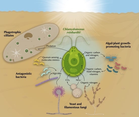
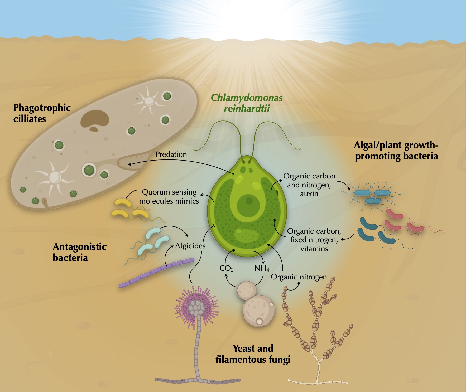
1. Why Microalgae and Why Chlamydomonas reinhardtii?

Microalgae are a highly diverse group of unicellular and photosynthetic eukaryotes that include major primary producers on our planet. Although highly abundant in aquatic ecosystems, microalgae can live in many habitats, including on the poles, below the ground within the crowded rhizosphere, or inside animal cells in coral reefs. These microbes can have photoautotrophic, heterotrophic, or mixotrophic lifestyles, show a broad range of cell sizes, morphologies, architectures, and exhibit a highly diverse metabolic capacity that offers several unique features for scientific research [1]. Responsible for a significant amount of total global carbon (C) fixation [2], microalgae are crucial to sustaining the ecosystems, but can also cause their disruption during algal blooms, which can pose a great ecological, economical, and health threat [3]. Furthermore, the use of microalgae in biotechnological applications has exploded in this century. For example, they are used in wastewater treatment, biofuel production, animal feed, or high-value compounds, and are promising organisms to increase biological C sequestration to mitigate global warming [4]. Therefore, microalgae are of tremendous ecological and economic interest.
Although polyphyletic, microalgal members are related by the ancestral origin of their plastid, which was initially the result of a primary endosymbiotic event involving a cyanobacterium that evolved into the plastid of the common ancestor of the Archaeplastida (i.e., glaucophytes, red and green algae, and land plants); and subsequently, expanded from red and green algae to unrelated eukaryotes through secondary and tertiary endosymbiosis, giving rise to the exceedingly colorful and metabolically diverse extant algal groups (e.g., diatoms and dinoflagellates) [5].
Over the centuries, microalgae have been isolated from nature—many of which have failed to grow axenically. Their reliance on other microorganisms is likely a consequence of the long-term co-evolution of microalgae with their symbiotic microbes [6]. Microalgae can form symbiotic relationships with a wide variety of organisms, including bacteria, fungi, plants, and animals [7]. When mutualistic, microalgae generally provide their partner(s) with fixed C and oxygen in exchange for limiting nutrients and essential molecules, such as available forms of vitamins and nitrogen (N) [8,9]. Additionally, a much broader set of molecules can be secreted, perceived, and utilized by the interacting partners. The elucidation of the intricacy and dynamics of many of these symbioses has only just begun [10,11].
The microalga C. reinhardtii (hereafter Chlamydomonas) was first isolated in 1945 from the soil of a potato field in Massachusetts (U.S.A.) and has since become a powerful model organism [12]. This green alga, sharing a common ancestor with land plants, has fundamental features related not only to plants but also to animals [13]. Some of the research topics that are being addressed using Chlamydomonas are photosynthesis, respiration, the metabolism of nitrogen (N), sulfur, phosphorus, amino acids and metals, the biosynthetic pathways of starch, lipids, carotenoids, hemes groups, glycerolipids, chlorophyll, and other fundamental aspects including the function of chaperones, proteases, thioredoxins, cilia biogenesis, and the responses to different stress conditions [14].
There are many features that make Chlamydomonas an excellent microbial model, as recently reviewed by Salomé et al. [15]. It grows well axenically, has a relatively rapid doubling time (~8–12 h), and the sequences of its nuclear, chloroplast, and mitochondrial genomes are available. In addition, The Chlamydomonas Sourcebook [12] provides a compendium of the major research lines, history, physiology and methodology of Chlamydomonas, and ‘The Chlamydomonas Resource Centre’ (www.chlamycollection.org, accessed on 7 February 2023) has numerous tools available, including biochemical assays, protocols, plasmids, and thousands of mapped mutant strains. Furthermore, the development of various techniques for gene editing in Chlamydomonas, such as CRISPR-Cas9, represents a decisive advance for addressing both fundamental questions and biotechnological applications [16,17].
Since its discovery, the number of reports studying Chlamydomonas interactions with diverse microorganisms has exponentially increased. Here, we summarize and categorize those reports into Chlamydomonas predators, fungal partners, and bacterial mutualists, and discuss the potential of microbial interactions for biotechnological applications.

2. Biodiversity in the Chlamydomonas Phycosphere

The phycosphere (Figure 1), the algal analog of the plant rhizosphere, has been defined as the region immediately surrounding algal cells, covering an undefined distance between the algal outer membrane and the aqueous zone in which bacterial growth is stimulated by extracellular algal products [18,19].
Microalgae produce large amounts of dissolved organic matter (DOM) that is released and accumulated in this microscopic environment, feeding the microbes inhabiting this niche. The bacterial communities in the phycosphere are taxonomically different to those in algal-free environments, similar to the differences observed between rhizosphere and bulk soil bacteria [20]. The delivery of DOM via photosynthate seems to be important for microalgae–bacterial interactions, and some other factors provided by the bacteria may facilitate and enhance their relationships [21]. Some studies have focused on characterizing the phycosphere microbiota to identify and understand the relevant interactions between microalgae and bacteria in their natural environment [22,23]. Thus, the phycosphere of different microalgae with great potential in the industry, including Chlamydomonas and other green algae such as Chlorella vulgaris, Scenedesmus sp., and Botryococcus braunii, has been characterized [24]. In this study, the phycosphere of Chlamydomonas was found to be dominated by Hyphomicrobiaceae sp. (44%), Mesorhizobium sp. (15%), and Rhizobium sp. (12%) [24].
Although bacterial associations can be species-specific, the analysis of the phycosphere of several phylogenetically distant green algae isolated from different environments revealed that most closely associated bacteria belonged to the phyla Bacteroidetes and α-Proteobacteria, which include members known to be symbiotic with plants. These observations suggested that at least some of these bacteria may have co-evolved with their green photosymbionts [6]. Supporting this idea, a more recent study comparing Chlamydomonas phycosphere microbiota with the root microbiota of Arabidopsis thaliana has shown an extensive taxonomic overlap, including Rhizobia, Pseudomonas, Burkholderia, and Xanthomonas spp. [23]. Moreover, the most abundant bacterial orders found in this comparison were shared between the root communities of different land plants and the phycosphere of several green algae, further supporting their possible co-evolution. This indicates that despite the vast evolutionary distance between Chlamydomonas (chlorophyte) and A. thaliana (embryophyte), the assembly of their microbiota might be driven by shared ecological principles that are not yet fully understood. Nevertheless, host specificity was also observed for these associated bacteria, as they can outcompete non-native strains only in the presence of their corresponding host. This study shows that establishing the phycosphere microbiota requires physical proximity between Chlamydomonas and its microbiota, probably because a bi-directional exchange of metabolites is required for their assembly [23].
Chlamydomonas has proven to sustain C cycling with a variety of taxonomically distinct bacteria isolated from soil [25]. Using Closed Microbial Ecosystems (CES) in which microbial communities are hermetically closed and maintained with only light, Chlamydomonas can feed the bacterial community with fixed C. In turn, the community produces CO2 to feed the alga, resulting in the self-organization of the consortia and robust C cycling to support the closed community for up to 50 days. In contrast, Chlamydomonas failed to persistently cycle C with Escherichia coli in the same closed system, likely due to E. coli’s inability to assimilate the starch produced by Chlamydomonas [25]. However, other studies using CES with the ciliate predator Tetrahymena thermophila in co-culture with Chlamydomonas and E. coli have shown that these three species can be maintained for years [26,27]. This suggests that introducing a phagotroph that can feed on the bacteria and, occasionally, on the algal population, allows for successful C cycling in this closed ecosystem. Additionally, Chlamydomonas can prevent bacterial invasion of T. thermophila cultures by inhibiting aggregation in E. coli—a defense mechanism to avoid predation—thus controlling the bacterial population by increasing predation pressure on E. coli, which is caused by the ciliate [28].
Even though the studies focused on characterizing Chlamydomonas’ microbiota are scarce and mostly limited to bacteria, this microalga shares a habitat with many other microbes, including other protists, fungi and viruses, that are likely relevant for Chlamydomonas’ biology in nature.

3. Chlamydomonas’ Predators

In addition, as a primary producer, Chlamydomonas not only feeds other organisms by secreting fixed C, but also by serving as food when lysed or ingested by predators. Chlamydomonas is a form of prey for a variety of different organisms, including rotifers such as Brachionus calyciflorus [29], crustaceans as Daphnia [30,31], protists such as Tetrahymena vorax [32] and Peranema [14], and soil bacteria such as Streptomyces iranensis [33] and Pseudomonas protegens [34,35,36] (Figure 1).
The bacterium P. protegens produces an arsenal of anti-algal compounds—secondary metabolites that induce morphological changes, inhibit growth, and can kill Chlamydomonas [35]. For instance, orfamide A induces an increase in cytosolic calcium levels in Chlamydomonas that causes deflagellation, whereas protegencin causes destruction of the carotenoids and eventually lyses algal cells [36] (Figure 1). This toxicity by protegencin also affects an eyeless mutant of Chlamydomonas and its colony-forming relative, Gonium pectorale. The protegencin biosynthesis gene cluster is ubiquitous in Pseudomonas genomes, which suggests that this antagonistic interaction is likely ecologically relevant between green algae and Pseudomonas species [36]. Interestingly, a P. protegens mutant impaired in secondary metabolism not only loses its anti-algal activity, but may promote Chlamydomonas growth [35].
The phagotrophic ciliate Paramecium bursaria, bearing the endosymbiotic green alga Chlorella, can feed on Chlamydomonas cells, which benefit both the host and endosymbiont by providing nutrients and clearing competitors for the latter [37] (Figure 1). To avoid being ingested by predators, Chlamydomonas can form cell aggregates of 4–16 cells, named palmelloids, and larger aggregates of up to thousands of cells [38]. The formation of palmelloids were the result of successive divisions induced by stress without degradation of the cell wall [39] and can be induced by predators such as the rotifer Brachionus calyciflorus [29] and the ciliate Paramecium tetraurelia [40,41]. Importantly, continuous exposure to predators repeatedly induced heritable genetic changes in Chlamydomonas, supporting the hypothesis that predation may have been one of the driving forces for the evolution of multicellularity [40,41,42].
The phagotrophic euglenoid Peranema trichophorum can also induce different Chlamydomonas spp. to form cell aggregates of up to 10,000 cells, composed of the same or different species, that dissociated when the predator was removed [43]. Interestingly, the culture filtrate of P. trichophorum also induces algal cell aggregation, likely in response to putative chemicals secreted by the predator that are still unknown (i.e., kairomones) [43]. Other kairomones produced by Daphnia and Daphnia-feeding fish can also alter Chlamydomonas behavior by generally inducing phototaxis (i.e., motility in response to light) [44]. The detection mechanism of kairomones in Chlamydomonas and whether it is predator-specific remains unclear.

4. Chlamydomonas’ Fungal Partners

Lichens, one of the most studied examples of obligate mutualisms, are formed by a self-sustaining association of fungi (mostly ascomycetes) with green algae, although they can also occur with cyanobacteria, as well as with bacteria and yeasts [45]. The study of synthetic consortia has revealed that Chlamydomonas can establish spontaneous obligate mutualisms with different fungal species—including the yeast Saccharomyces cerevisiae, and the filamentous fungi Neurospora crassa and Aspergillus nidulans—under specific conditions [46]. In closed systems, under illumination and in the presence of glucose as the sole C source, which cannot be used by Chlamydomonas, and nitrite as the only N source, which cannot be assimilated by the yeast S. cerevisiae, the alga and the yeast metabolize and cross-feed available sources of N and C; S. cerevisiae metabolizes the glucose, releasing carbon dioxide that is used by Chlamydomonas, which in turn reduces nitrite to feed the yeast with ammonium. This synthetic consortium has also proven relevant to assess the role of ecological interactions in the evolution of mutualism; the presence of the alga shifts natural selection to fix mutations in the yeast that strengthen their mutualistic interaction [10].
Chlamydomonas cells were found to physically interact with the filamentous fungi Neurospora crassa and Aspergillus nidulans, to likely facilitate nutrient exchange [46]. Moreover, Chlamydomonas and the ascomycetous fungus Alternaria infectoria can form long-living consortia that persist for up to several years in the absence of a N source other than atmospheric N2, which was fixed by the fungus and likely supplied to the alga as amino acids [47].
Additional beneficial interactions with the lichen forming fungus A. nidulans, which chemoattracts Chlamydomonas, include physical protection against the algicidal compound Azalomycin F produced by the bacterium Streptomyces iranensis in the presence of Chlamydomonas [33]. The shelter provided by the fungus suggests that the formation of algal–fungal associations may be a good strategy against harmful microorganisms and may have driven the evolution of lichen associations [33].
These systems have provided important clues about the possible abiotic and biotic conditions that initially led to the evolution of lichens and revealed valuable insights into the emergence of symbiosis and the design of synthetic biological systems.

5. Chlamydomonas’ Bacterial Partners

5.1. Vitamin B12 Production

Vitamin B12 (cobalamin) is an enzyme cofactor synthesized only by prokaryotes, yet more than half of microalgae require it for growth [8]. Therefore, this cofactor may act as a common bacterial currency for mutualism with algae in exchange for algal fixed C, and it may play a key role in microbial community structure and function [48]. Nevertheless, not all B12-producing bacteria and algal B12 auxotrophs establish mutualistic relationships, indicating a species-specificity that may be due to the amount of B12 released or the type of C fixed secreted by the alga [49,50]. While some studies have shown that bacterial growth was required to support algal B12 auxotrophs [51], others have reported that bacterial cell lysis was sufficient for that purpose [49]. In algae, the enzyme methionine synthase (METH) uses vitamin B12 as a cofactor, but some algae, such as Chlamydomonas, also have a B12-independent isoform (METE) that allows them to grow without the vitamin, albeit less efficiently [52]. Under heat stress, the Chlamydomonas METE gene is repressed, leading to chlorosis and cell death if this vitamin is unavailable. However, addition of exogenous B12 or co-culture with the B12-producing bacterium Ensifer meliloti allows Chlamydomonas to thrive under heat stress [53], indicating that abiotic stress could promote such beneficial interactions in nature [53] (Figure 1).
The loss of the METE gene, which leads to B12 auxotrophy, appears to be independent in the evolutionary history of algae, as it seems to have occurred several times [54,55]. Indeed, a B12 auxotroph of Chlamydomonas was generated via experimental evolution after ca. 500 generations in B12-replete media [52]. This newly evolved B12-dependent strain was then able to grow in the absence of this vitamin when co-cultured with B12-producing bacteria, including the rhizobium Mesorhizobium japonicum or even an E. coli strain engineered to produce and release B12 [50]. Nevertheless, it is unclear whether the B12-dependent strain would survive in nature. On the one hand, this strain grew more slowly in laboratory co-culture with bacteria than the B12-independent parental strain, but on the other hand, experimental co-evolution with bacteria improved the alga’s subsequent growth in co-culture and may suggest that prolonged coexistence with bacteria in the environment could favor the evolution of B12 auxotrophy [50].
Supporting this idea, Kazamia and collaborators found that M. japonicum established a mutualistic relationship based on B12-C exchange with the naturally B12-dependent green alga Lobomonas rostrata, which is closely related to Chlamydomonas [56]. Furthermore, proteomic analyses of this mutualism suggested that amino acids may be the reduced form of C that the algae were feeding the bacterium in exchange for B12 [57].
More recently, the integration of time-resolved isotope labeling followed by secondary ion mass spectrometry (SIMS) method with mechanistic modeling, has elucidated the role of nutrient exchange in controlling the inception and temporal onset of a mutualistic relationship between the B12 auxotrophic Chlamydomonas mutant mete7 and M. japonicum [58]. The results showed that these microbes, which were not given the time to co-evolve and whose growth does not seem to be induced by the interaction, established a mutualism based on the exchange of vitamin B12 and organic C. The combination of nutrient measurements and mathematical models might be a useful tool for the mechanistic understanding of more complex microbial interactions, such as those composed of more than two partners [58,59].

5.2. Nitrogen Fixation

N is highly abundant in the atmosphere as dinitrogen gas (N2), but it is only accessible to some prokaryotes (diazotrophs) which are capable of converting it to the fixed N form ammonium. Therefore, N-limited photoautotrophic eukaryotes often partner with diazotrophs that provide them with usable N sources, forming stable symbioses. One of the most extensively studied symbioses is that established between land plants (legumes) and symbiotic diazotrophs (rhizobia), which provide the plant with fixed N in exchange for nutrients and energy [60]. Although far less studied, diazotrophs can also form a symbiosis with different algae, including diatoms [61,62], haptophytes [63], and green algae [64,65].
The free-living symbiotic diazotroph Azotobacter chroococcum has been widely used as biofertilizer, successfully increasing the yields of a wide variety of crop plants [66]. Moreover, Azotobacter species can also support the growth of different algal species, including Chlamydomonas. In media without any N or C sources, spontaneous mutualisms have been observed by mixing different Azotobacter species (A. chroococcum, A. beljerinckii, A. agilis, A. vinelandii) with Chlamydomonas [67,68] (Figure 1). These algal–diazotroph co-cultures survived for several years, and algal photosynthetic efficiencies were observed to be similar to those growing with N, which strongly suggests that the diazotroph provides the alga with readily available N source(s) [69]. Additionally, these bacteria can provide the alga with amino acids, vitamins, and hormones, which can deliver proteins, polysaccharides, and glycolate to the bacterium in exchange [70]. While this association was extracellular, cell-to-cell fusion was artificially induced with polyethylene glycol to force the endosymbiotic interaction of Azotobacter cells within Chlamydomonas that were able to grow in N- and C-free medium for several years [71]. In this alga, Azotobacter cells have been found in organelle-resembling vesicles located in the cytoplasm, between the cell wall and plasma membrane, across the cell wall, and in the periplasmic space. The number of symbionts in these cellular locations was found to be regulated, and the ChlamydomonasAzotobacter volume ratios were nearly constant (47.2 ± 2.4), which may be key to the stabilization of the interaction [72]. To achieve the establishment of the co-culture, the depletion of C and N in the media seems to be critical, although the mechanism that controls the algal–bacterial volume ratio and the specific C and N compounds exchanged between these organisms remains elusive [65]. Although it seems clear that the C-fixing alga must be providing the heterotrophic bacterium with fixed C, and the N-fixing bacterium must be feeding the alga with fixed N, the exact C and N compounds that are exchanged are unknown. A. vinelandii excretes 17 amino acids [65], many of which can support Chlamydomonas growth after their deamination [73], while Chlamydomonas can secrete different C compounds that could serve as C and energy sources for the bacterium. Some of these compounds were glycolate; oxalate; pyruvate [74]; acetate, which was more efficiently used by Azotobacter than glucose [75]; several keto acids [76]; amino acids and sugars [77]; and various fermentation products such as acetic acid, formic acid, ethanol, and malic acid [78].
Resembling the plant symbioses with mycorrhizal fungi and diazotrophic bacteria, which improve their growth through the mobilization of nutrients [79], an artificial tripartite symbiosis involving Chlamydomonas, Azotobacter, and the fungus Alternaria has been found to support algal growth for three years on a N and C-free medium [80]. In this system, the fungus can secrete a wide range of amino acids, increasing the diversity of N compounds available for the alga, compared to those secreted by bacteria in the dual system, and positively impacting algal survival [68]. The sulfur-containing amino acid and metabolic intermediate cystathionine was found to be highly secreted by the fungus and increased in the tripartite system [80], likely providing sulfur to the alga and/or bacterium. Curiously, a cystathionine-β-synthase-like protein was required for rhizobial infection and symbiotic N fixation in Medicago truncatula [81]. However, whether the production of cystathionine by Alternaria is involved in the establishment of the tripartite association with Chlamydomonas and Azotobacter awaits further examination.

5.3. Amino Acids and Peptides Mineralization

In environments or conditions in which inorganic N is limiting but organic N is abundant, plant growth-promoting bacteria (PGPB) play a critical role in mineralizing organic N to inorganic forms, which are assimilable by photosynthetic organisms [82]. As previously mentioned, amino acids can act as a microbial currency to establish symbiotic interactions with Chlamydomonas. Although less efficiently than inorganic N forms such as ammonium or nitrate, this alga can metabolize a wide variety of amino acids and peptides as the sole N source [83]. This growth mainly relies on the extracellular activity of an L-amino acid oxidase encoded by the LAO1 gene and a specific high-affinity uptake system for L-arginine [73,84,85]. However, this alga cannot utilize the amino acid L-proline as well as short peptides as N sources [86]. The PGPB Methylobacterium species can “rescue” Chlamydomonas growth on L-proline as the only N source, establishing a mutualism. This mutualistic interaction is species-specific and based on a C-N exchange: the bacterium metabolizes L-proline, producing ammonium to feed Chlamydomonas, which releases the photosynthate glycerol to feed the bacterium [87] (Figure 1), although additional metabolites may also be exchanged. In addition to L-proline, Methylobacterium spp. were able to support algal growth on a variety of di- and tri-peptides containing L-alanine, glycine, L-proline and L-hydroxyproline [87]. Interestingly, the cell wall of Chlamydomonas is enriched in glycoproteins containing proline and hydroxyproline [88] and may secrete L-proline [80], which might be used to attract beneficial Methylobacterium spp. [87].
Although Chlamydomonas can grow on most free amino acids through the LAO1 enzyme, this extracellular activity may represent a beneficial strategy for microbial interactions [73]. LAO1 breaks down amino acids into the corresponding keto acid, H2O2 and ammonium [84], which is very efficiently taken up by the alga through high-affinity transport systems [89]. The remaining byproducts, keto acids and H2O2, were accumulated in the phycosphere and might serve as public goods for other microorganisms [73]. In the phycosphere, keto acids can be used as a C and an energy source by other microorganisms, play a role in solubilizing or chelating iron, buffer the toxic H2O2 [90], and help algal cells to establish interactions with other organisms. In fact, the keto acid indole-3-pyruvic acid, generated from L-tryptophan, was found to be a major intermediate of the auxin indole-3-acetic acid, and its impact on mutualism is discussed below (Section 5.4). The highly reactive H2O2 produced by LAO1 could aid Chlamydomonas to avoid competitors and promote the growth of beneficial bacteria that specifically contain H2O2-detoxifying enzymes such as catalase (e.g., Methylobacterium spp.). Supporting this idea, H2O2-consuming bacteria were present in the natural habitat of the diatom Amphiprora kufferathii, which can eliminate the H2O2 produced by the algal cells [91]. Similarly, other prokaryotes, such as Prochlorococcus spp., Sinorhizobium meliloti, and Vibrio fischeri, have shown mutualistic interactions based on H2O2 detoxification [92,93,94], suggesting that this is a widespread mechanism of symbiotic relationships.

5.4. Auxin Production and Degradation

The plant auxin indole-3-acetic acid (IAA) is a major signaling molecule involved in controlling plant–bacterial interactions [95]. Like plants, algae such as the marine diatom Pseudo-nitzschia multiseries can release L-tryptophan, which can be converted by bacteria into IAA to stimulate algal growth [96]. Chlamydomonas can secrete tryptophan [97], providing symbiotic microbes with the appropriate substrate for IAA production. Therefore, the role of IAA as a crosstalk molecule is not only restricted to plant–bacterial interactions, but extends to algae and is likely a widespread phenomenon in phototrophic–bacterial symbioses [96,98,99,100]. Notwithstanding, algae—including Chlamydomonas—can also synthesize IAA from L-tryptophan, and algal-derived auxin can also affect algal–bacterial interactions [101]. However, the role of auxin production played by the algal partner in these associations has been largely neglected. Algal-produced IAA has only recently been proposed to have a role in mutualistic interactions between Chlamydomonas and Methylobacterium spp. [102] (preprint) (Figure 1). Under N limitation, Chlamydomonas can produce IAA from L-tryptophan via a LAO1-mediated mechanism. The accumulation of this auxin in the medium causes the alga to arrest cell multiplication and chlorophyll degradation. This auxin-derived inhibitory effect can be relieved by the presence of Methylobacterium spp., which can feed on auxin to grow. Bacterial auxin degradation was promoted by the presence of the alga, which is, in turn, benefited by the reduction of auxin levels, and resumes growth. Since both Chlamydomonas and Methylobacterium spp. are found in the rhizosphere, this chemical crosstalk mediated by auxin production and degradation may impact plant fitness and could be used to improve crop yields in sustainable agriculture [102].

5.5. Quorum Quenching

Beyond nutrient exchange, microbial interactions are modulated by signal molecules. Using quorum sensing (QS), bacteria can sense and respond to changes in cell population density to control specific processes such as virulence factor expression, secondary metabolites production, biofilm formation, and other mechanisms involved in host–microbial interactions and microbial ecology [103]. These processes often impact their interaction with co-existing organisms and, consequently, these interacting organisms have developed the ability to interfere with bacterial quorum sensing by producing molecules that mimic QS and modulate their behavior; this process is known as quorum quenching (QQ) [104,105]. N-acyl-l-homoserine lactones (AHLs) are bacterial QS signals produced by over 50 different species [106]. The marine red alga Delisea pulchra secretes halogenated furanones that are structurally similar to AHL, which is the first eukaryote in which QS mimics were identified [107]. Similarly, the land plant Medicago truncatula can secrete more than a dozen compounds that stimulate or inhibit bacterial AHL-mediated QS [108], which suggests that QQ may be a widespread ability across photoautotrophic organisms to modulate their bacterial symbionts. In Chlamydomonas culture filtrates, at least a dozen AHL-mimicking molecules can stimulate QS in the soil bacterium Ensifer meliloti [109] (Figure 1). These AHL-like molecules have been chromatographically separated but have not been identified yet. Their production is critically dependent on the age and culture conditions; higher levels were produced under phototrophic than under mixotrophic conditions (with acetate) while the levels reduced in aged cultures. Interestingly, these molecules affect the accumulation of at least 16 S. meliloti proteins, including the regulatory protein PII, which is involved in the C/N balance in both prokaryotes and eukaryotes [110], and controls N assimilation and nodule formation in S. meliloti [111]. Since no homologs to the bacterial AHL synthases have been found in the genome of Chlamydomonas, the synthesis of the AHL-like molecules in Chlamydomonas molecules may involve different enzymes. A derivative of the vitamin riboflavin named lumichrome purified from Chlamydomonas culture filtrates was able to stimulate the QS LasR receptor in Pseudomonas aeruginosa [112] (Figure 1). This QS mimicking-signal has also been identified in rhizobial exudates that stimulate plant growth and seedling development [113]. Therefore, the production of this inter-kingdom signal molecule by Chlamydomonas might have a broader effect on the ecosystem, which is not only confined to bacteria, but also affects higher plants.
Figure 1. Interactions between Chlamydomonas reinhardtii and other microbes, illustrating the main topics covered in this review. This illustration represents a Chlamydomonas cell in the central part surrounded by its phycosphere populated by representative interacting microbes and indicating the main molecules involved in the interaction. Chlamydomonas is prey for a variety of different microbes, including protists such as Paramecium bursaria [37], Tetrahymena vorax [32], and Peranema [14], and soil bacteria such as Streptomyces iranensis [33] and Pseudomonas protegens [34,35,36]. The bacterium P. protegens produces an arsenal of anti-algal compounds and secondary metabolites such as orfamide A and protegencin that induce morphological changes, inhibit growth, and can kill Chlamydomonas [35,36]. S. iranensis produce the algicidal compound Orfamide A in the presence of Chlamydomonas, which can be physically protected from the algicide by the lichen-like fungi Aspergillus nidulans [33]. Chlamydomonas produces at least a dozen AHL mimics that can stimulate QS in the soil-dwelling bacterium Ensifer meliloti [106]; among them, lumichrome was able to stimulate the QS LasR receptor in Pseudomonas aeruginosa [109]. Chlamydomonas establishes spontaneous obligate mutualisms with fungal species under specific conditions [46]. In closed systems under illumination with glucose and nitrite, Saccharomyces cerevisiae metabolizes glucose to feed with CO2 Chlamydomonas, which provides the yeast with ammonium produced from nitrite. The ascomycetous fungus Alternaria infectoria fixes N2 that can be supplied to Chlamydomonas in the form of organic nitrogen, and they can form long-living consortia [47]. The rhizospheric bacteria Mesorhizobium japonicum and Ensifer meliloti can provide Chlamydomonas with vitamin B12 promoting algal growth in exchange for algal fixed C [50,58]. Nitrogen mineralizing bacteria such as Methylobacterium spp. can break down amino acids and peptides to feed Chlamydomonas with ammonium in exchange for the photosynthate glycerol [87]. Additionally, auxin produced by Chlamydomonas can feed Methylobacterium aquaticum, which in turn promotes algal growth [99]. Lastly, N2-fixing bacteria such as Azotobacter chroococcum, A. beljerinckii, A. agilis, and A. vinelandii can support Chlamydomonas growth with fixed nitrogen in exchange for fixed carbon [65,67,68,69,71]. This figure was generated using Biorender.com and Keynote software.
Plants 12 00788 g001

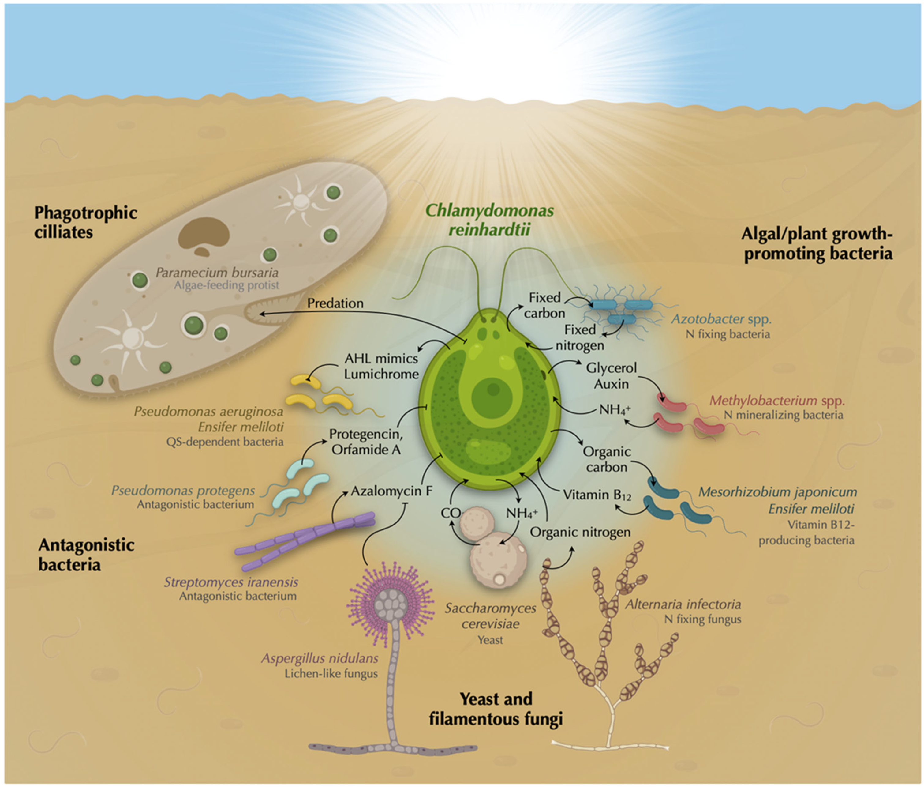
6. Harnessing Chlamydomonas—Microbial Interactions for Biotechnological Applications

The great metabolic diversity of microalgae, together with their rapid growth and low production costs, point to these organisms as promising resources for a wide range of biotechnological applications aimed at meeting urgent needs in industry and agriculture. For instance, microalgae have extensive potential as sustainable sources to produce bulk compounds such as biofuel, fertilizers or livestock feed, as well as for wastewater treatment and biological C sequestration [114]. Nevertheless, there are still many issues making the use of microalgae inefficient for industrial applications; thus, significant efforts are being devoted to achieving a better understanding of the biological activities related to industrial applications. The yield of many of these biotechnological processes conducted by microalgae can be increased by means of their interaction with other microorganisms, which mostly involve bacteria at present [115]. As a well-established model organism, Chlamydomonas has been extensively used axenically and improved with in co-culture for many of these applications that will be briefly summarized here.
The current climate crisis has precipitated the commitment of countries worldwide to transition from fossil fuels to alternative clean and renewable energies. Hydrogen (H2) is being pointed out as one of the most suitable renewable energy sources, since its combustion generates only water as product [116]. Biohydrogen production can be achieved using H2-producing bacteria or microalgae, while this production can be improved by their combination. The use of Chlamydomonas biomass can increase the efficiency of H2 production by different bacterial species such as Clostridium butyricum, Rhodobacter sphaeroides KD131 and Rhodospirillum rubrum [117,118]. In the case of R. rubrum, H2 production can be enhanced by using formate released by Chlamydomonas [119]. On the other hand, Chlamydomonas is also able to produce H2 by itself, which can be improved by the presence of some bacteria, as recently reviewed by Fakhimi and collaborators [120]. These bacteria include, among others, the soil bacteria Pseudomonas putida, P. stutzeri, Rhizobium etli, E. coli [121,122], P. fluorescens [123], Azotobacter chroococcum [124], Mesorhizobium sangaii [125], Bradyrhizobium japonicum [126], or Methylobacterium oryzae [127]. These bacterial-cocultures can improve Chlamydomonas biomass and avoid the accumulation of inhibitory waste compounds, such as oxygen or acetate, in the medium, which in turn increases H2 production yield [122,128]. Additionally, some bacteria can induce the accumulation of starch in Chlamydomonas, which may enhance H2 production [120,129]. Moreover, fermentative metabolites can be exchanged between bacteria and Chlamydomonas to improve synergic bacterial and algal H2 production [122,130] (Figure 2).
Microalgae have various advantages in biomass production: (i) they can be cultivated throughout the year, (ii) they do not compete for arable lands, and (iii) they are more productive per unit of land area than any plant system [131,132].
The co-culture of microalgae with other microorganisms increases growth stability and biomass productivity, and the biomass obtained can be used, for example, for biofuel production [7,133]. In Chlamydomonas, N deprivation greatly increases lipid accumulation, but its concomitant reduction in growth rate poses a challenge for biomass production. The diazotroph A. chroococcum can simultaneously enhance lipid and biomass productivity by promoting growth and inducing regulatory changes in the lipid metabolism of the alga [134]. The co-culture of Chlamydomonas with other plant/algal growth-promoting bacteria such as N-mineralizing Methylobacterium spp. may also be used to increase biomass, the production of algal-derived biofuels and other chemicals [135] (Figure 2). A mutualistic interaction between Chlamydomonas and the oleaginous yeast Lipomyces starkeyi, known for their oil productivity, increases lipid production and the resulting biomass was suitable for biofuel production [136]. Moreover, Chlamydomonas biomass can be digested by the non-reducing heterotrophic bacterium Geobacter sulfurreducens to convert light energy into electricity as microbial fuel cells [137] (Figure 2).
Bacteria are widely used in wastewater treatment plants, but the large O2 requirement is expensive. Therefore, the addition of oxygen-producing microalgae to the process has become a promising way to reduce costs in wastewater treatment. Moreover, microalgae make this process more environmentally friendly, since the bacterial release of the greenhouse gas CO2 is reduced by algal fixation [138,139], as recently reviewed by [140,141]. Importantly, bacteria naturally present in wastewater effluents promote the growth of microalgal species including Chlamydomonas, which in turn enhances the water treatment process [142]. In consortium with bacteria, Chlamydomonas can highly reduce the amount of N and phosphorous contaminant, and at the same time it can decrease most of the chemical oxygen demand in photobioreactors containing synthetic wastewater [127,143,144]. In addition to contaminant removal, the generated biomass can be used for lipid extraction used in biofuel production, while the remaining biomass may be applied in the generation of fertilizer or animal feed [145] (Figure 2).
Microalgae are also a great platform for the generation of high-value compounds, such as Biomimetic 3D living material [146]. The generation of a soft living matter based on cellulose has been achieved in a symbiotic relationship between Acetobacter aceti and Chlamydomonas [147]. This material was composed of bacterial cellulose produced in situ by A. aceti that benefits from the O2 produced by Chlamydomonas. The algal cells get embedded and immobilized in the cellulose gels and may be used as a new way to immobilize other commercially important microorganisms, improving product production and extraction.

7. Insights beyond Symbiosis

Due to the huge metabolic diversity of microalgae, a single species does not represent all microbial interactions, yet the use of Chlamydomonas reinhardtii provides a unique opportunity to learn about the principles that govern more complex symbioses. To date, most studies have focused on Chlamydomonas interactions with bacteria. Although there are some with fungi and ciliates, the potential impact on their physiology by other organisms that may co-exist with microalgae, such as viruses, plants, and animals, remains overlooked [148,149]. Although our knowledge of the metabolic exchange between microalgae and other microbes has greatly increased during the last decades, their regulation and dynamics, as well as the evolutionary drivers are only just starting to be elucidated [10,150]. Furthermore, horizontal gene transfer between microalgae and their interacting microbes may be a major driver for the evolution of new traits and their adaptation to different environments, which may improve some biotechnological applications. Moreover, the rapidly growing field of synthetic biology is critically increasing the efficiency of these processes. Current and future efforts to understand and predict microalgal–bacterial interactions will allow us to improve the control of many ecological and biotechnological processes, including biofuel production, as well as the development of new ones.

Author Contributions

A.L. and V.C. wrote the paper; M.T.-J., E.S.-L., E.F. and A.G. writing, review and editing; AL conceptualization and original draft preparation. All authors have read and agreed to the published version of the manuscript.

Funding

This work was funded by Gobierno de España, Ministerio de Ciencia e Innovacion (Grant PID2020-118398GB-I00), Junta de Andalucía (Grant ProyExcel_00483), the European FEDER program, the “Plan Propio” from University of Cordoba, and a grant awarded by the Torres-Gutierrez foundation.

Data Availability Statement

All data required to evaluate the conclusions of this paper are included in the main text.

Acknowledgments

We thank Freddy Bunbury (Carnegie Institution for Science) for proofreading this manuscript and his critical input in Section 5.1 (Vitamin B12 production), and Maribel Macias for technical assistance. The final sentence in the title was inspired by the song “Why can’t we be friends?” (1975) by the music band War (California, 1969). Harold Brown, member of the band said in an interview referring to this song: “we’re all connected by language, and by our food” (Interview by Songfacts, 2007). Thus, we believe that the message of this song is that despite seeming different, unrelated critters can come together and interact through their “language” (chemical cross-talk) and “food” (nutrients) and, if conditions allow for it, create beneficial relationships (to become “friends”)—i.e., the title does not imply that the interactions are negative, but rather dynamic and dependent on the environment.

Conflicts of Interest

The authors declare no conflict of interest.

References

  1. Gerotto, C.; Norici, A.; Giordano, M. Toward Enhanced Fixation of CO2 in Aquatic Biomass: Focus on Microalgae. Front. Energy Res. 2020, 8, 213. [Google Scholar] [CrossRef]
  2. Falkowski, P.G. The Role of Phytoplankton Photosynthesis in Global Biogeochemical Cycles. Photosynth. Res. 1994, 39, 235–258. [Google Scholar] [CrossRef] [PubMed]
  3. Kazmi, S.S.U.H.; Yapa, N.; Karunarathna, S.C.; Suwannarach, N. Perceived Intensification in Harmful Algal Blooms Is a Wave of Cumulative Threat to the Aquatic Ecosystems. Biology 2022, 11, 852. [Google Scholar] [CrossRef]
  4. Oliveira, C.Y.B.; Jacob, A.; Nader, C.; Oliveira, C.D.L.; Matos, Â.P.; Araújo, E.S.; Shabnam, N.; Ashok, B.; Gálvez, A.O. An Overview on Microalgae as Renewable Resources for Meeting Sustainable Development Goals. J. Environ. Manag. 2022, 320, 115897. [Google Scholar] [CrossRef]
  5. Sibbald, S.J.; Archibald, J.M. Genomic Insights into Plastid Evolution. Genome Biol. Evol. 2020, 12, 978–990. [Google Scholar] [CrossRef] [PubMed]
  6. Ramanan, R.; Kim, B.-H.; Cho, D.-H.; Oh, H.-M.; Kim, H.-S. Algae-Bacteria Interactions: Evolution, Ecology and Emerging Applications. Biotechnol. Adv. 2016, 34, 14–29. [Google Scholar] [CrossRef]
  7. Ray, A.; Nayak, M.; Ghosh, A. A Review on Co-Culturing of Microalgae: A Greener Strategy towards Sustainable Biofuels Production. Sci. Total Environ. 2022, 802, 149765. [Google Scholar] [CrossRef]
  8. Croft, M.T.; Lawrence, A.D.; Raux-Deery, E.; Warren, M.J.; Smith, A.G. Algae Acquire Vitamin B12 through a Symbiotic Relationship with Bacteria. Nature 2005, 438, 90–93. [Google Scholar] [CrossRef]
  9. Smith, M.J.; Francis, M.B. A Designed A. vinelandii-S. elongatus Coculture for Chemical Photoproduction from Air, Water, Phosphate, and Trace Metals. ACS Synth. Biol. 2016, 5, 955–961. [Google Scholar] [CrossRef] [PubMed]
  10. Venkataram, S.; Kuo, H.-Y.; Hom, E.F.Y.; Kryazhimskiy, S. Mutualism-Enhancing Mutations Dominate Early Adaptation in a Two-Species Microbial Community. Nat. Ecol. Evol. 2023, 7, 143–154. [Google Scholar] [CrossRef]
  11. Kazamia, E.; Helliwell, K.E.; Purton, S.; Smith, A.G.; Fussmann, G. How Mutualisms Arise in Phytoplankton Communities: Building Eco-Evolutionary Principles for Aquatic Microbes. Ecol. Lett. 2016, 19, 810–822. [Google Scholar] [CrossRef]
  12. Harris, E.H. Introduction into Chlamydomonas and Its Laboratory Use. In The Chlamydomonas Sourcebook; Oxford Academic Press: Oxford, UK, 2009. [Google Scholar]
  13. Merchant, S.S.; Prochnik, S.E.; Vallon, O.; Harris, E.H.; Karpowicz, S.J.; Witman, G.B.; Terry, A.; Salamov, A.; Fritz-Laylin, L.K.; Maréchal-Drouard, L.; et al. The Chlamydomonas Genome Reveals the Evolution of Key Animal and Plant Functions. Science 2007, 318, 245–250. [Google Scholar] [CrossRef] [PubMed]
  14. Sasso, S.; Stibor, H.; Mittag, M.; Grossman, A.R. From Molecular Manipulation of Domesticated Chlamydomonas reinhardtii to Survival in Nature. eLife 2018, 7, e39233. [Google Scholar] [CrossRef]
  15. Salomé, P.A.; Merchant, S.S. A Series of Fortunate Events: Introducing Chlamydomonas as a Reference Organism. Plant Cell 2019, 31, 1682–1707. [Google Scholar] [CrossRef]
  16. Scaife, M.A.; Nguyen, G.T.D.T.; Rico, J.; Lambert, D.; Helliwell, K.E.; Smith, A.G. Establishing Chlamydomonas reinhardtii as an Industrial Biotechnology Host. Plant J. 2015, 82, 532–546. [Google Scholar] [CrossRef] [PubMed]
  17. Kelterborn, S.; Boehning, F.; Sizova, I.; Baidukova, O.; Evers, H.; Hegemann, P. Gene Editing in Green Alga Chlamydomonas reinhardtii via CRISPR-Cas9 Ribonucleoproteins. Plant Synth. Biol. Methods Mol. Biol. 2022, 2379, 45–65. [Google Scholar]
  18. Bell, W.; Mitchell, R. Chemotactic And Growth Responses Of Marine Bacteria To Algal Extracellular Products. Biol. Bull. 1972, 143, 265–277. [Google Scholar] [CrossRef]
  19. Seymour, J.R.; Amin, S.A.; Raina, J.-B.; Stocker, R. Zooming in on the Phycosphere: The Ecological Interface for Phytoplankton–Bacteria Relationships. Nat. Microbiol. 2017, 2, 17065. [Google Scholar] [CrossRef]
  20. Ling, N.; Wang, T.; Kuzyakov, Y. Rhizosphere Bacteriome Structure and Functions. Nat. Commun. 2022, 13, 836. [Google Scholar] [CrossRef]
  21. Cooper, M.B.; Smith, A.G. Exploring Mutualistic Interactions between Microalgae and Bacteria in the Omics Age. Curr. Opin. Plant Biol. 2015, 26, 147–153. [Google Scholar] [CrossRef]
  22. Krohn-Molt, I.; Alawi, M.; Förstner, K.U.; Wiegandt, A.; Burkhardt, L.; Indenbirken, D.; Thieß, M.; Grundhoff, A.; Kehr, J.; Tholey, A.; et al. Insights into Microalga and Bacteria Interactions of Selected Phycosphere Biofilms Using Metagenomic, Transcriptomic, and Proteomic Approaches. Front. Microbiol. 2017, 8, 1941. [Google Scholar] [CrossRef] [Green Version]
  23. Durán, P.; Flores-Uribe, J.; Wippel, K.; Zhang, P.; Guan, R.; Melkonian, B.; Melkonian, M.; Garrido-Oter, R. Shared Features and Reciprocal Complementation of the Chlamydomonas and Arabidopsis Microbiota. Nat. Commun. 2022, 13, 406. [Google Scholar] [CrossRef]
  24. Kim, B.H.; Ramanan, R.; Cho, D.H.; Oh, H.M.; Kim, H.S. Role of Rhizobium, a Plant Growth Promoting Bacterium, in Enhancing Algal Biomass through Mutualistic Interaction. Biomass Bioenergy 2014, 69, 95–105. [Google Scholar] [CrossRef]
  25. de Jesus Astacio, L.M.; Prabhakara, K.H.; Li, Z.; Mickalide, H.; Kuehn, S. Closed Microbial Communities Self-Organize to Persistently Cycle Carbon. Ecol. Biophys. Comput. Biol. 2021, 118, 2013564118. [Google Scholar] [CrossRef] [PubMed]
  26. Hekstra, D.R.; Leibler, S. Contingency and Statistical Laws in Replicate Microbial Closed Ecosystems. Cell 2012, 149, 1164–1173. [Google Scholar] [CrossRef]
  27. Frentz, Z.; Kuehn, S.; Leibler, S. Strongly Deterministic Population Dynamics in Closed Microbial Communities. Phys. Rev. X 2015, 5, 041014. [Google Scholar] [CrossRef]
  28. Mickalide, H.; Kuehn, S. Higher-Order Interaction between Species Inhibits Bacterial Invasion of a Phototroph-Predator Microbial Community. Cell Syst. 2019, 9, 521–533. [Google Scholar] [CrossRef] [PubMed]
  29. Lurling, M.; Beekman, W. Palmelloids Formation in Chlamydomonas reinhardtii: Defence against Rotifer Predators? Ann. Limnol. 2006, 42, 65–72. [Google Scholar] [CrossRef]
  30. Van Donk, E.; Liirling, M.; Hessen, D.O.; Lokhorst, G.M. Altered Cell Wall Morphology in Nutrient-Deficient Phytoplankton and Its Impact on Grazers. Limnol. Ocean. 1997, 42, 357–364. [Google Scholar] [CrossRef]
  31. Gophen, M. Feeding of Daphnia on Chlamydomonas and Chlorobium. Nature 1977, 265, 271–273. [Google Scholar] [CrossRef]
  32. Taub, F.B.; Mckenzie, D.H. Continuous Cultures of an Alga and Its Grazer. Bull. Ecol. Res. Comm. 1973, 17, 371–377. [Google Scholar]
  33. Krespach, M.K.C.; García-Altares, M.; Flak, M.; Schoeler, H.; Scherlach, K.; Netzker, T.; Schmalzl, A.; Mattern, D.J.; Schroeckh, V.; Komor, A.; et al. Lichen-like Association of Chlamydomonas reinhardtii and Aspergillus nidulans Protects Algal Cells from Bacteria. ISME J. 2020, 14, 2794–2805. [Google Scholar] [CrossRef] [PubMed]
  34. Aiyar, P.; Schaeme, D.; García-Altares, M.; Flores, D.C.; Dathe, H.; Hertweck, C.; Sasso, S.; Mittag, M. Antagonistic Bacteria Disrupt Calcium Homeostasis and Immobilize Algal Cells. Nat. Commun. 2017, 8, 1756. [Google Scholar] [CrossRef] [PubMed]
  35. Rose, M.M.; Scheer, D.; Hou, Y.; Hotter, V.S.; Komor, A.J.; Aiyar, P.; Scherlach, K.; Vergara, F.; Yan, Q.; Loper, J.E.; et al. The Bacterium Pseudomonas protegens Antagonizes the Microalga Chlamydomonas reinhardtii Using a Blend of Toxins. Environ. Microbiol. 2021, 23, 5525–5540. [Google Scholar] [CrossRef]
  36. Hotter, V.; Zopf, D.; Joong Kim, H.; Silge, A.; Schmitt, M.; Aiyar, P.; Fleck, J.; Matthäus, C.; Hniopek, J.; Yan, Q.; et al. A Polyyne Toxin Produced by an Antagonistic Bacterium Blinds and Lyses a Chlamydomonas Alga. Proc. Natl. Acad. Sci. USA 2021, 118, 2107695118. [Google Scholar] [CrossRef]
  37. Iwai, S.; Fujita, K.; Takanishi, Y.; Fukushi, K. Photosynthetic Endosymbionts Benefit from Host’s Phagotrophy, Including Predation on Potential Competitors. Curr. Biol. 2019, 29, 3114–3119. [Google Scholar] [CrossRef]
  38. Iwasa, K.; Murakami, S. Palmelloid Formation of Chlamydomonas I. Palmelloid Induction by Organic Acids. Physiol. Plantarum 1968, 21, 1224–1233. [Google Scholar] [CrossRef]
  39. de Carpentier, F.; Lemaire, S.D.; Danon, A. When Unity Is Strength: The Strategies Used by Chlamydomonas to Survive Environmental Stresses. Cells 2019, 8, 1307. [Google Scholar] [CrossRef]
  40. Herron, M.D.; Borin, J.M.; Boswell, J.C.; Walker, J.; Chen, I.-C.K.; Knox, C.A.; Boyd, M.; Rosenzweig, F.; Ratcliff, W.C. De Novo Origins of Multicellularity in Response to Predation. Sci. Rep. 2019, 9, 2328. [Google Scholar] [CrossRef]
  41. Bernardes, J.P.; John, U.; Woltermann, N.; Valiadi, M.; Hermann, R.J.; Becks, L.; Wegener, A. The Evolution of Convex Trade-Offs Enables the Transition towards Multicellularity. Nat. Comun. 2021, 12, 4222. [Google Scholar] [CrossRef] [PubMed]
  42. Becks, L.; Ellner, S.P.; Jones, L.E.; Hairston, N.G. Reduction of Adaptive Genetic Diversity Radically Alters Eco-Evolutionary Community Dynamics. Ecol. Lett. 2010, 13, 980–997. [Google Scholar] [CrossRef] [PubMed]
  43. Sathe, S.; Durand, P.M. Cellular Aggregation in Chlamydomonas (Chlorophyceae) Is Chimaeric and Depends Ontraits like Cell Size and Motility. Eur. J. Phycol. 2016, 51, 129–138. [Google Scholar] [CrossRef] [Green Version]
  44. Latta, L.C.; O’donnell, R.P.; Pfrender, M.E.; O’Donnell, R.P.; Pfrender, M.E. Vertical Distribution of Chlamydomonas Changes in Response to Grazer and Predator Kairomones. Oikos 2009, 118, 853–858. [Google Scholar] [CrossRef]
  45. Grimm, M.; Grube, M.; Schiefelbein, U.; Zühlke, D.; Bernhardt, J.; Riedel, K. The Lichens’ Microbiota, Still a Mystery? Front. Microbiol. 2021, 12, 714. [Google Scholar] [CrossRef]
  46. Hom, E.F.Y.; Murray, A.W. Niche Engineering Demonstrates a Latent Capacity for Fungal-Algal Mutualism. Science 2014, 345, 94–98. [Google Scholar] [CrossRef]
  47. Simon, J.; Kósa, A.; Bóka, K.; Vági, P.; Simon-Sarkadi, L.; Mednyánszky, Z.; Horváth, A.N.; Nyitrai, P.; Böddi, B.; Preininger, E. Self-Supporting Artificial System of the Green Alga Chlamydomonas reinhardtii and the Ascomycetous Fungus Alternaria infectoria. Symbiosis 2017, 71, 199–209. [Google Scholar] [CrossRef]
  48. Sokolovskaya, O.M.; Shelton, A.N.; Taga, M.E. Sharing Vitamins: Cobamides Unveil Microbial Interactions. Science 2020, 369, eaba0165. [Google Scholar] [CrossRef] [PubMed]
  49. Nef, C.; Dittami, S.; Kaas, R.; Briand, E.; Noël, C.; Mairet, F.; Garnier, M. Sharing Vitamin B12 between Bacteria and Microalgae Does Not Systematically Occur: Case Study of the Haptophyte Tisochrysis lutea. Microorganisms 2022, 10, 1337. [Google Scholar] [CrossRef]
  50. Bunbury, F.; Deery, E.; Sayer, A.P.; Bhardwaj, V.; Harrison, E.L.; Warren, M.J.; Smith, A.G. Exploring the Onset of B12-Based Mutualisms Using a Recently Evolved Chlamydomonas Auxotroph and B 12-Producing Bacteria. Environ. Microbiol. 2022, 24, 3134–3147. [Google Scholar] [CrossRef]
  51. Grant, M.A.A.; Kazamia, E.; Cicuta, P.; Smith, A.G. Direct Exchange of Vitamin B12 Is Demonstrated by Modelling the Growth Dynamics of Algal–Bacterial Cocultures. ISME J. 2014, 8, 1418–1427. [Google Scholar] [CrossRef]
  52. Helliwell, K.E.; Collins, S.; Kazamia, E.; Purton, S.; Wheeler, G.L.; Smith, A.G. Fundamental Shift in Vitamin B12 Eco-Physiology of a Model Alga Demonstrated by Experimental Evolution. ISME J. 2015, 9, 1446–1455. [Google Scholar] [CrossRef]
  53. Xie, B.; Bishop, S.; Stessman, D.; Wright, D.; Spalding, M.H.; Halverson, L.J. Chlamydomonas reinhardtii Thermal Tolerance Enhancement Mediated by a Mutualistic Interaction with Vitamin B12-Producing Bacteria. ISME J. 2013, 7, 1544–1555. [Google Scholar] [CrossRef]
  54. Helliwell, K.E.; Wheeler, G.L.; Leptos, K.C.; Goldstein, R.E.; Smith, A.G. Insights into the Evolution of Vitamin B12 Auxotrophy from Sequenced Algal Genomes. Mol. Biol. Evol. 2011, 28, 2921–2933. [Google Scholar] [CrossRef] [PubMed]
  55. Ellis, K.A.; Cohen, N.R.; Moreno, C.; Marchetti, A. Cobalamin-Independent Methionine Synthase Distribution and Influence on Vitamin B12 Growth Requirements in Marine Diatoms. Protist 2017, 168, 32–47. [Google Scholar] [CrossRef] [PubMed]
  56. Kazamia, E.; Czesnick, H.; Thanh, T.; Nguyen, V.; Croft, M.T.; Sherwood, E.; Sasso, S.; Hodson, S.J.; Warren, M.J.; Smith, A.G. Mutualistic Interactions between Vitamin B12-Dependent Algae and Heterotrophic Bacteria Exhibit Regulation. Environ. Microbiol. 2012, 14, 1466–1476. [Google Scholar] [CrossRef] [PubMed]
  57. Helliwell, K.E.; Pandhal, J.; Cooper, M.B.; Longworth, J.; Kudahl, U.J.; Russo, D.A.; Tomsett, E.V.; Bunbury, F.; Salmon, D.L.; Smirnoff, N.; et al. Quantitative Proteomics of a B12-Dependent Alga Grown in Coculture with Bacteria Reveals Metabolic Tradeoffs Required for Mutualism. New Phytol. 2018, 217, 599–612. [Google Scholar] [CrossRef]
  58. Laeverenz, H.; Id, S.; Peaudecerf Id, J.; Bunbury, F.; Whitehouse, M.J.; Foster, R.A.; Smith, A.G.; Crozeid, O.A. Combining SIMS and Mechanistic Modelling to Reveal Nutrient Kinetics in an Algal-Bacterial Mutualism. PLoS ONE 2021, 16, e0251643. [Google Scholar] [CrossRef]
  59. Peaudecerf, F.J.; Bunbury, F.; Bhardwaj, V.; Bees, M.A.; Smith, A.G.; Goldstein, R.E.; Croze, O.A. Microbial Mutualism at a Distance: The Role of Geometry in Diffusive Exchanges. Phys. Rev. E 2018, 97, 022411. [Google Scholar] [CrossRef] [PubMed]
  60. Ferguson, B.J.; Mens, C.; Hastwell, A.H.; Zhang, M.; Su, H.; Jones, C.H.; Chu, X.; Gresshoff, P.M. Legume Nodulation: The Host Controls the Party. Plant Cell Environ. 2019, 42, 41–51. [Google Scholar] [CrossRef]
  61. Caputo, A.; Nylander, J.A.A.; Foster, R.A. The Genetic Diversity and Evolution of Diatom-Diazotroph Associations Highlights Traits Favoring Symbiont Integration. FEMS Microbiol. Lett. 2019, 366, fny297. [Google Scholar] [CrossRef]
  62. Schvarcz, C.R.; Wilson, S.T.; Caffin, M.; Stancheva, R.; Li, Q.; Turk-Kubo, K.A.; White, A.E.; Karl, D.M.; Zehr, J.P.; Steward, G.F. Overlooked and Widespread Pennate Diatom-Diazotroph Symbioses in the Sea. Nat. Commun. 2022, 13, 799. [Google Scholar] [CrossRef]
  63. Turk-Kubo, K.A.; Mills, M.M.; Arrigo, K.R.; van Dijken, G.; Henke, B.A.; Stewart, B.; Wilson, S.T.; Zehr, J.P. UCYN-A/Haptophyte Symbioses Dominate N2 Fixation in the Southern California Current System. ISME Commun. 2021, 1, 42. [Google Scholar] [CrossRef] [PubMed]
  64. Villa, J.A.; Ray, E.E.; Barney, B.M. Azotobacter vinelandii Siderophore Can Provide Nitrogen to Support the Culture of the Green Algae Neochloris oleoabundans and Scenedesmus sp. BA032. FEMS Microbiol. Lett. 2014, 351, 70–77. [Google Scholar] [CrossRef] [PubMed]
  65. Preininger, E.; Ponyi, T.; Sarkadi, L.; Nyitrai, P.; Gyurjan, I. Long-Living Azotobacter-Chlamydomonas Association as a Model System for Plant-Microbe Interactions. Symbiosis 2006, 42, 45–50. [Google Scholar]
  66. Romano, I.; Ventorino, V.; Pepe, O. Effectiveness of Plant Beneficial Microbes: Overview of the Methodological Approaches for the Assessment of Root Colonization and Persistence. Front. Plant Sci. 2020, 11, 6. [Google Scholar] [CrossRef] [PubMed]
  67. Gyurjan, I.; Turtoczky, I.; Toth, G.; Paless, G.; Nghia, N.H. Intercellular Symbiosis of Nitrogen-Fixing Bacteria and Green Alga. Acta Bot. Hung 1984, 30, 249–256. [Google Scholar]
  68. Gyurjan, I.; Nghia, N.H.; Tóth, G.; Turtozky, I.; Stefanovits, P. Photosynthesis, Nitrogen Fixation and Enzyme Activities in Chlamydomonas-Azotobacter Symbioses. Biochem. Physiol. Pflanz. 1986, 181, 147–153. [Google Scholar] [CrossRef]
  69. Nghia, N.H.; Gyurján, I.; Stefanovits, P.; Turtóczky, I. Establishment of Nitrogen-Fixing Chlamydomonas-Azotobacter Symbioses: Physiological, Biochemical and Morphological Examinations. Endocytobiosis Cell Res. 1986, 3, 179–188. [Google Scholar]
  70. Koranyi, P.; Gyurjan, I.; Stefanovits, P.; Boka, K.; Szigeti, Z. Characterisation of an Artificial Nitrogen-Fixing Alga-Bacterium Ectocytobiosis. Symbiosis 1990, 8, 175–187. [Google Scholar]
  71. Nghia, N.H.; Gyurján, I.; Stefanovits, P.; Paless, G.; Turtóczky, I. Uptake of Azotobacters by Somatic Fusion of Cell-Wall Mutants of Chlamydomonas reinhardtii. Biochem. Physiol. Pflanz. 1986, 181, 347–357. [Google Scholar] [CrossRef]
  72. Gyurjan, I.; Koranyi, P.; Paless, G.Y. Ultrastructural Analysis of an Artificial Alga-Bacterium Endosymbiosis After Prolonged Cultivation. Symbiosis 1992, 14, 475–484. [Google Scholar]
  73. Calatrava, V.; Hom, E.F.Y.; Llamas, Á.; Fernández, E.; Galván, A. Nitrogen Scavenging from Amino Acids and Peptides in the Model Alga Chlamydomonas reinhardtii. The Role of Extracellular L-Amino Oxidase. Algal Res. 2019, 38, 101395. [Google Scholar] [CrossRef]
  74. Allen, M.B. Excretion of Organic Compounds by Chlamydomonas. Arch. Mikrobiol. 1956, 24, 163–168. [Google Scholar] [CrossRef]
  75. Quiroz-Rocha, E.; Moreno, R.; Hernández-Ortíz, A.; Carlos Fragoso-Jiménez, J.; Felipe Muriel-Millán, L.; Guzmán, J.; Espín, G.; Rojo, F.; Núñez, C. Glucose Uptake in Azotobacter vinelandii Occurs through a GluP Transporter That Is under the Control of the CbrA/CbrB and Hfq-Crc Systems. Sci. Rep. 2001, 7, 858. [Google Scholar] [CrossRef]
  76. Collins, R.P.A.K.K. Keto Acids Produced by Chlamydomonas reinhardtii. Can. J. Microbiol. 1967, 13, 995–999. [Google Scholar] [CrossRef] [PubMed]
  77. Vogel, S.L.; Frisch, H.L.; Gotham, I.J. Qualitative Assay of Dissolved Amino Acids and Sugars Excreted by Chlamydomonas reinhardtii (Chlorophyceae) and Euglena gracilis (Euglenophyceae). J. Phycol. 1978, 14, 403–406. [Google Scholar] [CrossRef]
  78. Mus, F.; Dubini, A.; Seibert, M.; Posewitz, M.C.; Grossman, A.R. Anaerobic Acclimation in Chlamydomonas reinhardtii. J. Biol. Chem. 2007, 282, 25475–25486. [Google Scholar] [CrossRef] [PubMed] [Green Version]
  79. Van der Heijden, M.G.A.; De Bruin, S.; Luckerhoff, L.; van Logtestijn, R.S.P.; Schlaeppi, K. A Widespread Plant-Fungal-Bacterial Symbiosis Promotes Plant Biodiversity, Plant Nutrition and Seedling Recruitment. ISME J. 2016, 10, 389–399. [Google Scholar] [CrossRef] [PubMed]
  80. Lorincz, Z.; Preininger, E.; Kósa, A.; Pónyi, T.; Nyitrai, P.; Sarkadi, L.; Kovács, G.M.; Böddi, B.; Gyurján, I. Artificial Tripartite Symbiosis Involving a Green Alga (Chlamydomonas), a Bacterium (Azotobacter) and a Fungus (Alternaria): Morphological and Physiological Characterization. Folia Microbiol. 2010, 55, 393–400. [Google Scholar] [CrossRef]
  81. Sinharoy, S.; Liu, C.; Breakspear, A.; Guan, D.; Shailes, S.; Nakashima, J.; Zhang, S.; Wen, J.; Torres-Jerez, I.; Oldroyd, G.; et al. A Medicago truncatula Cystathionine-β-Synthase-like Domain-Containing Protein Is Required for Rhizobial Infection and Symbiotic Nitrogen Fixation. Plant Physiol. 2016, 170, 2204–2217. [Google Scholar] [CrossRef]
  82. Zhang, Y.; Gu, Z.; Ren, Y.; Wang, L.; Zhang, J.; Liang, C.; Tong, S.; Wang, Y.; Xu, D.; Zhang, X.; et al. Integrating Transcriptomics and Metabolomics to Characterize Metabolic Regulation to Elevated CO2 in Chlamydomonas reinhardtii. Mar. Biotechnol. 2021, 23, 255–275. [Google Scholar] [CrossRef]
  83. Chaiboonchoe, A.; Dohai, B.S.; Cai, H.; Nelson, D.R.; Jijakli, K.; Salehi-Ashtiani, K. Microalgal Metabolic Network Model Refinement through High-Throughput Functional Metabolic Profiling. Front. Bioeng. Biotechnol. 2014, 2, 68. [Google Scholar] [CrossRef]
  84. Vallon, O.; Bulté, L.; Kuras, R.; Olive, J.; Wollman, F.-A. Extensive Accumulation of an Extracellular L-Amino-Acid Oxidase during Gametogenesis of Chlamydomonas reinhardtii. Eur. J. Biochem. 1993, 215, 351–360. [Google Scholar] [CrossRef] [PubMed]
  85. Kirk, D.L.; Kirk, M.M. Carrier-Mediated Uptake of Arginine and Urea by Chlamydomonas reinhardtii. Plant Physiol. 1978, 61, 556–560. [Google Scholar] [CrossRef]
  86. Chaiboonchoe, A.; Ghamsari, L.; Dohai, B.; Ng, P.; Khraiwesh, B.; Jaiswal, A.; Jijakli, K.; Koussa, J.; Nelson, D.R.; Cai, H.; et al. Systems Level Analysis of the Chlamydomonas reinhardtii Metabolic Network Reveals Variability in Evolutionary Co-Conservation. Mol. BioSyst. 2016, 12, 2394–2407. [Google Scholar] [CrossRef] [PubMed]
  87. Calatrava, V.; Hom, E.F.Y.; Llamas, Á.; Fernández, E.; Galván, A. OK, Thanks! A New Mutualism between Chlamydomonas and Methylobacteria Facilitates Growth on Amino Acids and Peptides. FEMS Microbiol. Lett. 2018, 365, fny021. [Google Scholar] [CrossRef] [PubMed]
  88. Voigt, J. Biosynthesis and Turnover of Cell Wall Glycoproteins during the Vegetative Cell Cycle of Chlamydomonas reinhardtii. Z. Nat. 1986, 41, 885–896. [Google Scholar]
  89. Gonzalez-Ballester, D.; Camargo, A.; Fernandez, E. Ammonium Transporter Genes in Chlamydomonas: The Nitrate-Specific Regulatory Gene Nit2 Is Involved in Amt1;1 Expression. Plant Mol. Biol. 2004, 56, 863–878. [Google Scholar] [CrossRef]
  90. Kim, J.-G.; Park, S.-J.; Sinninghe Damsté, J.S.; Schouten, S.; Irene, W.; Rijpstra, C.; Jung, M.-Y.; Kim, S.-J.; Gwak, J.-H.; Hong, H.; et al. Hydrogen Peroxide Detoxification Is a Key Mechanism for Growth of Ammonia-Oxidizing Archaea. Proc. Natl. Acad. Sci. USA 2016, 113, 7888–7893. [Google Scholar] [CrossRef]
  91. Hünken, M.; Harder, J.; Kirst, G.O. Epiphytic Bacteria on the Antarctic Ice Diatom Amphiprora kufferathii Manguin Cleave Hydrogen Peroxide Produced during Algal Photosynthesis. Plant Biol. 2008, 10, 519–526. [Google Scholar] [CrossRef]
  92. Zinser, E.R. Cross-Protection from Hydrogen Peroxide by Helper Microbes: The Impacts on the Cyanobacterium Prochlorococcus and Other Beneficiaries in Marine Communities. Environ. Microbiol. Rep. 2018, 10, 399–411. [Google Scholar] [CrossRef] [PubMed]
  93. Jamet, A.; Sigaud, S.; Van De Sype, G.; Puppo, A.; Hérouart, D. Expression of the Bacterial Catalase Genes During Sinorhizobium meliloti-Medicago sativa Symbiosis and Their Crucial Role During the Infection Process. Mol. Plant-Microbe Interact. 2003, 16, 217–225. [Google Scholar] [CrossRef] [PubMed]
  94. Visick, K.L.; Ruby, E.G. The Periplasmic, Group III Catalase of Vibrio fischeri Is Required for Normal Symbiotic Competence and Is Induced Both by Oxidative Stress and by Approach to Stationary Phase. J. Bacteriol. 1998, 180, 2087–2092. [Google Scholar] [CrossRef] [PubMed]
  95. Keswani, C.; Singh, S.P.; Cueto, L.; García-Estrada, C.; Mezaache-Aichour, S.; Glare, T.R.; Borriss, R.; Singh, S.P.; Angel Blázquez, M.; Sansinenea, E. Auxins of Microbial Origin and Their Use in Agriculture. Appl. Microbiol. Biotechnol. 2020, 104, 8549–8565. [Google Scholar] [CrossRef] [PubMed]
  96. Amin, S.A.; Hmelo, L.R.; van Tol, H.M.; Durham, B.P.; Carlson, L.T.; Heal, K.R.; Morales, R.L.; Berthiaume, C.T.; Parker, M.S.; Djunaedi, B.; et al. Interaction and Signalling between a Cosmopolitan Phytoplankton and Associated Bacteria. Nature 2015, 522, 98–101. [Google Scholar] [CrossRef] [PubMed]
  97. Palombella, A.L.; Dutcher, S.K. Identification of the Gene Encoding the Tryptophan Synthase-Subunit from Chlamydomonas Reinhardtii. Plant Physiol. 1998, 117, 455–464. [Google Scholar] [CrossRef]
  98. De-Bashan, L.E.; Antoun, H.; Bashan, Y. Involvement of Indole-3-Acetic Acid Produced by the Growth-Promoting Bacterium Azospirillum spp. In Promoting Growth of Chlorella vulgaris. J. Phycol. 2008, 44, 938–947. [Google Scholar] [CrossRef]
  99. Segev, E.; Wyche, T.P.; Kim, K.H.; Petersen, J.; Ellebrandt, C.; Vlamakis, H.; Barteneva, N.; Paulson, J.N.; Chai, L.; Clardy, J.; et al. Dynamic Metabolic Exchange Governs a Marine Algal-Bacterial Interaction. eLife 2016, 5, e17473. [Google Scholar] [CrossRef]
  100. Palacios, O.A.; Gomez-Anduro, G.; Bashan, Y.; de-Bashan, L.E. Tryptophan, Thiamine and Indole-3-Acetic Acid Exchange between Chlorella sorokiniana and the Plant Growth-Promoting Bacterium Azospirillum brasilense. FEMS Microbiol. Ecol. 2016, 92, fiw077. [Google Scholar] [CrossRef] [PubMed]
  101. Labeeuw, L.; Khey, J.; Bramucci, A.R.; Atwal, H.; de la Mata, A.P.; Harynuk, J.; Case, R.J. Indole-3-Acetic Acid Is Produced by Emiliania huxleyi Coccolith-Bearing Cells and Triggers a Physiological Response in Bald Cells. Front. Microbiol. 2016, 7, 828. [Google Scholar] [CrossRef]
  102. Calatrava, V.; Hom, E.F.Y.; Llamas, A.; Fernández, E.; Galván, A. Auxin Production in the Green Alga Chlamydomonas involves an Extracellular L-Amino Acid Oxidase and Supports Algal-Bacterial Mutualism with Methylobacteria. bioRxiv 2022. [Google Scholar] [CrossRef]
  103. Miller, M.B.; Bassler, B.L. Quorum Sensing in Bacteria. Annu. Rev. Microbiol. 2001, 55, 165–199. [Google Scholar] [CrossRef]
  104. Li, Q.; Ren, Y.; Fu, X. Inter-Kingdom Signaling between Gut Microbiota and Their Host. Cell. Mol. Life Sci. 2019, 76, 2383–2389. [Google Scholar] [CrossRef]
  105. Fan, Q.; Wang, H.; Mao, C.; Li, J.; Zhang, X.; Grenier, D.; Yi, L.; Wang, Y. Structure and Signal Regulation Mechanism of Interspecies and Interkingdom Quorum Sensing System Receptors. J. Agric. Food Chem. 2022, 70, 429–445. [Google Scholar] [CrossRef]
  106. Gahan, C.G.; Patel, S.J.; Chen, L.M.; Manson, D.E.; Ehmer, Z.J.; Blackwell, H.E.; Van Lehn, R.C.; Lynn, D.M. Bacterial Quorum Sensing Signals Promote Large-Scale Remodeling of Lipid Membranes. Langmuir 2021, 37, 9120–9136. [Google Scholar] [CrossRef] [PubMed]
  107. Givskov, M.; De Nys, R.; Manefield, M.; Gram, L.; Maximilien, R.; Eberl, L.; Molin, S.; Steinberg, P.D.; Kjelleberg, S. Eukaryotic Interference with Homoserine Lactone-Mediated Prokaryotic Signalling. J. Bacteriol. 1996, 178, 6618–6622. [Google Scholar] [CrossRef] [PubMed]
  108. Gao, M.; Teplitski, M.; Robinson, J.B.; Bauer, W.D. Production of Substances by Medicago truncatula That Affect Bacterial Quorum Sensing. Mol. Plant-Microbe Interact. 2003, 16, 827–834. [Google Scholar] [CrossRef]
  109. Teplitski, M.; Chen, H.; Rajamani, S.; Gao, M.; Merighi, M.; Sayre, R.T.; Robinson, J.B.; Rolfe, B.G.; Bauer, W.D. Chlamydomonas reinhardtii Secretes Compounds That Mimic Bacterial Signals and Interfere with Quorum Sensing Regulation in Bacteria. Plant Physiol. 2004, 134, 137–146. [Google Scholar] [CrossRef] [PubMed] [Green Version]
  110. Selim, K.A.; Ermilova, E.; Forchhammer, K. From Cyanobacteria to Archaeplastida: New Evolutionary Insights into PII Signalling in the Plant Kingdom. New Phytol. 2020, 227, 722–731. [Google Scholar] [CrossRef]
  111. Arcondeguy, T.; Huez, I.; Tillard, P.; Gangneux, C.; de Billy, F.; Gojon, A.; Truchet, G.; Kahn, D. The Rhizobium Meliloti PII Protein, Which Controls Bacterial Nitrogen Metabolism, Affects Alfalfa Nodule Development. Genes Dev. 1997, 11, 1194–1206. [Google Scholar] [CrossRef]
  112. Rajamani, S.; Bauer, W.D.; Robinson, J.B.; Farrow, J.M.; Pesci, E.C.; Teplitski, M.; Gao, M.; Sayre, R.T.; Phillips, D.A. The Vitamin Riboflavin and Its Derivative Lumichrome Activate the LasR Bacterial Quorum-Sensing Receptor. Mol. Plant-Microbe Interact. 2008, 21, 1184–1192. [Google Scholar] [CrossRef] [PubMed]
  113. Saijo, Y.; Kossmann, J.; Dakora, F.D.; Matiru, V.N.; Kanu, A.S. Rhizosphere Ecology of Lumichrome and Riboflavin, Two Bacterial Signal Molecules Eliciting Developmental Changes in Plants. Front. Plant Sci. 2015, 6, 700. [Google Scholar] [CrossRef]
  114. Fabris, M.; Abbriano, R.M.; Pernice, M.; Sutherland, D.L.; Commault, A.S.; Hall, C.C.; Labeeuw, L.; McCauley, J.I.; Kuzhiuparambil, U.; Ray, P.; et al. Emerging Technologies in Algal Biotechnology: Toward the Establishment of a Sustainable, Algae-Based Bioeconomy. Front. Plant Sci. 2020, 11, 279. [Google Scholar] [CrossRef] [PubMed]
  115. Zhang, B.; Li, W.; Guo, Y.; Zhang, Z.; Shi, W.; Cui, F.; Lens, P.N.L.; Tay, J.H. Microalgal-Bacterial Consortia: From Interspecies Interactions to Biotechnological Applications. Renew. Sustain. Energy Rev. 2020, 118, 109563. [Google Scholar] [CrossRef]
  116. Panepinto, D.; Riggio, V.A.; Zanetti, M.; Musmarra, D. Analysis of the Emergent Climate Change Mitigation Technologies. Int. J. Environ. Res. Public Health 2021, 18, 6767. [Google Scholar] [CrossRef] [PubMed]
  117. Kim, M.S.; Baek, J.S.; Yun, Y.S.; Jun Sim, S.; Park, S.; Kim, S.C. Hydrogen Production from Chlamydomonas reinhardtii Biomass Using a Two-Step Conversion Process: Anaerobic Conversion and Photosynthetic Fermentation. Int. J. Hydrog. Energy 2006, 31, 812–816. [Google Scholar] [CrossRef]
  118. Melis, A.; Melnicki, M.R. Integrated Biological Hydrogen Production. Int. J. Hydrog. Energy 2006, 31, 1563–1573. [Google Scholar] [CrossRef]
  119. Miyamoto, K.; Ohta, S.; Nawa, Y.; Mori, Y.; Miura, Y. Hydrogen Production by a Mixed Culture of a Green Alga, Chlamydomonas reinhardtii and a Photosynthetic Bacterium, Rhodospirillum rubrum. Agric. Biol. Chem. 1987, 51, 1319–1324. [Google Scholar] [CrossRef]
  120. Fakhimi, N.; Gonzalez-Ballester, D.; Fernández, E.; Galván, A.; Dubini, A. Algae-Bacteria Consortia as a Strategy to Enhance H2 Production. Cells 2020, 9, 1353. [Google Scholar] [CrossRef]
  121. Fakhimi, N.; Dubini, A.; Tavakoli, O.; González-Ballester, D. Acetic Acid Is Key for Synergetic Hydrogen Production in Chlamydomonas-Bacteria Co-Cultures. Bioresour. Technol. 2019, 289, 121648. [Google Scholar] [CrossRef]
  122. Fakhimi, N.; Tavakoli, O.; Marashi, S.A.; Moghimi, H.; Mehrnia, M.R.; Dubini, A.; González-Ballester, D. Acetic Acid Uptake Rate Controls H2 Production in Chlamydomonas-Bacteria Co-Cultures. Algal Res. 2019, 42, 101605. [Google Scholar] [CrossRef]
  123. Edrei, J. Methods of Generating Hydrogen. Application US13/582,442, 27 December 2012. Available online: http://www.freepatentsonline.com/y2012/0329089.html (accessed on 27 December 2012).
  124. Xu, L.; Cheng, X.; Wang, Q. Effect of Co-Cultivation of Chlamydomonas Reinhardtii with Azotobacter chroococcum on Hydrogen Production. Int. J. Hydrog. Energy 2017, 42, 22713–22719. [Google Scholar] [CrossRef]
  125. Yu, Q.; He, J.; Zhao, Q.; Wang, X.; Zhi, Y.; Li, X.; Li, X.; Li, L.; Ge, B. Regulation of Nitrogen Source for Enhanced Photobiological H2 Production by Co-Culture of Chlamydomonas reinhardtii and Mesorhizobium sangaii. Algal Res. 2021, 58, 102422. [Google Scholar] [CrossRef]
  126. Wu, S.; Li, X.; Yu, J.; Wang, Q. Increased Hydrogen Production in Co-Culture of Chlamydomonas reinhardtii and Bradyrhizobium japonicum. Bioresour. Technol. 2012, 123, 184–188. [Google Scholar] [CrossRef] [PubMed]
  127. Torres, M.J.; Gonzalez-Ballester, D.; Gomez-Osuna, A.; Galván, A.; Fernandez, E.; Dubini, A. Chlamydomonas-Methylobacterium oryzae Cooperation Leads to Increased Biomass, Nitrogen Removal, and Hydrogen Production. Bioresour. Technol. 2022, 352, 127088. [Google Scholar] [CrossRef]
  128. Lakatos, G.; Deák, Z.; Vass, I.; Retfalvi, T.; Rozgonyi, S.; Rákhely, G.; Ördög, V.; Kondorosi, E.; Maróti, G. Bacterial Symbionts Enhance Photo-Fermentative Hydrogen Evolution of Chlamydomonas Algae. Green Chem. 2014, 16, 4716–4727. [Google Scholar] [CrossRef]
  129. Xu, L.; Li, D.; Wang, Q.; Wu, S. Improved Hydrogen Production and Biomass through the Co-Cultivation of Chlamydomonas reinhardtii and Bradyrhizobium japonicum. Int. J. Hydrog. Energy 2016, 41, 9276–9283. [Google Scholar] [CrossRef]
  130. Pachapur, V.L.; Sarma, S.J.; Brar, S.K.; Bihan, Y.L.; Soccol, C.R.; Buelna, G.; Verma, M. Co-Culture Strategies for Increased Biohydrogen Production. Int. J. Energy Res. 2015, 39, 1479–1504. [Google Scholar] [CrossRef]
  131. Sivaramakrishnan, R.; Suresh, S.; Kanwal, S.; Ramadoss, G.; Ramprakash, B.; Incharoensakdi, A. Microalgal Biorefinery Concepts’ Developments for Biofuel and Bioproducts: Current Perspective and Bottlenecks. Int. J. Mol. Sci. 2022, 23, 2623. [Google Scholar] [CrossRef] [PubMed]
  132. Chen, H.; Li, T.; Wang, Q. Ten Years of Algal Biofuel and Bioproducts: Gains and Pains. Planta 2019, 249, 195–219. [Google Scholar] [CrossRef] [PubMed]
  133. Fuentes, J.L.; Garbayo, I.; Cuaresma, M.; Montero, Z.; González-Del-Valle, M.; Vílchez, C. Impact of Microalgae-Bacteria Interactions on the Production of Algal Biomass and Associated Compounds. Mar. Drugs 2016, 14, 100. [Google Scholar] [CrossRef]
  134. Xu, L.; Cheng, X.; Wang, Q. Enhanced Lipid Production in Chlamydomonas reinhardtii by Co-Culturing With Azotobacter chroococcum. Front. Plant Sci. 2018, 9, 741. [Google Scholar] [CrossRef]
  135. Holland, M.; Di Bello, P.; Carlton, R.M. Method for Increasing Algae Growth and the Use Thereof in Production of Algae-Derived Biofuels and Other Chemical. U.S. Patent No. 8,778,660 B2, 15 July 2014. [Google Scholar]
  136. Zuccaro, G.; Steyer, J.P.; van Lis, R. The Algal Trophic Mode Affects the Interaction and Oil Production of a Synergistic Microalga-Yeast Consortium. Bioresour. Technol. 2019, 273, 608–617. [Google Scholar] [CrossRef]
  137. Nishio, K.; Hashimoto, K.; Watanabe, K. Digestion of Algal Biomass for Electricity Generation in Microbial Fuel Cells. Biosci. Biotechnol. Biochem 2013, 3, 670–672. [Google Scholar] [CrossRef]
  138. Bélanger-Lépine, F.; Tremblay, A.; Huot, Y.; Barnabé, S. Cultivation of an Algae-Bacteria Consortium in Wastewater from an Industrial Park: Effect of Environmental Stress and Nutrient Deficiency on Lipid Production. Bioresour. Technol. 2018, 267, 657–665. [Google Scholar] [CrossRef]
  139. Mantovani, M.; Marazzi, F.; Fornaroli, R.; Bellucci, M.; Ficara, E.; Mezzanotte, V. Outdoor Pilot-Scale Raceway as a Microalgae-Bacteria Sidestream Treatment in a WWTP. Sci. Total Environ. 2020, 710, 135583. [Google Scholar] [CrossRef] [PubMed]
  140. Oruganti, R.K.; Katam, K.; Show, P.L.; Gadhamshetty, V.; Upadhyayula, V.K.K.; Bhattacharyya, D. A Comprehensive Review on the Use of Algal-Bacterial Systems for Wastewater Treatment with Emphasis on Nutrient and Micropollutant Removal. Bioengineered 2022, 13, 10412–10453. [Google Scholar] [CrossRef] [PubMed]
  141. Viswanaathan, S.; Perumal, P.K.; Sundaram, S. Integrated Approach for Carbon Sequestration and Wastewater Treatment Using Algal–Bacterial Consortia: Opportunities and Challenges. Sustainability 2022, 14, 1075. [Google Scholar] [CrossRef]
  142. Toyama, T.; Kasuya, M.; Hanaoka, T.; Kobayashi, N.; Tanaka, Y.; Inoue, D.; Sei, K.; Morikawa, M.; Mori, K. Growth Promotion of Three Microalgae, Chlamydomonas reinhardtii, Chlorella vulgaris and Euglena gracilis, by in Situ Indigenous Bacteria in Wastewater Effluent. Biotechnol. Biofuels 2018, 11, 176. [Google Scholar] [CrossRef]
  143. Jia, H.; Yuan, Q. Removal of Nitrogen from Wastewater Using Microalgae and Microalgae–Bacteria Consortia. Cogent Environ. Sci. 2016, 2, 1275089. [Google Scholar] [CrossRef]
  144. Ashok, V.; Shriwastav, A.; Bose, P. Nutrient Removal Using Algal-Bacterial Mixed Culture. Appl. Biochem. Biotechnol. 2014, 174, 2827–2838. [Google Scholar] [CrossRef]
  145. Mironiuk, M.; Chojnacka, K.W. The Environmental Benefits Arising from the Use of Algae Biomass in Industry. Algae Biomass Charact. Appl. 2018, 8, 7–16. [Google Scholar]
  146. Wangpraseurt, D.; You, S.; Sun, Y.; Chen, S. Biomimetic 3D Living Materials Powered by Microorganisms. Trends Biotechnol. 2022, 40, 843–857. [Google Scholar] [CrossRef] [PubMed]
  147. Das, A.A.K.; Bovill, J.; Ayesh, M.; Stoyanov Bc, S.D.; Paunov, V.N. Fabrication of Living Soft Matter by Symbiotic Growth of Unicellular Microorganisms. J. Mater. Chem. B 2016, 4, 3685–3694. [Google Scholar] [CrossRef]
  148. D’Adamo, S.; Kormelink, R.; Martens, D.; Barbosa, M.J.; Wijffels, R.H. Prospects for Viruses Infecting Eukaryotic Microalgae in Biotechnology. Biotechnol. Adv. 2022, 54, 107790. [Google Scholar] [CrossRef] [PubMed]
  149. Alcántara-Martínez, N.; Figueroa-Martínez, F.; Rivera-Cabrera, F.; Volke-Sepúlveda, T. An Unexpected Guest: A Green Microalga Associated with the Arsenic-Tolerant Shrub Acacia farnesiana. FEMS Microbiol. Ecol. 2022, 98, fiac041. [Google Scholar] [CrossRef] [PubMed]
  150. Cirri, E.; Pohnert, G. Algae−bacteria Interactions That Balance the Planktonic Microbiome. New Phytologist 2019, 223, 100–106. [Google Scholar] [CrossRef] [Green Version]
Figure 2. Biotechnological applications using Chlamydomonas–microbial consortia. Main applications and bacterial species used for biotechnology are depicted. For detailed information, see Section 6 in the main text. This figure was generated using Biorender.com and Keynote software.
Plants 12 00788 g002
Disclaimer/Publisher’s Note: The statements, opinions and data contained in all publications are solely those of the individual author(s) and contributor(s) and not of MDPI and/or the editor(s). MDPI and/or the editor(s) disclaim responsibility for any injury to people or property resulting from any ideas, methods, instructions or products referred to in the content.

