Article

Characterization of Two AGAMOUS-like Genes and Their Promoters from the Cymbidium faberi (Orchidaceae)

College of Horticulture and Gardening, Yangtze University, Jingzhou 434025, China
*
Author to whom correspondence should be addressed.
These authors contributed equally to this work.
Plants 2023, 12(14), 2740; https://doi.org/10.3390/plants12142740
Submission received: 5 June 2023 / Revised: 9 July 2023 / Accepted: 21 July 2023 / Published: 24 July 2023
(This article belongs to the Special Issue Flower Development in Ornamental Plants)

Abstract

:
Arabidopsis AGAMOUS (AG) play roles in determining stamens’ and carpels’ identities, floral meristem determinacy, and repression of the A-function. Gynostemium fused by stamens and carpels is a characteristic reproductive structure in orchid flowers, which shows a considerable difference from the reproductive organs of eudicots and other monocot species. The molecular basis of orchid gynostemium development remains largely unknown. Here, we report the identification and functional characterization of two AG-like genes, CyfaAG1 and CyfaAG2, and their promoters from C. faberi. Both CyfaAG1 and CyfaAG2 are highly expressed in the anther cap, gynostemium, and ovary. Ectopic expression of CyfaAG1 and CyfaAG2 promotes early flowering of wild-type Arabidopsis. Moreover, ectopic expression of CyfaAG1 completely rescues floral defects in the Arabidopsis ag-1 mutant, while ectopic expression of CyfaAG2 only completes filament and carpel development. Our findings suggest that CyfaAG1 acts as an evolutionarily conserved C-function gene in determining reproductive organ identity and mediating floral meristem determinacy. CyfaAG2 redundantly mediates the C-function in floral meristem determinacy and gynostemium development. Our results provided more details to understand how the C-class function has been partitioned in orchids, and the roles of two AG orthologs in regulating gynostemium development in C. faberi.

1. Introduction

Cymbidium faberi Rolfe is a very popular potted orchid and has been cultivated for centuries in China, Japan, and Korea [1]. As a Chinese traditional famous flower, numerous varieties with diverse flower phenotypes have been developed during several thousand years of domestication. However, the mechanism for regulating the floral organ morphogenesis of C. faberi remains unclear. Previous studies suggested that different homeotic MADS transcription factors work together to specify the identities of different types of floral organs during flower development [2,3]. In Arabidopsis, the C-class MADS-box transcription factor AGAMOUS (AG) plays a key role in regulating stamen and carpel identities as well as meristem determinacy [4]. Moreover, most AG orthologs from other eudicots retain functional conservation in determining reproductive organ identity and meristem determinacy during flower development [5]. However, the C-class function has been partitioned among monocots, since the AG lineage is further duplicated within this taxa [6,7,8,9]. In rice, two C-class genes, OsMADS3 and OsMADS58, have diversified with separate functions: OsMADS3 is crucial for stamen identity and OSMADS58 is crucial for floral meristem determinacy and carpel morphogenesis [6,10]. Moreover, both genes share redundancy in the regulation of stamen and carpel identities [6,11,12]. The C-class function has also been partitioned in maize, ZMM2 and ZMM23 are orthologs of OsMADS3 and are crucial for organ (stamen and carpel) identity, while ZAG1 is an ortholog of OsMADS58 and crucial for floral meristem determinacy [13,14]. In orchids, the C-class gene DOAG1 from Dendrobium was proved to specify reproductive organs, mediate perianth development, and regulate floral meristem determinacy [7]. In C. sinense (Orchidaceae), two AG-like genes CsAG1 and CsAG2 also showed functional redundancy and divergence. CsAG1 expressed in the tepals, lips, and gynostemium, and was involved in reproductive organ development, while CsAG2 was highly expressed in the gynostemium but its function remained unclear [9]. In addition, an AG-like gene, CeMADS1, may have a pivotal C function in reproductive organ development in C. ensifolium, while the function of another C-class AG-like gene, CeMADS2, remains unclear [15].
The gynostemium, a column of fused stamens and carpels, is a characteristic reproductive structure in the orchid flower, which shows a considerable difference from the reproductive organ of eudicots and other monocot species. Moreover, two AG-like genes resulted from a duplication event predating the divergence of Orchidaceae species [9]. Although one AG lineage gene harboring C function was identified in Dendrobium, C. sinense, and C. ensifolium [7,9,15], the function of another AG lineage gene should be further extensively explored. In this study, we isolated two C-class genes, CyfaAG1 and CyfaAG2, and their promoters from C. faberi. The flower of C. faberi consists of three whorls of floral organs, with three sepals in whorl 1, two petals and a lip in whorl 2, and gynostemium-fused stamens and carpels in whorl 3 (Figure 1). In addition, we also characterized the functions of both genes and promoters. Our results provided more details to understand how C-class function has been partitioned in orchid, and the functions of two AG-like genes involved in the gynostemium development of C. faberi.

2. Results

2.1. Isolation and Characterization of CyfaAG1 and CyfaAG2 from Cymbidium faberi

The 987 bp CyfaAG1 cDNA contains a 702 bp ORF (open reading frame, ORF) encoding 233 amino acids (aa) (Genbank accession number: MH917913.1), while the 997 bp CyfaAG2 cDNA contains a 705 bp ORF (open reading frame, ORF) encoding 234 aa (Genbank accession number: MH917914.1). However, the transcription factors encoded by both genes showed only 85.90% identity. Phylogenetic tree analysis grouped CyfaAG1 and CyfaAG2 into monocots’ AG lineage (Figure 2), and the genes were designated as CyfaAG1 (Cymbidium faberi AGAMOUS) and CyfaAG2, respectively.

2.2. Isolation and Identification of CyfaAG1 and CyfaAG2 Promoters from Cymbidium faberi

A 1.4 kb CyfaAG1 promoter (pCyfaAG1) (−1207/+174) (Genbank accession number: OM032618.1) and a 1.6 kb CyfaAG2 promoter (pCyfaAG2) (−1473/+148) (Genbank accession number: OM032619.1) were separately isolated from C. faberi. The putative transcription start site and regulatory cis-elements of pCyfaAG1 and pCyfaAG2 are separately displayed in Figures S1 and S2. The pCyfaAG1 has a critical element (CArG-box) (−1203/−1194) for MADS-box transcription factor binding to regulate flower development, while pCyfaAG2 has two CArG-boxes (−1058/−1049 and +141/+150) [16]. In addition, pCyfaAG1 has five anther-specific elements (POLLEN1LELAT52-box) and five pollen-specific elements (GTGANTG10-box) [17,18], and pCyfaAG2 contains six POLLEN1LELAT52-boxes and a GTGANTG10-box, which suggests that CyfaAG1 and CyfaAG2 may work together to regulate gynostemium development in C. faberi. Moreover, three conserved binding sites (AACAAA-/TTTGTT- motif) for floral homeotic APETALA2 transcription factor are separately found in pCyfaAG1 and pCyfaAG2 [19]. In addition, some gibberellin-responsive elements (WRKY71OS-box and CAREOSREP1-box) [20,21], and abscisic acid-responsive elements (MYB1AT-box) [22] are separately found in pCyfaAG1 and pCyfaAG2. Moreover, auxin-responsive elements (D2GMAUX28-box) and gibberellin-responsive elements (PYRIMIDINEBOXOSRAMY1A-box) are found in pCyfaAG1 and pCyfaAG2, respectively [23,24]. In addition, cytokinin-responsive element ARR1AT motif is also found in pCyfaAG2 [25]. These data suggest that gibberellin (GA) and abscisic acid (ABA) signaling pathways may also directly regulate the floral homeotic genes CyfaAG1 and CyfaAG2 expressions, and the auxin and cytokinin signaling pathways may regulate CyfaAG1 and CyfaAG2 expressions, respectively. Furthermore, several mesophyll-specific elements (CACTFTPPCA1-box) are lying at pCyfaAG1 and pCyfaAG2, but two axillary bud-specific elements (SREATMSD-box) are found only in pCyfaAG1, which suggested that the corresponding gene expression may extend to bud, leaf, and rachis [26,27]. However, two recognizing and binding sites (CCAATBOX1) for the CONSTANS transcription factor to promote flowering are only found in pCyfaAG1 [28]. These suggest that pCyfaAG1 and pCyfaAG2 may drive the corresponding gene to regulate flowering and floral organ development in different ways.

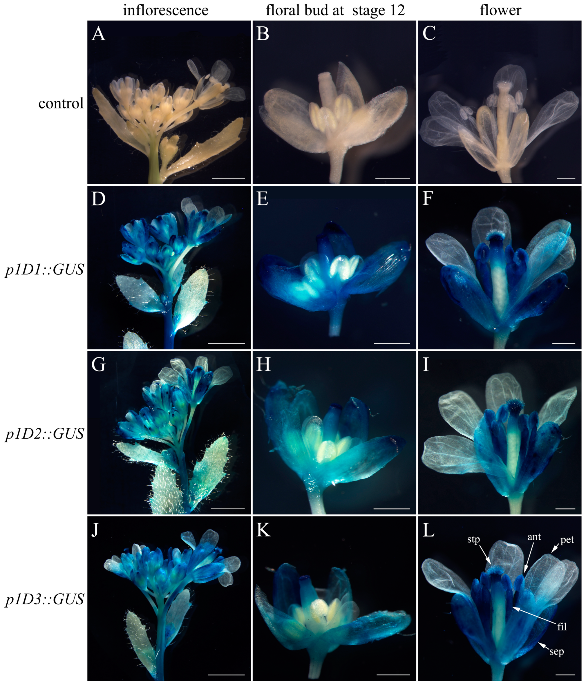
2.3. Deletion Analysis of pCyfaAG1 and pCyfaAG2 in Transgenic Arabidopsis

A GUS reporter gene separately driven by a series of 5′ deletions fragments of pCyfaAG1 and pCyfaAG2 was assayed in transgenic Arabidopsis to analyze the regulatory effect of different regions of the corresponding promoter (Figure 3, Figure 4 and Figure 5). GUS staining was separately examined in the T1 generation of different deletion constructs. Moreover, GUS staining was obviously observed in the inflorescence and mature flower where sepal, stamen (filament and anther), stigma, and stigmatic papillae staining were intense, but was almost absent in petal of p1D1::GUS, p1D2::GUS, and p1D3::GUS transgenic Arabidopsis (Figure 3D,F,G,I,J,L). However, GUS staining suggested that pCyfaAG1 drove GUS to extensively express in the sepal, filament, and gynoecium of stage 12 floral buds, but GUS expression was not observed in the anther and petal in p1D1::GUS transgenic Arabidopsis (Figure 3E) [29]. In addition, relatively weak GUS staining was separately observed in the sepal and gynoecium of stage 12 floral buds, but was almost absent in the petal and stamen of the stage 12 floral buds in p1D2::GUS and p1D3::GUS transgenic Arabidopsis (Figure 3H,K). In addition, GUS staining was obviously observed in the inflorescence and mature flower where sepal, stamen, and gynoecium were intense, but was absent in petal of p2D1::GUS and p2D2::GUS transgenic Arabidopsis, respectively (Figure 4D,F,G,I). Moreover, GUS staining was also observed in the sepal, filament, and gynoecia of stage 12 floral buds, but was absent in the anther and petal in p2D1::GUS and p2D2::GUS transgenic Arabidopsis, respectively (Figure 4E,H). However, GUS staining was only found in floral bud from appearance until stage 12 of p2D3::GUS transgenic Arabidopsis (Figure 4J). Furthermore, GUS staining was observed only in the gynoecium even in stage 12 of p2D3::GUS transgenic Arabidopsis (Figure 4K). A further deletion of the −956/−51 fragment from p2D2 (−956/+187) to produce p2D3 (−50/+187) caused obviously decreased GUS activity in transgenic Arabidopsis (Figure 4J,K and Figure 5B). These results suggested that the −956/−51 regions are capable of inducing pCyfaAG2 promoter activity in the sepal, stamen, and gynoecia, and a 1143 bp region (−956/+187) of pCyfaAG2 was sufficient for driving the CyfaAG2 gene to regulate stamen and gynoecium development.

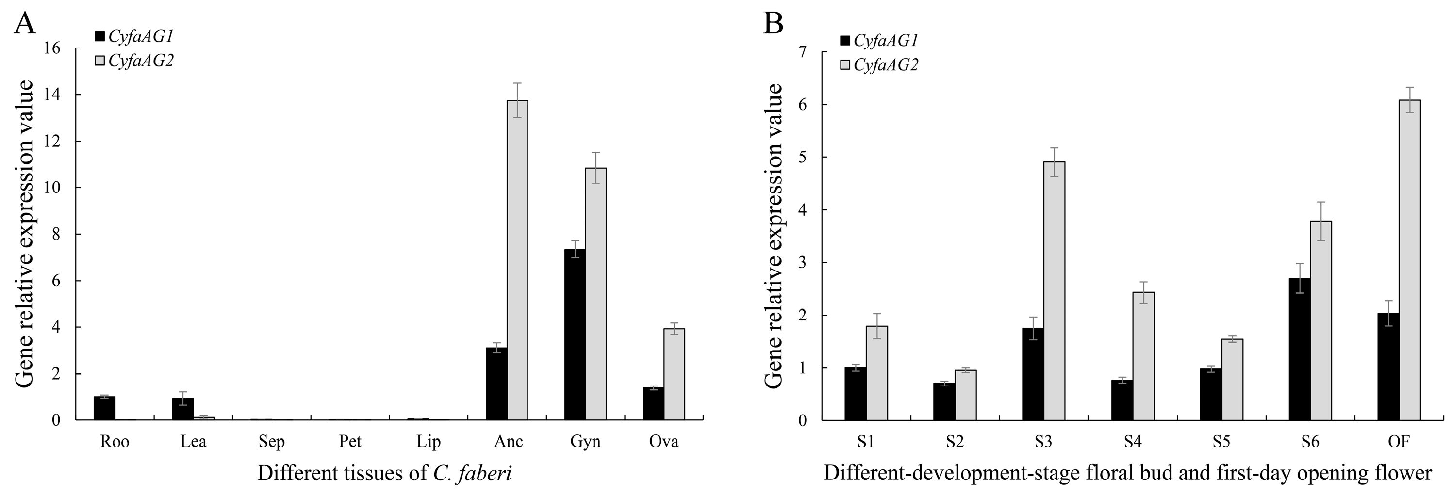
2.4. Expression Analysis of CyfaAG1 and CyfaAG2

CyfaAG1 was mainly expressed in root, leaf, anther cap, gynostemium, and ovary, while CyfaAG2 showed relatively narrower expression zones and was expressed only in the anther cap, gynostemium, and ovary of C. faberi (Figure 6A). In addition, the expression level of CyfaAG1 in the gynostemium was separately significantly higher than in the root, leaf, anther cap, and ovary (p < 0.05), while the expression level of CyfaAG2 in the anther cap was significantly higher than in the gynostemium and ovary (LSD, p < 0.05) (Figure 4A). Moreover, the expression level of CyfaAG2 in the anther cap or gynostemium was significantly higher than that of CyfaAG1 in the anther cap or gynostemium, respectively (p < 0.05) (Figure 6A). CyfaAG1 and CyfaAG2 transcripts became detectable after primodium emergence of floral buds (Figure 6B and Figure 7A). During the seasonal floral bud dormancy by winter chilling (S2), both genes were expressed at low levels (Figure 4B). Moreover, with the dormancy broken and microspore mother cell beginning meiosis, the expressions of CyfaAG1 and CyfaAG2 increased obviously (Figure 6B and Figure 7E,F). Furthermore, CyfaAG1 expression reached a peak at monokaryotic microspore within tetrads (S6), while CyfaAG2 expression reached a peak at 2-cell pollen within mature tetrads of pollinium at the first day of flower opening (Figure 6B and Figure 7N,O,Q,R). However, CyfaAG2 expression was separately significantly higher than that of CyfaAG1 in S3 and of the first-day opening flower (LSD, p < 0.05) (Figure 6B).

2.5. Ectopic Expression of CyfaAG1 and CyfaAG2 in Arabidopsis ag-1 Mutant

To further evaluate the functional divergence of CyfaAG1 and CyfaAG2, we attempted to rescue the loss-of-function Arabidopsis ag-1 mutant using CyfaAG1 and CyfaAG2. 35S::CyfaAG1 and 35S::CyfaAG2 constructs were separately introduced into heterozygote AG/ag-1 Arabidopsis to create complementation lines by agrobacterium-mediated transformation. Transgenic Arabidopsis mutant-allele lines were identified by dCAPS genotyping (Supplementary Figure S3) and were further confirmed by qRT−PCR. Moreover, sixteen 35S::CyfaAG1 lines under wild-type background and 13 independent 35S::CyfaAG1 lines under homozygous ag-1 mutant background were obtained, while ten 35S::CyfaAG2 lines under wild-type background and six independent 35S::CyfaAG2 lines under homozygous ag-1 mutant background were obtained. Furthermore, phenotypes of transgenic Arabidopsis lines were assayed in wild-type and homozygous ag-1 mutant backgrounds to evaluate whether CyfaAG1 and CyfaAG2 could substitute for the endogenous AG gene in determining stamen and carpel identities, respectively. Either 35S::CyfaAG1 or 35S::CyfaAG2 transgenic Arabidopsis under a wild-type background were observed obviously flowering early (Figure 8).
Among sixteen 35S::CyfaAG1 transgenic Arabidopsis under a wild-type background, one (6.25%) showed a strong phenotype with a sepal converted into a carpeloid organ and a new flower in the center (Figure 9C,D); two (12.50%) displayed a medium phenotype with a small sepal in whorl 1, stamenoid petal (filament with petal at the top) in whorl 2, and a big pistil in whorl 4 (Figure 9E,F); four (25.00%) showed a weak phenotype with a small sepal in whorl 1, a small petal in whorl 2, small stamens in whorl 3, and a big pistil in whorl 4 (Figure 9G,H); and the remaining nine lines (56.25%) had flowers similar to the wild-type Arabidopsis flowers.
Among ten 35S::CyfaAG2 transgenic Arabidopsis under a wild-type background, four (40.00%) produced flowers with a small sepal in whorl 1, a stamenoid petal (filament with petal at the top) in whorl 2, and a big pistil in whorl 4 (Figure 9I,J); three (30.00%) showed a weak phenotype with a small sepal in whorl 1, a small petal in whorl 2, a small stamen in whorl 3, and a big pistil in whorl 4 (Figure 9K,L); and the remaining two lines (20.00%) had flowers similar to the wild-type Arabidopsis flowers.
Among thirteen 35S::CyfaAG1 transgenic homozygous ag-1 Arabidopsis, one (7.69%) showed a strong complementation phenotype with a sepal in whorl 1, a petal in whorl 2, a stamen in whorl 3, and a pistil in whorl 4 (Figure 10C,D); three (23.08%) displayed a medium phenotype with many stamens in whorl 2 (Figure 10E,F) or sepals in whorl 1, a stamen in whorl 2, and a fat pistil in the interior whorl (Figure 10E,G); the remaining nine lines (69.23%) displayed no complementation and had flowers similar to the flower of the Arabidopsis ag-1 mutant.
Among six 35S::CyfaAG2 transgenic homozygous ag-1 Arabidopsis, one (16.67%) showed a strong complementation phenotype that consisted of flowers only with sepal and pistil whorls, or filament-like organs and pistil (Figure 10H), one (16.67%) produced flowers with a sepal in whorl 1, filament-like organs in whorl 2, and a fat pistil in the interior whorl (Figure 10I), and the remaining four lines (66.67%) displayed no complementation and had flowers similar with the flower of the Arabidopsis ag-1 mutant.

3. Discussion

Orchid reproductive organs (gynostemium) show dramatic changes in morphology compared to the reproductive organs (stamens and carpels) of other angiosperms. In the model species A. thaliana, the floral homeotic C-class gene AG is responsible for the specification of reproductive organ identity and the control of floral meristem determinacy, as well as the prevention of the misexpression of A-function genes in the reproductive organs [4,5,30]. AG expression starts at stage 3 of flower development in the domains of the floral meristem, where stamen and carpel primordia develop, and its expression remains during all stages of stamen and carpel development [11]. Other rosid species AG orthologs, such as PrseAG from Prunus lannesiana [31], KjAG from Kerria japonica [32] and EjAG from Eriobotrya japonica [33], were mainly expressed in stamens and carpels, and showed a conserved function for reproductive organ identity determination in core eudicot species. A duplication event of the AG lineage in the order Ranunculales, a sister lineage to all other eudicots, results in two functionally redundant but distinguishable AG-lineage members [34,35]. ThtAG1 and ThtAG2 are two AG orthologs from Thalictrum thalictroides (Ranunculaceae); ThtAG1 showed a highly conserved C- function, whereas ThtAG2 determined ovule identity [35]. Moreover, NdAG1 and NdAG2 are AG lineage genes from Nigella damascene (Ranunculaceae); NdAG1 confers C-function specifying stamens and carpels identities, while NdAG2 is involved in regulating carpel development and floral determinacy [36]. Two AG-lineage genes, MawuAG1 and MawuAG2, were also found in the basal angiosperm Magnolia wufengensis. MawuAG1 is mainly expressed in stamen, carpel, ovule, and seed, and shows highly conserved C-function in the determination of stamen, carpel, and ovule identity, while MawuAG2 is mainly expressed in stamen and carpel, and may be only involved in stamen development [37]. All these data suggest that gene duplication led to functional redundancy or sub-functionalization of both AG paralogs in eudicots.
In monocot rice, two AG lineage genes, OsMADS3 and OsMADS58, redundantly mediate the C-function: OsMADS3 plays a predominant role in stamen specification, but OSMADS58 plays a predominant role in floral meristem determinacy and carpel morphogenesis [11]. Three AG lineage genes, namely, ZMM2 and ZMM23 (both orthologs of rice OsMADS3), and ZAG1 (ortholog of rice OsMADS58) were found in Zea mays [13,14]. ZAG1 is expressed early in stamen and carpel primordia, and determines the floral meristem, ZMM2 is mainly expressed in the anthers and participates in regulating the formation of stamens and carpels [38]. In orchid C. sinense, CsAG1 expressed in the tepals, lips, and gynostemium, and conferred C-function in reproductive organ development, while CsAG2 was highly expressed in the gynostemium but its function remained unclear [9]. In addition, two AG-like genes, CeMADS1 and CeMADS2, were also found in C. ensifolium [15]. CeMADS1 was expressed only in the gynostemium and may have a pivotal C function in reproductive organ development, while CeMADS2 was expressed in all floral organs and predominantly in gynostemium and its function remained unclear [15].
Here, we have also isolated two AG-like genes (CyfaAG1 and CyfaAG2) from C. faberi. CyfaAG1 is mainly expressed in root, leaf, anther cap, gynostemium, and ovary, while CyfaAG2 was expressed only in anther cap, gynostemium, and ovary of C. faberi. Moreover, pCyfaAG1 and pCyfaAG2 contain CArG-box for MADS-box transcription factor binding and conserved binding sites (AACAAA-/TTTGTT-motif) for floral homeotic APETALA2 transcription factor recognition [16,19]. In addition, both pCyfaAG1 and pCyfaAG2 could drive GUS to extensively express in stamens and carpels of transgenic Arabidopsis. Furthermore, ectopic expressions of CyfaAG1 and CyfaAG2 separately promote early flowering phenotypes. Moreover, ectopic expression of CyfaAG1 produced flowers with sepal converted into carpeloid organ and a new flower in the center or a stamenoid petal (filament with petal at the top) in whorl 2 in wild-type Arabidopsis, while ectopic expression of CyfaAG2 produced flowers with a stamenoid petal (filament with petal at the top) in whorl 2. Furthermore, complementation of loss-of-function Arabidopsis ag-1 mutant suggested that CyfaAG1 could mimic the endogenous AG gene to specify stamen and carpel identity. However, ectopic expression of CyfaAG2 only completes filament and carpel development in the 35S::CyfaAG2 transgenic Arabidopsis ag-1 mutant. Based on these results, it seems that CyfaAG1 and CyfaAG2 work redundantly. CyfaAG1 may confer C-function specifying stamens and carpels identities, as well as floral meristem determinacy, while CyfaAG2 may only be involved in filament and carpel development. Both genes work together to regulate normal gynostemium and ovary development in C. faberi. Our data provide more details to understand how C-class function has been partitioned in orchid, and the functions of two AG-like genes regulating gynostemium development of C. faberi.

4. Materials and Methods
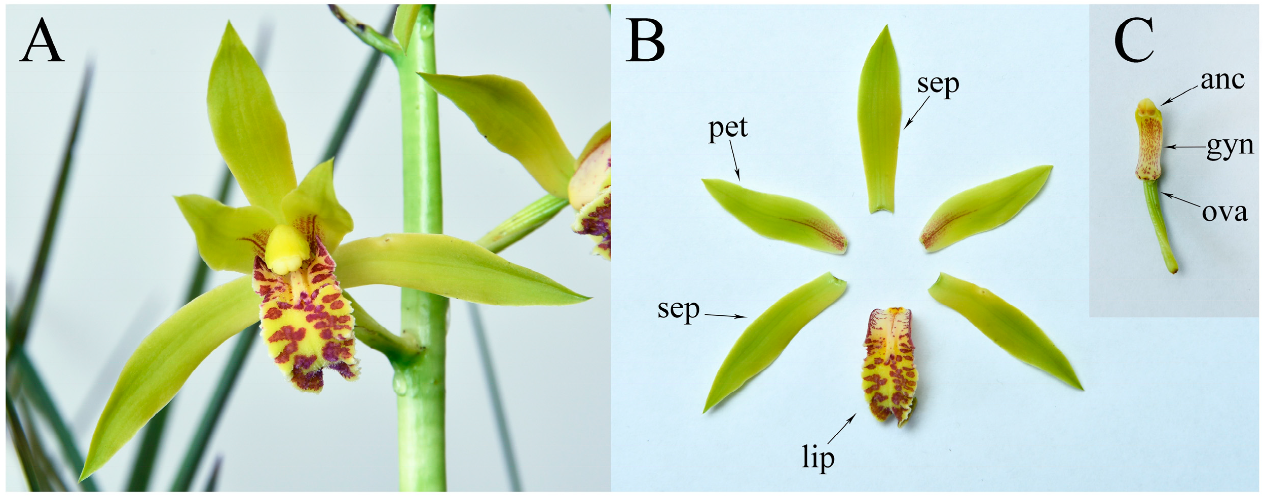
4.1. Plant Material

C. faberi plants were cultivated in redware flowerpots (30 cm × 18.5 cm × 28.2 cm) filled with orchid substrates in the botanical garden of Yangtze University under natural conditions in Jingzhou City, China. Juvenile leaf, sepal, petal, lip, anther, gynostemium, and ovary were separately dissected when the orchid began to bloom, immediately frozen in liquid nitrogen, and stored at −80 °C until used. Moreover, 5 mm (early stages), 8 mm (seasonal dormancy of floral bud), 10 mm (dormancy breaking and further development), 15 mm, 20 mm, and 25 mm long floral buds, and first-day opening flowers were also sampled.

4.2. Isolation and Characterization of CyfaAG1 and CyfaAG2 from Cymbidium faberi

Total RNA was extracted from floral buds using an EASYspin Plant RNA Kit (Aidlab China) according to the manufacturer’s protocol. The first-strand cDNA of 3′ RACE was prepared, and then the 3′end cDNA sequences of CyfaAG1 and CyfaAG2 were separately amplified with gene-specific forward primers 3RGSPAG1F and 3RGSPAG2F (Supplementary Table S1) using the 3-full RACE Core Set Ver. 2.0 kit (TaKaRa, Japan) according to the manufacturer’s protocol, but with 45 s annealing at 59 °C. The forward primers design was referenced the sequence of AG orthologs CsAG2 (Genbank accession numbers: MG021185.1) and CsAG1 (Genbank accession numbers: MG021184.1) from orchid’s close relative species C. ensifolium. In addition, the 5′ end cDNA sequences of CyfaAG1 were amplified through 5′RACE using the 5′RACE System for Rapid Amplification of cDNA Ends (Invitrogen, Carlsbad, CA, USA) following the manufacturer’s protocol and the gene-specific primers 5RAG1GSP1, 5RAG1GSP2, and 5RAG1GSP3 (Supplementary Table S1). The 5′ end cDNA sequences of CyfaAG2 were amplified according to the above method, but with the gene-specific primers 5RAG2GSP1, 5RAG2GSP2, and 5RAG2GSP3 (Supplementary Table S1). The phylogenetic tree was constructed by using MEGA version 5.05 with the neighbor-joining (NJ) method. The NJ tree was constructed with 1000 bootstrap replications. All the C-class and D-class MADS-box transcription factors (TF) with complete sequences were obtained from NCBI Genbank (Supplementary Table S2).

4.3. Isolation and Sequence Analysis of CyfaAG1 and CyfaAG2 Promoters from Cymbidium faberi

C. faberi genomic DNA was extracted from juvenile leaves using the CTAB Plant Genomic DNA Rapid Extraction Kit (Aidlab, Beijing, China) according to the manufacturer’s protocol. The CyfaAG1 5′ flanking regions were amplified from C. faberi genomic DNA using the Genome Walking Kit (TaKaRa, Japan) following the manufacturer’s protocol and with gene-specific primers pAG1SP1, pAG1SP2, and pAG1SP3 (Supplementary Table S1) for the first walking sequencing, and with the gene-specific primers pAG1SP4, pAG1SP5, and pAG1SP6 (Supplementary Table S1) for the second walking sequencing. Meanwhile, the CyfaAG2 5′ flanking regions were amplified according to the above method, but with the gene-specific primers pAG2SP1, pAG2SP2, and pAG2SP3 (Supplementary Table S1) for the first walking sequencing, and with the gene-specific primers pAG2SP4, pAG2SP5, and pAG2SP6 (Supplementary Table S1) for the second walking sequencing, and the gene-specific primers pAG2SP7, pAG2SP8, and pAG2SP9 (Supplementary Table S1) for the third sequencing. The primers designed for the first walking were separately based on the corresponding gene sequences CyfaAG1 (Genbank accession number: MH917913) and CyfaAG2 (Genbank accession number: MH917914). The putative transcription start sites of CyfaAG1 and CyfaAG2 were searched through 5′RACE according to the above method. The cis-acting elements located at the CyfaAG1 and CyfaAG2 promoter regions were separately searched in the PLACE database [39].

4.4. Characterization of pCyfaAG1 and pCyfaAG2 Activity from the 5′ Deleted Promoter Fragments in Transgenic Arabidopsis

We separately designed three forward primers (TpCyfaAG1F, TpCyfaAG1F1, and TpCyfaAG1F2) and a reversed primer TpCyfaAG1R (Supplementary Table S1) to amplify 5′-deletion fragments of pCyfaAG1. In addition, we also designed three forward primers (TpCyfaAG2F, TpCyfaAG2F1, and TpCyfaAG2F2) and a reversed primer TpCyfaAG2R (Supplementary Table S1) to amplify the promoter deletions of pCyfaAG2, respectively. Three 5′-deletion fragments of pCyfaAG1 were separately designated as p1D1 (−1207/+185), p1D2 (−1191/+185), and p1D3(−387/+185), and then cloned into the pCAMBIA1300 vector with Xba I and Sac I using the ClonExpress® Ultra One Step Cloning Kit (Vazyme, Nanjing, China) following the manufacturer’s protocol. In addition, three 5′-deletion fragments of pCyfaAG2 were separately designated as p2D1 (−1473/+187), p2D2 (−956/+187), and p2D3 (−50/+187), and then cloned into the pCAMBIA1300 vector by using the above method. All the constructs were separately transformed into A. thaliana Col-0 plants (ecotype Columbia) with the floral-dip method suggested by Clough and Bent [40]. Transgenic Arabidopsis seedlings were selected, cultivated, and prepared for histochemical GUS staining according to Zeng et al. [41]. The expression of the GUS gene controlled by different 5′-deletions of pCyfaAG1 and pCyfaAG2 were also separately confirmed via qRT−PCR in transgenic Arabidopsis with the primers qGUSF and qGUSR (Supplementary Table S1). An amplification fragment of A. thaliana Actin (Genbank accession numbers: AY114679.1) with the primers qActinF and qActinR (Supplementary Table S1) was used as the internal control.

4.5. Cytomorphological Examination and Expression Analysis of CyfaAG1 and CyfaAG2

The six stages of floral buds described above and first-day opening flower of C. faberi were separately sampled and fixed in FAA (38% formaldehyde: acetic acid: 70% ethanol = 1:1:18, by vol.). The samples were dehydrated in an ethanol series (1.5 h each), cleared in a xylene series (2 h each), infiltrated with a xylene and paraffin (12 h at 38 °C) series, followed by three changes of 100% molten paraffin at 60 °C (4 h each), then embedded into a paraffin block, which was serially sectioned at a thickness of 8 µm with a Leica RM2235 rotary microtome after two days. Subsequently, the sections were stained with safranin-fast green [42]. The sections were observed under a CAIKON RCK-40C microscope and photomicrographs were subsequently taken.
The total RNA of each sample was extracted according to the above method, but the first-strand cDNA was synthesized for quantitative real-time PCR (qRT-PCR) by using the HiScript® II Q RT SuperMix for qPCR kit (Vazyme, Nanjing, China) following the manufacturer’s protocol. The relative expressions of CyfaAG1 and CyfaAG2 were separately detected in root, juvenile leave, sepal, petal, lip, anther, gynostemium, and ovary of C. faberi plants according to Fei and Liu [1], but with the gene-specific forward primer qCyfaAG1F and reverse primer qCyfaAG1R for CyfaAG1, and with the gene-specific forward primer qCyfaAG2F and reverse primer qCyfaAG2R for CyfaAG2, respectively (Supplementary Table S1). The qRT−PCR was performed with three biological replicates and amplification of C. faberi actin (Genbank accession numbers: JN177719.1) fragment was used as the internal control with the forward primer qCyfaactinF and reverse qCyfaactinR (Supplementary Table S1).

4.6. Ectopic Expression of CyfaAG1/CyfaAG2 in Arabidopsis ag-1 Mutant and Function Analysis

Full-length CyfaAG1 cDNAs in the sense orientation were cloned into the pBI121 vector with Xba I and Sma I restriction enzymes, and the forward primer TCyfaAG1F and the reverse primer TCyfaAG1R (Supplementary Table S1) under control of the CaMV35S promoter using the ClonExpress® Ultra One Step Cloning Kit (Vazyme, Nanjing, China) according to the manufacturer’s protocol. Full-length CyfaAG2 cDNAs in the sense orientation were cloned into the pBI121 vector according to the above method, but with the forward primer TCyfaAG2F and the reverse primer TCyfaAG2R (Supplementary Table S1). The 35S::CyfaAG1 and 35S::CyfaAG2 constructs were separately transformed into heterozygous Ag/ag-1 Arabidopsis using the floral-dip method suggested by Clough and Bent [40]. Transgenic Arabidopsis seeds were selected and seedlings were subsequently transplanted into soil for cultivation in a growth chamber according to Li et al. [43]. Wild-type, heterozygous AG/ag-1 and homozygous ag-1 transgenic Arabidopsis lines were confirmed by the dCAPS finder program suggested by Neff et al. [44]. The phenotypes of transgenic Arabidopsis were analyzed after flowering. In addition, the complementation degrees of independent transgenic lines of 35S::CyfaAG1 and 35S::CyfaAG2 homozygous ag-1 Arabidopsis were categorized as ‘no complementation’, ‘weak complementation’, ‘medium complementation’ and ‘strong complementation’, respectively. Moreover, independent transgenic lines of each complementation degree were confirmed by qRT−PCR with the primers qCyfaAG1F and qCyfaAG1R for CyfaAG1 (Supplementary Table S1), and with the primers qCyfaAG2F and qCyfaAG2R suggested above for CyfaAG2 (Supplementary Table S1), respectively. An amplification fragment of A. thaliana Actin (Genbank accession numbers: AY114679.1) with the primers qActinF and qActinR was used as the internal control.

5. Conclusions

Orchid reproductive organs (gynostemium) show dramatic changes in morphology compared to reproductive organs (stamens and carpels) of other angiosperms. In Arabidopsis, AGAMOUS (AG) play roles in determining stamens and carpels identities, floral meristem determinacy, and repression of the A-function. However, the molecular basis of orchid gynostemium development remains largely unknown. In this study, two AG-like genes, CyfaAG1 and CyfaAG2, and their promoters were isolated and functionally characterized in a Chinese famous traditional orchid C. faberi. Both CyfaAG1 and CyfaAG2 are highly expressed in the anther cap, gynostemium, and ovary. Moreover, both pCyfaAG1 and pCyfaAG2 could drive GUS to extensively express in stamens and carpels of transgenic Arabidopsis. Ectopic expression of CyfaAG1 and CyfaAG2 promotes early flowering of wild-type Arabidopsis. Moreover, ectopic expression of CyfaAG1 completely rescues floral defects in the Arabidopsis ag-1 mutant, while ectopic expression of CyfaAG2 only completes filament and carpel development. Our findings suggest that CyfaAG1 acts as an evolutionarily conserved C-function gene in determining reproductive organ identity and mediating floral meristem determinacy. CyfaAG2 redundantly mediates the C-function in floral meristem determinacy and gynostemium development. Our results provide more details to understand how the C-class function has been partitioned in orchid, and the roles of two AG orthologs involved in the gynostemium development of C. faberi.

Supplementary Materials

The following supporting information can be downloaded at: https://www.mdpi.com/article/10.3390/plants12142740/s1. Table S1: Primers used in this study. Table S2: Information on Sequences selected for alignments and phylogenetic analyses from NCBI GenBank. Figure S1: CyfaAG1 promoter sequence. Figure S2: CyfaAG2 promoter sequence. Figure S3. Genotyping of wild-type, heterozygote and homozygous ag-1 mutant A. thaliana (Landsberg erecta) plants by dCAPS.

Author Contributions

J.L., writing—original draft preparation; L.W., X.C., L.Z. and Y.S., methodology; Z.L., writing—review and editing, supervision. All authors have read and agreed to the published version of the manuscript.

Funding

This research received no external funding.

Data Availability Statement

All data generated or analyzed during this study are included in this published article.

Conflicts of Interest

The authors declare no conflict of interest.

References

  1. Fei, Y.; Liu, Z. Isolation and Characterization of the PISTILLATA Ortholog Gene from Cymbidium faberi Rolfe. Agronomy 2019, 9, 425. [Google Scholar] [CrossRef] [Green Version]
  2. Stewart, D.; Graciet, E.; Wellmer, F. Molecular and regulatory mechanisms controlling floral organ development. FEBS J. 2016, 283, 1823–1830. [Google Scholar] [CrossRef] [PubMed]
  3. Theißen, G.; Melzer, R.; Rümpler, F. MADS-domain transcription factors and the floral quartet model of flower development: Linking plant development and evolution. Development 2016, 143, 3259–3271. [Google Scholar] [CrossRef] [Green Version]
  4. Pelayo, M.A.; Yamaguchi, N.; Ito, T. One factor, many systems: The floral homeotic protein AGAMOUS and its epigenetic regulatory mechanisms. Curr. Opin. Plant Biol. 2021, 61, 102009. [Google Scholar] [CrossRef] [PubMed]
  5. Dreni, L.; Kater, M.M. MADS reloaded: Evolution of the AGAMOUS subfamily genes. New Phytol. 2014, 201, 717–732. [Google Scholar] [CrossRef]
  6. Dreni, L.; Osnato, M.; Kater, M.M. The ins and outs of the rice AGAMOUS subfamily. Mol. Plant 2013, 6, 650–664. [Google Scholar] [CrossRef] [Green Version]
  7. Wang, Y.; Li, Y.; Yan, X.; Ding, L.; Shen, L.; Yu, H. Characterization of C- and D-Class MADS-Box Genes in Orchids. Plant Physiol. 2020, 184, 1469–1481. [Google Scholar] [CrossRef]
  8. Zhao, J.; Gong, P.; Liu, H.; Zhang, M.; He, C. Multiple and integrated functions of floral C-class MADS-box genes in flower and fruit development of Physalis floridana. Plant Mol. Biol. 2021, 107, 101–116. [Google Scholar] [CrossRef]
  9. Su, S.; Shao, X.; Zhu, C.; Xu, J.; Tang, Y.; Luo, D.; Huang, X. An AGAMOUS-like factor is associated with the origin of two domesticated varieties in Cymbidium sinense (Orchidaceae). Hortic. Res. 2018, 5, 48. [Google Scholar] [CrossRef] [Green Version]
  10. Yamaguchi, T.; Lee, D.Y.; Miyao, A.; Hirochika, H.; An, G.; Hirano, H.Y. Functional diversification of the two C-class MADS box genes OSMADS3 and OSMADS58 in Oryza sativa. Plant Cell 2006, 18, 15–28. [Google Scholar] [CrossRef] [Green Version]
  11. Dreni, L.; Pilatone, A.; Yun, D.; Erreni, S.; Pajoro, A.; Caporali, E.; Zhang, D.; Kater, M.M. Functional analysis of all AGAMOUS subfamily members in rice reveals their roles in reproductive organ identity determination and meristem determinacy. Plant Cell 2011, 23, 2850–2863. [Google Scholar] [CrossRef] [PubMed] [Green Version]
  12. Yoshida, H.; Nagato, Y. Flower development in rice. J. Exp. Bot. 2011, 62, 4719–4730. [Google Scholar] [CrossRef] [Green Version]
  13. Mena, M.; Ambrose, B.A.; Meeley, R.B.; Briggs, S.P.; Yanofsky, M.F.; Schmidt, R.J. Diversification of C-function activity in maize flower development. Science 1996, 274, 1537–1540. [Google Scholar] [CrossRef]
  14. Shen, C.; Li, G.; Dreni, L.; Zhang, D. Molecular Control of Carpel Development in the Grass Family. Front. Plant Sci. 2021, 12, 635500. [Google Scholar] [CrossRef]
  15. Wang, S.; Lee, P.F.; Lee, Y.I.; Hsiao, Y.Y.; Chen, Y.; Pan, Z.; Liu, Z.; Tsai, W.C. Duplicated C-class MADS-box genes reveal distinct roles in gynostemium development in Cymbidium ensifolium (Orchidaceae). Plant Cell Physiol. 2011, 52, 563–577. [Google Scholar] [CrossRef] [PubMed] [Green Version]
  16. De Folter, S.; Angenent, G.C. Trans meets cis in MADS science. Trends Plant Sci. 2006, 11, 224–231. [Google Scholar] [CrossRef] [PubMed]
  17. Filichkin, S.A.; Leonard, J.M.; Monteros, A.; Liu, P.; Nonogaki, H. A novel endo-beta-mannanase gene in tomato LeMAN5 is associated with anther and pollen development. Plant Physiol. 2004, 134, 1080–1087. [Google Scholar] [CrossRef] [PubMed] [Green Version]
  18. Rogers, H.J.; Bate, N.; Combe, J.; Sullivan, J.; Sweetman, J.; Swan, C.; Lonsdale, D.M.; Twell, D. Functional analysis of cis-regulatory elements within the promoter of the tobacco late pollen gene g10. Plant Mol. Biol. 2001, 45, 577–585. [Google Scholar] [CrossRef]
  19. Dinh, T.T.; Girke, T.; Liu, X.; Yant, L.; Schmid, M.; Chen, X. Development. The floral homeotic protein APETALA2 recognizes and acts through an AT-rich sequence element. Development 2012, 139, 1978–1986. [Google Scholar] [CrossRef] [Green Version]
  20. Zhang, Z.; Xie, Z.; Zou, X.; Casaretto, J.; Ho, T.H.; Shen, Q.J. A rice WRKY gene encodes a transcriptional repressor of the gibberellin signaling pathway in aleurone cells. Plant Physiol. 2004, 134, 1500–1513. [Google Scholar] [CrossRef] [Green Version]
  21. Sutoh, K.; Yamauchi, D. Two cis-acting elements necessary and sufficient for gibberellin-upregulated proteinase expression in rice seeds. Plant J. 2003, 34, 635–645. [Google Scholar] [CrossRef] [PubMed] [Green Version]
  22. Abe, H.; Urao, T.; Ito, T.; Seki, M.; Shinozaki, K.; Yamaguchi-Shinozaki, K. Arabidopsis AtMYC2 (bHLH) and AtMYB2 (MYB) function as transcriptional activators in abscisic acid signaling. Plant Cell 2003, 15, 63–78. [Google Scholar] [CrossRef] [PubMed] [Green Version]
  23. Nagao, R.T.; Goekjian, V.H.; Hong, J.C.; Key, J.L. Identification of protein-binding DNA sequences in an auxin-regulated gene of soybean. Plant Mol. Biol. 1993, 21, 1147–1162. [Google Scholar] [CrossRef]
  24. Mena, M.; Cejudo, F.J.; Isabel-Lamoneda, I.; Carbonero, P. A Role for the DOF Transcription Factor BPBF in the Regulation of Gibberellin-Responsive Genes in Barley Aleurone. Plant Physiol. 2002, 130, 111–119. [Google Scholar] [CrossRef] [Green Version]
  25. Ross, E.J.; Stone, J.M.; Elowsky, C.G.; Arredondo-Peter, R.; Klucas, R.V.; Sarath, G. Activation of the Oryza sativa non-symbiotic haemoglobin-2 promoter by the cytokinin-regulated transcription factor, ARR1. J. Exp. Bot. 2004, 55, 1721–1731. [Google Scholar] [CrossRef]
  26. Gowik, U.; Burscheidt, J.; Akyildiz, M.; Schlue, U.; Koczor, M.; Streubel, M.; Westhoff, P. cis-Regulatory elements for mesophyll-specific gene expression in the C4 plant Flaveria trinervia, the promoter of the C4 phosphoenolpyruvate carboxylase gene. Plant Cell 2004, 16, 1077–1090. [Google Scholar] [CrossRef] [PubMed] [Green Version]
  27. Tatematsu, K.; Ward, S.; Leyser, O.; Kamiya, Y.; Nambara, E. Identification of cis-elements that regulate gene expression during initiation of axillary bud outgrowth in Arabidopsis. Plant Physiol. 2005, 138, 757–766. [Google Scholar] [CrossRef] [Green Version]
  28. Wenkel, S.; Turck, F.; Singer, K.; Gissot, L.; Le Gourrierec, J.; Samach, A.; Coupland, G. CONSTANS and the CCAAT Box Binding Complex Share a Functionally Important Domain and Interact to Regulate Flowering of Arabidopsis. Plant Cell 2006, 18, 2971–2984. [Google Scholar] [CrossRef] [Green Version]
  29. Smyth, D.R.; Bowman, J.L.; Meyerowitz, E.M. Early flower development in Arabidopsis. Plant Cell 1990, 2, 755–767. [Google Scholar] [CrossRef] [Green Version]
  30. ÓMaoiléidigh, D.S.; Wuest, S.E.; Rae, L.; Raganelli, A.; Ryan, P.T.; Kwasniewska, K.; Das, P.; Lohan, A.J.; Loftus, B.; Graciet, E.; et al. Control of reproductive floral organ identity specification in Arabidopsis by the C function regulator AGAMOUS. Plant Cell 2013, 25, 2482–2503. [Google Scholar] [CrossRef] [Green Version]
  31. Liu, Z.; Zhang, D.; Liu, D.; Li, F.; Lu, H. Exon skipping of AGAMOUS homolog PrseAG in developing double flowers of Prunus lannesiana (Rosaceae). Plant Cell Rep. 2013, 32, 227–237. [Google Scholar] [CrossRef]
  32. Ma, J.; Shen, X.; Liu, Z.; Zhang, D.; Liu, W.; Liang, H.; Wang, Y.; He, Z.; Chen, F. Isolation and Characterization of AGAMOUS-Like Genes Associated With Double-Flower Morphogenesis in Kerria japonica (Rosaceae). Front. Plant Sci. 2018, 9, 959. [Google Scholar] [CrossRef] [Green Version]
  33. Xia, Y.; Shi, M.; Chen, W.; Hu, R.; Jing, D.; Wu, D.; Wang, S.; Li, Q.; Deng, H.; Guo, Q.; et al. Expression Pattern and Functional Characterization of PISTILLATA Ortholog Associated With the Formation of Petaloid Sepals in Double-Flower Eriobotrya japonica (Rosaceae). Front. Plant Sci. 2020, 10, 1685. [Google Scholar] [CrossRef] [Green Version]
  34. Kramer, E.M.; Jaramillo, M.A.; Di Stilio, V.S. Patterns of gene duplication and functional evolution during the diversification of the AGAMOUS subfamily of MADS box genes in angiosperms. Genetics 2004, 166, 1011–1023. [Google Scholar] [CrossRef] [PubMed]
  35. Galimba, K.D.; Di Stilio, V.S. Sub-functionalization to ovule development following duplication of a floral organ identity gene. Dev. Biol. 2015, 405, 158–172. [Google Scholar] [CrossRef] [PubMed] [Green Version]
  36. Wang, P.; Liao, H.; Zhang, W.; Yu, X.; Zhang, R.; Shan, H.; Duan, X.; Yao, X.; Kong, H. Flexibility in the structure of spiral flowers and its underlying mechanisms. Nat. Plants 2015, 2, 15188. [Google Scholar] [CrossRef] [PubMed]
  37. Ma, J.; Deng, S.; Jia, Z.; Sang, Z.; Zhu, Z.; Zhou, C.; Ma, L.; Chen, F. Conservation and divergence of ancestral AGAMOUS/SEEDSTICK subfamily genes from the basal angiosperm Magnolia wufengensis. Tree Physiol. 2020, 40, 90–107. [Google Scholar] [CrossRef] [PubMed]
  38. Callens, C.; Tucker, M.R.; Zhang, D.; Wilson, Z.A. Dissecting the role of MADS-box genes in monocot floral development and diversity. J. Exp. Bot. 2018, 69, 2435–2459. [Google Scholar] [CrossRef] [Green Version]
  39. Higo, K.; Ugawa, Y.; Iwamoto, M.; Korenaga, T. Plant cis-acting regulatory DNA elements (PLACE) database: 1999. Nucleic Acids Res. 1999, 27, 297–300. [Google Scholar] [CrossRef] [Green Version]
  40. Clough, S.J.; Bent, A.F. Floral dip: A simplified method for Agrobacterium-mediated transformation of Arabidopsis thaliana. Plant J. 1998, 16, 735–743. [Google Scholar] [CrossRef] [PubMed] [Green Version]
  41. Zeng, L.; Zhang, J.; Wang, X.; Liu, Z. Isolation and Characterization of APETALA3 Orthologs and Promoters from the Distylous Fagopyrum esculentum. Plants 2021, 10, 1644. [Google Scholar] [CrossRef]
  42. Liu, Z.; Xiong, H.; Li, L.; Fei, Y. Functional Conservation of an AGAMOUS Orthologous Gene Controlling Reproductive Organ Development in the Gymnosperm Species Taxus chinensis var. mairei. J. Plant Biol. 2018, 61, 50–59. [Google Scholar] [CrossRef]
  43. Li, L.; Fang, Z.; Li, X.; Liu, Z. Isolation and Characterization of the C-class MADS-box Gene from the Distylous Pseudo-cereal Fagopyrum esculentum. J. Plant Biol. 2017, 60, 189–198. [Google Scholar] [CrossRef]
  44. Neff, M.M.; Neff, J.D.; Chory, J. Pepper AE (1998) dCAPS, a simple technique for the genetic analysis of single nucleotide polymorphisms: Experimental applications in Arabidopsis thaliana genetics. Plant J. 1998, 14, 387–392. [Google Scholar] [CrossRef] [PubMed]
Figure 1. Flower of Cymbidium faberi. (A) Flower of C. faberi; (B) perianth (sepal, petal and lips) of C. faberi; (C) anther cap, gynostemium, and ovary of C. faberi. Sepals (sep), petals (pet), lips (lip), anther cap (anc), gynostemium (gyn), ovary (ova).
Plants 12 02740 g001
Figure 2. Phylogenetic tree of CyfaAG1, CyfaAG2, and other AG subfamily transcription factors from different clades of angiosperms.
Plants 12 02740 g002
Figure 3. Deletion analysis of the pCyfaAG1 promoter and histochemical GUS staining in the T1 generation transgenic Arabidopsis. (A) Wild-type Arabidopsis inflorescence; (B) stage 12 floral bud of wild-type Arabidopsis [29]; (C) wild-type Arabidopsis flower; (D) inflorescence of p1D1::GUS transgenic Arabidopsis; (E) stage 12 floral bud of p1D1::GUS transgenic Arabidopsis; (F) flower of p1D1::GUS transgenic Arabidopsis; (G) inflorescence of p1D2::GUS transgenic Arabidopsis; (H) stage 12 floral bud of p1D2::GUS transgenic Arabidopsis; (I) flower of p1D2::GUS transgenic Arabidopsis; (J) inflorescence of p1D3::GUS transgenic Arabidopsis; (K) stage 12 floral bud of p1D3::GUS transgenic Arabidopsis; (L) mature flower of p1D3::GUS transgenic Arabidopsis. Sepal (sep), petal (pet), anther (ant), filament (fil), stigmatic papillae (stp). Scale bars: (A,D,G,J) 2 mm; (C,F,I,L) 500 μm.
Plants 12 02740 g003
Figure 4. Deletion analysis of the pCyfaAG2 promoter and histochemical GUS staining in the T1 generation transgenic Arabidopsis. (A) Wild-type Arabidopsis inflorescence; (B) stage 12 floral bud of wild-type Arabidopsis [29]; (C) wild-type Arabidopsis flower; (D) inflorescence of p2D1::GUS transgenic Arabidopsis; (E) stage 12 floral bud of p2D1::GUS transgenic Arabidopsis; (F) flower of p2D1::GUS transgenic Arabidopsis; (G) inflorescence of p2D2::GUS transgenic Arabidopsis; (H) stage 12 floral bud of p2D2::GUS transgenic Arabidopsis; (I) flower of p2D2::GUS transgenic Arabidopsis; (J) inflorescence of p2D3::GUS transgenic Arabidopsis; (K) stage 12 floral bud of p2D3::GUS transgenic Arabidopsis; (L) flower of p2D3::GUS transgenic Arabidopsis. Sepal (sep), petal (pet), anther (ant), filament (fil), stigmatic papillae (stp). Scale bars: (A,D,G,J) 2 mm; (C,F,I,L) 500 μm.
Plants 12 02740 g004
Figure 5. GUS expression in different transgenic Arabidopsis was separately confirmed by qRT−PCR with actin as the internal control. (A) GUS expression in p1D1::GUS, p1D2::GUS, and p1D3::GUS transgenic Arabidopsis was separately confirmed by qRT−PCR. (B) GUS expression in p2D1::GUS, p2D2::GUS, and p2D3::GUS transgenic Arabidopsis was separately confirmed by qRT−PCR.
Plants 12 02740 g005
Figure 6. Expression patterns of CyfaAG1 and CyfaAG2. (A) CyfaAG1 and CyfaAG2 expression in the root (Roo), juvenile leaf (Lea), sepal (Sep), petal (Pet), lip (Lip), anther cap (Anc), gynostemium (Gyn), and ovary (Ova) were detected by qRT−PCR with Cyfaactin as the internal control; (B) CyfaAG1 and CyfaAG2 expression in different development stages of the floral bud and at the first day of flower opening.
Plants 12 02740 g006
Figure 7. Morphology of different development stages of floral bud, anther, and ovary of C. faberi. (A) Stage 1 floral bud, about 5 mm in length; (B) stage 2 floral bud, about 8 mm in length (seasonal floral bud dormancy); (C) anther, enlargement of (B), microspore mother cell; (D) ovary, enlargement of (B), ovule primordia (Ovp) formation; (E) stage 3 floral bud, about 10 mm in length (breaking dormancy and further development); (F) anther, enlargement of (E), microspore mother cell beginning meiosis; (G) ovary, enlargement of (E), ovule primordia and archesporial cells (Arc); (H) stage 4 floral bud, about 15 mm in length; (I) anther, enlargement of (H), microspore mother cell beginning meiosis; (J) ovule, enlargement of (H), megaspore mother cell (Mmc); (K) stage 5 floral bud, about 20 mm in length; (L) anther, enlargement of (K), formation of microspore tetrads; (M) ovule, enlargement of (K), meiosis of megaspore mother cell (Mem); (N) anther, longitudinal section of stage 6 floral bud, about 25 mm in length; (O) anther, enlargement of (N), monokaryotic microspore within tetrads; (P) ovule, enlargement of (N), meiosis of megaspore mother cell; (Q) anther, longitudinal section of the first-day opening flower; (R) pollinium, enlargement of (Q), 2-cell pollen within mature tetrads of pollinium; (S) ovule, enlargement of (Q), linear tetrad (Tet) of megaspores, functional megaspore (Fme) formation. Scale bar: (A,B,E,H,K,N,Q) 1 mm; (C,D,F,G,I,J,L,M,O,P,R,S) 100 μm.
Plants 12 02740 g007
Figure 8. Early flowering phenotypes of 35S::CyfaAG1 and 35S::CyfaAG2 under wild-type background. (A) mature wild-type A. thaliana with 10–12 rosette leaves stage and normal raceme. (B) early flowering 35S::CyfaAG1 transgenic Arabidopsis under wild-type background with 6 rosette leaves curled upwards and misshapen inflorescence. (C) early flowering 35S::CyfaAG2 transgenic Arabidopsis under wild-type background with 2 rosette leaves curled upwards and misshapen inflorescence.
Plants 12 02740 g008
Figure 9. Flower phenotypes comparison of the wild-type, 35S::CyfaAG1 transgenic Arabidopsis under wild-type background and 35S::CyfaAG2 transgenic Arabidopsis under wild-type background. (A) Wild-type Arabidopsis with normal raceme; (B) normal wild-type Arabidopsis flower (four sepals in whorl 1, four petals in whorl 2, six stamens in whorl 3, and pistil in whorl 4); (C,E,G) inflorescence of 35S::CyfaAG1 transgenic Arabidopsis under wild-type background; (D) flower of 35S::CyfaAG1 transgenic Arabidopsis under wild-type background with sepal converted into carpeloid organ and a new flower in the center; (F) flower of 35S::CyfaAG1 transgenic Arabidopsis under wild-type background with a small sepal in whorl 1, a stamenoid petal (filament with petal at the top) in whorl 2, and a big pistil in whorl 4; (H) flower of 35S::CyfaAG1transgenic Arabidopsis under wild-type background with a small sepal in whorl 1, a small petal in whorl 2, small stamens in whorl 3, and a big pistil in whorl 4; (I,K) inflorescence of 35S::CyfaAG2 transgenic Arabidopsis under wild-type background; (J) flower of 35S::CyfaAG2 transgenic Arabidopsis under wild-type background with a stamenoid petal (filament with petal at the top) in whorl 2; (L) flower of 35S::CyfaAG2 transgenic Arabidopsis under wild-type background with a small petal in whorl 2, small stamens in whorl 3, and a big pistil in whorl 4. Sepal (sep), petal (pet), filament (fil), anther (ant), stamenoid petal (stp), pistil (pis), carpeloid organ (cao). Scale bars: 500 μm.
Plants 12 02740 g009
Figure 10. Flower phenotypes comparison of the Arabidopsis ag-1 mutant, 35S::CyfaAG1 transgenic ag-1 Arabidopsis, and 35S::CyfaAG2 transgenic ag-1 Arabidopsis. (A) Inflorescence of Arabidopsis ag-1 mutant; (B) Arabidopsis ag-1 mutant flower (sepals in the first whorl, petals in the second and third whorls, and reiterations of this pattern in interior whorls); (C) inflorescence of 35S::CyfaAG1 transgenic ag-1 Arabidopsis; (D) flower of 35S::CyfaAG1 transgenic ag-1 Arabidopsis with small petal in whorl 2, stamens in whorl 3, and pistil in whorl 4; (E) inflorescence of 35S::CyfaAG1 transgenic ag-1 Arabidopsis that consists of a flower with many stamens in whorl 2; (F) flower of 35S::CyfaAG1 transgenic ag-1 Arabidopsis with sepal in whorl 1, stamen in whorl 2, and reiterations of this pattern in interior whorls; (G) flower of 35S::CyfaAG1 transgenic ag-1 Arabidopsis with sepal in whorl 1, stamen in whorl 2, and fat pistil in interior whorls; (H) inflorescence of 35S::CyfaAG2 transgenic ag-1 Arabidopsis that consists of flowers only with sepal and pistil whorls, or filament-like organs and pistil; (I) flower of 35S::CyfaAG2 transgenic ag-1 Arabidopsis with sepal in whorl 1, filament-like organs in whorl 2, and fat pistil in interior whorl. Sepal (sep), petal (pet), filament-like organ (fil), anther (ant), pistil (pis). Scale bars: 500 μm.
Plants 12 02740 g010
Disclaimer/Publisher’s Note: The statements, opinions and data contained in all publications are solely those of the individual author(s) and contributor(s) and not of MDPI and/or the editor(s). MDPI and/or the editor(s) disclaim responsibility for any injury to people or property resulting from any ideas, methods, instructions or products referred to in the content.

