Article

Genome-Wide Identification and Expression Analysis of the 14-3-3 (TFT) Gene Family in Tomato, and the Role of SlTFT4 in Salt Stress

1
Institute of Horticulture Crops, Xinjiang Academy of Agricultural Sciences (Key Laboratory of Genome Research and Genetic Improvement of Xinjiang Characteristic Fruits and Vegetables), Urumqi 830091, China
2
College of Life Science and Technology, Xinjiang University, Urumqi 830046, China
3
The State Key Laboratory of Genetic Improvement and Germplasm Innovation of Crop Resistance in Arid Desert Regions (Preparation), Urumqi 830091, China
4
College of Computer and Information Engineering, Xinjiang Agricultural University, Urumqi 830052, China
*
Authors to whom correspondence should be addressed.
These authors contributed equally to this work.
Plants 2022, 11(24), 3491; https://doi.org/10.3390/plants11243491
Submission received: 14 November 2022 / Revised: 5 December 2022 / Accepted: 10 December 2022 / Published: 13 December 2022

Abstract

:
The 14-3-3 proteins, which are ubiquitous and highly conserved in eukaryotic cells, play an essential role in various areas of plant growth, development, and physiological processes. The tomato is one of the most valuable vegetable crops on the planet. The main objective of the present study was to perform genome-wide identification and analysis of the tomato 14-3-3 (SlTFT) family to investigate its response to different abiotic stresses and phytohormone treatments in order to provide valuable information for variety improvement. Here, 13 SlTFTs were identified using bioinformatics methods. Characterization showed that they were categorized into ε and non-ε groups with five and eight members, accounting for 38.5% and 61.5%, respectively. All the SlTFTs were hydrophilic, and most of them did not contain transmembrane structural domains. Meanwhile, the phylogeny of the SlTFTs had a strong correlation with the gene structure, conserved domains, and motifs. The SlTFTs showed non-random chromosomal distribution, and the promoter region contained more cis-acting elements related to abiotic stress tolerance and phytohormone responses. The results of the evolutionary analysis showed that the SlTFTs underwent negative purifying selection during evolution. Transcriptional profiling and gene expression pattern analysis showed that the expression levels of the SlTFTs varied considerably in different tissues and periods, and they played a specific role under various abiotic stresses and phytohormone treatments. Meanwhile, the constructed protein-based interaction network systematically broadens our understanding of SlTFTs. Finally, the virus-induced gene silencing of SlTFT4 affected the antioxidant and reactive oxygen species defense systems, increased the degree of cellular damage, and reduced salt resistance in tomatoes.

1. Introduction

The 14-3-3 proteins are part of the G-box protein complex, which is widespread in eukaryotes and highly conserved in structure, mainly in the form of homodimers or heterodimers; every monomer consists of nine α-helices arranged in an antiparallel fashion, and acts as a dimeric phosphoserine binding protein that can interact with two target proteins simultaneously or with two structural domains of one target protein. After binding to a phosphorylated serine/threonine residue of a small shared sequence on the target protein, it mainly interacts with its target protein through three phosphorylation binding modes to ensure its important regulatory function in the organism [1,2,3,4,5,6,7]. The members of the 14-3-3 gene family are classified into ε and non-ε groups based on their gene structure and amino acid (AA) sequence similarity. Typically, the former gene structure contains six to seven exons and four to six introns, while the latter generally contains only four exons and three introns [8,9,10].
The nomenclature of higher plant 14-3-3 in different species has not yet been standardized, with some being referred to as G-box factor 14-3-3 homologs (GF14s) or general regulatory factors (GRFs) [8,11]. In tomatoes, the nomenclature Tomato Fourteen-Three-three (TFTs) is commonly used [12]. The 14-3-3 proteins serve as a link between protein interactions. They activate target proteins by recognizing specifically phosphorylated or non-phosphorylated sequences and interacting with a wide range of target proteins, including protein kinases, transporter proteins, metabolic enzymes, transcription factors (TFs), and ion channels [13]. They also modify their subcellular localization, regulate gene transcription, and affect their structural stability, thus playing key roles in a wide range of physiological processes, such as plant growth and development, signal transduction, cellular metabolism, post-harvest maturation, response to adversity and nutrient stress, and plant–pathogen interactions [14,15].
For example, in Arabidopsis thaliana, At14-3-3μ/ν affect flowering transition and early photosensitive response [16]. In pears, Pp14-3-3a is modulated during fruit ripening senescence and participates in the fruit response to salicylic acid (SA) and ethylene (ET) signaling [17]. In bananas, most 14-3-3s may be engaged in adjusting early fruit development, post-harvest ripening, and the response to abiotic stresses [18]. The overexpression of AtGRF9, At14-3-3λ, and ZmGF14-6/OsGF14c improved drought resistance in transgenic A. thaliana, cotton, and rice, respectively [19,20,21,22], while the silencing of AtGF14m improved drought resistance in A. thaliana at the seed germination and seedling stages [23]. In A. thaliana, At14-3-3λ/κ and AtRCI1A have been shown to negatively regulate salt tolerance and cold tolerance, respectively [20,24,25], and AtGRF5/9/11 play vital roles in the regulation of metabolic pathways during nitrogen deficiency, phosphorus starvation, and iron deficiency responses, respectively [26,27,28]. In soybean, SGF14c/l play a critical role in the early developmental stage of root nodules [29]. In rice, OsGF14b/c/d/e/f were significantly up-regulated in expression under low temperature stress [30].
In recent years, more and more studies on the interaction of 14-3-3 proteins with other proteins have been conducted, and more than 300 target proteins interacting with 14-3-3 proteins have been appraised in plants with the help of affinity chromatography and yeast two-hybrid [31,32]. For example, in A. thaliana, At14-3-3ω and AtCDPK16 can affect ET synthesis in plants by regulating the stability of AtACS7, the rate-limiting enzyme of ET synthesis [33]. At14-3-3S interacts with the transcription factor AtWRI1, and their transient co-expression promotes the biosynthesis of vegetable oil in tobacco leaves. The seed oil content of transgenic plants overexpressing At14-3-3 was increased [34]. Yeast two-hybrid and immunoprecipitation analysis confirmed that At14-3-3χ interacts with ATL31, which targets and ubiquitinates the 14-3-3 protein for degradation through the ubiquitin-proteasome system and thus participates in plant responses to cellular carbon (C)/nitrogen (N) stress [35]. The yeast two-hybrid technique confirmed that the rice 14-3-3 protein can interact with 1-aminocyclopropane-1-carboxylic acid synthase, which is involved in ET synthesis [36]. In barley, the transcription factor HvABI5 of the abscisic acid (ABA)-responsive element binding factor protein family was found to bind to the cis-acting element of the ABA-inducible HVA1 promoter and to interact with three 14-3-3 proteins by yeast two-hybrid experiments, demonstrating that the 14-3-3 junctional protein is a key component of the seed intermediate in ABA signaling during germination [37].
In tomatoes, it has been shown that the 14-3-3 protein acts as an intracellular receptor for Hd3a florigenin, which affects the florigenin flowering pathway in tomatoes, and tomato productivity can be fine-tuned and optimized by using combinations of selected mutations in multiple florigens in pathway components [38]. Similarly, FTL1, a FLOWERING LOCUS T paralog, interacts with 14-3-3/2 to form a flowering activation complex, with SELF-PRUNING-interacting G-BOX, regulating tomato flowering [39]. During blue light-induced photomorphogenesis, the expression of SlTFT9, but not SlTFT6, is regulated by phytohormone cytokinins.
Many studies have found that multiple abiotic stresses alter the transcriptional and protein expression levels of SlTFTs, suggesting that SlTFT transcription and translation can respond to abiotic stresses [15,31,40,41]. For example, SlTFT7 may mediate the interactions between the signal transduction pathways in response to salt stress, potassium deficiency, and iron deficiency in tomatoes [12]. Arbuscular mycorrhizal (AM) symbiosis may regulate stomatal behavior and maintain water use efficiency (WUE) by regulating the 14-3-3 gene in the ABA signaling pathway, thereby improving drought tolerance in tomatoes [42]. The growth of transgenic A. thaliana overexpressing SlTFT4 was significantly enhanced under alkaline stress compared with the wild type (WT), and SlTFT4 acts as a regulator of H+ efflux, basal IAA transport, and PKS5-J3 pathway integration in the root alkaline stress response and coordinates the root tip response to alkaline stress to maintain primary root elongation [43]. Transgenic A. thaliana overexpressing SlTFT6 and SlTFT7 both showed a great promotion of growth under low phosphorus (LP) stress compared to the WT. The former showed reduced starch synthase activity, reduced starch content, and increased sucrose loading in the phloem under LP stress. In the latter, root tip H+ flux and root plasma membrane (PM) H+-ATPase activity were significantly increased under LP stress. SlTFT6 acts mainly on leaves to promote root growth by regulating leaf carbon allocation and increasing sucrose transport in the phloem and is involved in the systemic response to LP, while SlTFT7 acts directly on roots by activating the root PM H+-ATPase, which releases more protons under LP [44].
Meanwhile, a growing body of evidence supports the prominent role of SlTFTs in regulating tomato immunity to various pathogens. Many studies have shown that SlTFTs can alter their transcript levels or properties in response to pathogen infection [15,31,40,41]. The SlTFTs were down-regulated in the interaction of resistant tomatoes with Verticillium dahliae. However, SlTFT1/4/6 were induced after treatment with a fungal toxin, Fusicoccin [40]. The silencing of SlTFT7 in leaves did not affect tomato susceptibility to Macrosiphum euphorbiae, but it increased the longevity and fertility of Aphis gossypii [45]. XopN interacts with SlTFT1, which functions in PAMP-triggered immunity and is a virulence target of XopN. The silencing of SlTFT1 in leaves resulted in the increased growth of Xanthomonas campestris pv vesicatoria, indicating that SlTFT1 is required to suppress Xcv proliferation. The induction of PTI5, GRAS4, WRKY28, and LRR22 mRNAs by Xcv requires the expression of SlTFT1 [46]. The Hrp outer protein Q effector from the Pseudomonas syringae pv tomato strain DC3000 binds to a variety of tomato 14-3-3 proteins, including SlTFT1 and SlTFT5.
Currently, with the continuous release of the genome sequences of different species, the genome-wide identification and analysis of the 14-3-3 gene family has been completed within several species, including model crops (A. thaliana and rice) [8,9,11,30], grain crops (wheat and millet) [47,48], cash crops (soybean and cotton) [49,50], fruit crops (banana, grape, apple, and mango) [18,51,52,53], and other plants (poplar, rubber tree, and alfalfa) [10,54,55].
The tomato is an important vegetable crop and a model plant of the Solanaceae family. The cloning and the identification of SlTFTs have also attracted the attention of researchers. They appear to play a regulatory role in abiotic stresses (such as drought, salt, alkali, and low phosphorus) and biotic stresses (such as Xanthomonas euvesicatoria and M. euphorbiae) [12,42,43,44,45,56]. Nevertheless, the functions of the majority members of the SlTFT family remain unclear. With the rapid development of bioinformatics and modern molecular biology technical approaches, it is necessary to further analyze and refine the properties and functions of the entire family.
Presently, we conducted the genome-wide identification of SlTFTs and the systematic analysis of their phylogeny, gene structure, conserved domains, motifs, phylogenetic trees among different species, chromosome distribution, purification pressure, replication events, synteny and divergence time among different species, cis-acting elements, transcriptional profiles, expression patterns under various abiotic stresses and phytohormone treatments, VIGS, and protein interaction networks. The main purpose of the present study is to provide new and comprehensive information on the SlTFT family and to provide a reference value for gene molecular and biological functions and genetic breeding studies.

2. Results

2.1. Identification and Characterization of SlTFTs

In the present study, a total of 13 TFTs, named SlTFT1–SlTFT13, were identified from the tomato genome, of which SlTFT13 is a newly identified SlTFT. Meanwhile, the genomic, the coding region (CDS), and the protein sequence information of all the SlTFTs are shown in Supplementary Table S1. The SlTFTs as a whole could be divided into ε and non-ε groups, with five and eight members, accounting for 38.5% and 61.5%, respectively. Their AA lengths ranged from 243 AA (SlTFT7) to 401 AA (SlTFT2), with an average of 278.9 AA. The molecular weight (MW) ranged from 27.76 kDa (SlTFT7) to 45.15 kDa (SlTFT72), and the isoelectric point (pI) ranged from 4.61 (SlTFT8) to 5.4 (SlTFT3), all of which were acidic proteins (pI < 7). The instability coefficients ranged from 39.24 (SlTFT6) to 54.17 (SlTFT2), and all the SlTFTs except SlTFT1 and SlTFT6 had low instability indices (<40), indicating that 84.6% of the SlTFTs were unstable at the theoretical level (Table 1). The grand average of the hydropathy (GRAVY) index indicated that all the SlTFTs were hydrophilic (GRAVY < 0). The predicted possible subcellular localization of all the SlTFTs revealed that all members were localized in the cytoplasm, except SlTFT7, which is presumably beneficial for strengthening the interaction between the nucleus and the cell membrane, thus establishing a protective mechanism to effectively respond to various external stressful environments (Table 1).
The model with the largest global model quality estimate (GMQE) score (between 0 and 1), i.e., the most reliable model, was selected as the tertiary structure model for the SlTFTs. They are mainly formed by secondary structures such as α-helix, β-folding, β-turning, and irregular coiling by further coiling and folding through the interaction of side chain groups with the help of secondary bond maintenance. Apart from SlTFT8, the remaining members of the SlTFTs have the same or similar typical groove structures, suggesting that they may perform similar functions. The NetPhos 3.1 server uses neural network integration to forecast the Ser, Thr, or tyrosine (Tyr) phosphorylation locations in eukaryotic proteins. We found that SlTFTs generally have multiple different phosphorylation sites on their AA sequences, and the results are generally scored (0–1) with a multiple above 0.5, which shows that the results are positive, indicating that general and kinase specificity is also predicted. It is hypothesized that the proteins with different structures and kinase-specific phosphorylation sites determine the functional diversity of the SlTFTs (Figure 1). The Tied Mixture Hidden Markov Model (TMHMM) is a program for predicting transmembrane helices based on the Hidden Markov Model (HMM) model, which integrates the properties of the hydrophobicity, charge bias, helix length, and topological constraints of the membrane proteins in the transmembrane region and the inner and outer membrane regions as a whole. Because it is particularly good at distinguishing soluble proteins from membrane proteins, it is often used to determine whether a protein is a membrane protein. Our prediction results show that all the SlTFTs have no transmembrane helices, except for SlTFT12 which predicts two transmembrane helices, implying that the majority of them are membrane proteins that do not contain transmembrane structural domains (Supplementary Table S2).

2.2. Phylogeny, Gene Structure, Conserved Domains, and Motif Analysis of SlTFTs

To investigate the homology and similarity of the identified SlTFTs, an unrooted phylogenetic tree was constructed according to the alignment of all the SlTFT protein sequences. Phylogenetic analysis indicated that they could be categorized into two groups belonging to three major branches, i.e., the first two major branches constituted the non-ε group, while the last major branch constituted the ε group (Figure 2a). Meanwhile, the phylogeny of the SlTFTs had a strong correlation with the gene structure, conserved domains, and motifs. Gene structure analysis revealed that the SlTFTs have different exons, introns, and structural features. We found that the SlTFTs contained four to seven exons and three to six introns. The non-ε groups, except for SlTFT2, all contained only four exons and three to four introns. In contrast, the members of the ε group had five to approximately seven exons and six introns (Figure 2b). It is hypothesized that there is intron loss and additional exon gain for genes in the tomato ε group and non-ε group, and that this exon–intron structural diversity also indicates differences in the amplification and evolution of the genes in the different groups. Conserved domain analysis revealed that members of the SlTFTs subgroup all contain only the same protein domain, implying that their primary functions may be consistent (Figure 2c). Our further conserved motif analysis showed that the motif composition of all the SlTFT subgroup members was relatively similar except for SlTFT2 and SlTFT12 (Figure 2d). Each SlTFT subgroup member contains several identical motifs specific to the group, and this taxon specificity implies that the same taxon has similar biological functions. Motifs 1, 2, 3, 4, and 6 are present in all SlTFTs and are highly conserved, suggesting that they may be required for protein function. Moreover, the formation of motif patterns suggests that SlTFTs are actively involved in various biological processes.

2.3. Phylogenetic Tree Analysis of 14-3-3 Proteins among Different Model Species

To better understand the evolutionary relationships of the SlTFT family members among different species, we used the ML method to construct a phylogenetic tree of the SlTFT family using 14-3-3 protein sequences from two monocots (rice and maize) and four dicots (A. thaliana, grape, potato, and soybean) species (Figure 3). Based on the phylogenetic relationships, the 170 14-3-3 proteins were divided into two main groups: the ε group and the non-ε group, each consisting of 56 and 114 members. The members in the common group imply that they have comparable origin and evolutionary relationships. The different groups all contain members of 14-3-3s from multiple species, and their origins presumably appeared before the divergence of monocotyledons and dicotyledons. The SlTFTs and StGRFs were mostly clustered next to each other in a branch, indicating that their AA sequence similarity was closer. Some subbranches contain members of only one species, while others contain members of multiple species; presumably, the 14-3-3s are more readily divergent in structure and function, suggesting a diversity of the gene expression and function of the family members.

2.4. Chromosome Distribution, Replication Events, Divergence Time, and Synteny Analysis of SlTFTs

The genomic distribution of SlTFTs varies depending on the chromosome; they are distributed on the remaining eight chromosomes except chromosomes 6, 8, 9, and 10, with one member on each of the chromosomes 1, 2, 3, 5, and 7, two members on each of the chromosomes 11 and 12, and four members on chromosome 4. Almost all the SlTFTs are distributed at both ends of the chromosome (Figure 4a). The results of the intra-genomic replication events analysis of all the SlTFTs showed that we did not find tandem replication gene pairs, while 12 SlTFTs were found to exhibit seven pairs of segmental duplication events scattered on the corresponding chromosomes (Figure 4b). It may be that SlTFTs belonging to the same group in the phylogenetic tree are not localized on the same chromosome; rather, they are arranged in different positions on the same chromosome or scattered on different chromosomes. It is implied that segmental duplication rather than tandem replication plays a critical role in the amplification of the SlTFT family.
Our assessment of species selection and evolution showed that the nonsynonymous substitutions (Ka)/synonymous substitutions (Ks) ratios of the segmental duplication events in the SlTFTs fell in the range of 0.04 to 0.14, with a mean value of 0.09. As the ratios of all the segmental duplication gene pairs were much less than 1, this suggests that SlTFTs have undergone a strong purifying selection during evolution. Then, we used the Ks values to predict the divergence time of the duplication events in SlTFTs. Segmental duplication in SlTFTs occurred between 15.61 and 54.19 million years ago (MYA), with a mean value of 31.51 MYA (Supplementary Table S3).
The divergence time is a current hot topic in macroevolutionary analysis. Usually, the genetic distance between two species is proportional to the divergence time of the species. Synteny analysis is an essential analytical strategy in comparative genomics. This is because it allows the analysis of molecular evolutionary events between species at large scales (estimates of rearrangement and duplication events within the genome) and small scales (for base substitution rates as well as insertion and deletion events at the genome level). In summary, it is important to investigate the divergence time and synteny analysis in species.
By constructing the divergence time trees and synteny profiles among the SlTFTs, three monocotyledons (rice, maize, and wheat), and six dicotyledons (watermelon, melon potato, grape, poplar, and A. thaliana), we further understood the divergence time and synteny relationships of the SlTFTs with other species and 14-3-3s among other species (Figure 5). The results showed that the 10 species were mainly divided into two evolutionary branches at around 108 MYA, with tomato and potato having similar divergence times with rice and maize at around 32 MYA, while the divergence times with other species were farther apart. In monocotyledonous plants of rice, wheat, and maize, in addition to the synteny between the 14-3-3s (shown by red lines), there is also synteny between the homologous genes (shown by green lines). More synteny relationships between each of the 14-3-3s exist in dicotyledonous plants. Some 14-3-3s form two to four synteny gene pairs, and they may have played an essential role in the evolution of the 14-3-3 family. Between tomato and potato, 14-3-3s have the most synteny relationships, probably because they belong to the same family of Solanaceae. Notably, no synteny relationships of 14-3-3s were found between tomato and wheat, rice, and maize, and between melon and rice, suggesting that there may not be synteny relationships of 14-3-3s between monocotyledons and dicotyledons, indirectly indicating that there is variability and diversity in the structure and function of 14-3-3s (Supplementary Figure S1).

2.5. Analysis of Cis-Acting Elements of SlTFTs

Cis-acting elements are found in sequences next to genes that can affect gene expression, and they include core promoters, enhancers, and silencers, whose role is to participate in the regulation of gene expression. The cis-acting element itself does not encode any protein but only provides an action site to act by interacting with the trans-acting factor. The prediction of cis-acting elements and the identification and characterization of cis-regulatory sequences and their functional studies in co-regulating development and accomplishing environmental responses are essential for botany.
We discovered that in addition to the common cis-acting elements, TATA box and CAAT box, in the promoter regions of SlTFTs, the other cis-acting elements present can be divided into four categories (Figure 6). The first category comprises the light-responsive cis-acting elements. All SlTFTs contain 18 such cis-acting elements, including Box4, GT1-motif, chs-CMA2a, and G-box, and all the promoter regions of SlTFTs contain at least three or more light-responsive cis-acting elements. The second category comprises the cis-acting elements associated with responses to abiotic stress environments or external stresses. Eleven such cis-acting elements, including MYB, MYC, and LTR, suggest that SlTFTs may play a key role in processes such as drought, salt, or low-temperature abiotic stress defense and stress. The third category comprises the phytohormone response cis-acting elements, including 12 such cis-acting elements, such as ABRE, TGACG-motif, and P-box, which are associated with the response of different hormones, such as ABA, methyl jasmonate (MeJA), and gibberellin (GA), respectively. The fourth group is involved in the plant growth and development of cis-acting elements, such as GCN4 motif, CAT box, and circadian, and seven such cis-acting elements are associated with growth and developmental processes, such as the endosperm expression, meristem expression, and circadian rhythm in tomato. In addition, some SlTFTs also contain F-box with substrate recognition properties during ubiquitin-mediated protein hydrolysis and W-box with binding sites for WRKY TFs.

2.6. Transcription Profiling of SlTFTs

To explore the potential functions of SlTFTs, the transcript profile expression data for each SlTFT were systematically analyzed. The expression modes of the SlTFTs in the various tissues were presented as clustered heat maps (Figure 7). We found that SlTFTs were expressed in the multiple tissues examined and that the expression levels varied greatly among the tissues and periods. SlTFTs generally have low expression in seeds, flowers, and roots and generally have high expression in leaves, meristematic tissues, and fruits. Among them, certain genes exhibited unique expression profiles. For example, SlTFT1/2/4/5/7/8/11 are highly expressed in tomato fruits and possibly participated in the growth and development process from immaturity to maturity. SlTFT3/4/6/8/9/10/11 are highly expressed in leaves (4/5/11/14/17 DAPL) and meristematic tissues (4/5/11/14 DAPL) and may play a regulatory effect in these periods. In addition, SlTFT3/12/13 and SlTFT13 are highly expressed in the flower bud (3 mm) and flowering stage, respectively, and may play a regulatory role in the early and full periods of flower development. Among them, SlTFT4/8/11 are highly expressed in leaves, meristematic tissues, and fruits; SlTFT3 was highly expressed in leaves, meristematic tissues, and flower buds; and SlTFT13 is highly expressed in flower buds and anthesis, and they possibly closely participated in these growth and developmental processes. The above analysis of the expression patterns of SlTFTs provides additional insight into their possible roles.

2.7. Analysis of Expression Patterns of SlTFTs in Response to Abiotic Stresses and Phytohormone Treatments

We also investigated the general expression characteristics of SlTFTs in response to different abiotic stresses and phytohormone treatments to better understand the possible roles of various SlTFT family members. We observed that under regular circumstances, no significant differences occurred in the expression changes of all the SlTFTs at all the periods under the control treatment (Supplementary Figure S2). However, under simulated high salt stress, the expression of most of the SlTFTs was significantly down-regulated, whereas the expression of SlTFT4/7/10/12 was significantly up-regulated. SlTFT4/7 remained significantly highly expressed during all periods. SlTFT7/10 showed a highly significant increase in expression of about 2.5-fold at the mid-stage (12 h) and the SlTFT12 expression increased extremely significantly by about 2-fold at the later stage (24 h) (Figure 8a). Under simulated drought stress, the expression of SlTFT11/12 continued to be significantly down-regulated throughout all periods, and the expression of SlTFT5/13 was significantly down-regulated at the initial stage (3 h) and at 24 h, respectively. The expression of SlTFT6 did not alter significantly over the entire period. The expression of SlTFT4/7 continued to be significantly up-regulated throughout the entire period, with SlTFT7 at 12 h showing a highly significant increase in expression of more than 3-fold. The expression of SlTFT10 was significantly up-regulated at 12 h and significantly down-regulated at 24 h. The expression of SlTFT1/2/3/8/9 was significantly up-regulated at 3 h and significantly down-regulated at 12 or 24 h (Figure 8b).
Similarly, under the control treatment of double-distilled water (ddH2O) spraying, no significant difference in expression changes occurred for all the SlTFTs over the full time period (Supplementary Figure S3). However, under the phytohormone ABA treatment, the expression of SlTFT4/7/11 continued to be significantly up-regulated throughout all the periods, with peak expression at 12 h. The expression of SlTFT5/8 was significantly up-regulated at 3 h and 12 h, and significantly down-regulated at 24 h. The expression of SlTFT2/9 was significantly up-regulated at 3 h. The expression of SlTFT1/2/6/10 was significantly up-regulated at 3 h and 12 h. No significant difference in the expression changes of SlTFT12/13 occurred in all the periods (Figure 9a). Under the phytohormone MeJA treatment, the expression of SlTFT3/12 continued to be significantly down-regulated for the entire period. The expression of SlTFT1/5/6/8 was significantly down-regulated in some periods. The expression of SlTFT4 was significantly up-regulated at 12 h and 24 h. The expression of SlTFT11 was significantly up-regulated at 12 h. The expression of SlTFT10 was significantly up-regulated at 3 h and 24 h. The expression of SlTFT2/7/9/13 did not change significantly in all the periods (Figure 9b).
The different expression patterns of these SlTFTs described above suggest that they have specific roles under different stresses and phytohormone treatments. Overall, the expression of SlTFTs was more sensitive to drought stress and ABA treatment. We found that some SlTFTs showed similar expression profiles in response to different abiotic stresses, with SlTFT4/7/10 being up-regulated in response to both high salt and drought stresses. Similarly, SlTFT4/10/11 showed a similar up-regulated expression pattern in response to different phytohormone treatments. SlTFT4/10 can respond to all of these treatment conditions simultaneously, and they may be key genes in the SlTFT family involved in defense and the response to external stresses.

2.8. Correlation between Cis-Acting Elements and Expression Patterns under Different Abiotic Stresses or Phytohormone Treatments

As mentioned above, certain SlTFTs exhibit similar up-regulated expression patterns in response to different abiotic stresses and phytohormone treatments, and it is necessary to further explore their regulatory mechanisms. We analyzed the correlation between the number of cis-acting elements involved in defense, the response to external stress, and the phytohormone response possessed by SlTFT4/7/10/11 and the different expression patterns under abiotic stresses or phytohormone treatments (Supplementary Table S4).
The numbers of cis-acting elements (MYB, Myb, MYC, and MBS) possessed by SlTFT4/7/10/11 in response to salt and drought were 12, 11, 9, and 7, respectively, which were closely related to the significant changes in their expression patterns under salt and drought stresses. Notably, SlTFT4/7/10 showed an up-regulated expression pattern, whereas SlTFT11 showed a down-regulated expression pattern, suggesting that it may be involved in negative regulation. In addition, SlTFT4/7/10/11 possessed one, five, four, and two STRE elements, respectively, presumably in response to high-temperature stress, and SlTFT10 also possessed an LTR element, which may also respond to low-temperature stress. The numbers of cis-acting elements (ABRE and AAGAA-motif) possessed by SlTFT4/10/11 in response to ABA were two, one, and five, respectively, which was closely related to the significant change in their expression patterns under ABA treatment. Meanwhile, SlTFT4/11 had two cis-acting elements (TGACG-motif and CGTCA-motif) in response to MeJA, respectively, which were closely associated with their significant changes in expression patterns under MeJA treatment. SlTFT7/10 both had no cis-acting elements in response to MeJA, but SlTFT10 changed significantly under MeJA treatment at 3 h and 24 h, which may be related to the presence of the phytohormone-responsive element ERE. In addition, SlTFT4/11 had one cis-acting element (TGA-element) in response to auxin (IAA), SlTFT7/10 had one cis-acting element (TCA-element) in response to SA, and SlTFT10 had two cis-acting elements (P-box) in response to GA, respectively; presumably, they may respond to the above phytohormone treatments.

2.9. Interaction Network Analysis of 14-3-3 Proteins

Based on String 11.5 and Cytoscape 3.9.1, we constructed the interaction network between the SlTFTs and the other proteins. We found that all 13 SlTFTs interacted with five (SlBSL3, SlPP2C, SlBSU1, SlCDC25, and SlBSL1) other proteins, belonging to dual-specificity tyrosine-phosphatase, serine-/threonine-protein phosphatase, and protein kinase A-like kinase. Among the SlTFTs, SlTFT7 interacted with SlTFT5 and SlTFT6, respectively. Among the five other proteins, SlBSL1, SlBSL3, and SlBSU1 interacted with SlPP2C and SlCDC25, respectively, and SlPP2C and SlCDC25 interacted with each other and with all the other proteins (Figure 10a). As the model plant A. thaliana was more intensively studied, we also constructed the interaction networks between the AtGRFs and the other proteins, and we observed that all 14 AtGRFs were associated with five (AtBZR1, AtIDH-III, AtCNI1, AtCDC25, and ATACS7) other proteins belonging to the brassinosteroid signaling positive regulator family protein, isocitrate dehydrogenase III, RING-type ubiquitin ligase, rhodanese/cell cycle control phosphatase superfamily protein, and 1-amino-cyclopropane-1-carboxylate synthase 7. The five other proteins did not have interactions with each other. Within the AtGRFs, they did have interactions with each other, except for AtGRF1 and AtGRF6 (Figure 10b). These findings demonstrate the importance of the phosphorylation binding mode of the 14-3-3s and their interactions with their target proteins to ensure their regulatory functions in the organism, and they deepen the comprehension of the functional proteome-wide regulatory network.

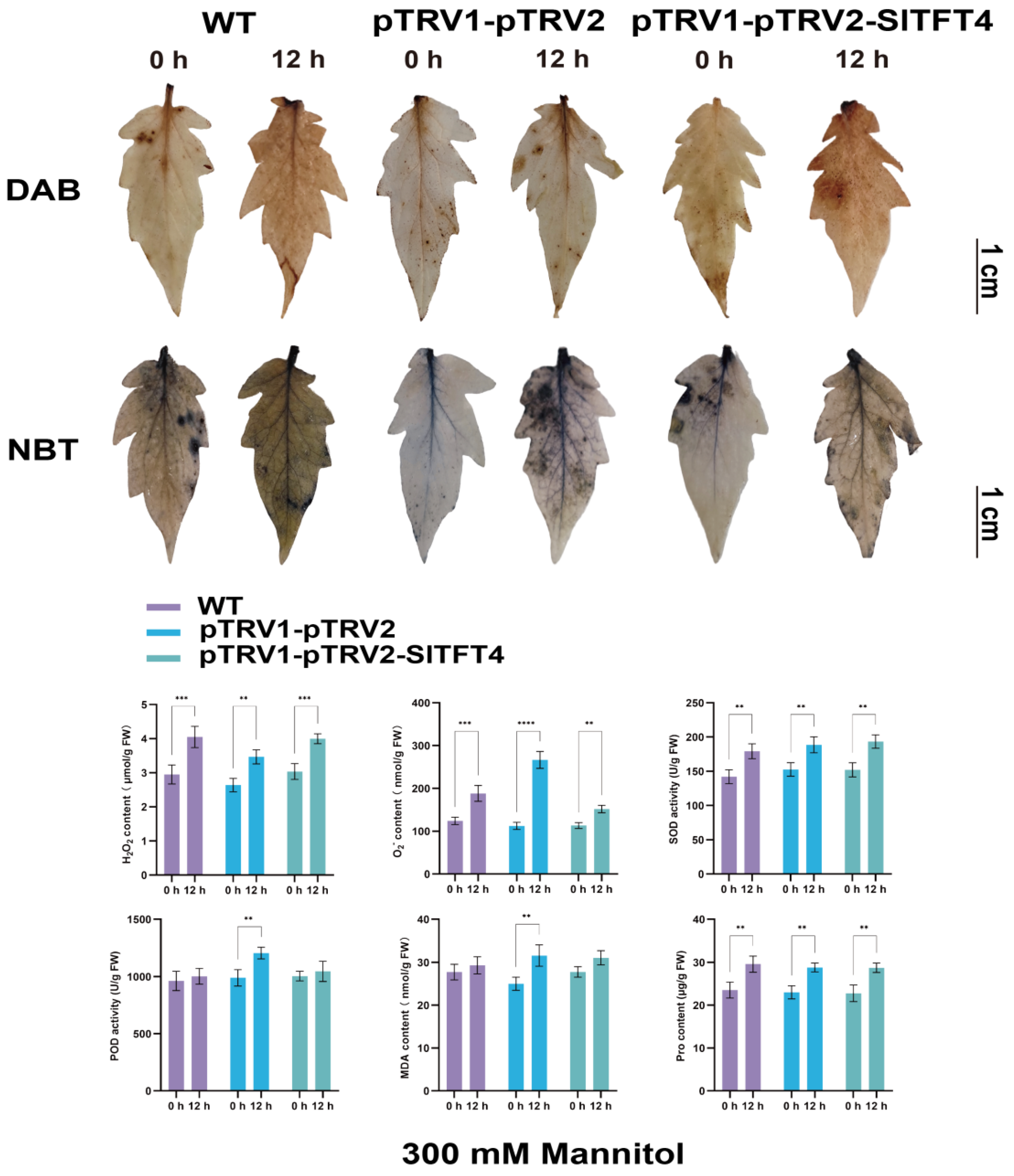
2.10. Subcellular Localization and Gene Silencing of SlTFT4

We performed the subcellular localization of SlTFT4 using tobacco leaves as material to further explore the role of SlTFT4. The results of the localization of the SlTFT4-GFP signal, determined by confocal microscopy, showed that the SlTFT4-GFP fusion protein was detected to emit intense fluorescence in both the cytoplasm and the PM, using the membrane localization marker (CBL) as a reference. Thus, SlTFT4 appears to be co-localized in the cytoplasm and the PM (Figure 11). This is largely consistent with the previous results in which SlTFT4 was predicted to be located mainly in the cytoplasm without a transmembrane helix, indicating the reliability of the electronic predictions and again suggesting the possibility of SlTFT4 as a membrane protein.
VIGS was developed to exploit dsRNA-mediated disease resistance defense mechanisms in plants, and it is one of the most effective reverse genetics techniques used to characterize gene function. The results of our silencing and functional analysis of SlTFT4 using VIGS technology showed that 14 d after inoculation, SlTFT4 underwent gene silencing at the post-transcriptional level, with a highly significant 65% decrease in expression compared to the negative control (Supplementary Figure S4a). After 12 h of drought stress and salt stress, all the plants showed some degree of phenotypic changes compared to 0 h. Among them, the pTRV1-pTRV2-SlTFT4 plants showed more severe yellowing and curling of leaves than the negative control and the positive control plants; in particular, the degree of wilting was more severe under salt stress (Supplementary Figure S4b).
We further collected the top leaves of plants after drought and salt stress for DAB and NBT staining and measured the enzymatic activities and physiological index contents of their biological antioxidant and reactive oxygen species (ROS) scavenging systems to systematically determine the effect of silencing SlTFT4 on the drought and salt tolerance of the tomato plant. The results showed that after 12 h of drought stress, the contents of superoxide anion (O2), hydrogen peroxide (H2O2), and proline (Pro) were increased in the leaves of all the plants compared to 0 h. However, the O2 content produced by the pTRV1-pTRV2-SlTFT4 plants was less than that of the control by about 20%, and the superoxide dismutase (SOD) activity increased by about 10% without significant differences in the peroxidase (POD) activity and malondialdehyde (MDA) content, implying that their antioxidant defense system functioned relatively normally under drought stress and that the cell membrane did not suffer much damage (Figure 12).
Interestingly, after 12 h of salt stress, although the H2O2 and O2 contents of both the control and the pTRV1-pTRV2-SlTFT4 plants increased with significant differences, the increase in the contents of both was highest in the pTRV1-pTRV2-SlTFT4 plants and both were 30% and 50% higher than the control, respectively. The pTRV1-pTRV2-SlTFT4 plants showed no significant change in SOD activity, and the Pro content did not change significantly, while the POD activity was decreased by about 25% and the MDA content was increased by about 35%, indicating that the antioxidant and the ROS defense systems of the pTRV1-pTRV2-SlTFT4 plants were greatly affected, resulting in their inability to catalyze the timely degradation and enhanced oxidation, which caused more serious damage to the cells (Figure 13). The above phenotypic, physiological, and biochemical measurements suggest that silencing SlTFT4 decreases the tolerance of tomato plants to salt stress and that it may be a candidate gene involved in the salt stress response.

3. Discussion

3.1. Significance of the Study of SlTFTs and Its Structural Characterization

It is well known that 14-3-3 proteins are a family of proteins widely expressed in eukaryotes, and different 14-3-3 proteins play different regulatory roles in various physiological processes and tissues of plants. Therefore, it is important to explore the functions of plant 14-3-3 proteins. Due to the deterioration of the environment, abiotic stresses have an increasing impact on plant physiology and crop yield, and the study of plant abiotic stress mechanisms is fundamental to enhancing and improving plant stress tolerance. The 14-3-3 proteins play critical functions in abiotic stresses, and the research targeting them has been an important breakthrough in solving stress problems [57,58].
In the present study, we identified 13 SlTFTs from the tomato genome, and we found one more SlTFT compared with the results of the previous studies [12]. The updated version of the plant genome database, the continuous improvement of the annotation, and more refined gene identification and classification are the most likely reasons for such discrepant results [59,60]. We found that the vast majority of SlTFTs are unstable, with their protein MW mostly around 30 kD, and they are all acidic and hydrophilic proteins, which is generally consistent with the published findings [10]. As with the results of previous studies on A. thaliana [4], rice [48], soybean [49], and cotton [50], SlTFTs can be classified into two groups, i.e., the ε group and the non-ε group. They have different structural features of exons and introns, and the intron phase in the ε group is diverse and relatively less conserved than that in the non-ε group, but both contain only the same protein structural domain, suggesting that their main functions may be consistent. However, although the central region of each SlTFT subgroup member is highly conserved, there are variations in the motifs within the N-terminal (affecting the binding to different membranes) and the C-terminal (directly involved in interactions with target proteins) and the structural domains, which may generate functional specificity while forming many heterodimers due to their being the core structures of the 14-3-3 proteins as they perform their different functions [61,62]. Our predicted subcellular localization results indicate that SlTFTs are mainly located in the cytoplasm, with individual distribution in the nucleus, while our transmembrane helix prediction results indicate that the majority of SlTFTs have no transmembrane helix and are probably membrane proteins. For example, SlTFT4 was predicted to be located mainly in the cytoplasm without transmembrane helices. To further confirm the reliability of the electronic prediction results, we conducted molecular experiments to validate the subcellular localization of SlTFT4. The results showed that SlTFT4 co-localized on the cytoplasm and PM. This indicates that the predicted results are still informative, although with some deviations. The previous findings suggest that 14-3-3 proteins are mainly distributed in the cytoplasm but are also present in the PM, nucleus, Golgi apparatus, chloroplasts, and mitochondria [63,64], which also provides indirect support for our findings. In addition, the remaining members of the SlTFTs, except SlTFT8, have the same or similar typical groove space structure, suggesting that they may have similar functions. SlTFTs generally have multiple distinct phosphorylation sites on their AA sequences. It is hypothesized that proteins with different structures and kinase-specific phosphorylation sites determine the functional diversity of SlTFTs.
In the present study, a phylogenetic tree analysis of 14-3-3 proteins among different species was performed to facilitate a better understanding of the evolutionary relationships of the 14-3-3 family between tomatoes and six other species. All the 14-3-3 proteins were divided into two major groups: the ε group and the non-ε group. The clustering of members into the same group in different species implies that they have similar origins and evolutionary relationships. The different groups contain members of 14-3-3s from multiple species; presumably, their origins appeared before the divergence of monocotyledons and dicotyledons. The results of our evolutionary analysis also suggest that certain subbranches in the different groups may have evolved to produce more loss and amplification events, and presumably, the structure and function of 14-3-3s are more susceptible to divergence, suggesting a diversity of gene expression and function in members of this family [58,65].

3.2. Evolutionary Characterization of SlTFTs

Notably, we observed that all 13 SlTFTs showed a non-random distribution pattern on the tomato chromosomes. They were present on only 8 of the 12 chromosomes, with only one and two members on each of the remaining 5 and 2 chromosomes, except for four members on chromosome 4. Interestingly, almost all of the SlTFTs were distributed at the ends of the chromosomes. This layout is a possible reason for the absence of tandem repeat gene pairs. Usually, the source drivers of gene family amplification mainly include segmental duplication, tandem repeats, and transposition events [66]. Segmental duplication plays a vital part in the amplification of the SlTFT family and is the main mechanism leading to the increase in the number of SlTFTs, a result which is identical to that of the GmGF14 family in soybean [49]. The emergence of new genes is inseparable from the generation of replication events, and the diversity of gene functions requires the continuous replenishment of new genes, which can further help plants to resist the stress of adversity in the process of continuous evolution [67].
Generally, the Ka/Ks ratio can be used as a measure of selective pressure, which can provide important clues to species selection and evolutionary processes [68]. Our results show that the Ka/Ks ratios of the segmental replication gene pairs of SlTFTs are all much smaller than 1, and their average value is only 0.09, indicating that SlTFTs have undergone extremely strong purifying selection during evolution, a result that is essentially the same as the evolutionary process of 14-3-3s in other plants, implying that 14-3-3s evolve very slowly at the plant protein level and have a conserved evolutionary mode [49]. This probably plays an essential part in maintaining the long-term stability of the biological structure of SlTFTs.
It has been shown that at least two large-scale replication events have occurred during the evolution of tomatoes. First, chromosome block replication occurred after monocotyledon–dicotyledon differentiation (approximately 170-235 MYA) [69], followed by polyploid replication after A. thaliana–tomato division (approximately 90 MYA) [70]. The second scenario is consistent with our analysis of the divergence times of 14-3-3s among the 10 species, with A. thaliana and tomato 14-3-3s producing divisions at 90.21 MYA. Previous authors also estimated that another large-scale replication event occurred within the plant kingdom within the last 30 million years [69], which also coincides with the time of the segmental replication of SlTFTs and the time of divergence of 14-3-3s in tomato and potato. Considering the similar number of 14-3-3 family members in A. thaliana, tomato, and potato, it suggests that there may have been a horizontal doubling of the AtGRF family during this period, while no large-scale gene duplication or loss occurred in the SlTFT family.
By constructing the synteny profiles separately, we further understood the synteny relationships between SlTFTs and other species and among their respective species in 14-3-3s. Compared with monocotyledons, dicotyledons had more synteny relationships of the 14-3-3s with each other. Between tomato and potato, the most synteny relationships of 14-3-3s were observed, probably because they belong to the same family of Solanaceae. Notably, no synteny of the 14-3-3s was found between tomato and wheat, rice, and maize and between melon and rice, suggesting that 14-3-3s may not be syntenic between monocotyledons and dicotyledons, indirectly indicating the variability and diversity of 14-3-3s in structure and function.

3.3. Analysis of Cis-Acting Elements of SlTFTs and Correlation with Expression Patterns

It has been shown that in A. thaliana, 14-3-3 proteins can interact with photoperiodic regulatory proteins and bind specifically to phototropin 1 (PHOT1) under red and blue light, respectively [71,72]. In A. thaliana, 14-3-3 proteins can also regulate IAA concentration and maintain ET levels [73,74]. In tobacco, 14-3-3 proteins can negatively regulate GA expression [75]. In barley, 14-3-3 protein is induced by ABA signaling and is involved in ABA signaling [37]. Cis-elements in the promoter region are required for gene regulation and expression. In particular, the cis-acting elements are essential for the rapid and accurate recognition of the genes involved in specific functions in processes associated with plant growth and development and resistance to stress [76]. The cis-acting elements we screened in the promoter region of the SlTFTs were classified into four major classes. In addition, some SlTFTs also contain F-boxes with substrate recognition properties during ubiquitin-mediated protein hydrolysis and W-boxes with binding sites for WRKY TFs. The cis-acting elements suggest that members of the SlTFT family are possibly related to specific growth and development processes, regulated by multiple stress signals and associated with multiple phytohormones responses.
The results of the correlation analysis between the number of cis-acting elements and the expression patterns under different abiotic stresses or phytohormone treatments also showed that SlTFT4/7/10/11, whose expression pattern changed significantly under salt and drought stress, had many cis-acting elements that responded to salt and drought. SlTFT4/10/11 had cis-acting elements that responded to ABA; so, they showed significant changes in expression patterns upon ABA treatment. SlTFT7 did not contain a cis-acting element in response to MeJA; so, its expression pattern did not change significantly in response to the MeJA treatment. Of course, there was also an exception that SlTFT10 did not contain a cis-acting element in response to MeJA; yet, its expression pattern changed significantly under the MeJA treatment. It also implies the incomplete uniformity between the number of cis-acting elements and expression patterns and the complexity of the regulatory mechanism of gene expression.
These inferences are supported by the previous findings on cis-acting elements in the promoter regions of the 14-3-3 family members in soybean [49] and mango [53]. It further suggests that SlTFTs play a very important role in growth and development and response to adversity and deserve to be studied in depth.

3.4. Unique Transcriptional Profiles and Expression Patterns of SlTFTs

In addition, we found that the SlTFTs were expressed in several tissues and that the expression levels varied greatly between the tissues and periods. This presence of tissue-specific expression during specific growth and development is generally consistent with the previously reported expression patterns of the 14-3-3 family members in cottons [50], apples [52], bananas [18], grapes [51], and mangoes [53]. It is speculated that the reason for this phenomenon is related to their possible role as tissue-specific regulators. Furthermore, in our results, the SlTFTs were generally expressed at low levels in seeds, flowers, and roots and at high levels in leaves, meristematic tissues, and fruits. It is suggested that during fruit development, certain members differ in protein function and may play specific roles. In grapes, most VviGRFs are expressed to a considerable extent in different tissues and organs, but some genes show very high or low expression in certain tissues. For example, VviGRF12 usually has a low expression in most tissues and organs but not in floral organs such as stamens, buds, flowers, and pollen. VviGRF15 is expressed at high levels in most tissues and organs except leaves, petals, and pollen. VviGRFs may be involved in the regulation of grape development and fruit ripening [51]. In cottons, 31 GhGRFs also showed different expression levels in the 15 examined tissues. Nine GhGRFs were expressed at relatively low levels in all tissues, while another fifteen GhGRFs were highly expressed in all test tissues, indicating that they are involved in the whole process of cotton growth and development. In addition, the expression levels of GhGRF8-A/6-A/6-D in ovules at 0 and 1 d after anthesis were similar to those of these 15 genes, and they may play important roles as tissue-specific regulators [50]. The above findings provide a strong corroboration of our findings.
Meanwhile, substantial evidence suggests that 14-3-3s are involved in plant responses to various stresses. The 14-3-3 family members similarly exhibit differential transcript accumulation patterns in response to abiotic stresses, suggesting that they play potentially different roles in regulating abiotic stress responses. For example, in grapes, six VviGRFs were significantly up- or down-regulated in their expression in response to cold and heat stresses, suggesting a possible role in abiotic stress responses [51]. In mangoes, drought, salt, and low-temperature stresses affected the expression levels of Mi14-3-3s, and different Mi14-3-3s responded differently to these stresses [53]. In bananas, MaGRF2/3/25 showed similar expression patterns after salt, cold, and osmotic treatments, respectively, indicating that they function similarly in the two cultivars (BX and FJ) under cold, salt, and osmotic treatments. However, six, ten, and six genes exhibited differential expression patterns between BX and FJ under cold, salt, and osmotic stresses, respectively [18]. In tomatoes, SlTFT1/4/7/10 were significantly up-regulated in young tomato roots under salt stress [12].
We found that the expression pattern of SlTFTs in leaves was affected by different abiotic stresses and phytohormone treatments, and certain SlTFTs were rapidly induced and persistently highly expressed during specific periods, suggesting that they have specific roles under different stresses and phytohormone treatments. SlTFT4/10 can simultaneously respond to all processing conditions in this study, and they may be key genes in the SlTFT family, involved in defense and response to external stress. In addition, we also noticed that the expression of SlTFTs tended to be more responsive to drought stress and ABA treatment compared to the rest of the treatments. It has been shown that under drought stress, for the tomato ABA-deficient mutant notabilis, inoculation with AM significantly up-regulated the expression of SlTFT5/7/9/10 to maintain WUE and improve drought tolerance [42]. In conclusion, all the above reports provide indirect support for our findings.

3.5. The Constructed Protein Network That Interacts with SlTFTs Deepens the Knowledge

Generally, 14-3-3 proteins are required to respond to adversity stress by combining with target proteins. As 14-3-3 proteins require a low specificity of recognition sequences and diverse recognition modes in recognizing target proteins, the types of target proteins regulated by 14-3-3 proteins are abundant [31,77]. Our constructed interaction network between SlTFTs and the other proteins showed that all 13 SlTFTs interacted with five (SlBSL3, SlPP2C, SlBSU1, SlCDC25, and SlBSL1) other proteins belonging to dual-specificity tyrosine-phosphatase, serine/threonine-protein phosphatase, and protein kinase A-like kinase. This is consistent with the study that 14-3-3 proteins mainly combine with their interacting target proteins through phosphorylation, which in turn regulates the function of the target protein, and their structural requirement for the interacting target protein is to contain a phosphorylated Ser or Thr sequence [77,78]. Interestingly, there are fewer interactions within the SlTFTs and more interactions in the five other proteins. It is hypothesized that they exert their regulatory effects mainly by binding to phosphorylated target proteins rather than by their phosphorylation.
Subsequently, we also constructed the interaction network between AtGRFs and other proteins, and we observed that all 14 AtGRFs interacted with five (AtBZR1, AtIDH-III, AtCNI1, AtCDC25, and AtACS7) other proteins belonging to the brassinosteroid signaling positive regulator family protein, isocitrate dehydrogenase III, RING-type ubiquitin ligase, the rhodanese/cell cycle control phosphatase superfamily protein, and 1-amino-cyclopropane-1-carboxylate synthase 7. In particular, there were more interactions within the AtGRFs, while there were no interactions among the five other proteins. Although 14-3-3 proteins mainly exert their regulatory effects by interacting with target proteins, the interactions between the different internal members may enhance their regulatory effects in some physiological processes [31,58]. As different family members of 14-3-3 proteins have their specific regulatory functions in rice, for example, OsGF14b is associated with drought resistance [79], OsGF14c is considered as a negative regulator of flowering [80], and OsGF14e negatively regulates cell death and the disease resistance response [81]. In maize, ZmGF14-4 specifically binds to disease and resistance-related proteins during endosperm development, while ZmGF14-6 mainly interacts with metabolic and cytoarchitecture-related proteins [82]. Therefore, our findings corroborate the importance of the phosphorylation binding mode of 14-3-3 proteins and their interactions with their target proteins to ensure their regulatory functions in organisms, and they deepen the understanding of the functional proteome-wide regulatory network. They also lay the groundwork for further investigations into the regulatory mechanism of phosphorylation and phytohormones signaling pathways in 14-3-3 proteins and their roles in plant development and the response to adversity stress.

3.6. Functional Analysis Revealed That SlTFT4 Is a Candidate Gene for Salt Tolerance

Typically, the consideration of the overexpression of 14-3-3s in transgenic plants has been used to elucidate the contribution of the corresponding proteins in stress resistance [43,44]. For example, the overexpression of AtGRF9, At14-3-3λ, and ZmGF14-6/OsGF14c improved drought resistance in transgenic A. thaliana, cotton, and rice, respectively [19,20,21,22], while the silencing of AtGF14m improved drought resistance in A. thaliana at the seed germination and seedling stages [23]. The At14-3-3λ/κ have been shown to negatively regulate salt tolerance [20,24]. The transgenic A. thaliana overexpressing SlTFT6 and SlTFT7, respectively, both showed great growth promotion under LP stress [44]. Moreover, the transgenic A. thaliana overexpressing SlTFT4 showed significantly enhanced growth under alkaline stress [43]. In the present study, we found that silencing SlTFT4 significantly increased the content of H2O2, O2, and MDA in leaves, while significantly decreasing POD activities, which caused a greater impact on the biological antioxidant and ROS defense systems, increased the oxidation, thereby increasing the degree of cellular damage, and led to an increase in leaf wilting, reducing the tolerance of tomato plants to salt stress. Therefore, SlTFT4, identified by VIGS technology, can be used as a candidate gene for subsequent studies to improve the understanding of the salt tolerance function studies on tomatoes.

4. Materials and Methods

4.1. Identification and Characterization of SlTFT Family

The genome-wide data were downloaded from the International Tomato Annotation Group website (https://www.sgn.cornell.edu/organism/Solanum_lycopersicum/genome, accessed on 23 January 2022), and the HMM profile (PF00244), downloaded from the protein families database (http://pfam.xfam.org/, accessed on 23 January 2022), was loaded into the HMMER search (https://www.ebi.ac.uk/Tools/hmmer/, accessed on 23 January 2022) at the default inclusion threshold. The potential SlTFTs identified in the genomic database were retrieved by the above steps (Supplementary Table S5). All predicted protein sequences of SlTFTs were uploaded to the Simple Modular Architecture Research Tool database (http://smart.embl-heidelberg.de/, accessed on 24 January 2022) and HMMER to verify the 14-3-3 domain presence. Subsequently, sequences that did not contain the 14-3-3 domain (Solyc02g086840.3.1, Solyc08g008455.1.1, Solyc09g065270.3.1, and Solyc03g114320.3.1) were artificially discarded (Supplementary Table S6). The physicochemical parameters of the SlTFTs were measured using the ExPASy server tool (http://web.expasy.org/protparam/, accessed on 24 January 2022). In addition, the SubCELlular LOcalization predictor website (http://cello.life.nctu.edu.tw/, accessed on 25 January 2022) was used to forecast the possible subcellular localization results of the SlTFTs. The tertiary structure of the SlTFTs was forecasted using the SWISS-MODEL server (https://swissmodel.expasy.org/interactive, accessed on 25 January 2022). The identification of possible phosphorylation sites of the SlTFTs was conducted using NetPhos 3.1 server (https://services.healthtech.dtu.dk/service.php?NetPhos-3.1, accessed on 26 January 2022). The prediction of the transmembrane helices of the SlTFTs was performed using the TMHMM online software tool (https://services.healthtech.dtu.dk/service.php?TMHMM-2.0, accessed on 29 January 2022). The details of the bioinformatics analysis are as follows: after opening the homepage of the above website, click on the corresponding input box or analysis button, paste the AA sequence of each SlTFT for analysis, and then obtain bioinformatics information, such as various physicochemical properties, subcellular localization, tertiary structure, possible phosphorylation sites, and transmembrane helices of the protein.

4.2. Phylogenetic Tree, Gene Structure, Conserved Domains, and Motif Analysis of SlTFTs

The protein sequences of SlTFTs were used to create a phylogenetic tree, and the CDS and genomic base sequences of SlTFTs were used to analyze the gene structure. The conserved domains and motifs of SlTFTs were evaluated using the HMMER search and the Multiple Expectation Maximization for Motif Elicitation website (http://meme-suite./org/tools/meme, accessed on 27 January 2022), respectively. Lastly, the above data were visualized using TBtools [83].

4.3. Phylogenetic Tree Analysis of SlTFTs among Different Species

To explore the phylogenetic relationships among SlTFT families in different species, we first obtained the 14-3-3 protein sequences of six other species using HMMER search screening. Then, the 14-3-3 protein sequences of all seven species were imported into MEGA 11 software, and the phylogenetic tree analysis was performed by the maximum likelihood method with bootstrap analysis of 1000 replicates.

4.4. Chromosome Distribution, Replication Events, and Synteny Analysis of SlTFTs

The distribution of SlTFTs on chromosomes was calculated and plotted using TBtools. Gene replication events were calculated and plotted for SlTFTs using the MCScanX toolkit [84]. The ratio of Ka and Ks in duplicated gene pairs in SlTFTs was calculated by the KaKs_Calculator 3.0 software [68]. The replication date in each gene pair was evaluated using the equation T = Ks/2λ, where λ was assumed to be 1.5 × 10⁻⁸ synonymous/substitution site/year [69]. Single-copy homologous genes were obtained using diamond comparison in the OrthoFinder 2.5.2 software [85], and evolutionary divergence trees of different species were constructed using the RAxML 8.0 tool [86], and fossil times were added to the evolutionary trees using the TimeTree database (http://www.timetree.org/, accessed on 7 February 2022). The fossil calibration times were 40.3 to 51.9 MYA for rice, maize, and wheat, and 109.0 to 123.5 MYA for A. thaliana, grapes, and poplars. Then, the mcmctree command in the PAML 4.9 software [87] was used to estimate the divergence times of different species. In addition, the synteny of 14-3-3s between different species was calculated, analyzed, and visualized using the dual synteny plotter module in TBtools.

4.5. Analysis of Cis-Acting Elements in the Promoter Region of SlTFTs

The sequences within 2000 base pairs upstream of each SlTFT initiation codon were extracted from the tomato genome database and subsequently collated and submitted to the PlantCARE database (http://bioinformatics.psb.ugent.be/webtools/plantcate/html, accessed on 8 February 2022) for cis-acting element screening and identification. Visualization was performed using TBtools.

4.6. Transcriptional Profiling of SlTFTs

The transcriptional profiling data of SlTFTs in different tissues, such as seeds, roots, meristematic tissues, leaves, flowers, and fruits, were analyzed using the TomExpress database (http://tomexpress.toulouse.inra.fr/, accessed on 10 February 2022), a unified tomato RNA-Seq platform. Cluster heat maps were drawn using the OmicShare tool (https://www.omicshare.com/tools, accessed on 10 February 2022), a free online data analysis platform.

4.7. Plant Material and Treatments

Seeds (Solanum lycopersicum var. M82 is a representative material of the processing of tomato, which is collected and preserved by Horticultural Crops Research, Xinjiang Academy of Agricultural Sciences) were planted in potting soil (1:3 ratio of vermiculite to nutrient soil) and grown in an artificial climate chamber at the appropriate temperature and humidity, with the light and dark cycles set to 16 h/8 h. After 4 weeks, seedlings of uniform growth were watered with 300 mM D-mannitol (CM7091-1kg, Coolaber, Beijing, China) and 200 mM NaCl (CS9971-1kg, Coolaber, Beijing, China) solutions to simulate drought and salt stress responses, respectively, and seedlings without any treatment under the above standard degree conditions were used as controls. The leaves of seedlings with essentially the same growth were uniformly sprayed with 100 μM ABA (PH109X-2×1 mL, Coolaber, Beijing, China) and 100 μM MeJA (CJ6691-5 mL, Coolaber, Beijing, China) solutions, respectively, for phytohormone treatment response analysis, with leaves of seedlings sprayed with ddH2O as controls. In each of the above treatments, the top first fully expanded leaves of three plants were mixed and harvested as one biological replicate and three technical replicates, and all the abiotic stress and phytohormone experimental samples were collected and obtained at 0 h, 3 h, 12 h, and 24 h post-treatment, respectively. The samples were first rapidly frozen in liquid nitrogen and subsequently stored in an ultra-low temperature refrigerator at −80 °C until they were used.

4.8. Total RNA Extraction, Quantitative Real-Time PCR (qRT-PCR), and Statistical Analysis

Total RNA was extracted from the lyophilized samples using the Polysaccharide Polyphenol Plant Total RNA Extraction Kit (DP441, Tiangen, Beijing, China) and processed with RNase-free DNAase following the manufacturer’s protocol. Total RNA was reverse transcribed into cDNA using 5 × All-In-One RT MasterMix (with AccuRT Genomic DNA Removal Kit) (G492, ABM, Vancouver, BC, Canada). qRT-PCR was performed on a LightCycler®® real-time fluorescent quantitative PCR system (Roche, Basel, Switzerland) using ChamQ Universal SYBR qPCR Master Mix (Q711, Vazyme, Nanjing, China). The amplification process was pre-denaturation: 95 °C, 30 s, one cycle; followed by denaturation: 95 °C, 5 s; annealing extension: 60 °C, 30 s, 40 cycles; and finally a melting curve was obtained: 95 °C, 15 s, 60 °C, 60 s, 95 °C, 15 s, one cycle. The specific primers and the internal reference sequence of Actin (Solyc03g078400.2) used in this study for SlTFTs were designed using NCBI Primer-BLAST (https://www.ncbi.nlm.nih.gov/tools/primer-blast/index.cgi?LINK_LOC=BlastHome, accessed on 13 February 2022) and synthesized by Sangon Biotech (Shanghai, China) Co., Ltd., and then, the amplification validity study was performed first using RT-PCR, and primers with unique specific bands for the target fragment (80–150 bp range) were selected for subsequent qRT-PCR assay analysis, and the details are shown in Supplementary Table S7. All qRT-PCR assays were performed three times as independent biological replicates, and the mean of the cycle threshold (Ct) was obtained. Relative gene expression levels were calculated using the 2−ΔΔCt method [88]. Three independent biological replicates were averaged and analyzed for each assay. One-way analysis of variance was performed using GraphPad Prism for Windows (version 9.0.0, GraphPad Software, San Diego, CA, USA), followed by Dunnett’s multiple comparison test.

4.9. Protein Interaction Network Analysis

Protein interactions were queried according to String 11.5 (https://cn.string-db.org/, accessed on 15 February 2022). In the basic settings, select the full STRING network (the edges indicate both functional and physical protein associations) as the network type. The line color indicates the type of interaction evidence chosen to indicate the meaning of the network edges. Active interaction sources include text mining, experiments, databases, co-expression, neighborhood, gene fusion, and co-occurrence. The minimum required interaction score is medium confidence (0. 40). The network display mode in the advanced settings is set to interactive SVG (the network is a scalable vector graphic [SVG]; interactive). Select hide disconnected nodes in the network in the network display option. Thereafter, the interaction network of SlTFTs with other proteins was visualized using Cytoscape 3.9.1 (https://cytoscape.org//, accessed on 15 February 2022).

4.10. Subcellular Localization

Briefly, the method of subcellular localization adopted in the present study comprised the following. The CDS of SlTFT4 was amplified using cDNA as a template by the upstream primer 5′- GCGTCGACATGGCTGACTCTTCGCGTGA-3′ (Sal I enzyme cut site) and the downstream primer 5′-GCTCTCTAGACTGCTGCCGCCTCGCCTGAC-3′ (Xba I enzyme cut site), respectively. After correct sequencing, the agarose gel recovery product of the target fragment was ligated into the pCAMBIA super1300-GFP plant expression vector, which was also double-cleaved with Sal I and Xba I, and after correct sequencing again, the pCAMBIA super1300-SlTFT4-GFP vector was constructed. The pCAMBIA super1300-GFP empty was used as a control, and the CBL was used as a reference. The vector plasmid was transformed into Agrobacterium tumefaciens GV3101, and after overnight incubation, it was collected by centrifugation and suspended using the suspension, and the concentration was adjusted to OD600 = 0.8. They were then mixed separately at a 1:1 (v/v) ratio and incubated at room temperature for 3 h in the dark before infiltration from the abaxial side of Nicotiana benthamiana leaves using a needleless 1 mL syringe, and the infiltrated plants were incubated in the dark for 12 h and then grown for an additional 5 d under a 16 h/8 h light/dark cycle. Lastly, confocal laser scanning microcopy (LSM 800, Zeiss, Germany) was used for observation and photography.

4.11. VIGS Vector Construction and the Process of Gene Silencing and Physiological Indices Determination

The VIGS vector for the target gene was designed and constructed using the SGN VIGS tool (https://vigs.solgenomics.net/, accessed on 17 February 2022). The pTRV2 recombinant vector was transformed into A. tumefaciens GV3101 (pSoup-p19). A. tumefaciens containing pTRV1, pTRV2 empty, and pTRV2 recombinant vectors were incubated in a rotary shaker (28 °C, 200 rpm). The cells were collected by centrifugation at 4000 rpm for 10 min and resuspended in appropriate volumes of resuspension solution (10 mM MgCl2, 10 mM MES, and 150 μM acetosyringone) to a final concentration of OD600 = 1.0. The pTRV1-pTRV2 empty and pTRV1-pTRV2 recombinant vector resuspensions were mixed at a ratio of 1:1 (v/v) and incubated for 3 h at 28 °C, protected from light, and then infiltrated into the abaxial surface of the leaves of 4-week-old tomato plants using a de-needled 1 mL syringe. After 12 h of dark treatment, a normal 16 h/8 h photoperiod was performed. Plants without treatment were used as negative controls, and plants inoculated with pTRV1-pTRV2 empty were used as positive controls. After 14 d, salt and drought stresses were applied as described previously, respectively, and the top leaves were collected from the same parts at 0 h and 12 h post-stress and stained using DAB solution (1 mg/mL, pH 3.8) (SL1805, Coolaber, Beijing, China) and NBT solution (0.1%, pH 7.8) (SL18061, Coolaber, Beijing, China), respectively. According to the manufacturer’s instructions, six test kits (SA-1-G, H2O2-1-Y, MDA-1-Y, PRO-1-Y, SOD-1-Y, and POD-1-Y, Comin, Suzhou, China) were used sequentially to detect O2, H2O2, MDA, Pro, SOD, and POD activity/content, and three replicates were performed.

5. Conclusions

In summary, this research provides a comprehensive and systematic description of the SlTFT family. We found 13 SlTFTs divided into two groups. The SlTFTs were non-randomly distributed on chromosomes, and the promoter regions contained more cis-acting elements involved in abiotic stress tolerance and phytohormone responses. Gene expression pattern analysis indicated specific roles for certain SlTFTs under different abiotic stresses and phytohormone treatments. SlTFT4 may be a candidate gene involved in the salt stress response. Our results provide comprehensive information on the characteristic structure, evolutionary history, expression patterns, and protein interactions of the SlTFT family, which is a significant guide for the further elucidation of this family. It also lays the foundation for the subsequent in-depth study of the molecular and biological functions of this family and the screening of family members for future application in variety improvement.

Supplementary Materials

The following supporting information can be downloaded at: https://www.mdpi.com/article/10.3390/plants11243491/s1, Figure S1: Synteny maps between SlTFTs and 14-3-3s of other four species, respectively. The different colored rectangles represent the chromosomes of different species. The gray lines indicate synteny between tomato genome and other species. The red line indicates the synteny between SlTFTs and 14-3-3s of other species; Figure S2: Expression patterns of SlTFTs under normal conditions (control for stress treatments). A to M represent SlTFT1 to SlTFT13, respectively. The horizontal coordinates in the figure are the four different treatment periods, and the vertical coordinates are the relative expression of each SlTFT gene. Error bars are standard deviations (SD) of three independent biological replicates, presented as mean ± SD. Statistical significance of differences was confirmed by Dunnett's multiple comparison test (* p < 0.05, ** p < 0.01, *** p < 0.001, and **** p < 0.0001); Figure S3: Expression patterns of SlTFTs in response to ddH2O treatment (control for phytohormone treatments). A to M represent SlTFT1 to SlTFT13, respectively. The horizontal coordinates in the figure are the four different treatment periods, and the vertical coordinates are the relative expression of each SlTFT gene. Error bars are SD of three independent biological replicates, presented as mean ± SD. Statistical significance of differences was confirmed by Dunnett's multiple comparison test (* p < 0.05, ** p < 0.01, *** p < 0.001, and **** p < 0.0001); Figure S4: Relative expression of SlTFT4 after silencing and phenotypic changes of tomato plants under stress conditions. (a) qRT-PCR to verify the relative expression of SlTFT4 after silencing. The horizontal coordinates in the figure represent the different plant types. WT: untreated wild type plants, pTRV1-pTRV2: control plants injected with a empty vector, pTRV1-pTRV2-SlTFT4: treated plants injected with a SlTFT4 silencing vector. The vertical coordinates represent the relative expression of SlTFT4 in different plant types. Error bars are SD of three independent biological replicates, presented as mean ± SD. Statistical significance of differences was confirmed by Dunnett's multiple comparison test (* p < 0.05, ** p < 0.01, *** p < 0.001, and **** p < 0.0001). (b) Phenotypic changes in different categories of tomato plants under drought and salt stress conditions. The figure represents different plant types and the same treatment period horizontally, and the same plant types and different treatment periods vertically. The upper two columns show the phenotypic changes of different plant types under 300 mM D-mannitol simulated drought stress treatment at 0 h and 12 h treatment periods. The following two columns show the phenotypic changes of different plant types under 200 mM NaCl simulated salt stress treatment at 0 h and 12 h treatment periods. The scale bar of the plants is 5 cm.; Table S1: Sequence information of the 13 identified SlTFTs; Table S2: Results of the TMHMM in this study; Table S3: The list of 7 pairs of fragment duplication events, Ka/Ks ratios, and their replication date for SlTFTs; Table S4: Number of cis-acting elements involved in the response to different abiotic stresses and phytohormone treatments; Table S5: Results of the HMMER search in this study; Table S6: Results of verifying the presence of 14-3-3 domain in SMART and HMMER; Table S7: The primer sequences used in this study for qRT-PCR.

Author Contributions

Q.Y. and J.W. designed the experiment and methodology; C.J. and B.G. carried out most of the experimental work; B.W. and X.L. conducted data analysis; N.L. and T.Y. finished the plant material treatment; Q.Y., J.W., C.J. and B.G. wrote the manuscript. All authors contributed to the experimental design and data analysis, commented on the manuscript, and gave final approval for publication. All authors have read and agreed to the published version of the manuscript.

Funding

This work was supported by the Key Programs for Crop Important Traits Formation and Cutting-Edge Technologies in Biological Breeding (xjnkywdzc-2022001-8), the Special Incubation Project of Science & Technology Renovation of Xinjiang Academy of Agricultural Sciences (xjkcpy-2021001), and the Key Research and Development Task Special Project of Xinjiang Uygur Autonomous Region (2022B02002).

Institutional Review Board Statement

Not applicable.

Informed Consent Statement

Not applicable.

Data Availability Statement

Not applicable.

Conflicts of Interest

The authors declare no conflict of interest.

References

  1. Thomas, D.; Guthridge, M.; Woodcock, J.; Lopez, A. 14-3-3 protein signaling in development and growth factor responses. Curr. Top. Dev. Biol. 2005, 67, 285–303. [Google Scholar]
  2. Rooney, M.F.; Ferl, R.J. Sequences of three Arabidopsis general regulatory factor genes encoding GF14 (14-3-3) proteins. Plant Physiol. 1995, 107, 283–284. [Google Scholar] [CrossRef] [PubMed] [Green Version]
  3. Daugherty, C.J.; Rooney, M.F.; Miller, P.W.; Ferl, R.J. Molecular organization and tissue-specific expression of an Arabidopsis 14-3-3 gene. Plant Cell 1996, 8, 1239–1248. [Google Scholar]
  4. Wu, K.; Rooney, M.F.; Ferl, R.J. The Arabidopsis 14-3-3 multigene family. Plant Physiol. 1997, 114, 1421–1431. [Google Scholar] [CrossRef] [PubMed] [Green Version]
  5. Yaffe, M.B.; Rittinger, K.; Volinia, S.; Caron, P.R.; Aitken, A.; Leffers, H.; Gamblin, S.J. The structural basisfor 14-3-3:phosphopeptide binding specificity. Cell 1997, 91, 961–971. [Google Scholar] [CrossRef] [PubMed] [Green Version]
  6. Ottmann, C.; Marco, S.; Jaspert, N.; Marcon, C.; Schauer, N.; Weyand, M.; Vandermeeren, C.; Duby, G.; Boutry, M.; Wittinghofer, A.; et al. Structure of a 14-3-3 coordinated hexamer of the plant plasma membrane H+ -ATPase by combining X-ray crystallography and electron cryomicroscopy. Mol. Cell 2007, 25, 427–440. [Google Scholar] [CrossRef] [Green Version]
  7. Taoka, K.; Ohki, I.; Tsuji, H.; Furuita, K.; Hayashi, K.; Yanase, T.; Yamaguchi, M.; Nakashima, C.; Purwestri, Y.A.; Tamaki, S.; et al. 14-3-3 proteins act as intracellular receptors for rice Hd3a florigen. Nature 2011, 476, 332–335. [Google Scholar] [CrossRef]
  8. Delille, J.M.; Sehnke, P.C.; Ferl, R.J. The Arabidopsis 14-3-3 family of signaling regulators. Plant Physiol. 2001, 126, 35–38. [Google Scholar] [CrossRef] [Green Version]
  9. Rosenquist, M.; Alsterfjord, M.; Larsson, C.; Sommarin, M. Data mining the Arabidopsis genome reveals fifteen 14-3-3 genes. Expression is demonstrated for two out of five novel genes. Plant Physiol. 2001, 127, 142–149. [Google Scholar] [CrossRef] [Green Version]
  10. Tian, F.; Wang, T.; Xie, Y.; Zhang, J.; Hu, J. Genome-wide identification, classification, and expression analysis of 14-3-3 gene family in Populus. PLoS ONE 2015, 10, e0123225. [Google Scholar] [CrossRef] [Green Version]
  11. Chen, F.; Li, Q.; Sun, L.; He, Z. The rice 14-3-3 gene family and its involvement in responses to biotic and abiotic stress. DNA Res. 2006, 13, 53–63. [Google Scholar] [CrossRef] [Green Version]
  12. Xu, W.F.; Shi, W.M. Expression profiling of the 14-3-3 gene family in response to salt stress and potassium and iron deficiencies in young tomato (Solanum lycopersicum) roots: Analysis by real-time RT-PCR. Ann. Bot. 2006, 98, 965–974. [Google Scholar] [CrossRef] [PubMed]
  13. Cotelle, V.; Leonhardt, N. 14-3-3 proteins in guard cell signaling. Front. Plant Sci. 2015, 6, 1210. [Google Scholar] [CrossRef] [PubMed] [Green Version]
  14. Camoni, L.; Visconti, S.; Aducci, P.; Marra, M. 14-3-3 proteins in plant hormone signaling: Doing several things at once. Front. Plant Sci. 2018, 9, 297. [Google Scholar] [CrossRef]
  15. Liu, Q.; Zhang, S.; Liu, B. 14-3-3 proteins: Macro-regulators with great potential for improving abiotic stress tolerance in plants. Biochem. Biophys. Res. Commun. 2016, 477, 9–13. [Google Scholar] [CrossRef] [PubMed]
  16. Mayfield, J.D.; Folta, K.M.; Paul, A.L.; Ferl, R.J. The 14-3-3 Proteins μ and ν influence transition to flowering and early phytochrome response. Plant Physiol. 2007, 145, 1692–1702. [Google Scholar] [CrossRef] [PubMed] [Green Version]
  17. Shi, H.Y.; Zhang, Y.X. Pear 14-3-3a gene (Pp14-3-3a) is regulated during fruit ripening and senescense, and involved in response to salicylic acid and ethylene signalling. J. Genet. 2014, 93, 747–753. [Google Scholar] [CrossRef]
  18. Li, M.; Ren, L.; Xu, B.; Yang, X.; Xia, Q.; He, P.; Xiao, S.; Guo, A.; Hu, W.; Jin, Z. Genome-wide identification, phylogeny, and expression analyses of the 14-3-3 family reveal their involvement in the development, ripening, and abiotic stress response in banana. Front. Plant Sci. 2016, 7, 1442. [Google Scholar] [CrossRef] [Green Version]
  19. He, Y.; Wu, J.; Lv, B.; Li, J.; Gao, Z.; Xu, W.; Baluska, F.; Shi, W.; Shaw, P.C.; Zhang, J. Involvement of 14-3-3 protein GRF9 in root growth and response under polyethylene glycol-induced water stress. J. Exp. Bot. 2015, 66, 2271–2281. [Google Scholar] [CrossRef] [Green Version]
  20. Yan, J.; He, C.; Jing, W.; Mao, Z.; Holaday, S.A.; Allen, R.D.; Zhang, H. Overexpression of the Arabidopsis 14-3-3 protein GF14λ in cotton leads to a "stay-green" phenotype and improves stress tolerance under moderate drought conditions. Plant Cell Physiol. 2004, 45, 1007–1014. [Google Scholar] [CrossRef]
  21. Campo, S.; Peris-Peris, C.; Montesinos, L.; Penas, G.; Messeguer, J.; San Segundo, B. Expression of the maize ZmGF14-6 gene in rice confers tolerance to drought stress while enhancing susceptibility to pathogen infection. J. Exp. Bot. 2012, 63, 983–999. [Google Scholar] [CrossRef] [PubMed] [Green Version]
  22. Ho, S.L.; Huang, L.F.; Lu, C.A.; He, S.L.; Wang, C.C.; Yu, S.P.; Chen, J.; Yu, S.M. Sugar starvation- and GA-inducible calcium-dependent protein kinase 1 feedback regulates GA biosynthesis and activates a 14-3-3 protein to confer drought tolerance in rice seedlings. Plant Mol. Biol. 2013, 81, 347–361. [Google Scholar] [CrossRef] [PubMed]
  23. Sun, X.; Luo, X.; Sun, M.; Chen, C.; Ding, X.; Wang, X.; Yang, S.; Yu, Q.; Jia, B.; Ji, W.; et al. A Glycine soja 14-3-3 protein GsGF14o participates in stomatal and root hair development and drought tolerance in Arabidopsis thaliana. Plant Cell Physiol. 2014, 55, 99–118. [Google Scholar] [CrossRef] [Green Version]
  24. Zhou, H.; Lin, H.; Chen, S.; Becker, K.; Yang, Y.; Zhao, J.; Kudla, J.; Schumaker, K.S.; Guo, Y. Inhibition of the Arabidopsis salt overly sensitive pathway by 14-3-3 proteins. Plant Cell 2014, 26, 1166–1182. [Google Scholar] [CrossRef] [PubMed] [Green Version]
  25. Catala, R.; Lopez-Cobollo, R.; Mar Castellano, M.; Angosto, T.; Alonso, J.M.; Ecker, J.R.; Salinas, J. The Arabidopsis 14-3-3 protein RARE COLD INDUCIBLE 1A links low-temperature response and ethylene biosynthesis to regulate freezing tolerance and cold acclimation. Plant Cell 2014, 26, 3326–3342. [Google Scholar] [CrossRef] [Green Version]
  26. Vercruyssen, L.; Tognetti, V.B.; Gonzalez, N.; Van Dingenen, J.; De Milde, L.; Bielach, A.; De Rycke, R.; Van Breusegem, F.; Inze, D. GROWTH REGULATING FACTOR5 stimulates Arabidopsis chloroplast division, photosynthesis, and leaf longevity. Plant Physiol. 2015, 167, 817–832. [Google Scholar] [CrossRef] [Green Version]
  27. Cao, A.; Jain, A.; Baldwin, J.C.; Raghothama, K.G. Phosphate differentially regulates 14-3-3 family members and GRF9 plays a role in Pi-starvation induced responses. Planta 2007, 226, 1219–1230. [Google Scholar] [CrossRef]
  28. Yang, J.L.; Chen, W.W.; Chen, L.Q.; Qin, C.; Jin, C.W.; Shi, Y.Z.; Zheng, S.J. The 14-3-3 protein GENERAL REGULATORY FACTOR11 (GRF11) acts downstream of nitric oxide to regulate iron acquisition in Arabidopsis thaliana. New Phytol. 2013, 197, 815–824. [Google Scholar] [CrossRef]
  29. Radwan, O.; Wu, X.; Govindarajulu, M.; Libault, M.; Neece, D.J.; Oh, M.H.; Berg, R.H.; Stacey, G.; Taylor, C.G.; Huber, S.C.; et al. 14-3-3 proteins SGF14c and SGF14l play critical roles during soybean nodulation. Plant Physiol. 2012, 160, 2125–2136. [Google Scholar] [CrossRef] [Green Version]
  30. Yao, Y.; Du, Y.; Jiang, L.; Liu, J.Y. Molecular analysis and expression patterns of the 14-3-3 gene family from Oryza Sativa. J. Biochem. Mol. Biol. 2007, 40, 349–357. [Google Scholar] [CrossRef]
  31. Denison, F.C.; Paul, A.L.; Zupanska, A.K.; Ferl, R.J. 14-3-3 proteins in plant physiology. Semin. Cell Dev. Biol. 2011, 22, 720–727. [Google Scholar] [CrossRef] [PubMed]
  32. Wilson, R.S.; Swatek, K.N.; Thelen, J.J. Regulation of the regulators: Post-translational modifications, subcellular, and spatiotemporal distribution of plant 14-3-3 proteins. Front. Plant Sci. 2016, 7, 611. [Google Scholar] [CrossRef] [PubMed]
  33. Ormancey, M.; Thuleau, P.; Mazars, C.; Cotelle, V. CDPKs and 14-3-3 proteins: Emerging duo in signaling. Trends Plant Sci. 2017, 22, 263–272. [Google Scholar] [CrossRef]
  34. Kong, Q.; Ma, W. WRINKLED1 as a novel 14-3-3 client: Function of 14-3-3 proteins in plant lipid metabolism. Plant Signal. Behav. 2018, 13, e1482176. [Google Scholar] [CrossRef]
  35. Sato, T.; Maekawa, S.; Yasuda, S.; Domeki, Y.; Sueyoshi, K.; Fujiwara, M.; Fukao, Y.; Goto, D.B.; Yamaguchi, J. Identification of 14-3-3 proteins as a target of ATL31 ubiquitin ligase, a regulator of the C/N response in Arabidopsis. Plant J. 2011, 68, 137–146. [Google Scholar] [CrossRef]
  36. Yao, Y.; Du, Y.; Jiang, L.; Liu, J.Y. Interaction between ACC synthase 1 and 14-3-3 proteins in rice: A new insight. Biochemistry 2007, 72, 1003–1007. [Google Scholar] [CrossRef]
  37. Schoonheim, P.J.; Sinnige, M.P.; Casaretto, J.A.; Veiga, H.; Bunney, T.D.; Quatrano, R.S.; de Boer, A.H. 14-3-3 adaptor proteins are intermediates in ABA signal transduction during barley seed germination. Plant J. 2007, 49, 289–301. [Google Scholar] [CrossRef]
  38. Park, S.J.; Jiang, K.; Tal, L.; Yichie, Y.; Gar, O.; Zamir, D.; Eshed, Y.; Lippman, Z.B. Optimization of crop productivity in tomato using induced mutations in the florigen pathway. Nat. Genet. 2014, 46, 1337–1342. [Google Scholar] [CrossRef]
  39. Song, J.; Zhang, S.; Wang, X.; Sun, S.; Liu, Z.; Wang, K.; Wan, H.; Zhou, G.; Li, R.; Yu, H.; et al. Variations in both FTL1 and SP5G, two tomato FT paralogs, control day-neutral flowering. Mol. Plant 2020, 13, 939–942. [Google Scholar] [CrossRef] [PubMed]
  40. Zhao, X.; Li, F.; Li, K. The 14-3-3 proteins: Regulators of plant metabolism and stress responses. Plant Biol. 2021, 23, 531–539. [Google Scholar] [CrossRef] [PubMed]
  41. Huang, Y.; Wang, W.; Yu, H.; Peng, J.; Hu, Z.; Chen, L. The role of 14-3-3 proteins in plant growth and response to abiotic stress. Plant Cell Rep. 2022, 41, 833–852. [Google Scholar] [CrossRef]
  42. Xu, L.; Li, T.; Wu, Z.; Feng, H.; Yu, M.; Zhang, X.; Chen, B. Arbuscular mycorrhiza enhances drought tolerance of tomato plants by regulating the 14-3-3 genes in the ABA signaling pathway. Appl. Soil Ecol. 2018, 125, 213–221. [Google Scholar] [CrossRef]
  43. Xu, W.; Jia, L.; Shi, W.; Baluska, F.; Kronzucker, H.J.; Liang, J.; Zhang, J. The tomato 14-3-3 protein TFT4 modulates H+ efflux, basipetal auxin transport, and the PKS5-J3 pathway in the root growth response to alkaline stress. Plant Physiol. 2013, 163, 1817–1828. [Google Scholar] [CrossRef] [Green Version]
  44. Xu, W.; Shi, W.; Jia, L.; Liang, J.; Zhang, J. TFT6 and TFT7, two different members of tomato 14-3-3 gene family, play distinct roles in plant adaption to low phosphorus stress. Plant Cell Environ. 2012, 35, 1393–1406. [Google Scholar] [CrossRef] [PubMed]
  45. Chaudhary, R.; Peng, H.C.; He, J.; MacWilliams, J.; Teixeira, M.; Tsuchiya, T.; Chesnais, Q.; Mudgett, M.B.; Kaloshian, I. Aphid effector Me10 interacts with tomato TFT7, a 14-3-3 isoform involved in aphid resistance. New Phytol. 2019, 221, 1518–1528. [Google Scholar] [CrossRef] [Green Version]
  46. Taylor, K.W.; Kim, J.G.; Su, X.B.; Aakre, C.D.; Roden, J.A.; Adams, C.M.; Mudgett, M.B. Tomato TFT1 is required for PAMP-triggered immunity and mutations that prevent T3S effector XopN from binding to TFT1 attenuate Xanthomonas virulence. PLoS Pathog. 2012, 8, e1002768. [Google Scholar] [CrossRef] [Green Version]
  47. Shao, W.; Chen, W.; Zhu, X.; Zhou, X.; Jin, Y.; Zhan, C.; Liu, G.; Liu, X.; Ma, D.; Qiao, Y. Genome-wide identification and characterization of wheat 14-3-3 genes unravels the role of TaGRF6-A in salt stress tolerance by binding MYB transcription factor. Int. J. Mol. Sci. 2021, 22, 1904. [Google Scholar] [CrossRef]
  48. Liu, J.; Jiang, C.; Kang, L.; Zhang, H.; Song, Y.; Zou, Z.; Zheng, W. Over-expression of a 14-3-3 protein from foxtail millet improves plant tolerance to salinity stress in Arabidopsis thaliana. Front. Plant Sci. 2020, 11, 449. [Google Scholar] [CrossRef] [Green Version]
  49. Wang, Y.; Ling, L.; Jiang, Z.; Tan, W.; Liu, Z.; Wu, L.; Zhao, Y.; Xia, S.; Ma, J.; Wang, G.; et al. Genome-wide identification and expression analysis of the 14-3-3 gene family in soybean (Glycine max). PeerJ 2019, 7, e7950. [Google Scholar] [CrossRef] [PubMed] [Green Version]
  50. Sang, N.; Liu, H.; Ma, B.; Huang, X.; Zhuo, L.; Sun, Y. Roles of the 14-3-3 gene family in cotton flowering. BMC Plant Biol. 2021, 21, 162. [Google Scholar] [CrossRef]
  51. Cheng, C.; Wang, Y.; Chai, F.; Li, S.; Xin, H.; Liang, Z. Genome-wide identification and characterization of the 14-3-3 family in Vitis vinifera L. during berry development and cold- and heat-stress response. BMC Genom. 2018, 19, 579. [Google Scholar] [CrossRef] [PubMed] [Green Version]
  52. Zuo, X.; Wang, S.; Xiang, W.; Yang, H.; Tahir, M.M.; Zheng, S.; An, N.; Han, M.; Zhao, C.; Zhang, D. Genome-wide identification of the 14-3-3 gene family and its participation in floral transition by interacting with TFL1/FT in apple. BMC Genom. 2021, 22, 41. [Google Scholar] [CrossRef] [PubMed]
  53. Xia, L.; He, X.; Huang, X.; Yu, H.; Lu, T.; Xie, X.; Zeng, X.; Zhu, J.; Luo, C. Genome-wide identification and expression analysis of the 14-3-3 gene family in mango (Mangifera indica L.). Int. J. Mol. Sci. 2022, 23, 1593. [Google Scholar] [CrossRef] [PubMed]
  54. Yang, Z.P.; Li, H.L.; Guo, D.; Tang, X.; Peng, S.Q. Identification and characterization of the 14-3-3 gene family in Hevea brasiliensis. Plant Physiol. Biochem. 2014, 80, 121–127. [Google Scholar] [CrossRef]
  55. Qin, C.; Cheng, L.; Shen, J.; Zhang, Y.; Cao, H.; Lu, D.; Shen, C. Genome-wide identification and expression analysis of the 14-3-3 family genes in Medicago truncatula. Front. Plant Sci. 2016, 7, 320. [Google Scholar] [CrossRef] [Green Version]
  56. Dubrow, Z.; Sunitha, S.; Kim, J.G.; Aakre, C.D.; Girija, A.M.; Sobol, G.; Teper, D.; Chen, Y.C.; Ozbaki-Yagan, N.; Vance, H.; et al. Tomato 14-3-3 proteins are required for Xv3 disease resistance and interact with a subset of Xanthomonas euvesicatoria effectors. Mol. Plant Microbe Interact. 2018, 31, 1301–1311. [Google Scholar] [CrossRef] [Green Version]
  57. Aitken, A. Post-translational modification of 14-3-3 isoforms and regulation of cellular function. Semin. Cell Dev. Biol. 2011, 22, 673–680. [Google Scholar] [CrossRef]
  58. Obšlova, V.; Šilhan, J.; BouŔa, E.; Teisinger, J.; Obšil, T. 14-3-3 proteins: A family of versatile molecular regulators Physiol. Res. 2008, 57, S11–S21. [Google Scholar] [CrossRef]
  59. Wang, Y.; Xu, L.; Thilmony, R.; You, F.M.; Gu, Y.Q.; Coleman-Derr, D. PIECE 2.0: An update for the plant gene structure comparison and evolution database. Nucleic Acids Res. 2017, 45, 1015–1020. [Google Scholar] [CrossRef] [Green Version]
  60. Mukherjee, S.; Stamatis, D.; Bertsch, J.; Ovchinnikova, G.; Sundaramurthi, J.C.; Lee, J.; Kandimalla, M.; Chen, I.M.A.; Kyrpides, N.C.; Reddy, T.B.K. Genomes online database (GOLD) v.8: Overview and updates. Nucleic Acids Res. 2021, 49, D723–D733. [Google Scholar] [CrossRef]
  61. Testerink, C.; van Zeijl, M.J.; Drumm, K.; Palmgren, M.G.; Collinge, D.B.; Kijne, J.W.; Wang, M. Post-translational modification of barley 14-3-3A is isoform-specific and involves removal of the hypervariable C-terminus. Plant Mol. Biol. 2002, 50, 535–542. [Google Scholar] [CrossRef] [PubMed]
  62. Börnke, F. The variable C-terminus of 14-3-3 proteins mediates isoform-specific interaction with sucrose-phosphate synthase in the yeast two-hybrid system. J. Plant Physiol. 2005, 162, 161–168. [Google Scholar] [CrossRef]
  63. Aducci, P.; Camoni, L.; Marra, M.; Visconti, S. From cytosol to organelles: 14-3-3 proteins as multifunctional regulators of plant cell. IUBMB Life 2002, 53, 49–55. [Google Scholar] [CrossRef]
  64. Waese, J.; Fan, J.; Pasha, A.; Yu, H.; Fucile, G.; Shi, R.; Cumming, M.; Kelley, L.A.; Sternberg, M.J.; Krishnakumar, V.; et al. ePlant: Visualizing and exploring multiple levels of data for hypothesis generation in plant biology. Plant Cell 2017, 29, 1806–1821. [Google Scholar] [CrossRef] [Green Version]
  65. Paul, A.-L.; Denison, F.C.; Schultz, E.R.; Zupanska, A.K.; Ferl, R.J. 14-3-3 phosphoprotein interaction networks—Does isoform diversity present functional interaction specification? Front. Plant Sci. 2012, 3, 190. [Google Scholar] [CrossRef] [PubMed] [Green Version]
  66. Kong, H.; Landherr, L.L.; Frohlich, M.W.; Leebens-Mack, J.; Ma, H.; dePamphilis, C.W. Patterns of gene duplication in the plant SKP1 gene family in angiosperms: Evidence for multiple mechanisms of rapid gene birth. Plant J. 2007, 50, 873–885. [Google Scholar] [CrossRef] [PubMed]
  67. Flagel, L.E.; Wendel, J.F. Gene duplication and evolutionary novelty in plants. New Phytol. 2009, 183, 557–564. [Google Scholar] [CrossRef]
  68. Zhang, Z. KaKs_calculator 3.0: Calculating selective pressure on coding and non-coding sequences. Genom. Proteom. Bioinf. 2022; in press. [Google Scholar] [CrossRef]
  69. Blanc, G.; Wolfe, K.H. Widespread paleopolyploidy in model plant species inferred from age distributions of duplicate genes. Plant Cell 2004, 16, 1667–1678. [Google Scholar] [CrossRef] [PubMed] [Green Version]
  70. Ku, H.M.; Vision, T.; Liu, J.; Tanksley, S.D. Comparing sequenced segments of the tomato and Arabidopsis genomes: Large-scale duplicationfollowed by selective gene loss creates anetwork of synteny. Proc. Natl. Acad. Sci. USA 2000, 97, 9121–9126. [Google Scholar] [CrossRef] [Green Version]
  71. Folta, K.M.; Paul, A.-L.; Mayfield, J.D.; Ferl, R.J. 14-3-3 isoforms participate in red light signaling and photoperiodic flowering. Plant Signal Behav. 2014, 3, 304–306. [Google Scholar] [CrossRef] [Green Version]
  72. Sullivan, S.; Thomson, C.E.; Kaiserli, E.; Christie, J.M. Interaction specificity of Arabidopsis14-3-3 proteins with phototropin receptor kinases. FEBS Lett. 2009, 583, 2187–2193. [Google Scholar] [CrossRef] [PubMed]
  73. Keicher, J.; Jaspert, N.; Weckermann, K.; Möller, C.; Throm, C.; Kintzi, A.; Oecking, C. Arabidopsis 14-3-3 epsilon members contribute to polarity of PIN auxin carrier and auxin transport-related development. eLife 2017, 6, e24336. [Google Scholar] [CrossRef] [PubMed]
  74. Yoon, G.M.; Kieber, J.J. 14-3-3 regulates 1-aminocyclopropane-1-carboxylate synthase protein turnover in Arabidopsis. Plant Cell 2013, 25, 1016–1028. [Google Scholar] [CrossRef] [Green Version]
  75. Ishida, S.; Fukazawa, J.; Yuasa, T.; Takahashi, Y. Involvement of 14-3-3 signaling protein binding in the functional regulation of the transcriptional activator REPRESSION OF SHOOT GROWTH by gibberellins. Plant Cell 2004, 16, 2641–2651. [Google Scholar] [CrossRef] [PubMed] [Green Version]
  76. Mehrotra, R.; Sethi, S.; Zutshi, I.; Bhalothia, P.; Mehrotra, S. Patterns and evolution of ACGT repeat cis-element landscape across four plant genomes. BMC Genom. 2013, 14, 203. [Google Scholar] [CrossRef] [Green Version]
  77. Coblitz, B.; Shikano, S.; Wu, M.; Gabelli, S.B.; Cockrell, L.M.; Spieker, M.; Hanyu, Y.; Fu, H.; Amzel, L.M.; Li, M. C-terminal Recognition by 14-3-3 proteins for surface expression of membrane receptors. J. Biol. Chem. 2005, 280, 36263–36272. [Google Scholar] [CrossRef] [PubMed] [Green Version]
  78. de Boer, A.H.; van Kleeff, P.J.M.; Gao, J. Plant 14-3-3 proteins as spiders in a web of phosphorylation. Protoplasma 2012, 250, 425–440. [Google Scholar] [CrossRef]
  79. Liu, J.; Sun, X.; Liao, W.; Zhang, J.; Liang, J.; Xu, W. Involvement of OsGF14b adaptation in the drought resistance of rice plants. Rice 2019, 12, 82. [Google Scholar] [CrossRef] [Green Version]
  80. Purwestri, Y.A.; Ogaki, Y.; Tamaki, S.; Tsuji, H.; Shimamoto, K. The 14-3-3 protein GF14c acts as a negative regulator of flowering in rice by interacting with the florigen Hd3a. Plant Cell Physiol. 2009, 50, 429–438. [Google Scholar] [CrossRef] [Green Version]
  81. Manosalva, P.M.; Bruce, M.; Leach, J.E. Rice 14-3-3 protein (GF14e) negatively affects cell death and disease resistance. Plant J. 2011, 68, 777–787. [Google Scholar] [CrossRef]
  82. Dou, Y.; Liu, X.; Yin, Y.; Han, S.; Lu, Y.; Liu, Y.; Hao, D. Affinity chromatography revealed insights into unique functionality of two 14-3-3 protein species in developing maize kernels. J. Proteom. 2015, 114, 274–286. [Google Scholar] [CrossRef] [PubMed]
  83. Chen, C.; Chen, H.; Zhang, Y.; Thomas, H.R.; Frank, M.H.; He, Y.; Xia, R. TBtools: An integrative toolkit developed for interactive analyses of big biological data. Mol. Plant 2020, 13, 1194–1202. [Google Scholar] [CrossRef] [PubMed]
  84. Wang, Y.; Tang, H.; Debarry, J.D.; Tan, X.; Li, J.; Wang, X.; Lee, T.H.; Jin, H.; Marler, B.; Guo, H.; et al. MCScanX: A toolkit for detection and evolutionary analysis of gene synteny and collinearity. Nucleic Acids Res. 2012, 40, e49. [Google Scholar] [CrossRef] [Green Version]
  85. Emms, D.M.; Kelly, S. OrthoFinder: Phylogenetic orthology inference for comparative genomics. Genome Biol. 2019, 20, 238. [Google Scholar] [CrossRef] [PubMed] [Green Version]
  86. Stamatakis, A. RAxML version 8: A tool for phylogenetic analysis and post-analysis of large phylogenies. Bioinformatics 2014, 30, 1312–1313. [Google Scholar] [CrossRef] [Green Version]
  87. Yang, Z. PAML 4: Phylogenetic analysis by maximum likelihood. Mol. Biol. Evol. 2007, 24, 1586–1591. [Google Scholar] [CrossRef] [PubMed] [Green Version]
  88. Livak, K.J.; Schmittgen, T.D. Analysis of relative gene expression data using real-time quantitative PCR and the 2−ΔΔCT Method. Methods 2001, 25, 402–408. [Google Scholar] [CrossRef]
Figure 1. The predicted phosphorylation sites and protein tertiary structure prediction of SlTFTs. The vertical red, green, and blue lines in the middle graph represent the predicted phosphorylation site scores for serine, threonine, and tyrosine, respectively. The horizontal pink line represents the average of the predicted score threshold of 0.5. The gradient colors in the tertiary structure of the protein on the right indicate amino acids from the N-terminal to the C-terminal, different helices, and folded regions with color transitions from blue to red colors.
Plants 11 03491 g001
Figure 2. Phylogeny, gene structure, conserved domains, and motif of SlTFTs. (a) Phylogenetic tree of SlTFTs. The blue-purple area represents clustering to the 8 members of the non-ε group, and the orange area represents clustering to the 5 members of the ε group. (b) Structure of exons, introns, and untranslated regions (UTR) in SlTFTs. The green box represents the UTR region in the gene structure, the yellow box represents the CDS region in the gene structure, and the thin gray line represents the intron region in the gene structure. (c) Distribution of conserved domains of SlTFTs. The gray boxes represent protein regions composed of AA, and the orange boxes indicate that they contain 14-3-3 conserved domains. (d) Different motifs contained in SlTFTs. Each of the 20 different gradient colors represents 20 different motifs.
Plants 11 03491 g002
Figure 3. Phylogenetic analysis among SlTFTs and six other species. All 170 14-3-3s were compared in multiple sequences and a phylogenetic tree was constructed using the maximum likelihood method and 1000 bootstrap resamples in MEGA 11 software. The different color shapes represent the 14-3-3s in different species. Red and blue colors in the outer circle represent different groups, respectively.
Plants 11 03491 g003
Figure 4. Chromosomal localization and gene duplication events in SlTFTs. (a) Distribution of genes on tomato chromosomes. The green rectangular squares represent chromosomes with specific chromosome numbers and the location and number of SlTFTs distributed on the chromosomes, with a scale of chromosome lengths on the left. (b) Duplicate pairs of SlTFTs in tomato. From outside to inside, the first circle is the chromosome number and density, and the second circle is the ideogram of the chromosome, with the scale on top indicating the coordinate position of the chromosome. Different colored lines indicate synteny pairs of SlTFTs, while gray lines indicate synteny pairs of all tomato genes.
Plants 11 03491 g004
Figure 5. Divergence times and synteny of 14-3-3s among the 10 species. The left side of the figure shows the constructed evolutionary divergence tree with the divergence time labeled above the branches. In the middle are the Latin names of 10 different species. The long bars of different colors on the right side represent the chromosomes of different species. The gray line indicates the synteny of all genes between different species. The red line indicates the synteny of 14-3-3s between different species. The green line indicates the synteny between the 14-3-3s of one species and the non-14-3-3s of another species.
Plants 11 03491 g005
Figure 6. Predicted cis-acting elements in the promoter regions of SlTFTs. Gene names are arranged according to the order of the phylogenetic tree. The different color fonts on the left side are the grouping of SlTFTs and the corresponding members. The thin gray line in the middle represents sequences within 2000 bp upstream of the initiation codon of each SlTFTs, and the scale of base lengths at the bottom, where different colors and shapes of patterns represent different classes of cis-acting elements. A detailed division of the four major categories of cis-acting elements is provided on the right.
Plants 11 03491 g006
Figure 7. Clustering heat map of the expression patterns of SlTFTs in various tissues. The top of the figure indicates the hierarchical clustering results, and the bottom indicates the seeds, roots, meristematic tissues, leaves, flowers, and fruits at different periods in cultivated tomatoes. The left side of the horizontal coordinate shows the hierarchical clustering results, and the right side shows the expression patterns of all 13 SlTFTs. The gradient from blue to red indicates the expression from low to high.
Plants 11 03491 g007
Figure 8. Responses of 13 SlTFTs to different abiotic stress salt (a) or drought (b) treatments. (a) A to M represent SlTFT1 to SlTFT13, respectively. The horizontal coordinates in the figure are the four different treatment periods under 200 mM NaCl treatment, and the vertical coordinates are the relative expression of each SlTFT gene. (b) a to m represent SlTFT1 to SlTFT13, respectively. The horizontal coordinates in the figure are the four different treatment periods under 300 mM D-mannitol treatment, and the vertical coordinates are the relative expression of each SlTFT gene. Error bars are the standard deviations (SD) of three independent biological replicates, presented as mean ± SD. The statistical significance of differences was confirmed by Dunnett’s multiple comparison test (* p < 0.05, ** p < 0.01, *** p < 0.001, and **** p < 0.0001).
Plants 11 03491 g008
Figure 9. Responses of 13 SlTFTs to different phytohormone ABA (a) or MeJA (b) treatments. (a) A to M represent SlTFT1 to SlTFT13, respectively. The horizontal coordinates in the figure are the four different treatment periods under 100 µM ABA treatment, and the vertical coordinates are the relative expression of each SlTFT gene. (b) a to m represent SlTFT1 to SlTFT13, respectively. The horizontal coordinates in the figure are the four different treatment periods under 100 µM MeJA treatment, and the vertical coordinates are the relative expression of each SlTFT gene. Error bars are the standard SD of three independent biological replicates, presented as mean ± SD. The statistical significance of differences was confirmed by Dunnett’s multiple comparison test (* p < 0.05, ** p < 0.01, *** p < 0.001, and **** p < 0.0001).
Plants 11 03491 g009
Figure 10. Protein interaction network diagram. (a) Protein interaction network between SlTFTs and other proteins in tomato. (b) Protein interaction network between AtGRFs and other proteins in A. thaliana. Lines of different colors and thicknesses represent the interactions between different proteins. Network construction was performed using String 11.5. In the basic settings, select the full STRING network (the edges indicate both functional and physical protein associations) as the network type. The line color indicates the type of interaction evidence chosen to indicate the meaning of network edges. Active interaction sources include text mining, experiments, databases, co-expression, neighborhood, gene fusion, and co-occurrence. The minimum required interaction score is medium confidence (0.40). The network display mode in the advanced settings is set to interactive SVG (network is a scalable vector graphic [SVG]; interactive). In the network display option, click the “hide disconnected nodes in the network” button.
Plants 11 03491 g010
Figure 11. Subcellular localization of SlTFT4. The name of the constructed vector is shown on the left. Images under bright field and fluorescence are displayed. The scale bar is 20 μm.
Plants 11 03491 g011
Figure 12. DAB and NBT staining and physiological indices in leaves of different tomato plants under drought stress. WT: untreated wild type plants; pTRV1-pTRV2: control plants injected with an empty vector; pTRV1-pTRV2-SlTFT4: treated plants injected with an SlTFT4 silencing vector. The upper part of the figure shows the DAB and NBT staining of the leaves under different treatment periods of drought stress. The scale bar of the leaves is 1 cm. The physiological indexes of the leaves under different treatment periods of drought stress are measured below. Error bars are the SD of three independent biological replicates, presented as mean ± SD. The statistical significance of differences was confirmed by Dunnett’s multiple comparison test (** p < 0.01, *** p < 0.001, and **** p < 0.0001).
Plants 11 03491 g012
Figure 13. DAB and NBT staining and physiological indices in leaves of different tomato plants under salt stress. WT: untreated wild type plants, pTRV1-pTRV2: control plants injected with an empty vector, pTRV1-pTRV2-SlTFT4: treated plants injected with an SlTFT4 silencing vector. The upper part of the figure shows the DAB and NBT staining of the leaves under different treatment periods of salt stress. The scale bar of the leaves is 1 cm. The physiological indexes of the leaves under different treatment periods of salt stress are measured below. Error bars are the standard SD of three independent biological replicates, presented as mean ± SD. The statistical significance of differences was confirmed by Dunnett’s multiple comparison test (** p < 0.01, *** p < 0.001, and **** p < 0.0001).
Plants 11 03491 g013
Table 1. Identification and characterization of SlTFTs.
GroupNameIDLength (AA)MW (Da)pIGRAVYInstability IndexSubcellular Localization
non-εSlTFT1Solyc11g010470.2.124928,201.164.76−0.026137.97 (Stable)Cytoplasmic (2.968)
non-εSlTFT2Solyc12g057120.2.140145,149.054.67−0.68954.17 (Unstable)Cytoplasmic (2.695)
non-εSlTFT3Solyc04g074510.3.126830,439.915.4−0.35342.24 (Unstable)Cytoplasmic (2.492)
non-εSlTFT4Solyc02g063070.3.126029,338.814.66−0.51743.74 (Unstable)Cytoplasmic (3.355)
non-εSlTFT5Solyc04g012120.3.125528,795.314.74−0.52541.08 (Unstable)Cytoplasmic (3.528)
non-εSlTFT6Solyc11g010200.2.125929,107.684.76−0.49439.24 (Stable)Cytoplasmic (3.666)
εSlTFT7Solyc04g074230.3.124327,757.465.31−0.34452.12 (Unstable)Nuclear (1.160)
εSlTFT8Solyc12g010860.2.126129,478.944.61−0.53143.40 (Unstable)Cytoplasmic (2.519)
εSlTFT9Solyc07g053260.3.126129,431.934.74−0.53847.55 (Unstable)Cytoplasmic (2.867)
non-εSlTFT10Solyc04g076060.3.129533,701.285.14−0.39740.59 (Unstable)Cytoplasmic (2.356)
non-εSlTFT11Solyc03g034180.3.125829,122.594.69−0.49244.61 (Unstable)Cytoplasmic (3.792)
εSlTFT12Solyc05g012420.3.136742,073.875.33−0.38750.02 (Unstable)Cytoplasmic (1.076)
εSlTFT13Solyc01g010360.3.124928,193.674.95−0.71851.66 (Unstable)Cytoplasmic (2.979)
Length (AA): the length of the protein in AA; MW (Da): molecular weight in Da; pI: isoelectric point; GRAVY: grand average of hydropathy.
Publisher’s Note: MDPI stays neutral with regard to jurisdictional claims in published maps and institutional affiliations.

