Article

Agrobacterium-Mediated Genetic Transformation of Taiwanese Isolates of Lemna aequinoctialis

1
Department of Aquaculture, National Pingtung University of Science and Technology, Pingtung 91201, Taiwan
2
Department and Graduate Institute of Aquaculture, National Kaohsiung University of Science and Technology, Kaohsiung 81157, Taiwan
*
Author to whom correspondence should be addressed.
Plants 2021, 10(8), 1576; https://doi.org/10.3390/plants10081576
Submission received: 5 July 2021 / Revised: 28 July 2021 / Accepted: 28 July 2021 / Published: 30 July 2021

Abstract

:
Duckweed (Lemna aequinoctialis) is one of the smallest flowering plants in the world. Due to its high reproduction rate and biomass, duckweeds are used as biofactors and feedstuff additives for livestock. It is also an ideal system for basic biological research and various practical applications. In this study, we attempt to establish a micropropagation technique and Agrobacterium-mediated transformation in L. aequinoctialis. The plant-growth regulator type and concentration and Agrobacterium-mediated transformation were evaluated for their effects on duckweed callus induction, proliferation, regeneration, and gene transformation efficiency. Calli were successfully induced from 100% of explants on Murashige and Skoog (MS) medium containing 25.0 μM 2,4-dichlorophenoxyacetic acid (2,4-D) and 2.0 μM thidiazuron (TDZ). MS medium containing 4.5 μM 2,4-D and 2.0 μM TDZ supported the long-lasting growth of calli. Fronds regenerated from 100% of calli on Schenk and Hildebrandt (SH) medium containing 1.0 μM 6-benzyladenine (6-BA). We also determined that 200 μM acetosyringone in the cocultivation medium for 1 day in the dark was crucial for transformation efficiency (up to 3 ± 1%). Additionally, we propose that both techniques will facilitate efficient high-throughput genetic manipulation in Lemnaceae.

1. Introduction

The genus Lemna is a member of the Lemnaceae or duckweed family. In nature, there are five genera within this family: Landoltia, Lemna, Spirodela, Wolffiella, and Wolffia [1]. Duckweed is the smallest and fastest-growing aquatic plant and is widely distributed on the surface of still or slowly flowing water around the world [2,3]. The biomass doubles in 48 h under controlled and axenic conditions, which raises the possibility of using duckweed in bioreactors and feedstock [4,5,6]. In addition, several studies have shown that duckweed has a high starch accumulation rate, low lignin content, and pollution-absorbing ability. It can be used as a reliable material for bioethanol fermentation [7,8,9,10] and could be widely used as an ideal aquatic plant for phytoremediation [11]. Moreover, duckweed is an ideal plant-based system for producing recombinant proteins due to its high protein content. For instance, Biolex Therapeutics produced Locteron®, a recombinant IFN-α2b synthesized by a duckweed-based expression system that uses Lemna minor, for the large-scale production of recombinant pharmaceuticals and interferons [12].
To date, an Agrobacterium-mediated transformation system has been developed for several duckweeds, including Lemna gibba [13,14], Lemna minor [5,14,15,16,17,18,19,20,21], Lemna turionifera [10], Landoltia punctata (formerly Spirodela oligorrhiza) [22,23], Spirodela polyrhiza [5], Wolffia arrhizal [24,25], and Wolffia globosa [26]. In addition, Liu et al. [27] reported efficient genetic transformation and CRISPR/Cas9-mediated genome editing in Lemna aequinoctialis 6002, which is one of the most widely distributed duckweed species in Asia [28,29]. However, the micropropagation and genetic transformation protocol was adopted from that reported in L. minor [13,15]. The conditions influencing the virulence and host tissue competence of Agrobacterium and consequently the transformation frequencies were not optimized in detail. The plant material of this research was a Taiwanese isolate of L. aequinoctialis. It is known that different duckweed isolates might vary in their physiological properties, including hormonal requirements, regeneration potential, and susceptibility to A. tumefaciens [15]. Consequently, the protocols of tissue culture and genetic transformation should be revised to the characteristics of duckweed isolates [15].
To facilitate efficient high-throughput genetic manipulation in Lemnaceae, our study focuses on micropropagation and Agrobacterium-mediated transformation methodology to better define the optimal basal-medium formulation, plant-growth regulator type and concentration, light, cocultivation time, and acetosyringone (AS) concentration for callus induction, proliferation, regeneration, and genetic transformation in the Taiwanese isolate of L. aequinoctialis.

2. Results

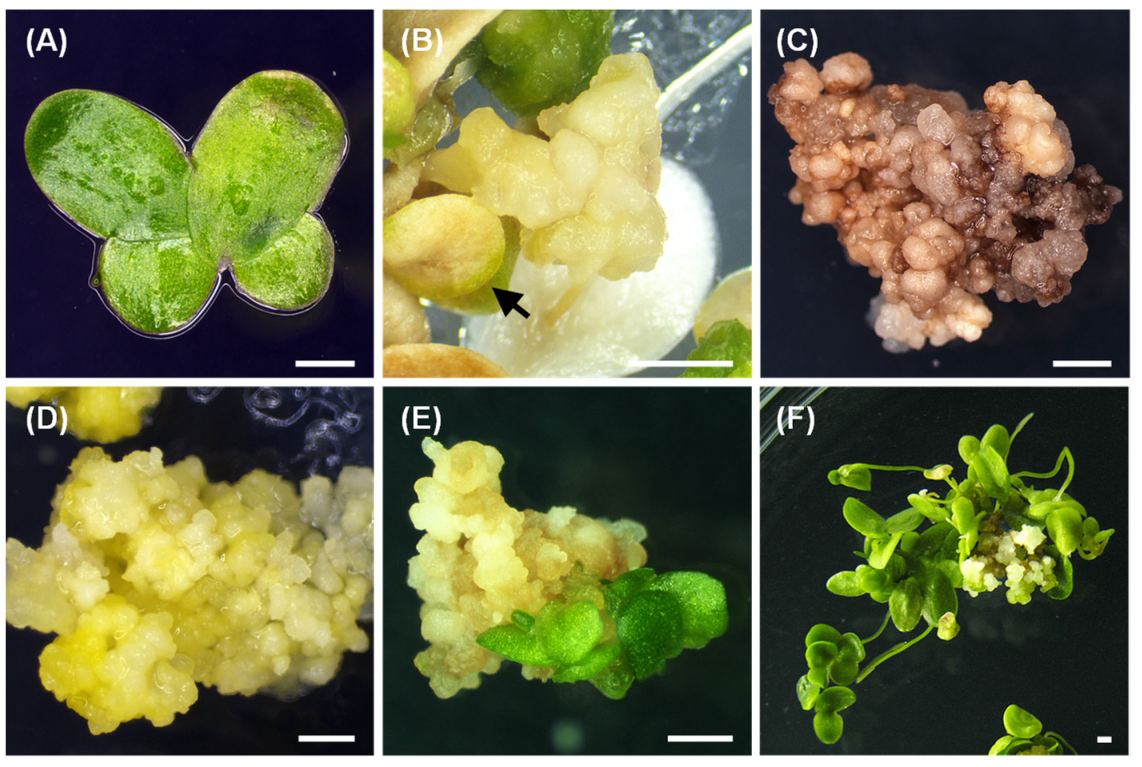
2.1. Callus Induction

Two to three-week-old fronds of Lemna aequinoctialis (Figure 1A) were used to investigate the effects of various combinations of plant-growth regulators (PGRs) on callus induction. The types and concentrations of PGRs had a significant (p < 0.05) effect on callus induction (Table 1). Frond proliferation was significantly retarded during the first 2 weeks, fronds started to curl irregularly in all treatments, and white or yellow senescent fronds were observed after 4 weeks of cultivation. Callus induction was slow and first visible after 8 weeks in culture. The callus arose most often from the meristematic region on the ventral surface of the frond. After 10 weeks in culture, different types of callus were obtained. The calli on media supplemented solely with 2,4-D were friable, nodular and whitish to light yellowish-colored (Figure S1A). The calli developed on media containing 2,4-D combined with cytokinins were also friable and nodular, but yellowish and greenish-colored (Figure S1B). Calli could be induced in each treatment, whereas 2,4-D combined with both cytokinins could significantly increase the induction rate, especially with the addition of TDZ. The most successful callus induction was observed on callus-induction medium (CIM) with 25.0 µM 2,4-D combined with TDZ (0.5 or 2.0 µM) or 6-BA (2.0 µM), where 100% of explants produced calli. However, more calli induced from 25.0 µM 2,4-D/2.0 µM TDZ showed a friable hard texture and golden yellow morphology (Figure 1B). The calli that were subcultured in the same medium showed retarded growth and browning phenomena (Figure 1C). Thus, the callus-proliferation medium was determined. In summary, the most effective CIM was MS medium containing 3% (w/v) sucrose, 0.3% (w/v) Phytagel, and 2,4-D (25.0 μM) combined with TDZ (2.0 μM) at pH 5.7.

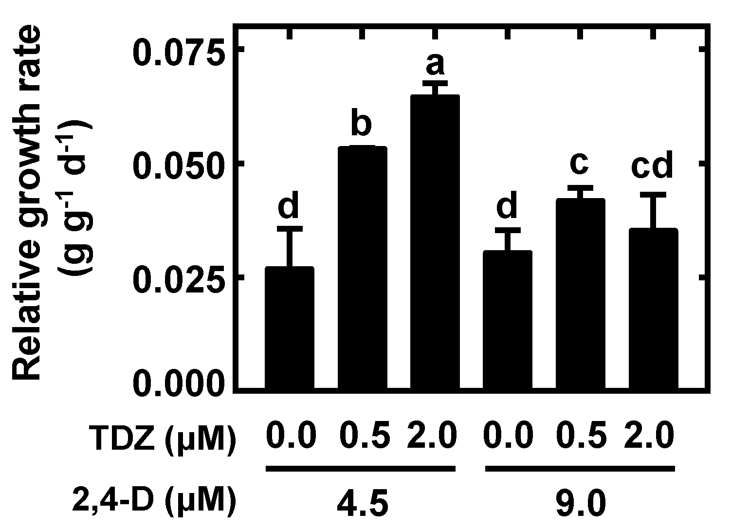
2.2. Nodular Callus Proliferation

Because callus proliferation was very slow on CIM, six combinations of 2,4-D with TDZ were tested to determine if they would enhance the rate of callus growth. Nodular callus-proliferation medium (NCPM) with 4.5 µM 2,4-D and 2.0 µM TDZ supported the significant growth rate of calli (Figure 2), which prevented growth cessation and browning. The morphology of calli derived from the effective NCPM was golden yellow and friable with a hard texture and consisted of small, connected amorphous masses (Figure 1D). Furthermore, the abovementioned medium supported callus proliferation for over 6 months by subculturing periodically. Additionally, the most effective NCPM was MS medium containing 3% (w/v) sucrose, 0.3% (w/v) Phytagel, and 2,4-D (4.5 μM) combined with TDZ (2.0 μM) at pH 5.7.

2.3. Frond Regeneration

Both SH and MS basal media supplemented with 6-BA were evaluated for their ability to regenerate fronds using nodular calli derived from the most effective NCPM (Table 2). The results showed that SH medium was superior to MS medium independent of cytokinin addition, whereas a 60 ± 5% regeneration rate was obtained in SH medium without the addition of 6-BA. In contrast, a ½ MS and MS medium poorly supported frond regeneration (0%). The formation of frond-like clusters and green structures was observed within 2–3 weeks after transferring nodular calli from NCPM to frond regeneration medium (FRM) (Figure 1E). The highest frond regeneration rate (100%) and number of fronds regenerated per callus (42 ± 4) was observed on FRM composed of SH medium containing 1.0 µM 6-BA after 4 weeks of cultivation. An example of L. aequinoctialis regenerating from callus tissue is shown in Figure 1F. Eventually, the most effective FRM was SH medium containing 1.0% (w/v) sucrose, 0.3% (w/v) Phytagel, and 6-BA (1.0 μM) at pH 5.7.

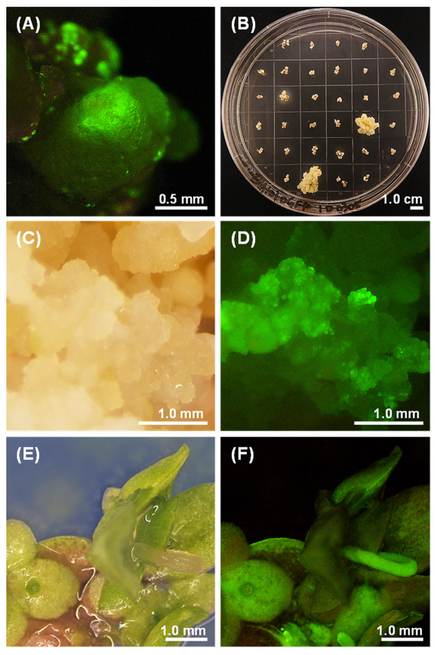
2.4. Optimization of the Transformation Protocol

To develop an efficient procedure for the stable transformation of Lemna aequinoctialis, we elucidated the effect of altering the concentrations of acetosyringone (AS), coculture days, and photoperiods on transformation efficiency (Table 3). We monitored GFP expression in nodules throughout the selection process after transferring the cocultured calli to the selection medium (NCPM-CH), which contained 50 mg L−1 hygromycin and 275 mg L−1 cefotaxime. The results showed that the maximum transformation frequency was observed in nodular calli cocultivated with the pD-clone1-harboring Agrobacterium in NCPM containing 200 µM AS in the dark for 1 day. During the early stage of selection, many GFP-expressing calli (Figure 3A) were observed, and a 49 ± 4% transformation frequency was obtained under NCPM-CH on day 9. In addition, we observed GFP-positive cells on many nodules that appeared dead following the selection process. After 10 weeks of selection, the newly divided calli showed GFP expression (Figure 3B–D). Furthermore, relatively few stable transformants (3 ± 1%) were recovered compared with the large number of GFP-positive cells at early stages. These observations indicated that only a small fraction of cells transiently expressing GFP became stably transformed. Subsequently, we transferred the resistant calli into FRM containing 50 mg L−1 hygromycin for frond regeneration. Regenerated fronds with GFP expression could be found at a 100% rate of regeneration after 4 weeks of regeneration. Finally, the regenerated fronds were transferred to SH medium and subcultured periodically. Transformants that had been propagated vegetatively for up to 1 year exhibited a uniform GFP signal, indicating the long-term stability of GFP expression (Figure 3E,F). Accordingly, the optimal transformation condition for L. aequinoctialis was observed in nodular calli cocultured in NCPM containing 200 µM AS for 1 day in the dark.

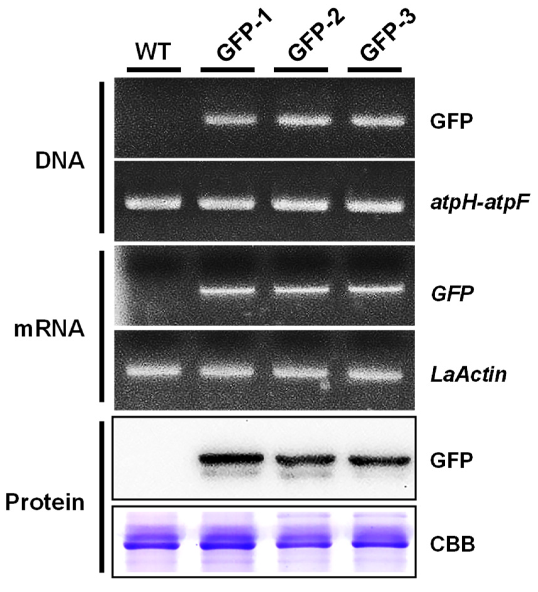
2.5. Molecular Analysis of Putative Transformants

To verify the putative transformants with the stable integration of the transgene, DNA, mRNA, and protein levels of the transgene (GFP) were evaluated in the three transgenic lines with the strongest GFP fluorescence (Figure 4). First, we verified the incorporation of the transgene using PCR-based analysis. According to the genomic PCR analysis, wild type and transformants showed atpH-atpF fragments, but GFP signals were only detected in the three transgenic lines. The positive RT-PCR results indicated that GFP transcripts were also detected in the three transgenic lines. Western blot analysis revealed the presence of a ~27-kDa band corresponding to the GFP protein in the three transgenic lines. Overall, the transformants were confirmed by the molecular analytical results above.

3. Discussion

Previous studies have implemented callus induction on species within Lemnoideae and have revealed that both the type and concentration of PGRs can significantly enhance the callus induction rate [25,30,31]. A crucial role is played by 2,4-D in callus induction in many plant species [32,33,34]. In 1997, Moon and Stomp [35] reported that MS medium supplemented with 20.0 or 50.0 µM 2,4-D alone could induce callus formation in Lemna gibba, but only ~10% of fronds produced calli. In this study, we found that L. aequinoctialis calli could be induced on MS medium containing 25.0 µM 2,4-D and 2.0 µM TDZ at a rate of 100%, which was better than the results from Liu et al. [27]. Our results showed that the optimal balance between auxin and cytokinin could significantly enhance the callus-induction rate. Several studies have demonstrated similar results in other duckweeds. For example, Chhabra et al. [15] found that B5S1.0 medium containing 50.0 µM 2,4-D and 5.0 µM TDZ could induce hard nodular calli in L. minor. In addition, Huang et al. [30] reported that the optimum medium for callus induction was MS basal medium supplemented with 15 mg L−1 2,4-D (~67.9 µM) and 2 mg L−1 6-BA (~9.01 µM) for Landoltia punctata; Yang et al. [5] reported that the optimum medium for callus induction was half-strength MS basal medium containing 22.62 µM 2,4-D and 8.88 µM 6-BA in Spirodela polyrhiza; and Khvatkov et al. [26] reported that the optimum medium for callus induction was SH medium containing 5.0 mg L−1 2,4-D (~22.6 µM) and 0.5 mg L−1 6-BA (~2.25 µM) in Wolffia arrhiza.
Our results showed that nodular callus-production medium (NCPM) with 4.5 μM 2,4-D and 2.0 μM TDZ could significantly improve callus proliferation. For L. minor, Cantó-Pastor et al. [13] transferred calli to culture medium containing 1.0 μM 2,4-D and 2.0 μM 6-BA, which had lower 2,4-D concentrations than the callus-induction medium. Firsov et al. [16] transferred calli to culture medium with 2.0 mg L−1 2,4-D (~9.04 μM) for subsequent growth and proliferation in L. gibba. Therefore, we found that FRM with 1.0 μM 6-BA in SH medium had a 100% rate of regeneration. For L. gibba, a 100% plant regeneration rate was observed on FRM with 1.0 μM 6-BA in SH medium [36]. For Spirodela punctata, a 95–98% plant regeneration rate was attained on FRM with 1.0 mg L−1 2ip in McCown Woody Plant medium [37]. It has been previously reported that L. gibba G3 and W. arrhiza fronds could also regenerate on PGR-free SH medium [25,35,36]. This indicates that SH medium is crucial for duckweed frond regeneration, which could be promoted by cytokinin addition. Above all, the selection and concentration of hormones could affect the formation of callus proliferation and frond regeneration in different duckweed species.
Here, by establishing the transformation protocol, we achieved a 3 ± 1% stable transformation success rate for Agrobacterium-mediated transformation in L. aequinoctialis. In addition, the stable transformation efficiency obtained was approximately 3.8% [15] and 4% [20] in L. minor, a different species of the same genus, or 0.5–5% in Spirodela oligorrhiza [23] and 0.4% in W. arrhizal [24,38]. Thus, we developed a protocol that obtained a similarly stable transformation efficiency that is feasible for Agrobacterium-mediated transformation in L. aequinoctialis. Acetosyringone (AS) has previously been shown to aid in the transformation of several crops, such as cotton, rice, and wheat [23]. Researchers have also found that adding AS at a low pH to cocultivation medium was effective in enhancing T-DNA transfer. For example, Cantó-Pastor et al. [13] used 200 µM AS during the Agrobacterium transformation of L. minor. In addition, Vunsh et al. [23] supplemented S. oligorrhiza with 100 µM AS at pH 5.6. These conditions had similar results to those found in the transformation protocol of S. oligorrhiza, L. gibba and L. minor [13,14,15]. Our study showed that the addition of 200 µM AS to the cocultivation medium at pH 5.2 could result in the maximum transformation rate. The cocultivation period and photoperiod were also important factors that could affect the transformation efficiency, while shorter or longer cocultivation periods led to unsuccessful transformation [39]. In our study, we determined 1 day in the dark to be the optimal duration for the cocultivation of L. aequinoctialis and Agrobacterium. Vunsh et al. [23] and Yang et al. [21] showed the same cocultivation conditions for S. oligorrhiza and S. polyrhiza. We know that longer cocultivation periods of approximately 4–5 days could cause the over-proliferation of Agrobacterium around the plant materials, ultimately leading to the death of the plant tissue [26]. Interestingly, Chhabra et al. [15] showed that maximum transformation frequencies were observed in nodular calli of L. minor cocultured for 3 days under a 16 h light/8 h dark photoperiod. They suggested that the possible reason for the difference was due to different physiological properties among duckweed species. Eventually, we successfully regenerated fronds from the transgenic calli. These fronds stably expressed green fluorescence through vegetative proliferation for several generations (up to 1 year). To date, transgenic fronds have shown uniform GFP activity, which suggests their ability to stably inherit transgenes through mitotic divisions.
However, several factors greatly affect the transformation efficiency, including plant species, type of explants, A. tumefaciens strain, binary vector types, infection methods, surfactant addition, and so on [39]. In several previous studies, such as on banana [40], chickpea [41], and cowpea [42], it has been shown that the combination of vacuum infiltration and sonication can greatly improve the efficiency of Agrobacterium infection. In comparison to previous studies of Agrobacterium-mediated transformation in duckweeds [13,14,15,16,17], Liu et al. [27] also suggested that the increased transformation efficiency likely resulted from the use of sonication coupled with vacuum infiltration infection. We believe the two strategies can be used to enhance the transformation efficiency in Taiwanese isolates of L. aequinoctialis in the future.

4. Materials and Methods

4.1. Plant Material and Growth Conditions

Lemna aequinoctialis was collected from a pool at National Pingtung University of Science and Technology (NPUST), Pingtung, Taiwan. The duckweed species was identified by an atpF-atpH noncoding spacer [43]. After sterilization using 0.6% NaHClO for 2 to 3 min and washing 6 to 8 times with sterilized water, the plants were cultured in 90 × 20 mm Perti dishes containing 50 mL Schenk and Hildebrandt (SH) medium with 1% sucrose and 0.30% (w/v) Phytagel (Sigma, St. Louis, MO, USA) at pH 5.7. All cultures were maintained at 27 ± 1 °C under a 12 h photoperiod of cool-white fluorescent light of 80 μmol photons m−2 s−1 intensity. An axenic clone was produced and maintained in our laboratory for 4 years. Two to three-week-old axenic fronds were used for callus induction.

4.2. Callus Induction

To evaluate the optimal conditions for callus induction, fronds (~3.5 mm in length) were grown on MS medium containing 3% (w/v) sucrose and 0.3% (w/v) Phytagel at pH 5.7. Different combinations of plant-growth regulators were added in the medium. For cytokinins, we used either 6-benzyladenine (6-BA) or thidiazuron (TDZ) at 0, 0.5, 1 and 2 μM; for auxins, we used 2,4-dichlorophenoxyacetic acid (2,4-D) at 5, 10, 25 and 50 μM. Thirty-two fronds were cultured in sterilized 150 × 20 mm Petri dishes containing 100 mL solid medium for callus induction. The explants were maintained under a 12/12 h photoperiod at 27 ± 1 °C (80 μmol photons m−2 s−1). Each treatment was performed in triplicate. The efficiency of callus induction on each plate was calculated after 10 weeks of cultivation.

4.3. Nodular Callus Proliferation

To evaluate the optimal conditions for callus proliferation, calli (~5 mm in diameter) derived from the most effective callus-induction medium (CIM) were cultured on MS medium containing 3% (w/v) sucrose, 0.3% (w/v) Phytagel, and either 2,4-D at 4.5 or 9.0 μM combined with TDZ (0, 0.5 and 2 μM) at pH 5.7. Five calli were cultured in sterilized 90 × 20 mm Petri dishes containing 50 mL solid medium under a 12/12 h photoperiod at 27 ± 1 °C (80 μmol photons/m2/s). Each treatment was performed in triplicate. The relative growth rate (RGR) of calli during 4 weeks of cultivation was calculated with the following equation [44]: RGR = (lnWt − lnW0)/(tt − t0), where Wt is the fresh weight at time tt and W0 is the initial fresh weight at the beginning of the treatment. The nodular calli derived from the most effective nodular callus-proliferation medium (NCPM) were then used for frond regeneration and gene transformation.

4.4. Frond Regeneration

To determine the optimal conditions for frond regeneration, calli (~5 mm in diameter) derived from the most effective NCPM were cultured on both MS and SH media containing 3% and 1% (w/v) sucrose, respectively, 0.3% (w/v) Phytagel, and 6-BA (0, 1, 2 μM) at pH 5.7. Five calli were cultured in sterilized 90 × 20 mm Petri dishes containing 50 mL solid medium under a 12/12 h photoperiod at 27 ± 1 °C (80 μmol photons m−2 s−1). Each treatment was performed in triplicate. The efficiency of frond regeneration on each plate was calculated after 4 weeks of cultivation.

4.5. Agrobacterium Strain and Binary Vector

The green fluorescence protein (GFP) expression vector pD-clone1 [45] was introduced into Agrobacterium strain EHA101 and used for genetic transformation. pD-clone1 contained two gene expression cassettes, including the GFP protein with the hygromycin selection marker (hygromycin phosphotransferase gene, Hpt). GFP was driven by the maize ubiquitin1 promoter together with its intron (Ubi1) promoter and terminated by the Nos poly (A) signal.

4.6. Optimization of the Transformation Protocol

To develop a genetic transformation protocol for Lemna aequinoctialis, various parameters influencing transformation efficiency were evaluated. Three days prior to the transformation experiment, Agrobacterium tumefaciens (pD-clone1) from a freshly streaked plate was inoculated in 3 mL of liquid YEP medium (yeast extract 1.0 g L−1, bactopeptone 10.0 g L−1, NaCl 5 g L−1) containing ampicillin (100 mg L−1) and kanamycin (50 mg L−1) and grown overnight on a rotary shaker (180 rpm) at 27 °C. The next day, 0.5 mL of this primary culture was inoculated in 50 mL subculture medium (3.0 g L−1 K2HPO4, 1.0 g L−1 NaH2PO4, 1.0 g L−1 NH4Cl, 0.3 g L−1 MgSO4·7H2O, 0.15 g L−1 KCl, 0.01 g L−1 CaCl2, 2.5 mg L−1 FeSO4·7H2O, and 5 g L−1 glucose) containing ampicillin (100 mg L−1) and kanamycin (50 mg L−1). The culture was grown at 27 °C with shaking (180 rpm) until the OD600 reached 0.6–0.8. The bacteria were harvested by centrifuging at 5000 rpm for 7 min at 4 °C and resuspended in 10 mL MS liquid medium. After soaking for 2 min, the calli (~100 calli/treatment) were blotted dry on sterile filter paper and cocultivated on the most effective NCPM (pH 5.2) supplemented with 0, 50, 100, and 200 µM acetosyringone (AS) for 1–3 days at 27 ± 1 °C under a 12/12 h photoperiod or dark conditions.
After cocultivation, nodular calli from each treatment were collected and washed with sterile distilled water at least 20 times, followed by washing with MS liquid medium containing 275 mg L−1 cefotaxime once per hour for five hours on a shaker at 110 rpm. The mixture was left to culture overnight. After washing, the calli were blotted dry and cultured on selection medium (the most effective NCPM containing 50 mg L−1 hygromycin and 275 mg L−1 cefotaxime) (NCPM-CH) under a 12/12 h photoperiod at 27 ± 1 °C (80 μmol photons/m2/s).
After culturing on the selection medium for 3 days, we started to screen calli showing GFP signals under a fluorescence stereomicroscope (SteREO Discovery. V8, Carl Zeiss Microscopy, Pleasanton, CA, USA) until the nodular calli began to proliferate, which were regarded as putative transformants. The frequency of GFP expression was calculated as the number of calli showing GFP out of the total number of treated cells. Hygromycin-resistant calli could be found at approximately 10 weeks. The putative transformants were transferred to regeneration medium containing 50 mg L−1 hygromycin for frond regeneration.

4.7. Genomic PCR Analysis of Transgene Integration

Genomic DNA was extracted from wild-type and putative transformants by using an RBC Real Genomics Genomic DNA Extraction Kit (YGP50, RBCBioscience) and used as a template for PCR analysis to test the presence for GFP using the following specific primers: GFP-F: 5′-CAAGGGCGAGGAGCTGTT-3′; and GFP-R: 5′-CTTGTACAGCTCGTCCATGC-3′. The chloroplast marker atpF-atpH was amplified as an endogenous control [43]. The PCR program was as follows: 94 °C denaturation for 10 min, followed by 30 cycles at 94 °C for 30 s, 50 °C for 30 s, and 72 °C for 40 s, and a final extension at 72 °C for 7 min. The PCR products were resolved by electrophoresis in 1.2 % (w/v) agarose gel and stained with SafeView™ Classic (Applied Biological Materials Inc., Richmond, BC, Canada).

4.8. RT- PCR Analysis of Transgenic Duckweed

Total RNA was isolated from wild-type and putative transformants using TRIzol reagent (Invitrogen, CA, USA), and cDNA synthesis involved the use of an MMLV reverse transcriptase first-strand cDNA synthesis kit (Epicentre Biotechnologies, Madison, WI, USA). cDNAs were used as a template for RT-PCR analysis to determine GFP and Lemna aequinoctialis Actin (LaActin, GenBank accession no. MZ394729) expression using the following specific primers: the same primer pairs of GFP; LaActin-F: 5′-GACGCAGATCATGTTCGAGA-3′; and LaActin-R: 5′-TGGTTCCACCACTAAGCACA-3′. PCR amplification was performed as mentioned above. Three batches of independently isolated total RNA samples underwent three rounds of RT-PCR. All tests were repeated at least three times, and the results from one repeat are shown in the figures. LaActin was used as the internal control.

4.9. Western Blot Analysis of Transgenic Duckweed

Total protein extracted from the fronds of wild-type plants and putative transformants (15 μg each lane) was subjected to SDS-PAGE using a 12% polyacrylamide gel and Western blot analysis with a monoclonal antibody against GFP (sc-9996, Santa Cruz Biotechnology, Inc., Dallas, TX, USA) as described in [46]. Immunoreactivity was detected by the enhanced chemiluminescence method (Amersham International).

4.10. Statistical Analysis

A one-way analysis of variance (ANOVA) was used to analyze the data. When the ANOVA identified differences among the groups, Duncan’s multiple range test was conducted to examine significant differences among the treatments in SAS software (SAS Institute, Cary, NC, USA). The level of significance was p < 0.05.

5. Conclusions

In conclusion, we developed an efficient micropropagation technique and achieved stable transformation in Lemna aequinoctialis (Figure S2). Our results showed that a 100% callus induction rate was observed under CIM with 25.0 µM 2,4-D and 2 µM TDZ, the best callus proliferation was observed under NCPM with 4.5 μM 2,4-D and 2.0 μM TDZ, and a 100% frond regeneration rate was observed under frond regeneration medium (FRM) with 1 μM 6-BA. We achieved the stable transformation of calli derived from the optimal NCPM using Agrobacterium tumefaciens strain EHA101 harboring pD-clone1. A stable transformation frequency (3.13 ± 1.08%) was observed in calli cocultivated with Agrobacterium on NCPM containing 200 μM AS for 1 day in the dark. Therefore, the protocol for micropropagation and Agrobacterium-mediated transformation could be a good foundation for the transgenic engineering of L. aequinoctialis.

Supplementary Materials

The following are available online at https://www.mdpi.com/article/10.3390/plants10081576/s1, Figure S1: The callus appearances of Lemna aequinoctialis, Figure S2: The scheme for Agrobacterium-mediated genetic transformation of Taiwanese isolates of Lemna aequinoctialis.

Author Contributions

K.-T.W. and T.-M.W. conceived the original idea. K.-T.W. performed the experiments, analyzed data, and co-wrote the manuscript. M.-C.H. and Y.-S.W. advised and helped supervise the project. T.-M.W. supervised the project and co-wrote the manuscript. All authors have read and agreed to the published version of the manuscript.

Funding

This work was supported by the Ministry of Science and Technology (MOST) (MOST 104-2311-B-020-003 and MOST 106-2313-B-020-010) of Taiwan to T.-M.W.

Data Availability Statement

The authors confirm that the data supporting the findings of this study are available within the article.

Acknowledgments

We thank Chwan-Yang Hong (Department of Agricultural Chemistry, National Taiwan University, Taiwan) for kindly providing the pD-clone1.

Conflicts of Interest

The authors declare no conflict of interest.

References

  1. Appenroth, K.; Borisjuk, N.; Lam, E. Telling duckweed apart: Genotyping technologies for the Lemnaceae. Chin. J. Appl. Environ. Biol. 2013, 19, 1–10. [Google Scholar]
  2. Landolt, E. The family of Lemnaceae-a monographic study. In Biosystematic Investigations in the Family of Duckweeds (Lemnaceae); Veröffentlichungen des Geobotanischen Institutes der ETH; Stiftung Rübel: Zurich, Switzerland, 1986; Volume 1. [Google Scholar]
  3. Sree, K.S.; Bog, M.; Appenroth, K.J. Taxonomy of duckweeds (Lemnaceae), potential new crop plants. Emir. J. Food Agric. 2016, 28, 291–302. [Google Scholar] [CrossRef] [Green Version]
  4. Stomp, A.M. The duckweeds: A valuable plant for biomanufacturing. Biotechnol. Annu. Rev. 2005, 11, 69–99. [Google Scholar]
  5. Yang, J.; Li, G.; Hu, S.; Bishopp, A.; Heenatigala, P.; Kumar, S.; Duan, P.; Yao, L.; Hou, H. A protocol for efficient callus induction and stable transformation of Spirodela polyrhiza (L.) Schleiden using Agrobacterium tumefaciens. Aquat. Bot. 2018, 151, 80–86. [Google Scholar] [CrossRef]
  6. Ziegler, P.; Adelmann, K.; Zimmer, S.; Schmidt, C.; Appenroth, K.J. Relative in vitro growth rates of duckweeds (Lemnaceae)—The most rapidly growing higher plants. Plant Biol. 2015, 17, 33–41. [Google Scholar] [CrossRef]
  7. Xu, J.; Cheng, J.J.; Stomp, A.M. Growing Spirodela polyrrhiza in swine wastewater for the production of animal feed and fuel ethanol: A pilot study. CLEAN–Soil Air Water 2012, 40, 760–765. [Google Scholar] [CrossRef]
  8. Xu, J.; Cui, W.; Cheng, J.J.; Stomp, A.M. Production of high-starch duckweed and its conversion to bioethanol. Biosyst. Eng. 2011, 110, 67–72. [Google Scholar] [CrossRef]
  9. Khellaf, N.; Zerdaoui, M. Growth response of the duckweed Lemna minor to heavy metal pollution. J. Environ. Health Sci. Eng. 2009, 6, 161–166. [Google Scholar]
  10. Yang, L.; Han, Y.; Wu, D.; Yong, W.; Liu, M.; Wang, S.; Liu, W.; Lu, M.; Wei, Y.; Sun, J. Salt and cadmium stress tolerance caused by overexpression of the Glycine max Na+/H+ Antiporter (GmNHX1) gene in duckweed (Lemna turionifera 5511). Aquat. Toxicol. 2017, 192, 127–135. [Google Scholar] [CrossRef] [PubMed]
  11. Zhao, H.; Appenroth, K.; Landesman, L.; Salmeán, A.A.; Lam, E. Duckweed rising at Chengdu: Summary of the 1st International conference on duckweed application and research. Plant Mol. Biol. 2012, 78, 627–632. [Google Scholar] [CrossRef] [PubMed]
  12. Makhzoum, A.; Benyammi, R.; Moustafa, K.; Trémouillaux-Guiller, J. Recent advances on host plants and expression cassettes’ structure and function in plant molecular pharming. BioDrugs 2014, 28, 145–159. [Google Scholar] [CrossRef]
  13. Cantó-Pastor, A.; Mollá-Morales, A.; Ernst, E.; Dahl, W.; Zhai, J.; Yan, Y.; Meyers, B.; Shanklin, J.; Martienssen, R. Efficient transformation and artificial miRNA gene silencing in Lemna minor. Plant Biol. 2015, 17, 59–65. [Google Scholar] [CrossRef] [Green Version]
  14. Yamamoto, Y.T.; Rajbhandari, N.; Lin, X.; Bergmann, B.A.; Nishimura, Y.; Stomp, A.M. Genetic transformation of duckweed Lemna gibba and Lemna minor. Vitr. Cell. Dev. Biol.-Plant 2001, 37, 349–353. [Google Scholar] [CrossRef]
  15. Chhabra, G.; Chaudhary, D.; Sainger, M.; Jaiwal, P.K. Genetic transformation of Indian isolate of Lemna minor mediated by Agrobacterium tumefaciens and recovery of transgenic plants. Physiol. Mol. Biol. Plants 2011, 17, 129–136. [Google Scholar] [CrossRef] [Green Version]
  16. Firsov, A.; Tarasenko, I.; Mitiouchkina, T.; Ismailova, N.; Shaloiko, L.; Vainstein, A.; Dolgov, S. High-yield expression of M2e peptide of avian influenza virus H5N1 in transgenic duckweed plants. Mol. Biotechnol. 2015, 57, 653–661. [Google Scholar] [CrossRef] [PubMed]
  17. Firsov, A.; Tarasenko, I.; Mitiouchkina, T.; Shaloiko, L.; Kozlov, O.; Vinokurov, L.; Rasskazova, E.; Murashev, A.; Vainstein, A.; Dolgov, S. Expression and immunogenicity of M2e peptide of avian influenza virus H5N1 fused to ricin toxin b chain produced in duckweed plants. Front. Chem. 2018, 6, 22. [Google Scholar] [CrossRef] [PubMed]
  18. Ko, S.M.; Sun, H.J.; Oh, M.J.; Song, I.J.; Kim, M.J.; Sin, H.S.; Goh, C.H.; Kim, Y.W.; Lim, P.O.; Lee, H.Y. Expression of the protective antigen for PEDV in transgenic duckweed, Lemna minor. Hortic. Environ. Biotechnol. 2011, 52, 511–515. [Google Scholar] [CrossRef]
  19. Sun, Y.; Cheng, J.J.; Himmel, M.E.; Skory, C.D.; Adney, W.S.; Thomas, S.R.; Tisserat, B.; Nishimura, Y.; Yamamoto, Y.T. Expression and characterization of Acidothermus cellulolyticus E1 endoglucanase in transgenic duckweed Lemna minor 8627. Bioresour. Technol. 2007, 98, 2866–2872. [Google Scholar] [CrossRef] [PubMed]
  20. Yang, G.L.; Fang, Y.; Xu, Y.L.; Tan, L.; Li, Q.; Liu, Y.; Lai, F.; Jin, Y.-L.; Du, A.-P.; He, K.-Z. Frond transformation system mediated by Agrobacterium tumefaciens for Lemna minor. Plant Mol. Biol. 2018, 98, 319–331. [Google Scholar] [CrossRef] [PubMed]
  21. Yang, L.; Han, H.; Liu, M.; Zuo, Z.; Zhou, K.; Lü, J.; Zhu, Y.; Bai, Y.; Wang, Y. Overexpression of the Arabidopsis photorespiratory pathway gene, serine: Glyoxylate aminotransferase (AtAGT1), leads to salt stress tolerance in transgenic duckweed (Lemna minor). Plant Cell Tissue Organ Cult. (PCTOC) 2013, 113, 407–416. [Google Scholar] [CrossRef]
  22. Rival, S.; Wisniewski, J.P.; Langlais, A.; Kaplan, H.; Freyssinet, G.; Vancanneyt, G.; Vunsh, R.; Perl, A.; Edelman, M. Spirodela (duckweed) as an alternative production system for pharmaceuticals: A case study, aprotinin. Transgenic Res. 2008, 17, 503–513. [Google Scholar] [CrossRef] [PubMed]
  23. Vunsh, R.; Li, J.; Hanania, U.; Edelman, M.; Flaishman, M.; Perl, A.; Wisniewski, J.P.; Freyssinet, G. High expression of transgene protein in Spirodela. Plant Cell Rep. 2007, 26, 1511–1519. [Google Scholar] [CrossRef] [PubMed]
  24. Khvatkov, P.; Chernobrovkina, M.; Okuneva, A.; Pushin, A.; Dolgov, S. Transformation of Wolffia arrhiza (L.) Horkel ex Wimm. Plant Cell Tissue Organ Cult. (PCTOC) 2015, 123, 299–307. [Google Scholar] [CrossRef]
  25. Khvatkov, P.; Chernobrovkina, M.; Okuneva, A.; Shvedova, A.; Chaban, I.; Dolgov, S. Callus induction and regeneration in Wolffia arrhiza (L.) Horkel ex Wimm. Plant Cell Tissue Organ Cult. (PCTOC) 2015, 120, 263–273. [Google Scholar] [CrossRef]
  26. Heenatigala, P.P.M.; Yang, J.; Bishopp, A.; Sun, Z.; Li, G.; Kumar, S.; Hu, S.; Wu, Z.; Lin, W.; Yao, L.; et al. Development of efficient protocols for stable and transient gene transformation for Wolffia globosa using Agrobacterium. Front. Chem. 2018, 6, 227. [Google Scholar] [CrossRef] [Green Version]
  27. Liu, Y.; Wang, Y.; Xu, S.; Tang, X.; Zhao, J.; Yu, C.; He, G.; Xu, H.; Wang, S.; Tang, Y. Efficient genetic transformation and CRISPR/Cas9-mediated genome editing in Lemna aequinoctialis. Plant Biotechnol. J. 2019, 17, 2143–2152. [Google Scholar] [CrossRef] [PubMed] [Green Version]
  28. Tang, J.; Li, Y.; Ma, J.; Cheng, J. Survey of duckweed diversity in Lake Chao and total fatty acid, triacylglycerol, profiles of representative strains. Plant Biol. 2015, 17, 1066–1072. [Google Scholar] [CrossRef]
  29. Xu, Y.; Ma, S.; Huang, M.; Peng, M.; Bog, M.; Sree, K.S.; Appenroth, K.-J.; Zhang, J. Species distribution, genetic diversity and barcoding in the duckweed family (Lemnaceae). Hydrobiologia 2015, 743, 75–87. [Google Scholar] [CrossRef]
  30. Huang, M.; Fu, L.; Sun, X.; Di, R.; Zhang, J. Rapid and highly efficient callus induction and plant regeneration in the starch-rich duckweed strains of Landoltia punctata. Acta Physiol. Plant. 2016, 38, 122. [Google Scholar] [CrossRef]
  31. Wang, Y. Callus induction and frond regeneration in Spirodela polyrhiza. Czech J. Genet. Plant Breed. 2016, 52, 114–119. [Google Scholar] [CrossRef] [Green Version]
  32. Ball, S.T.; Zhou, H.; Konzak, C.F. Influence of 2,4-D, IAA, and duration of callus induction in anther cultures of spring wheat. Plant Sci. 1993, 90, 195–200. [Google Scholar] [CrossRef]
  33. Adil, M.; Ren, X.; Kang, D.I.; Jeong, B.R. Effect of explant type and plant growth regulators on callus induction, growth and secondary metabolites production in Cnidium officinale Makino. Mol. Biol. Rep. 2018, 45, 1919–1927. [Google Scholar] [CrossRef]
  34. Zheng, M.; Konzak, C. Effect of 2,4-dichlorophenoxyacetic acid on callus induction and plant regeneration in anther culture of wheat (Triticum aestivum L.). Plant Cell Rep. 1999, 19, 69–73. [Google Scholar] [CrossRef]
  35. Moon, H.; Stomp, A. Effects of medium components and light on callus induction, growth, and frond regeneration in Lemna gibba (Duckweed). Vitr. Cell. Dev. Biol.-Plant 1997, 33, 20–25. [Google Scholar] [CrossRef]
  36. Moon, H.K.; Yang, M.S. Nodular somatic embryogenesis and frond regeneration in duckweed, Lemna gibba G3. J. Plant Biol. 2002, 45, 154–160. [Google Scholar] [CrossRef]
  37. Li, J.; Jain, M.; Vunsh, R.; Vishnevetsky, J.; Hanania, U.; Flaishman, M.; Perl, A.; Edelman, M. Callus induction and regeneration in Spirodela and Lemna. Plant Cell Rep. 2004, 22, 457–464. [Google Scholar] [CrossRef] [PubMed]
  38. Khvatkov, P.; Firsov, A.; Shvedova, A.; Shaloiko, L.; Kozlov, O.; Chernobrovkina, M.; Pushin, A.; Tarasenko, I.; Chaban, I.; Dolgov, S. Development of Wolffia arrhiza as a producer for recombinant human granulocyte colony-stimulating factor. Front. Chem. 2018, 6, 304. [Google Scholar] [CrossRef]
  39. Aileni, M.; Abbagani, S.; Zhang, P. Highly efficient production of transgenic Scoparia dulcis L. mediated by Agrobacterium tumefaciens: Plant regeneration via shoot organogenesis. Plant Biotechnol. Rep. 2011, 5, 147–156. [Google Scholar] [CrossRef]
  40. Subramanyam, K.; Subramanyam, K.; Sailaja, K.; Srinivasulu, M.; Lakshmidevi, K. Highly efficient Agrobacterium-mediated transformation of banana cv. Rasthali (AAB) via sonication and vacuum infiltration. Plant Cell Rep. 2011, 30, 425–436. [Google Scholar] [CrossRef] [PubMed]
  41. Indurker, S.; Misra, H.S.; Eapen, S. Agrobacterium-mediated transformation in chickpea (Cicer arietinum L.) with an insecticidal protein gene: Optimisation of different factors. Physiol. Mol. Biol. Plants 2010, 16, 273–284. [Google Scholar] [CrossRef] [Green Version]
  42. Bakshi, S.; Sadhukhan, A.; Mishra, S.; Sahoo, L. Improved Agrobacterium-mediated transformation of cowpea via sonication and vacuum infiltration. Plant Cell Rep. 2011, 30, 2281–2292. [Google Scholar] [CrossRef]
  43. Wang, W.; Wu, Y.; Yan, Y.; Ermakova, M.; Kerstetter, R.; Messing, J. DNA barcoding of the Lemnaceae, a family of aquatic monocots. BMC Plant Biol. 2010, 10, 205. [Google Scholar] [CrossRef] [PubMed] [Green Version]
  44. Watanabe, Y.; Sawa, Y.; Nagaoka, N.; Kozai, T. Photoautotrophic growth of Pleioblastus pygmaea plantlets in vitro and ex vitro as affected by types of supporting material in vitro. In Transplant Production in the 21st Century; Springer: Dordrecht, The Netherlands, 2000; pp. 226–230. [Google Scholar]
  45. Wu, T.M.; Lin, K.C.; Liau, W.S.; Chao, Y.Y.; Yang, L.H.; Chen, S.Y.; Lu, C.A.; Hong, C.Y. A set of GFP-based organelle marker lines combined with DsRed-based gateway vectors for subcellular localization study in rice (Oryza sativa L.). Plant Mol. Biol. 2016, 90, 107–115. [Google Scholar] [CrossRef] [PubMed]
  46. Benke, D.; Cicin-Sain, A.; Mertens, S.; Mohler, H. Immunochemical identification of the α1-and α3–subuntis of the GabaA-receptor in rat brain. J. Recept. Res. 1991, 11, 407–424. [Google Scholar] [CrossRef] [PubMed]
Figure 1. Lemna aequinoctialis. (A) Fronds of L. aequinoctialis; (B) fronds were cultured for 8 weeks on MS medium containing 3% sucrose, 25 μM 2,4-D, and 2.0 μM TDZ to induce callus formation. The arrow indicates the senescent frond; (C) browning phenomenon of callus cultured on previous CIM; (D) callus that was separated from the bleached frond explant and grown for an additional 4 weeks on MS medium containing 3% sucrose, 4.5 μM 2,4-D, and 2.0 μM TDZ for callus proliferation; (E,F) plants regenerating from callus incubated for 2 (E) and 4 (F) weeks on SH medium containing 1% sucrose and 1.0 μM 6-BA. Bar = 1.0 mm.
Plants 10 01576 g001
Figure 2. The effects of 2,4-D and TDZ on callus proliferation. Calli were cultured on the MS medium containing 3% sucrose and different plant growth regulators. Data (mean ± SD) with different letters among treatments significantly differ (p < 0.05).
Plants 10 01576 g002
Figure 3. Stable transformation and regeneration of L. aequinoctialis callus with a GFP expression construct. (A) Transformed nodular calli showed GFP expression under selection medium at day 9; (B) newly divided calli were observed after 10 weeks of selection; (C) newly divided calli were observed under bright-field and (D) GFP channel with long-pass filter; (E) transformed fronds were observed under bright-field and (F) GFP channel with long-pass filter.
Plants 10 01576 g003
Figure 4. Molecular analysis of putative transformants from DNA, mRNA, and protein. The atpF-atpH marker, LaActin, and coomassie brilliant blue (CBB) staining were used as internal controls for each molecular analysis, respectively.
Plants 10 01576 g004
Table 1. The effects of the type and concentration of plant growth regulators on callus induction from fronds after 10 weeks in culture.
2,4-D 1 (μM) Cytokinin 2 (μM)% Fronds Forming Callus 3
5 0.057 ± 6 gf
1065 ± 5 ef
2550 ± 9 g
5027 ± 6 h
56-BA0.528 ± 14 h
1.055 ± 5 gf
2.055 ± 5 gf
100.565 ± 5 ef
1.067 ± 3 ef
2.073 ± 14 e
250.587 ± 3 bcd
1.095 ± 5 abc
2.0100 ± 0 a
500.572 ± 3 e
1.093 ± 6 abc
2.087 ± 6 bcd
5TDZ0.575 ± 5 de
1.088 ± 3 bc
2.085 ± 5 cd
100.593 ± 6 abc
1.095 ± 5 abc
2.087 ± 12 bcd
250.5100 ± 0 a
1.095 ± 5 abc
2.0100 ± 0 a
500.592 ± 10 abc
1.098 ± 3 ab
2.097 ± 3 abc
1 2,4-D, 2,4-dichlorophenoxyacetic acid. 2 TDZ, thidiazuron; 6-BA, 6-benzylaminopurine. 3 Data (mean ± SD) with different letters among treatments significantly differ (p < 0.05).
Table 2. The effects of basal medium type and strength and 6-BA concentration on the frequency and efficiency of frond regeneration from callus after 4 weeks in culture.
Basal Medium 16-BA (μM)% Callus Regenerating Fronds 2Number of Fronds Regenerated/Callus Piece 2
00 d0 D
½SH10 d0 D
261 ± 5 b9 ± 2 C
060 ± 5 b10 ± 2 C
SH1100 ± 0 a42 ± 4 A
247 ± 2 c18 ± 3 B
00 d0 D
½MS10 d0 D
20 d0 D
00 d0 D
MS10 d0 D
20 d0 D
1 SH, Schenk and Hildebrandt (1972); MS, Murashige and Skoog (1962); ½, half-strength. 2 Data (mean ± SD) within each column followed by different letters are significantly different (p < 0.05).
Table 3. The effects of different transformation parameters on GFP expression in nodular calli of L. aequinoctialis cocultivated with A. tumefaciens strain EHA 101 harboring binary vector pD-clone1.
AS 1
(µM)
Coculture
(Day)
PhotoperiodGFP Expression 2 (%)
Day 9Week 10
0112 h light/12 h dark0 e0 B
24 h dark0 e0 B
212 h light/12 h dark0 e0 B
24 h dark0 e0 B
312 h light/12 h dark0 e0 B
24 h dark0 e0 B
50112 h light/12 h dark0 e0 B
24 h dark0 e0 B
212 h light/12 h dark0 e0 B
24 h dark0 e0 B
312 h light/12 h dark0 e0 B
24 h dark0 e0 B
100112 h light/12 h dark0 e0 B
24 h dark3 ± 3 de0 B
212 h light/12 h dark0 e0 B
24 h dark0 e0 B
312 h light/12 h dark0 e0 B
24 h dark0 e0 B
200112 h light/12 h dark2 ± 2 de0 B
24 h dark49 ± 4 a3 ± 1 A
212 h light/12 h dark2 ± 2 de0 B
24 h dark14 ± 2 c0 B
312 h light/12 h dark4 ± 2 d0 B
24 h dark25 ± 5 b0 B
1 AS, acetosyringone. 2 Data (mean ± SD) within each column followed by different letters are significantly different (p < 0.05).
Publisher’s Note: MDPI stays neutral with regard to jurisdictional claims in published maps and institutional affiliations.

