
Molecular Engineering of Curcumin, an Active Constituent of Curcuma longa L. (Turmeric) of the Family Zingiberaceae with Improved Antiproliferative Activity

 , , and
Abstract
:1. Introduction
2. Methods and Materials
2.1. Procurement of Chemicals and Crude Turmeric
2.2. Isolation of Curcumin
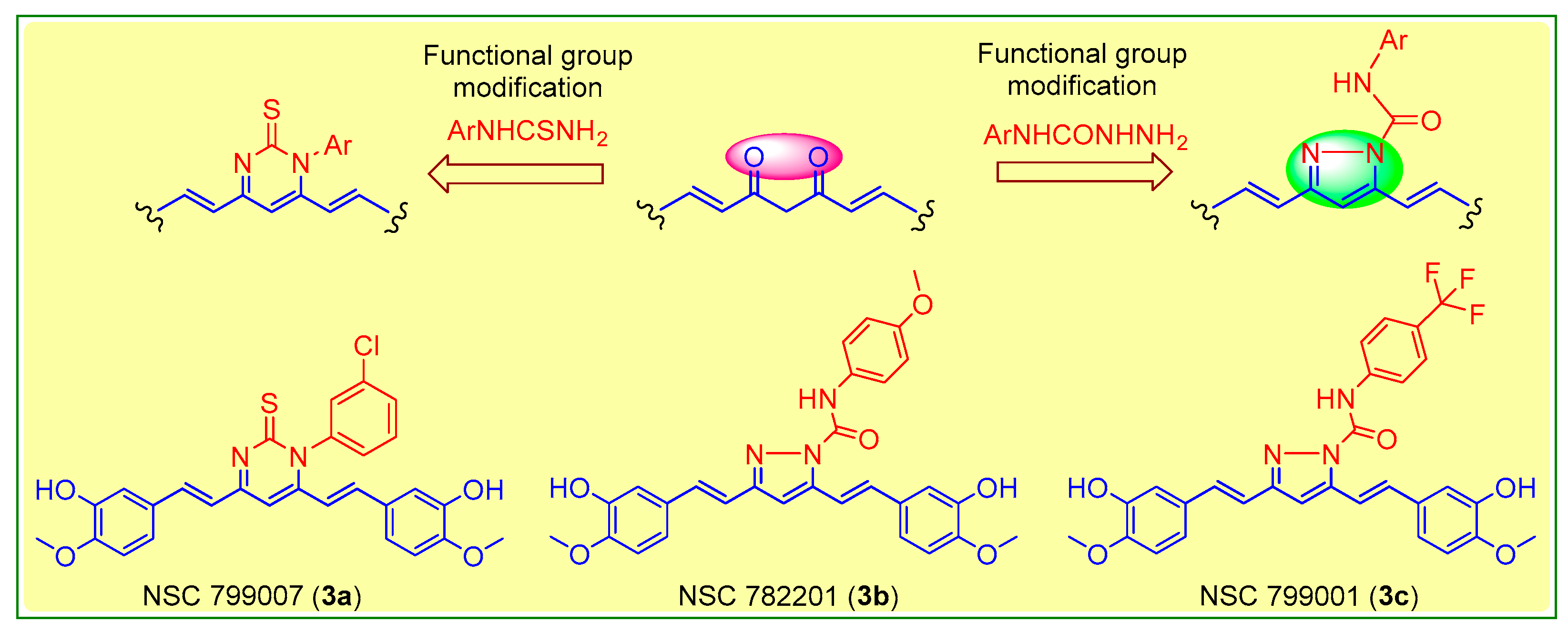
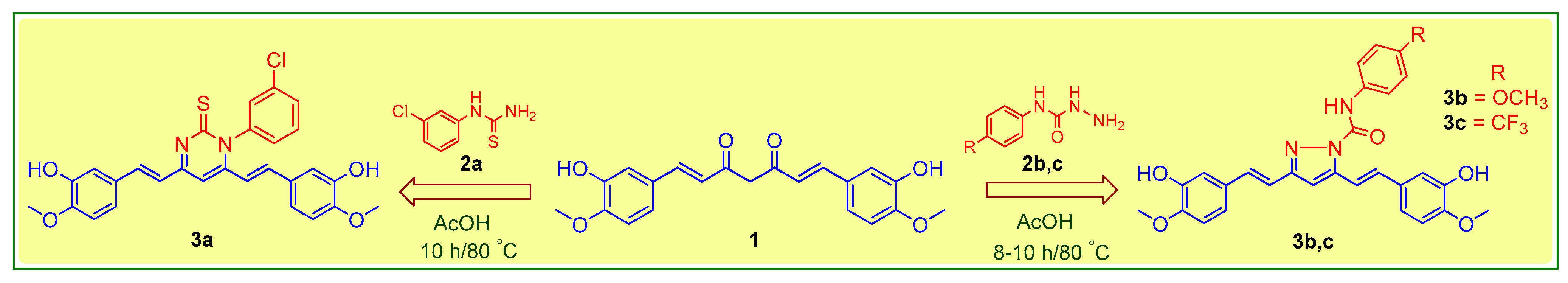
2.3. Preparation of Curcumin Analogues
2.4. Molecular Docking Studies
2.5. Antiproliferative Activity
2.6. Anti-EGFR Activity
3. Results
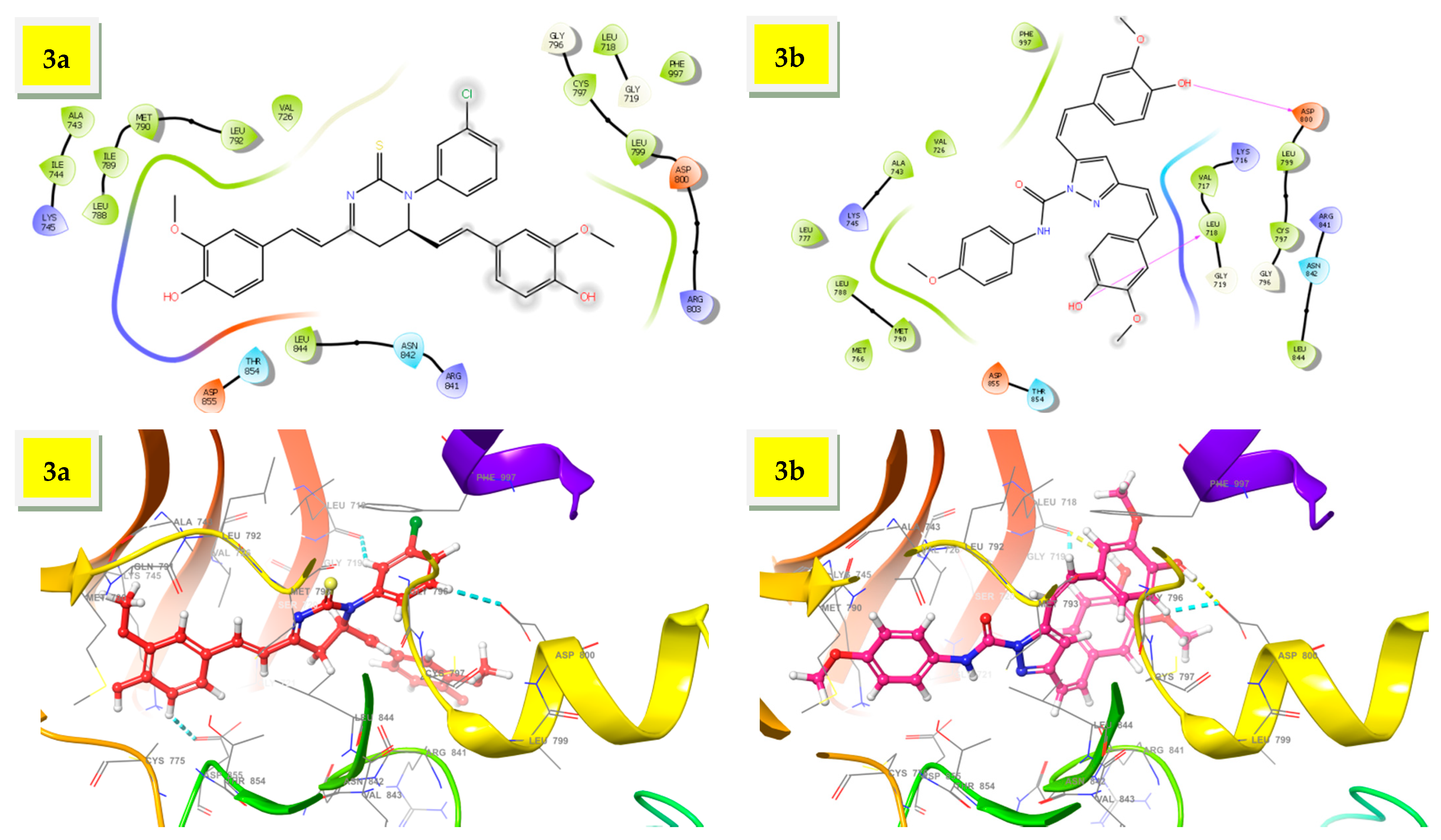
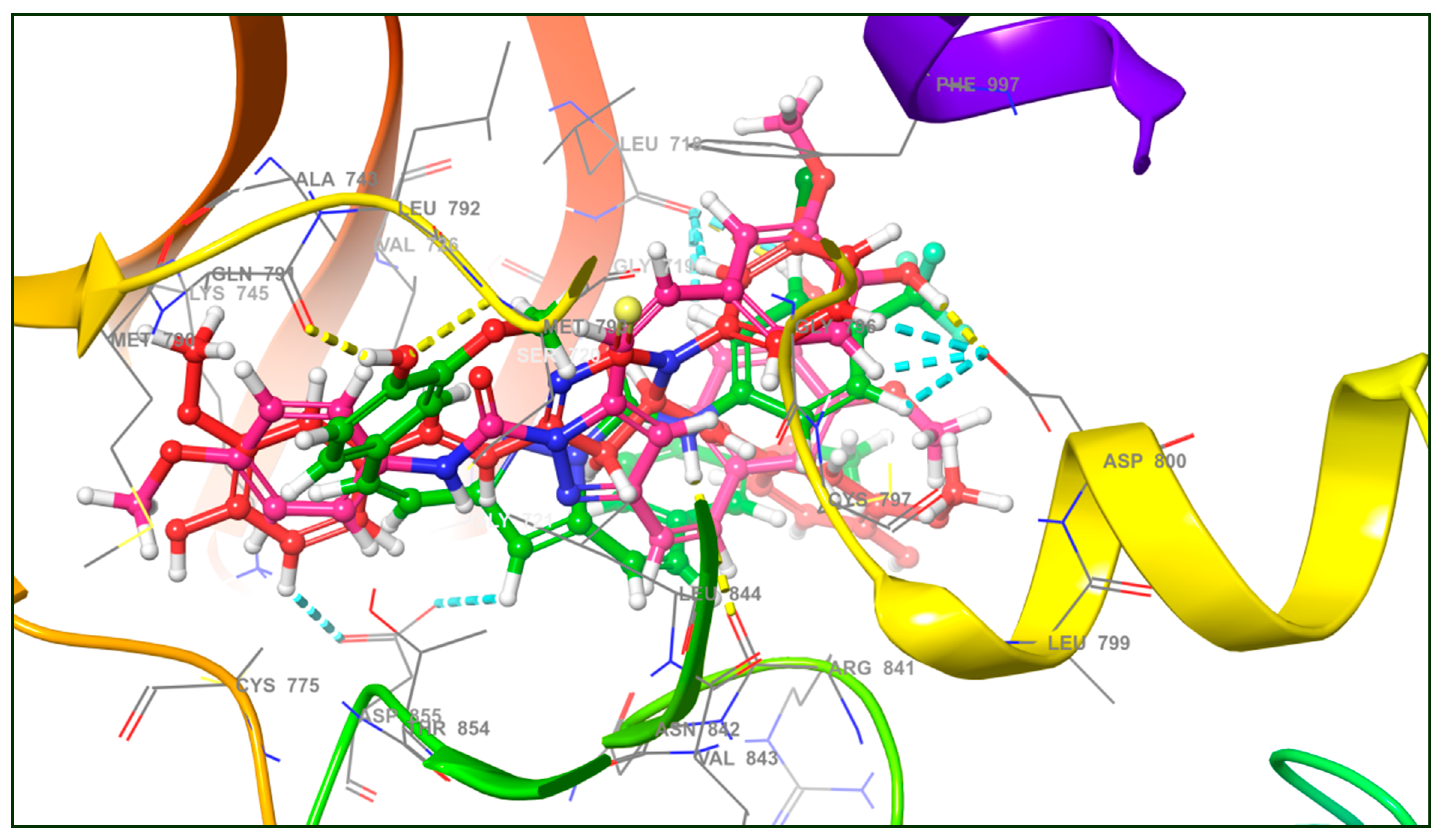
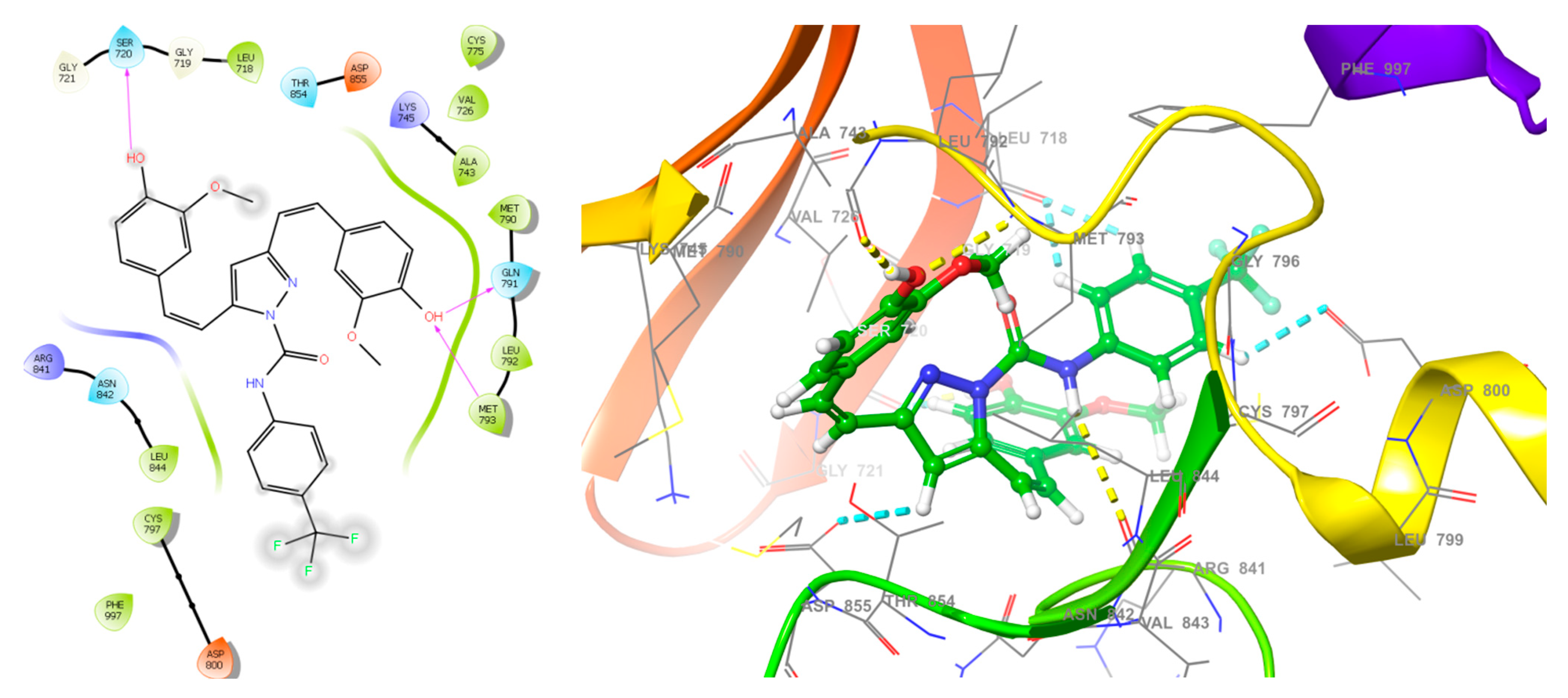
3.1. Molecular Docking Studies
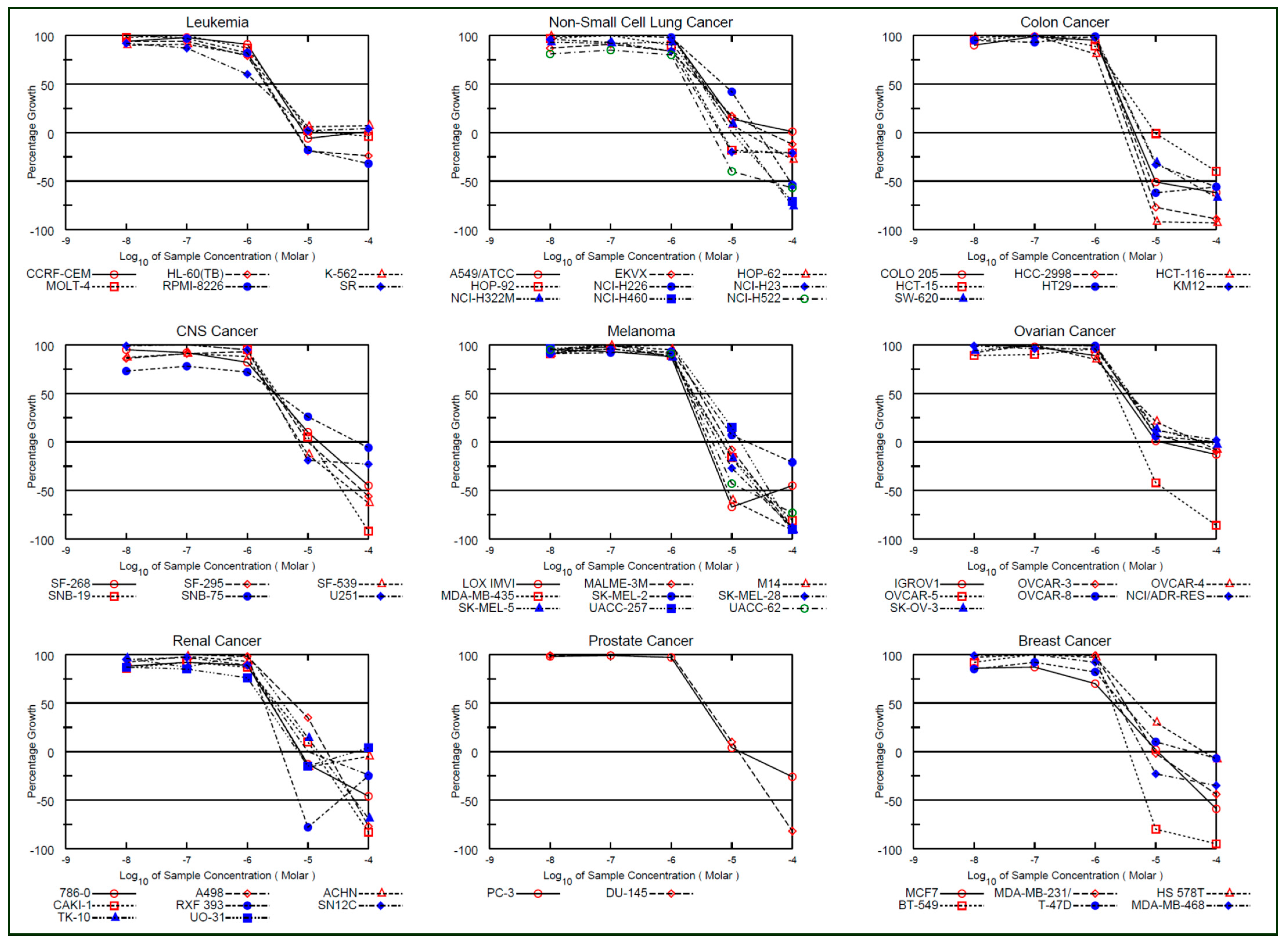
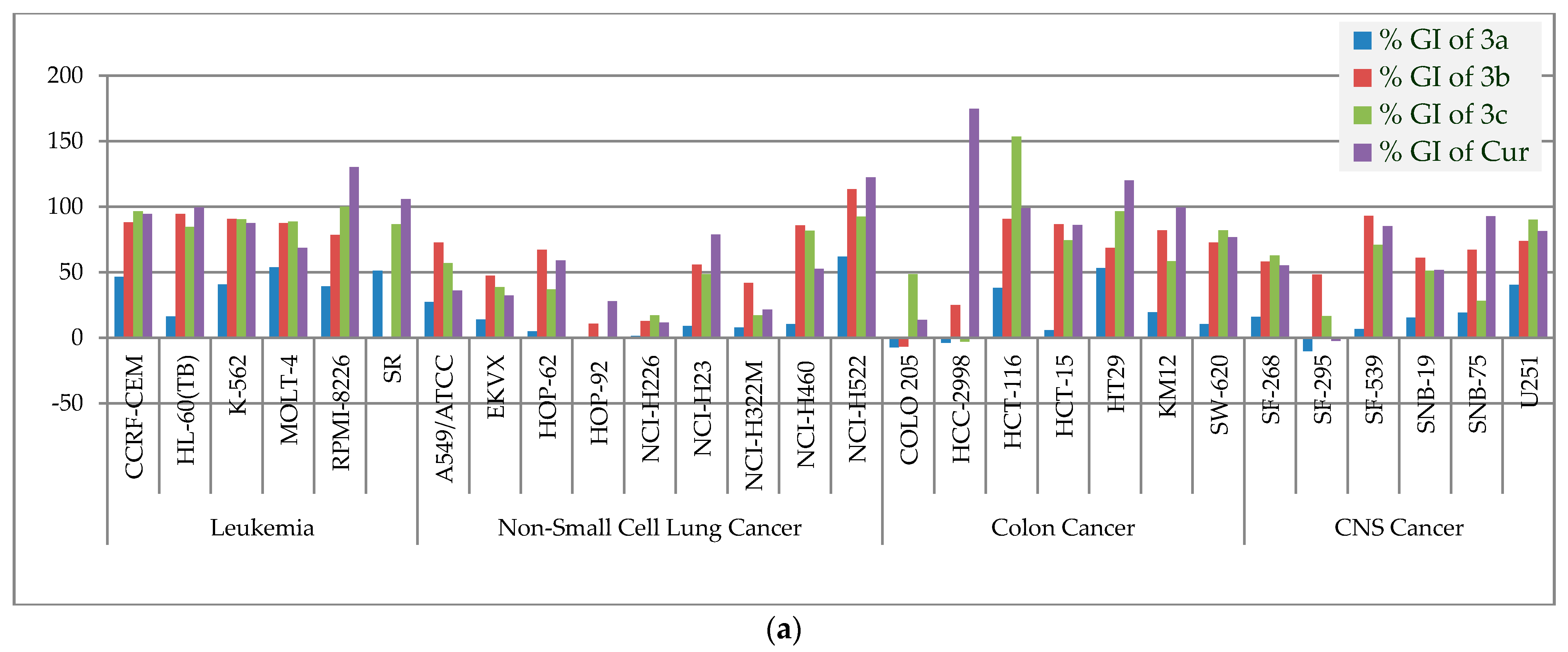
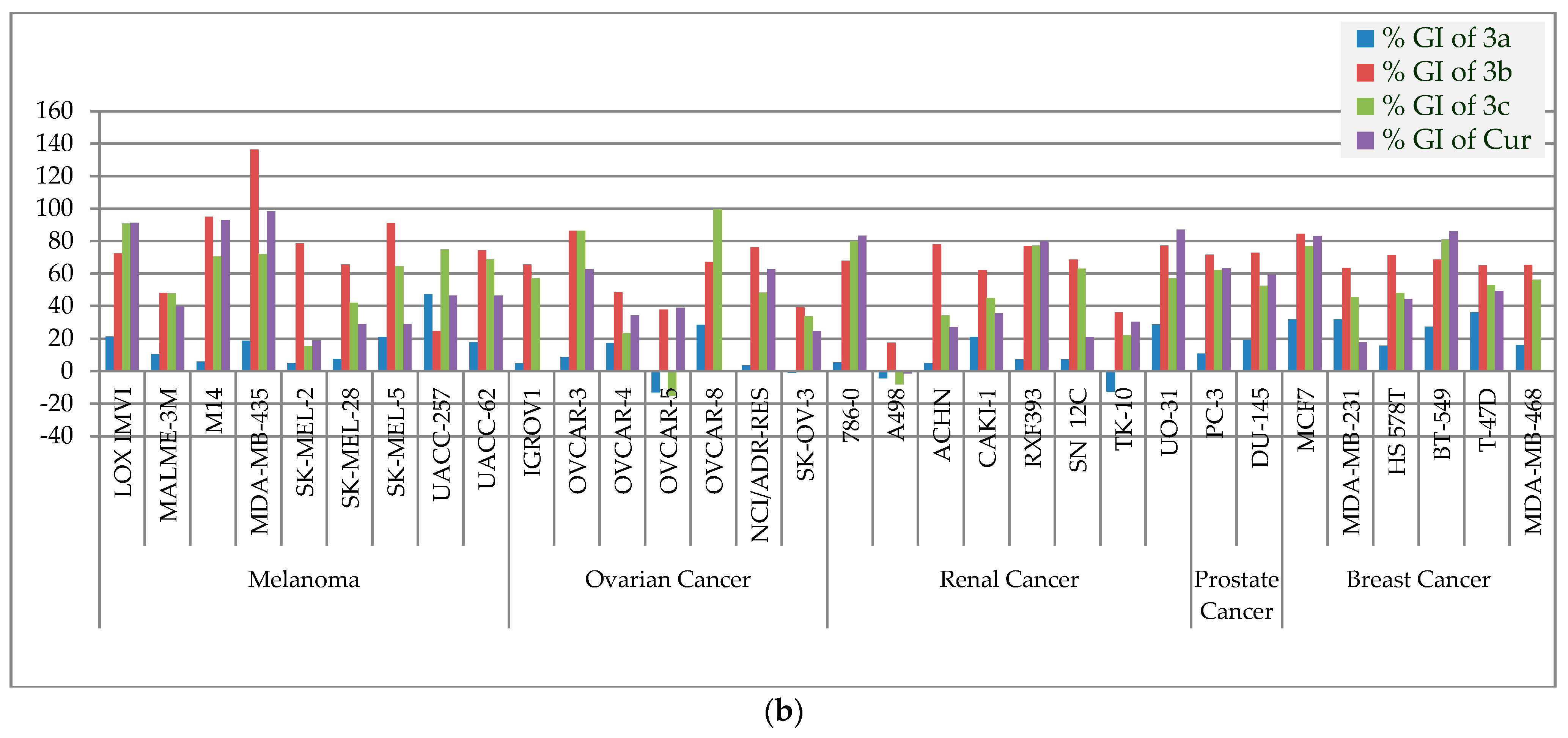
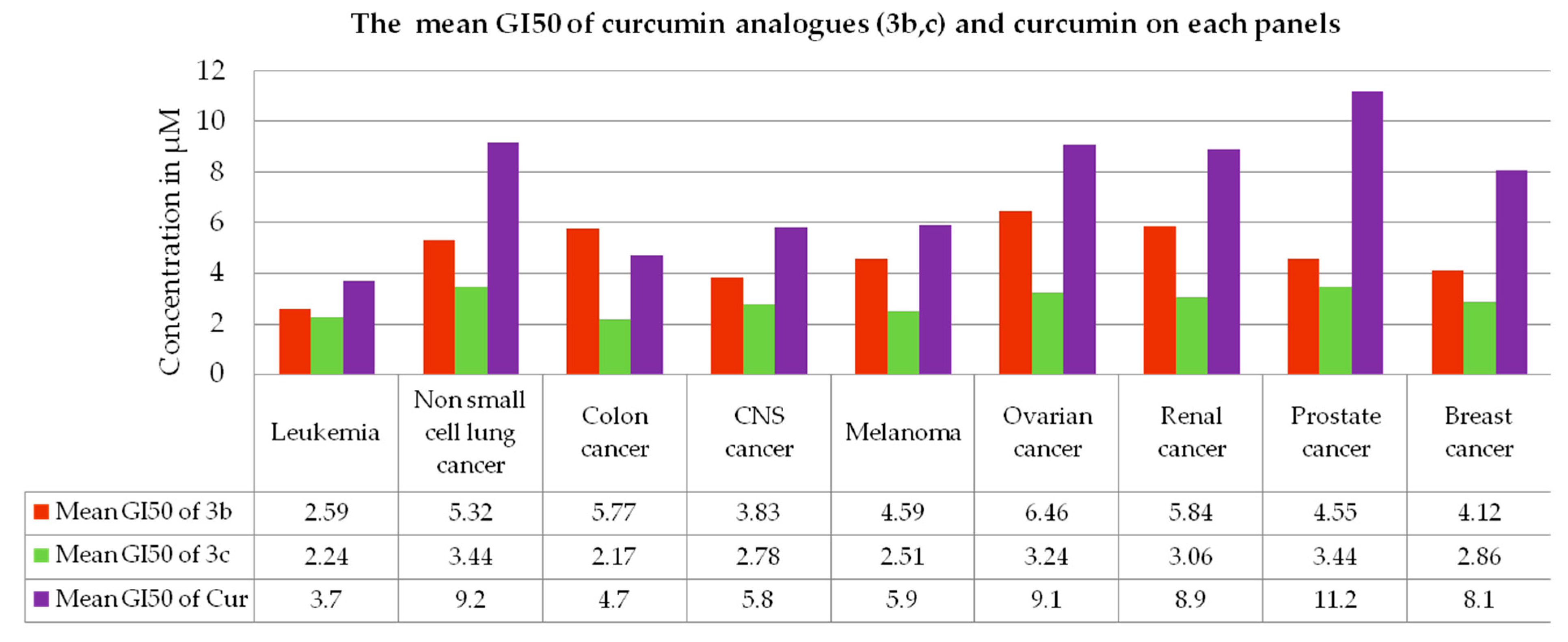
3.2. Antiproliferative Activity
3.3. Anti-EGFR Activity
4. Discussion
5. Conclusions
Supplementary Materials
Author Contributions
Funding
Institutional Review Board Statement
Informed Consent Statement
Data Availability Statement
Acknowledgments
Conflicts of Interest
References
- Koehn, F.E.; Carter, G.T. The evolving role of natural products in drug discovery. Nat. Rev. Drug Dis. 2005, 4, 206–220. [Google Scholar] [CrossRef] [PubMed]
- Mishra, B.B.; Tiwari, V.K. Natural products: An evolving role in future drug discovery. Eur. J. Med. Chem. 2011, 46, 4769–4807. [Google Scholar] [CrossRef]
- Newman, D.J.; Cragg, G.M. Natural products as sources of new drugs from 1981 to 2014. J. Nat. Prod. 2016, 79, 629–661. [Google Scholar] [CrossRef] [Green Version]
- Joo, E. Natural Product-Derived Drugs for the Treatment of Inflammatory Bowel Diseases. Intest. Res. 2014, 12, 103–109. [Google Scholar] [CrossRef] [Green Version]
- Conlin, A.; de Azambuja, E.; Lago, L.D. Current perspectives of epothilones in breast cancer. Eur. J. Cancer 2008, 44, 341–352. [Google Scholar]
- Grossman, S.A.; Carson, K.A.; Phuphanich, S.; Batchelor, T.; Peereboom, D.; Nabors, L.B.; Lesser, G.; Hausheer, F. Phase I and pharmacokinetic study of karenitecin in patients with recurrent malignant gliomas. Neuro-oncology 2008, 10, 608–616. [Google Scholar] [CrossRef]
- Butler, M.S. Natural products to drugs: Natural product-derived compounds in clinical trials. Nat. Prod. Rep. 2008, 25, 475. [Google Scholar] [CrossRef] [PubMed]
- Sessa, C.; Cresta, S.; Cerny, T.; Baselga, J.; Caremoli, E.R.; Malossi, A.; Hess, D.; Trigo, J.; Zucchetti, M.; D’Incalci, M.; et al. Concerted escalation of dose and dosing duration in a phase I study of the oral camptothecingimatecan (ST1481) in patients with advanced solid tumors. Ann. Oncol. 2007, 18, 561–568. [Google Scholar] [CrossRef]
- Sergent, J.M.; Elgie, A.W.; Williamson, C.J.; Hill, B.T. Ex vivo effects of the dual topoisomerase inhibitor tafluposide (F 11782) on cells isolated from fresh tumor samples taken from patients with cancer. Anti-Cancer. Drug 2003, 14, 467–473. [Google Scholar] [CrossRef]
- David-Cordonnier, M.H.; Laine, W.; Lansiaux, A.; Kouach, M.; Briand, G.; Pierré, A.; Hickman, J.A.; Bailly, C. Alkylation of Guanine in DNA by S23906-1, a Novel Potent Antitumor Compound Derived from the Plant Alkaloid Acronycine. Biochemistry 2002, 41, 9911–9920. [Google Scholar] [CrossRef]
- Tron, G.C.; Pirali, T.; Sorba, G.; Pagliai, F.; Bussacca, S.; Genzzani, A.A. Medicinal Chemistry of Combretastatin A4: Present and Future Directions. J. Med. Chem. 2006, 49, 3033–3044. [Google Scholar] [CrossRef] [PubMed]
- Pettit, G.R.; Lippert, J.W.; Naraynan, V.R.; Varma, R.; Simpson, M.J.; Boyd, M.R.; Rener, G.A.; Bansal, N. Antineoplastic agents 322. synthesis of combretastatin A-4 prodrugs. Anti-Cancer Drug Des. 1995, 10, 299–309. [Google Scholar]
- Salmon, H.W.; Siemann, D.W. Effect of the Second-Generation Vascular Disrupting Agent OXi4503 on Tumor Vascularity. Clin. Cancer Res. 2006, 12, 4090–4094. [Google Scholar] [CrossRef] [Green Version]
- Grossman, S.A.; Ye, X.; Peereboom, D.; Rosenfeld, M.R.; Mikkelsen, T.; Supko, J.G.; Desideri, S. Phase I study of terameprocol in patients with recurrent high-grade glioma. Neuro. Oncol. 2012, 14, 511–517. [Google Scholar] [CrossRef] [PubMed] [Green Version]
- Taylor, C.G.; Feitelson, A.K.; Taylor, D.D. Inhibitory effect of genistein and daidzein on ovarian cancer cell growth. Anticancer Res. 2004, 24, 795–800. [Google Scholar]
- Saif, M.W.; Tytler, E.; Lansigan, F.; Brown, D.M. Husband, A.J. Flavonoids, phenoxodiol, and a novel agent, triphendiol, for the treatment of pancreaticobiliary cancers. Expert Opin. Investig. Drugs 2009, 18, 469–479. [Google Scholar] [CrossRef] [PubMed]
- Rodrigues, F.C.; Kumar, N.V.A.; Thakur, G. The potency of heterocyclic curcumin analogues: An evidence-based review. Pharmacol. Res. 2021, 166, 105489. [Google Scholar] [CrossRef]
- Ahsan, M.J.; Khalilullah, H.; Yasmin, S.; Jadav, S.S.; Govindasamy, J. Synthesis, characterisation, and in vitro anticancer activity of curcumin analogues bearing pyrazole/pyrimidine ring targeting EGFR tyrosine kinase. BioMed Res Int. 2013, 2013, 239354. [Google Scholar] [CrossRef] [PubMed]
- Ahsan, M.J.; Choudhary, K.; Jadav, S.S.; Yasmin, S.; Ansari, M.Y.; Sreenivasulsu, R. Synthesis, anticancer activity and molecular docking studies of curcumin analogues bearing pyrazole ring. Med. Chem. Res. 2015, 24, 4166–4180. [Google Scholar] [CrossRef]
- Sharma, R.; Singh, S.; Yasmin, S.; Bhatia, S.; Khalilullah, H.; Ahsan, M.J. Simple, efficient, and improved synthesis of Biginel-li-type compounds of curcumin as anticancer agents. Med. Chem. Res. 2015, 24, 636–644. [Google Scholar] [CrossRef]
- Mishra, S.; Karmodiya, K.; Surolia, N.; Surolia, A. Synthesis and exploration of novel curcumin analogues as anti-malarial agents. Bioorg. Med. Chem. 2008, 16, 2894–2902. [Google Scholar] [CrossRef] [PubMed]
- Lal, J.; Gupta, S.K.; Thavaselvam, D.; Agrawal, D.D. Design, synthesis, synergistic antimicrobial activity and cytotoxicity of 4-aryl substituted 3,4-dihydropyrimidinones of curcumin. Bioorg. Med. Chem. 2012, 22, 2872–2876. [Google Scholar] [CrossRef]
- Sahu, P.K.; Sahu, P.K.; Gupta, S.K.; Thavaselvam, D.; Agarwal, D.D. Synthesis and evaluation of antimicrobial activity of 4H-pyrimido[2,1-b]benzothiazole, pyrazole and benzylidene derivatives of curcumin. Eur. J. Med. Chem. 2012, 54, 366–378. [Google Scholar] [CrossRef] [PubMed]
- Saja, K.; Babu, M.S.; Karunagaran, D.; Sudhakaran, P.R. Anti-inflammatory effect of curcumin involves down regulation of MMP-9 in blood mononuclear cells. Int. Immunopharm. 2007, 7, 1659–1667. [Google Scholar] [CrossRef] [PubMed]
- Singh, R.K.; Rai, D.; Yadav, D.; Bhargava, A.; Balzarini, J.; DeClercq, E. Synthesis, antibacterial and antiviral properties of curcumin bioconjugates bearing dipeptide, fatty acids and folic acid. Eur. J. Med. Chem. 2010, 45, 1078–1086. [Google Scholar] [CrossRef] [PubMed]
- Zhichang, L.; Yinghong, W.; Yuanqin, Z.; Qinxiang, X. Synthesis and antibacterial activities of N-Substituted pyrazole curcumin derivatives. Chin. J. Org. Chem. 2012, 32, 1487–1492. [Google Scholar]
- Lee, W.H.; Loo, C.Y.; Bebawy, M.; Luk, F.; Mason, R.S.; Rohanizadeh, R. Curcumin and its Derivatives: Their Application in Neuropharmacology and Neuroscience in the 21st Century. Curr. Neuropharmacol. 2013, 11, 338–378. [Google Scholar] [CrossRef] [Green Version]
- Yadav, I.S.; Nandekar, P.P.; Shrivastava, S.; Sanganwar, A.; Choudhry, A.; Agarwal, S.M. Ensemble docking and molecular dynamics identify knoevenagel curcumin derivatives with potent anti-EGFR activity. Gene 2014, 539, 82–90. [Google Scholar] [CrossRef]
- Sung, H.; Ferley, J.; Siegel, R.L.; Laversanne, M.; Soerjomartaram, I. Jemal, A.; Bray, F. Global cancer statistics 2020: GLOBOCAN estimates of incidence and mortality worldwide for 36 cancers in 185 countries. CA Cancer J. Clin. 2021, 71, 209–249. [Google Scholar] [CrossRef]
- Xu, H.; Yu, Y.; Marciniak, D.; Rishi, A.K.; Sarkar, F.H.; Kucuk, O.; Majumdar, A.P.N. Epidermal growth factor receptor (EGFR)–related protein inhibits multiple members of the EGFR family in colon and breast cancer cells. Mol. Can. Ther. 2005, 4, 435–442. [Google Scholar] [CrossRef]
- Hoadley, K.A.; Weigman, V.J.; Fan, C.; Sawyer, L.R.; He, X.; Troester, M.A.; Sartor, C.I.; Rieger-House, T.; Bernard, P.S.; Carey, L.A.; et al. EGFR associated expression profiles vary with breast tumor subtype. BMC Genom. 2007, 8, 258. [Google Scholar] [CrossRef] [Green Version]
- Rusnak, D.W.; Alligood, K.J.; Mullin, R.J.; Spehar, G.M.; Arenas-Elliott, C.; Martin, A.M.; Degenhardt, Y.; Rudolph, S.K.; Haws, T.F., Jr.; Hudson-Curtis, B.L.; et al. Assessment of epidermal growth factor receptor (EGFR, ErbB1) and HER2 (ErbB2) protein expression levels and response to lapatinib (Tykerb®, GW572016) in an expanded panel of human normal and tumour cell lines. Cell Prolif. 2007, 40, 580–594. [Google Scholar] [CrossRef]
- Anderson, N.G.; Ahmad, T.; Chan, K.; Dobson, R.; Bundred, N.J. ZD1839 (Iressa), a novel epidermal growth factor receptor (EGFR) tyrosine kinase inhibitor, potently inhibits the growth of EGFR-positive cancer cell lines with or without erbb2 over-expression. Int. J. Can. 2001, 94, 774–782. [Google Scholar] [CrossRef] [Green Version]
- Corkey, B.; Crown, J.; Clynes, M.; O’Donovan, N. Epidermal growth factor receptor as a potential therapeutic target in triple-negative breast cancer. Annals Oncol. 2009, 20, 862–867. [Google Scholar] [CrossRef] [PubMed]
- Kruwel, T.; Nevoltris, D.; Bode, J.; Dullin, C.; Baty, D.; Chames, P.; Alves, F. In vivo detection of small tumour lesions by multi-pinhole SPECT applying a 99mTc-labelled nanobody targeting the Epidermal Growth Factor Receptor. Sci. Rep. 2016, 6, 21834. [Google Scholar] [CrossRef] [PubMed]
- Shaik, N.A.; Al-Kreathy, H.M.; Ajabnoor, G.M.; Verma, P.K.; Banaganapalli, B. Molecular designing, virtual screening and docking study of novel curcumin analogue as mutation (S769L and K846R) selective inhibitor for EGFR. Saudi J. Biol. Sci. 2019, 26, 439–448. [Google Scholar] [CrossRef] [PubMed]
- Jakhar, R.; Dangi, M.; Khichi, A.; Chhillar, A.K. Relevance of Molecular Docking Studies in Drug Designing. Curr. Bioinform. 2020, 15, 270–278. [Google Scholar] [CrossRef]
- Morris, G.M.; Lim-Wilby, M. Molecular Docking. In Molecular Modeling of Proteins; Humana Press: Totowa, NJ, USA, 2008; pp. 365–382. [Google Scholar] [CrossRef]
- Matter, H.; Sotriffer, C. Applications and success stories in virtual screening. In Virtual Screening: Principles, Challenges, and Practical Guidelines; Sotriffer, C., Ed.; Wiley: Weinheim, Germany, 2011; pp. 319–358. [Google Scholar]
- Anderson, A.M.; Mitchell, M.S.; Mohan, R.S. Isolation of Curcumin from Turmeric. J. Chem. Edu. 2000, 77, 59–60. [Google Scholar] [CrossRef]
- X-ray Crystal Structure of EGFR. Available online: https://www.rcsb.org/structure/3W2R (accessed on 24 May 2021).
- Sogabe, S.; Kawakita, Y.; Igaki, S.; Iwata, H.; Miki, H.; Cary, D.R.; Takagi, T.; Takagi, S.; Ohta, Y.; Ishikawa, T. Structure-Based Approach for the Discovery of Pyrrolo[3,2-d]pyrimidine-Based EGFR T790M/L858R Mutant Inhibitors. ACS Med. Chem. Lett. 2013, 4, 201–205. [Google Scholar] [CrossRef] [Green Version]
- DTP Developmental Therapeutic Programs. Available online: http://dtp.nci.nih.gov (accessed on 9 May 2021).
- Monks, A.; Scudiero, D.; Skehan, P.; Shoemaker, R.; Paull, K.; Vistica, D.; Hose, C.; Langley, J.; Cronise, P.; Vaigro-Wolff, A.; et al. Feasibility of a highflux anticancer drug screening using a diverse panel of cultured human tumor cell lines. J. Nat. Cancer Inst. 1991, 83, 757–766. [Google Scholar] [CrossRef]
- Boyd, M.R.; Paull, K.D. Some practical considerations and applications of the National Cancer Institute in vitro anticancer drug discovery screen. Drug Dev. Res. 1995, 34, 91–109. [Google Scholar] [CrossRef]
- Shoemaker, R.H. The NCI60 human tumour cell line anticancer drug screen. Nat. Rev. Cancer 2006, 6, 813–823. [Google Scholar] [CrossRef] [PubMed]
- Grever, M.R.; Schepartz, S.A.; Chabner, B.A. The National Cancer Institute: Cancer drug discovery and development program. Sem. Oncol. 1992, 19, 622–638. [Google Scholar]
- Nawaz, F.; Alam, A.; Perwez, A.; Rizvi, A.R.; Naim, M.J.; Siddiqui, N.; Firdaus, J.; Rahman, S.; Jha, M.; Sheikh, A.A. Design, synthesis, molecular docking, and anticancer evaluation of pyrazole linked pyrazoline derivatives with carbothioamide tail as EGFR kinase inhibitors. Anti-Cancer Agent Med. Chem. 2021, 21, 42–60. [Google Scholar] [CrossRef]
- Modjtahedi, H.; Essapen, S. Epidermal growth factor receptor inhibitors in cancer treatment: Advances, challenges and opportunities. Anticancer Drugs 2009, 20, 851–855. [Google Scholar] [CrossRef] [PubMed]
- Rostom, S.A.F. Synthesis and in vitro antitumor evaluation of some indeno[1,2-c]pyrazol(in)es substituted with sulfonamide, sulfonylurea(-thiourea) pharmacophores, and some derived thiazole ring systems. Bioorg. Med. Chem. 2006, 14, 6475–6485. [Google Scholar] [CrossRef]
- Corona, P.; Carta, A.; Loriga, M.; Vitale, G.; Paglietti, G. Synthesis and in-vitro antitumor activity of new quinoxaline derivatives. Eur. J. Med. Chem. 2009, 44, 1579–1591. [Google Scholar] [CrossRef]
- Kocaadam, B.; Şanlier, N. Curcumin, an active component of turmeric (Curcuma longa), and Its effects on health. Crit. Rev. Food Sci. Nutr. 2017, 57, 2889–2895. [Google Scholar] [CrossRef]
- Starok, M.; Preira, P.; Vayssade, M.; Haupt, K.; Salome, L.; Rossi, C. EGFR Inhibition by Curcumin in Cancer Cells: A Dual Mode of Action. Biomacromolecules 2015, 16, 1634–1642. [Google Scholar] [CrossRef]
- Sahu, P.K. Design, structure activity relationship, cytotoxicity and evaluation of antioxidant activity of curcumin derivatives/analogues. Eur. J. Med. Chem. 2016, 121, 510–516. [Google Scholar] [CrossRef]
- Seghetti, F.; Di Martino, R.M.C.; Catanzaro, E.; Bisi, A.; Gobbi, S.; Rampa, A.; Canonico, B.; Montanari, M.; Krysko, D.V.; Papa, S.; et al. Curcumin-1,2,3-Triazole Conjugation for Targeting the Cancer Apoptosis Machinery. Molecules 2020, 25, 3066. [Google Scholar] [CrossRef] [PubMed]


| S. No. | Structure | Docking Score | Interaction |
|---|---|---|---|
| 1 |  | −6.593 | π–π stacking (Asp855); π–π stacking (Asp800); π–π stacking (Leu718) |
| 2 | 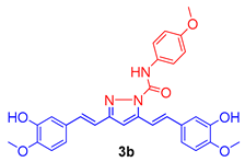 | −6.337 | H-bond (Leu718); H-bond (Asp800); π-π stacking (Leu718); π–π stacking (Asp800) |
| 3 | 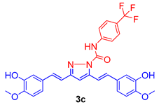 | −6.452 | H-bond (Met793); H-bond (Gln791); H-bond (Ser720); π–π stacking (Asp800); π–π stacking (Asp855); π–π stacking (Leu718) |
| Panel | Cell Line | GP and %GI at 10 µM | 3b (NSC 782201) | 3b (NSC 799011) | |||||||||||||
|---|---|---|---|---|---|---|---|---|---|---|---|---|---|---|---|---|---|
| 3a (NSC 799007) | 3b (NSC 782201) | 3c (NSC 799011) | GI50 | Sub Panel MID b | Selectivity Ratio (MID a:MID b) | TGI | LC50 | GI50 | Sub Panel MID b | Selectivity Ratio (MID a:MID b) | TGI | LC50 | |||||
| GP | %GI | GP | %GI | GP | %GI | ||||||||||||
| Leukemia | CCRF-CEM | 53.43 | 46.57 | 11.77 | 88.23 | 3.42 | 96.58 | 2.34 | 2.59 | 1.89 | 32.7 | >100 | 2.63 | 2.24 | 1.27 | - | >100 |
| HL-60(TB) | 83.48 | 16.52 | 5.32 | 94.68 | 15.17 | 84.83 | 2.55 | 6.69 | >100 | 1.98 | 6.45 | >100 | |||||
| K-562 | 59.23 | 40.77 | 9.25 | 90.75 | 9.45 | 90.55 | 2.87 | 16.9 | >100 | 2.53 | >100 | >100 | |||||
| MOLT-4 | 46.22 | 53.78 | 12.24 | 87.76 | 11.09 | 88.91 | 2.87 | 13.3 | >100 | 2.73 | 20.0 | >100 | |||||
| RPMI-8226 | 60.59 | 39.41 | 21.30 | 78.70 | −0.21 | 100.21 | 3.38 | 29.2 | >100 | 2.09 | 6.59 | >100 | |||||
| SR | 48.72 | 51.28 | - | - | 13.21 | 86.79 | 1.52 | 12.8 | >100 | 1.48 | >100 | >100 | |||||
| Non-Small Cell Lung Cancer | A549/ATCC | 72.56 | 27.44 | 27.29 | 72.71 | 42.95 | 57.05 | 5.50 | 5.32 | 0.92 | 97.9 | >100 | 4.11 | 3.44 | 0.82 | >100 | >100 |
| EKVX | 85.81 | 14.19 | 52.39 | 47.61 | 61.25 | 38.75 | 6.62 | 83.7 | >100 | 3.22 | 38.4 | >100 | |||||
| HOP-62 | 95.06 | 4.94 | 32.75 | 67.25 | 62.87 | 37.13 | 3.65 | 42.8 | >100 | 3.60 | 16.2 | >100 | |||||
| HOP-92 | - | - | 89.07 | 10.93 | - | - | 7.77 | >100 | >100 | 2.35 | 6.79 | >100 | |||||
| NCI-H226 | 98.54 | 1.46 | 87.15 | 12.85 | 82.67 | 17.33 | 10.7 | 78.6 | >100 | 7.29 | 27.6 | 91.7 | |||||
| NCI-H23 | 90.95 | 9.05 | 43.94 | 56.06 | 51.29 | 48.71 | 3.81 | 63.8 | >100 | 2.13 | 6.48 | >100 | |||||
| NCI-H322M | 91.93 | 8.07 | 58.14 | 41.86 | 82.70 | 17.30 | 3.87 | 72.5 | >100 | 3.25 | 12.9 | 49.6 | |||||
| NCI-H460 | 89.51 | 10.49 | 14.22 | 85.78 | 18.29 | 81.71 | 4.24 | 31.4 | >100 | 3.22 | 9.95 | 50.7 | |||||
| NCI-H522 | 37.89 | 62.11 | −13.52 | 113.52 | 7.32 | 92.68 | 1.76 | 12.7 | >100 | 1.78 | 4.65 | 40.8 | |||||
| Colon Cancer | COLO 205 | 107.08 | −7.08 | 106.58 | −6.58 | 51.27 | 48.73 | 16.2 | 5.77 | 0.85 | 44.3 | >100 | 2.05 | 2.17 | 1.31 | 4.50 | 9.91 |
| HCC-2998 | 103.70 | −3.70 | 74.91 | 25.09 | 102.63 | −2.63 | 5.78 | >100 | >100 | 2.04 | 3.82 | 71.2 | |||||
| HCT-116 | 61.88 | 38.12 | 9.13 | 90.87 | −53.67 | 153.67 | 3.22 | 42.9 | >100 | 1.51 | 2.94 | 5.70 | |||||
| HCT-15 | 93.93 | 6.07 | 13.17 | 86.87 | 25.51 | 74.49 | 2.98 | >100 | >100 | 2.69 | 9.63 | >100 | |||||
| HT29 | 46.57 | 53.43 | 31.26 | 68.74 | 3.50 | 96.50 | 3.52 | 11.0 | >100 | 2.02 | 4.13 | 8.44 | |||||
| KM12 | 80.33 | 19.67 | 17.89 | 82.11 | 41.38 | 58.62 | 3.19 | 20.7 | >100 | 2.33 | 5.59 | 54.0 | |||||
| SW-620 | 89.48 | 10.52 | 27.10 | 72.90 | 18.00 | 82.00 | 5.51 | 61.6 | >100 | 2.56 | 5.90 | 33.6 | |||||
| CNS Cancer | SF-268 | 83.76 | 16.24 | 41.81 | 58.19 | 37.12 | 62.88 | 3.21 | 3.83 | 1.28 | 36.4 | >100 | 2.80 | 2.78 | 1.02 | 15.4 | >100 |
| SF-295 | 109.94 | −9.94 | 51.62 | 48.38 | 83.42 | 16.58 | 6.25 | 53.2 | >100 | 2.89 | 9.85 | 76.5 | |||||
| SF-539 | 93.11 | 6.89 | 6.72 | 93.28 | 29.01 | 70.99 | 2.35 | 13.6 | >100 | 2.37 | 7.45 | 56.0 | |||||
| SNB-19 | 84.55 | 15.45 | 38.85 | 61.15 | 48.68 | 51.32 | 5.28 | 86.7 | >100 | 3.16 | 11.2 | 36.9 | |||||
| SNB-75 | 80.78 | 19.22 | 32.61 | 67.39 | 71.80 | 28.20 | 2.35 | 16.3 | >100 | 2.99 | 66.0 | >100 | |||||
| U251 | 59.41 | 40.59 | 26.03 | 73.97 | 9.71 | 90.29 | 3.55 | 20.9 | >100 | 2.49 | 6.86 | >100 | |||||
| Melanoma | LOX IMVI | 78.64 | 21.36 | 27.59 | 72.41 | 9.11 | 90.89 | 3.95 | 4.59 | 1.06 | >100 | >100 | 1.77 | 2.51 | 1.31 | 3.71 | - |
| MALME-3M | 89.46 | 10.54 | 51.86 | 48.14 | 52.27 | 47.73 | 3.66 | 38.6 | >100 | 2.89 | 8.40 | 32.6 | |||||
| M14 | 94.04 | 5.96 | 4.97 | 95.03 | 29.48 | 70.52 | 3.15 | 41.5 | >100 | 1.94 | 4.08 | 8.58 | |||||
| MDA-MB-435 | 81.32 | 18.68 | −36.25 | 136.25 | 27.85 | 72.15 | 1.25 | 4.68 | >100 | 2.35 | 7.06 | 33.4 | |||||
| SK-MEL-2 | 95.15 | 4.85 | 21.38 | 78.62 | 84.47 | 15.53 | 3.23 | 18.1 | >100 | 3.15 | 17.3 | >100 | |||||
| SK-MEL-28 | 92.44 | 7.56 | 34.53 | 65.47 | 57.99 | 42.01 | 3.52 | 92.7 | >100 | 2.19 | 5.90 | 23.9 | |||||
| SK-MEL-5 | 78.99 | 21.01 | 9.04 | 90.96 | 35.40 | 64.60 | 3.67 | 16.1 | >100 | 2.27 | 6.89 | 28.0 | |||||
| UACC-257 | 52.91 | 47.09 | 75.22 | 24.78 | 25.01 | 74.99 | 16.7 | 64.8 | >100 | 4.01 | 14.0 | 42.0 | |||||
| UACC-62 | 82.19 | 17.81 | 25.47 | 74.53 | 31.10 | 68.90 | 2.14 | 6.91 | >100 | 2.02 | 4.76 | 16.7 | |||||
| Ovarian Cancer | IGROV1 | 95.26 | 4.74 | 34.39 | 65.61 | 42.82 | 57.18 | 4.49 | 6.46 | 0.76 | 57.0 | >100 | 2.77 | 3.24 | 0.88 | 11.6 | >100 |
| OVCAR-3 | 91.30 | 8.70 | 13.62 | 86.38 | 13.76 | 86.24 | 2.98 | 8.40 | >100 | 3.41 | 26.7 | >100 | |||||
| OVCAR-4 | 82.55 | 17.45 | 51.51 | 48.49 | 76.71 | 23.29 | 5.72 | 90.7 | >100 | 3.51 | 52.7 | >100 | |||||
| OVCAR-5 | 112.92 | −12.92 | 62.30 | 37.70 | 115.18 | −15.16 | 18.8 | 54.1 | >100 | 2.16 | 4.97 | 15.2 | |||||
| OVCAR-8 | 71.43 | 28.57 | 32.79 | 67.21 | 0.43 | 99.57 | 5.32 | 59.9 | >100 | 3.33 | >100 | >100 | |||||
| NCI/ADR-RES | 96.37 | 3.63 | 23.89 | 76.11 | 51.65 | 48.35 | 4.28 | >100 | >100 | 3.51 | >100 | >100 | |||||
| SK-OV-3 | 100.88 | −0.88 | 60.74 | 39.26 | 66.09 | 33.91 | 3.66 | 27.5 | >100 | 4.02 | 66.7 | >100 | |||||
| Renal Cancer | 786–0 | 94.62 | 5.38 | 32.08 | 67.92 | 20.07 | 79.93 | 4.00 | 5.84 | 0.84 | 24.3 | >100 | 2.42 | 3.06 | 0.93 | 7.52 | >100 |
| A498 | 104.38 | −4.38 | 82.47 | 17.53 | 108.03 | -8.03 | 17.2 | 56.4 | >100 | 5.81 | 2.06 | 57.3 | |||||
| ACHN | 95.01 | 4.99 | 26.03 | 77.93 | 65.82 | 34.19 | 4.18 | >100 | >100 | 2.48 | 7.08 | >100 | |||||
| CAKI-1 | 79.04 | 20.96 | 38.06 | 61.94 | 54.99 | 45.01 | 3.60 | >100 | >100 | 3.02 | 12.7 | 44.1 | |||||
| RXF 393 | 92.63 | 7.37 | 23.01 | 76.99 | 22.66 | 77.34 | 3.02 | 8.87 | >100 | 1.98 | 3.73 | - | |||||
| SN 12C | 92.61 | 7.39 | 31.45 | 68.55 | 36.92 | 63.08 | 3.05 | 33.7 | >100 | 2.73 | 9.90 | >100 | |||||
| TK-10 | 112.60 | −12.60 | 63.84 | 36.16 | 77.69 | 22.31 | 10.2 | 95.3 | >100 | 4.13 | 14.8 | 59.4 | |||||
| UO-31 | 71.26 | 28.74 | 22.67 | 77.33 | 42.85 | 57.15 | 1.52 | 36.4 | >100 | 1.94 | - | >100 | |||||
| Prostate Cancer | PC-3 | 89.21 | 10.79 | 28.43 | 71.57 | 38.03 | 61.97 | 5.11 | 4.55 | 1.07 | >100 | >100 | 3.21 | 3.44 | 0.82 | 13.5 | >100 |
| DU-145 | 80.84 | 19.16 | 27.23 | 72.77 | 47.55 | 52.45 | 3.99 | 24.6 | >100 | 3.67 | 13.0 | 45.3 | |||||
| Breast Cancer | MCF7 | 67.91 | 32.09 | 15.63 | 84.37 | 22.92 | 77.08 | 3.30 | 4.12 | 1.19 | 17.0 | >100 | 1.97 | 2.86 | 0.99 | 10.7 | 72.0 |
| MDA-MB-231 | 68.25 | 31.75 | 36.53 | 63.47 | 54.79 | 45.21 | 4.94 | 83.5 | >100 | 3.06 | 9.62 | >100 | |||||
| HS 578T | 84.33 | 15.67 | 28.71 | 71.29 | 51.89 | 48.11 | 6.77 | 61.9 | >100 | 5.10 | 60.6 | >100 | |||||
| BT-549 | 72.52 | 27.48 | 31.37 | 68.63 | 18.97 | 81.03 | 2.49 | 29.5 | >100 | 1.96 | 3.67 | 6.86 | |||||
| T-47D | 63.82 | 36.18 | 34.83 | 65.17 | 47.40 | 52.60 | 3.68 | 25.8 | >100 | 2.81 | 39.0 | >100 | |||||
| MDA-MB-468 | 83.81 | 16.19 | 34.68 | 65.32 | 43.79 | 56.21 | 3.55 | 21.6 | >100 | 2.30 | 6.25 | >100 | |||||
| Mean | 82.32 | 17.68 | 33.54 | 66.46 | 40.74 | 59.26 | |||||||||||
| Total cell line and sum of concentration | 60 | 293.96 | 170.18 | ||||||||||||||
| MID a | 4.89 | 2.84 | |||||||||||||||
| Panel | 3a | 3b | 3c | Cur * | Gefitinib # |
|---|---|---|---|---|---|
| Leukemia | 41.39 | 88.02 | 91.31 | 97.76 | 79.68 |
| Non-Small cell lung cancer | 17.22 | 56.51 | 48.83 | 49.27 | 63.97 |
| Colon Cancer | 16.72 | 60 | 73.05 | 95.76 | 52.19 |
| CNS Cancer | 14.74 | 67.06 | 53.38 | 60.75 | 46.13 |
| Melanoma | 17.21 | 76.24 | 60.81 | 54.63 | 44.99 |
| Ovarian Cancer | 7.04 | 60.11 | 47.62 | 44.66 | 60.93 |
| Renal Cancer | 7.23 | 60.54 | 46.37 | 45.35 | 77.89 |
| Prostate Cancer | 14.98 | 72.17 | 57.21 | 61.3 | 59.6 |
| Breast Cancer | 26.56 | 69.71 | 60.04 | 56.1 | 52.88 |
Publisher’s Note: MDPI stays neutral with regard to jurisdictional claims in published maps and institutional affiliations. |
© 2021 by the authors. Licensee MDPI, Basel, Switzerland. This article is an open access article distributed under the terms and conditions of the Creative Commons Attribution (CC BY) license (https://creativecommons.org/licenses/by/4.0/).
Share and Cite
Ali, A.; Ali, A.; Tahir, A.; Bakht, M.A.; Salahuddin; Ahsan, M.J. Molecular Engineering of Curcumin, an Active Constituent of Curcuma longa L. (Turmeric) of the Family Zingiberaceae with Improved Antiproliferative Activity. Plants 2021, 10, 1559. https://doi.org/10.3390/plants10081559
Ali A, Ali A, Tahir A, Bakht MA, Salahuddin, Ahsan MJ. Molecular Engineering of Curcumin, an Active Constituent of Curcuma longa L. (Turmeric) of the Family Zingiberaceae with Improved Antiproliferative Activity. Plants. 2021; 10(8):1559. https://doi.org/10.3390/plants10081559
Chicago/Turabian StyleAli, Amena, Abuzer Ali, Abu Tahir, Md. Afroz Bakht, Salahuddin, and Mohamed Jawed Ahsan. 2021. "Molecular Engineering of Curcumin, an Active Constituent of Curcuma longa L. (Turmeric) of the Family Zingiberaceae with Improved Antiproliferative Activity" Plants 10, no. 8: 1559. https://doi.org/10.3390/plants10081559
APA StyleAli, A., Ali, A., Tahir, A., Bakht, M. A., Salahuddin, & Ahsan, M. J. (2021). Molecular Engineering of Curcumin, an Active Constituent of Curcuma longa L. (Turmeric) of the Family Zingiberaceae with Improved Antiproliferative Activity. Plants, 10(8), 1559. https://doi.org/10.3390/plants10081559