Review

The Role of Tetrapyrrole- and GUN1-Dependent Signaling on Chloroplast Biogenesis

Graduate School of Arts and Sciences, The University of Tokyo, 3-8-1 Komaba, Meguro-ku, Tokyo 153-8902, Japan
*
Author to whom correspondence should be addressed.
Plants 2021, 10(2), 196; https://doi.org/10.3390/plants10020196
Submission received: 15 December 2020 / Revised: 15 January 2021 / Accepted: 18 January 2021 / Published: 21 January 2021
(This article belongs to the Special Issue Molecular Biology of Plastids)

Abstract

:
Chloroplast biogenesis requires the coordinated expression of the chloroplast and nuclear genomes, which is achieved by communication between the developing chloroplasts and the nucleus. Signals emitted from the plastids, so-called retrograde signals, control nuclear gene expression depending on plastid development and functionality. Genetic analysis of this pathway identified a set of mutants defective in retrograde signaling and designated genomes uncoupled (gun) mutants. Subsequent research has pointed to a significant role of tetrapyrrole biosynthesis in retrograde signaling. Meanwhile, the molecular functions of GUN1, the proposed integrator of multiple retrograde signals, have not been identified yet. However, based on the interactions of GUN1, some working hypotheses have been proposed. Interestingly, GUN1 contributes to important biological processes, including plastid protein homeostasis, through transcription, translation, and protein import. Furthermore, the interactions of GUN1 with tetrapyrroles and their biosynthetic enzymes have been revealed. This review focuses on our current understanding of the function of tetrapyrrole retrograde signaling on chloroplast biogenesis.

1. Introduction

Tetrapyrroles are involved in various functions critical to whole organisms’ viability, including light absorption, electron transfer, and oxygen binding [1,2]. Thus, they are essential components of primary metabolism, such as respiration and photosynthesis. Tetrapyrroles contain four pyrroles, aromatic rings containing four carbon atoms and one nitrogen atom, in linear (e.g., bilins) or cyclic (e.g., porphyrins) chemical structures. Porphyrins often chelate central metal ions, such as Co2+, Fe2+ or Fe3+, or Mg2+ ions. Meanwhile, fully conjugated (pigmented) porphyrin rings possess photodynamic properties: they can generate reactive oxygen species (ROS), primarily singlet oxygen under light excitation, which cause photooxidative damage and cell death [3]. Therefore, organisms must strictly regulate tetrapyrrole biosynthesis. In plants and algae, tetrapyrroles’ main end products are siroheme, heme, phytochromobilin, chlorophyll (Chl) a, and Chl b. Although they are synthesized in plastids, these tetrapyrroles are widely distributed, with the exception of Chls. Especially, heme is found throughout the cell. In addition to prosthetic groups’ function, tetrapyrroles have been proposed as signaling molecules that control transcription and intracellular signaling. This review focuses on the signaling function of tetrapyrroles on chloroplast biogenesis in Arabidopsis thaliana. We inform interested readers of several comprehensive reviews on the signaling function of tetrapyrroles on other aspects of plant physiology [4,5,6].

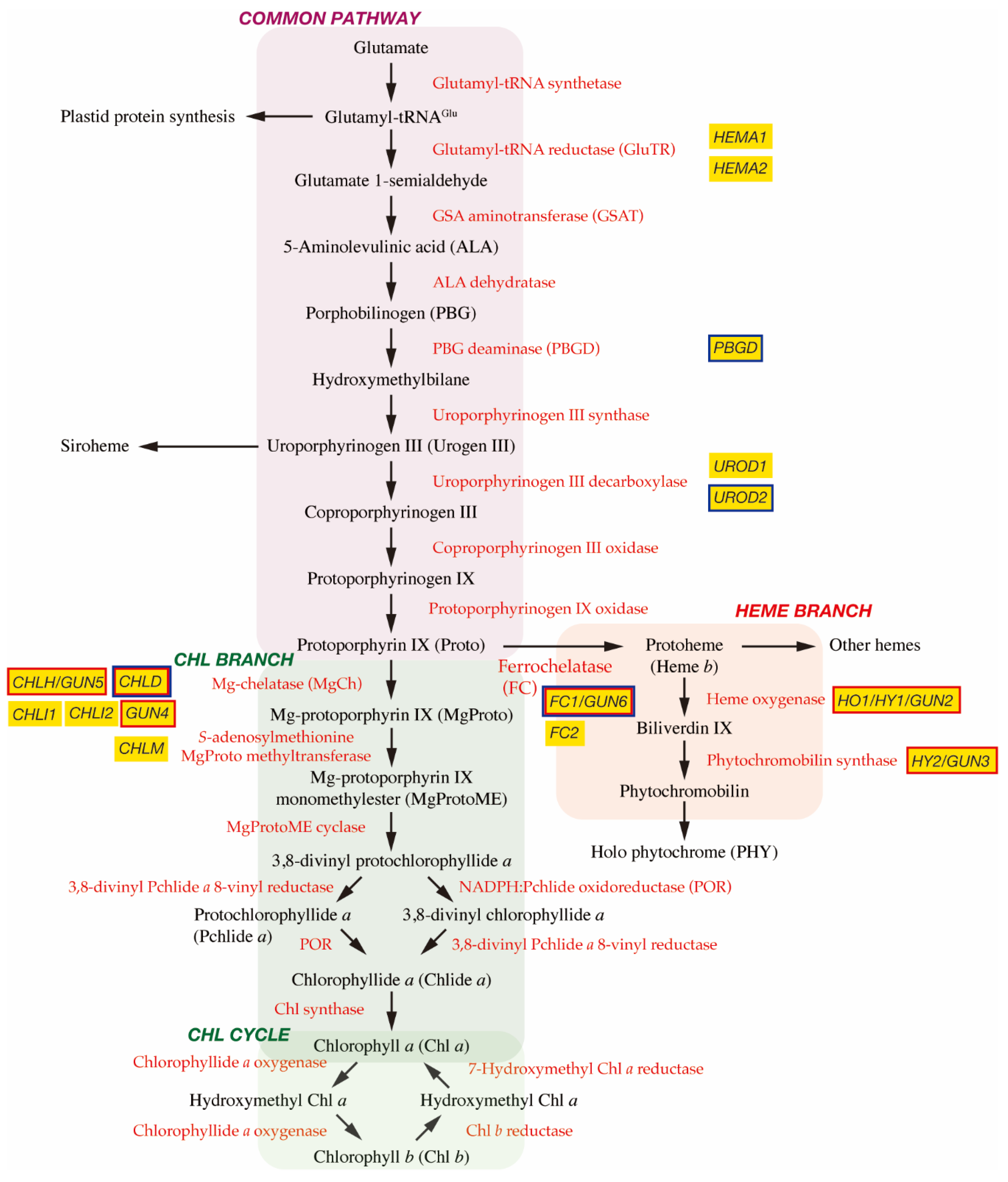
2. Biosynthesis of Tetrapyrroles in Plants

2.1. The C5 Pathway and the Common Pathway

In plant cells, tetrapyrrole biosynthesis takes place entirely in the plastid (Figure 1). The first committed precursor for all tetrapyrroles is 5-aminolevulinic acid (ALA). In plants, algae, and many bacteria, ALA is synthesized from glutamate via the C5 pathway [7]. In this pathway, glutamate is first ligated with plastid-encoded tRNAGlu to form glutamyl-tRNAGlu, a substrate for plastid protein biosynthesis. The following two enzymes, glutamyl-tRNA reductase (GluTR) and glutamate 1-semialdehyde aminotransferase (GSAT), synthesize ALA from glutamyl-tRNAGlu. In particular, the step of GluTR is the rate-limiting step of total tetrapyrrole biosynthesis, the activity of which is controlled by transcriptional and post-transcriptional regulations [8,9]. Arabidopsis has two paralogous genes for GluTR, HEMA1 and HEMA2. HEMA1 is light-responsive, is actively expressed in green tissues, and contributes predominantly to Chl biosynthesis [10,11]. ALA dehydratase condenses two molecules of ALA onto the monopyrrole to form porphobilinogen (PBG). PBG deaminase assembles four PBG molecules, which are further assembled onto the tetrapyrrole precursor uroporphyrinogen III (Urogen III). Three stepwise oxidation steps oxidatively decarboxylate Urogen III and the final step enzyme, protoporphyrinogen IX oxidase, oxidizes the colorless protoporphyrinogen IX to fully conjugated and pigmented protoporphyrin IX (Proto). Alternatively, Urogen III can be directed to siroheme biosynthesis. Since the pathway from ALA to Proto is conserved among most organisms, this pathway is called ‘the common pathway’.

2.2. Chl Branch and Chl Cycle

The next branchpoint involves the insertion of either Mg2+ or Fe2+ by Mg-chelatase (MgCh) or ferrochelatase (FC), respectively, directing Proto into the Chl or heme biosynthetic pathways. MgCh consists of three subunits, CHLI, CHLD, and CHLH in plants. In Arabidopsis, CHLD and CHLH are encoded by a single gene, and CHLI is encoded by two isoforms, CHLI1 and CHLI2. CHLI1 is essential for photosynthesis [12,13], whereas CHLI2 has a minor role in assembling the MgCh complex [14]. Additionally, GUN4 enhances MgCh activity by mediating substrate or product channeling [15,16,17]. In the Chl branch, MgCh catalyzes the formation of Mg-protoporphyrin IX (MgProto), which is methylated by S-adenosylmethionine MgProto methyltransferase (encoded by CHLM) to form MgProto methyl ester (MgProtoME). MgProtoME cyclase catalyzes the formation of the fifth ring of the tetrapyrrole ring structure, which is further converted to 3,8-divinyl protochlorophyllide a (DV-Pchlide a). DV-Pchlide a is further converted to Pchlide a by DV-Pchlide a 8-vinyl reductase. Pchlide a accumulates in dark-grown angiosperms because the next enzyme, light-dependent NADPH:Pchlide oxidoreductase (POR), requires light to reduce Pchlide a to chlorophyllide a (Chlide a). Depending on the plant species, it is considered that the step of DV-Pchlide a 8-vinyl reductase occurs after POR reaction (Figure 1) [9]. Then, Chlide a is esterified with a geranylgeraniol or phytol by Chl synthase to form Chl a, some of which is reversibly converted to Chl b via the Chl cycle [8].

2.3. Heme Branch

In the heme branch, FC inserts Fe2+ into Proto to produce protoheme (heme b), which is the prosthetic group of b-type cytochromes and proteins, such as catalase and peroxidase. In these hemoproteins, the heme is noncovalently bound via coordination to the Fe atom by histidine and/or cysteine residues [18]. There are two isoforms of FC (FC1 and FC2) in Arabidopsis and cucumber, which show differential tissue-specific and development-dependent expression profiles: FC2 is light-dependent and mainly expressed in photosynthetic tissues, whereas FC1 is stress responsive and ubiquitously expressed in all tissues [19,20]. Some protoheme is further metabolized into other hemes, such as heme a and heme c. Protoheme is also substrate for bilins. Heme oxygenase oxidatively cleaves protoheme to biliverdin IX. Then, phytochromobilin synthase converts biliverdin IX to 3Z-phytochromobilin. Finally, 3Z-phytochromobilin is isomerized to 3E-phytochromobilin, which functions as the chromophore for the phytochromes (PHYs) [21].

3. Coupling of Two Genomes Is Required for Chloroplast Biogenesis

In plant cells, the chloroplast is one of the differentiated states in which plastids have a photosynthetic function [22]. In the meristem of angiosperms, plastids exist as undifferentiated proplastids, and chloroplasts can directly form from the proplastids with developmental cues and light signals. This process is called chloroplast biogenesis. During chloroplast biogenesis, thylakoids are formed and stacked into defined grana. The thylakoids are the internal lipid membranes interlaced with protein complexes, which provide the platform for the light reactions of photosynthesis [22,23]. In the absence of light, proplastids differentiate into etioplasts with unique lattice membrane structures called prolamellar bodies (PLBs), which accumulate Pchlide a and POR. Once the etiolated seedlings are exposed to light, most Pchlide a molecules in PLBs are immediately converted to Chlide a by POR, and then to Chl a via enzymatic processes [24].
Plastids originate from a free-living cyanobacterium in a process known as endosymbiosis [25]. A primitive cyanobacterium was engulfed by a non-photosynthetic eukaryotic cell and coexisted in ancient times. Many genes of the cyanobacterium endosymbiont are thought to be lost or transferred to the nucleus of the host cell following endosymbiosis. Despite this, some genes involved in photosynthesis, transcription, and translation were retained in plastid genomic DNA [26,27,28]. Photosynthesis in chloroplasts is a reaction that uses light in the photochemical system at the level of thylakoids. The carbon fixation system (Calvin cycle) present in the soluble stroma fraction. Since the protein complexes responsible for these two reaction systems are composed of proteins encoded by nuclear and chloroplast genes, coordinated gene expression between the two components is necessary for functional chloroplast biogenesis.
Thus, for efficient chloroplast biogenesis, communication between the nucleus and the plastids is paramount. The nucleus controls most aspects of chloroplast biogenesis (“anterograde signaling”) [29,30], while plastids are also thought to emit signals that alter nuclear gene expression (“retrograde signaling”). So far, multiple signaling pathways have been proposed to be plastid-to-nucleus communication. In general, the retrograde signals are categorized into two classes: (i) “biogenic control” signals that mainly act during the initial stage of chloroplast development, and (ii) “operational control” signals that are primarily generated in response to environmental stimuli in matured chloroplasts [31]. For evaluation of the biogenic control, the relationship between chloroplast function and nuclear gene expression at the initial stage of seedling development has been mainly evaluated. Meanwhile, the operational control is occurred in matured chloroplasts. This control is proposed to include three chloroplast redox signals: (i) the redox states of components of the photosynthetic electron transport (PET) chain, primarily plastoquinone, (ii) redox-active thiol group-containing proteins and antioxidants couples to PET, and (iii) the generation of ROS [31]. As this review focuses the biogenic control, so interested readers are encouraged to see several comprehensive reviews about the operational control [32,33,34].
Important insights into biogenic control of retrograde signals have come from the finding that the expression of many photosynthesis-associated nuclear genes (PhANGs) is dependent on the presence of functional chloroplasts [35,36]. The perturbation of chloroplast function by mutations or treatments with inhibitors results in the strong down-regulation of many PhANGs [37]. Subsequently, a set of mutants, called genomes uncoupled (gun) mutants, which have a reduced ability to coordinate this nuclear response to the chloroplast function, were identified through the retention of PhANGs, such as Lhcb gene expression after treatment with norflurazon (NF) [37]. So far, two major categories of mutants have been identified: mutants affected in tetrapyrrole metabolism [15,37,38,39] and mutants in the light signaling components [40].

4. Identification of Gun Mutants

The original gun mutant screening isolated five mutants (gun1 to gun5) that retained the expression of PhANGs after NF treatment [37]. gun2, gun3, gun4, and gun5 are the four mutants of tetrapyrrole biosynthetic genes and encode heme oxygenase, phytochromobilin synthase, and the regulator and the CHLH subunit of MgCh, respectively [38] (Figure 1). These results suggest the involvement of tetrapyrrole metabolism in biogenic retrograde signaling.
As discussed below, researchers of the retrograde signaling field related to gun mutants have struggled with some of the proposed signals and components of the signaling pathway. These discrepancies may be caused by phenotypic analysis of mutants and transgenic lines involved in retrograde signaling mainly via molecular genetic approaches. These approaches were: knockout or knockdown mutants or transgenic lines of Arabidopsis seedlings, developmental and growth (light intensity and sugar concentration) conditions, type and concentration of inhibitors used, and sensitivity of detection methods (RNA gel blot or quantitative reverse transcription-polymerase chain reaction (qRT-PCR)) in the gun phenotype evaluation (derepression of PhANG expression). In gun mutant screening, NF, an inhibitor of the carotenoid biosynthesis enzyme phytoene desaturase, is mainly used to block chloroplast functions that result in the intense repression of many PhANGs. Inhibition of carotenoid biosynthesis by NF may cause photooxidative stress during the conversion of proplastids to chloroplasts [35,36]. In general, Arabidopsis seedlings are grown on agar plates containing 1–5 µM NF for 4–10 days under illumination (~100 µmol m−2 s−1) for scoring of the gun phenotype. It is assumed that during growth on the NF-containing plates, free Chl or its precursors accumulate without the concomitant accumulation of carotenoids. In such a situation, ROS (singlet oxygen) are generated, which cause the photooxidative block of chloroplast biogenesis [35,36]. However, it is not conclusive whether singlet oxygen is generated transiently or consistently, or whether this ROS is directly involved in photooxidative bleaching by NF [6,41]. It is presumed that NF’s inhibitory mechanism on chloroplast biogenesis may be complex, making it difficult to evaluate the phenotype [42,43]. Light intensity also affects the ability of NF to repress PhANG expression [42]. Furthermore, as there is no clear threshold for determining the gun phenotype. It is sometimes difficult to distinguish whether tested lines are real gun mutants or not, and if the changes in PhANG expression are marginal or rare, but significant.

4.1. The Function of MgProto as a Negative Mobile Signal

The gun4 and gun5 mutations directly affect MgCh activity, and gun2 and gun3 mutants are unable to metabolize heme that may cause feedback inhibition of GluTR on ALA synthesis. These results led the hypothesis that the first intermediates of the Chl branch, MgProto and/or MgProtoME, function as mobile retrograde signals between the chloroplast and the nucleus. The signaling role of MgProto has been suggested in algae [44]. In the green alga Chlamydomonas reinhardtii, exogenous treatment of MgProto or MgProtoME induced the nuclear heat shock protein 70 (HSP70A) gene [45]. Additionally, in the red alga Cyanidioschyzon merolae, MgProto is proposed to function as a coordinator of the cell cycle from plastid-to-nuclear DNA replication [46].
In Arabidopsis, the chld [47,48] and chli1/chli2 double mutant [48] were also shown to exhibit a gun phenotype. Furthermore, higher accumulation of MgProto after NF treatment in the wild type than in the gun2 and gun5 mutants was detected [47]. A treatment with MgProto but not protoheme, Proto, or PBG repressed Lhcb1 expression in leaf protoplasts [47]. Subsequently, using confocal fluorescent microscopy, an accumulation of MgProto in the cytosol was observed in NF- and ALA-fed Arabidopsis seedlings [49]. These observations led to the proposal that MgProto functions as a negative mobile signal emitted from the chloroplast to repress PhANG expression [47].
Opponents have argued that Arabidopsis mutants deficient in the MgProto methyltransferase (chlm) [50] and MgProtoME cyclase (chl27) [49] accumulate high levels of MgProto, but neither mutant exhibits a gun phenotype. In barley, LHCB expression was greatly reduced in NF-treated seedlings, but no accumulation of MgProto was detected [51]. Furthermore, a detailed quantification of the tetrapyrrole intermediates in NF-treated Arabidopsis did not support any relationship between the levels of MgProto and gun phenotype in several of the gun mutants [52,53].
Meanwhile, a transient increase in MgProto that might regulate nuclear gene expression has been proposed [54,55,56]. In 3-week-old Arabidopsis seedlings, the levels of MgProto and MgProtoME increased, peaking 72 h after NF treatment—the profile of which is opposite to that of Lhcb expression in the wild type [56]. Using methyl viologen (MV) as an inhibitor, accumulation of MgProto and MgProtoME was reported in Arabidopsis after 3.5 h of treatment [54,55]. Contrastingly, using the dexamethasone-inducible RNAi system, CHLH/GUN5, CHLM, and CHL27 were repressed in 10-day-old Arabidopsis seedlings, which caused a transient increase in the levels of MgProto within 24 h [57]. However, such a temporary increase in MgProto did not affect the expression of PhANGs, suggesting photooxidative damage is necessary to exhibit a gun phenotype. Concerning the cytosolic receptor of MgProto, the proteomic analysis identified heat shock proteins, especially in HEAT SHOCK PROTEIN 90 (HSP90), as MgProto-binding proteins [54,55]. In RNAi lines of HSP90 genes in the gun5 background, significantly decreased levels of derepression of PhANG expression were observed when compared to gun5 in NF- or MV-treated seedlings [54,55].
It is currently unknown how MgProto and/or MgProtoME can be exported from the dysfunctional chloroplast to the cytosol. Since MgProto and MgProtoME contain a fully conjugated ring structure, which has photodynamic properties, deregulated accumulation of these intermediates may cause phototoxicity to the cell [4]. In addition, these unstable intermediates can be rapidly degraded under illumination. Considering derepression of PhANGs is observed in the gun mutants after several days on NF-containing plates when the levels of MgProto are quite low, other mechanisms than MgProto signaling are likely to be involved.

4.2. Function of Heme as a Positive Mobile Signal

An alternative hypothesis is that another metabolite, protoheme (hereafter just “heme”), functions as a positive signal. Heme has been proposed to be a regulatory factor in controlling transcription and intercellular signaling in yeast, animals [58,59], and algae [60,61,62]. In Ch. reinhardtii, heme is proposed as a signaling molecule that may substitute for light [60], and the expression of hundreds of genes was affected by exogenous heme treatment. However, only a few of them have been associated with photosynthesis [61]. In Cy. merolae, abscisic acid (ABA) induced heme-scavenging tryptophan-rich sensory protein-related protein (TSPO), resulting in inhibition of the cell cycle G1/S transition [62]. Since the addition of exogenous heme canceled the ABA-dependent inhibition of DNA replication, ABA and heme are assumed to have a regulatory role in algal cell cycle initiation [63]. It is noted that a homolog of TSPO in Arabidopsis showed heme-binding properties and was induced by ABA treatment [64]. However, Arabidopsis TSPO localized to the secretory pathway [64].
A dominant gun mutation (gun6-1D) resulting in the overexpression of FC1 has led to a model in which FC1-derived heme mediates plastid signaling [39]. Although the FC1-overexpressing line (gun6-1D) exhibited the gun phenotype, the FC2-overexpressing line did not exhibit gun phenotypes, suggesting increased flux of the FC1-derived heme may act as a signaling molecule that controls the PhANGs [39]. In plants, the main FC activity is detected in chloroplasts and negligible activity is observed in mitochondria [65,66], although the possibility of mitochondrial localization of FC cannot be excluded [67]. In tobacco, the overexpression of FC1 resulted in the detection of FC1 protein in mitochondria with a concomitant increase in mitochondrial FC activity [67]. A functional analysis of FC1 and FC2 has suggested that FC1 is key to providing non-photosynthetic heme required for extraplastidic organelles, while FC2 produces photosynthetic heme [68]. Partial compensation of fc1 and fc2 by the FC2 and FC1 genes, respectively, confirmed distinct functions of these FC isoforms [69]. The overexpression of FC1 in plastids but not in mitochondria resulted in the gun phenotype, supporting the role of FC1-derived heme as a plastid-derived retrograde signal [70]. However, endogenous levels of the total [39,42] and free [71] heme did not correlate to a gun phenotype. Since FC1 and FC2 are colocalized to the plastid, the importance of clarifying the precise localization of FC1 and intracellular heme trafficking mechanism is emphasized [39]. Proteomic analysis of heme-binding proteins has identified several novel extraplastidic proteins, including nuclear-localized transcription factors, histone deacetylases, and RNA helicases from Arabidopsis and Cy. merolae [72]. Further clarification of these putative heme-binding proteins may be necessary for elucidating the heme signaling pathway.

4.3. Function of Other Signaling Components

ABSCISIC ACIC INSENSITIVE 4 (ABI4) is assumed to be the transcription factor that mediates retrograde signaling [73]. Actually, ABI4 is indeed featured prominently in published models [33,34,74,75]. However, this model is challenged by independent observations that abi4 does not show a gun phenotype [76,77,78,79]. It is also proposed that PHD TYPE TRANSCRIPTION FACTOR WITH TRANSMEMBRANE DOMAINS (PTM) mediates retrograde signaling [80]. However, careful analysis has shown no significant involvement of PTM in retrograde signaling [81]. From these results, it is recommended to omit PTM and ABI4 in the model of biogenic retrograde signaling [76,77,78,79,81,82].
Contrastingly, it looks promising that biogenic retrograde signaling is closely linked to light signaling [78,83]. Screens for a gun mutant phenotype identified multiple alleles of the blue light photoreceptor cryptochrome 1 (CRY1). They also suggested a role for the red-light photoreceptor PhyB and the transcription factor ELONGATED HYPOCOTYL 5 (HY5) [40]. HY5 is one of the potent transcription factors that functions downstream of photoreceptors [84]. In the dark, HY5 is ubiquitinated and degraded by CONSTITUTIVE PHOTOMORPHOGENIC 1 (COP1), a ubiquitin E3 ligase that regulates the abundance of various light-signaling components in association with DE-ETIOLATED1 (DET1) [85]. When chloroplast biogenesis was blocked, CRY1 became a negative regulator of Lhcb1 expression, because HY5 was converted from a positive to a negative regulator [40]. Meanwhile, gun1 cry1 and gun1 hy5 synergistically attenuated the plastid regulation of PhANG expression and chloroplast biogenesis, consistent with the integration of light and plastid-to-nucleus signaling [40].
GOLDEN2-LIKE (GLK) also contributes to the retrograde signaling. Many plant species have GLK genes in pairs (GLK1 and GLK2). In Arabidopsis, GLK1 and GLK2 are functionally equivalent, and only the double knockout mutant (glk1 glk2) showed perturbed chloroplast development [86]. GLK1/2 is a key transcriptional regulator of photomorphogenesis that positively regulates the expression of a large number of PhANGs during chloroplast biogenesis [86,87,88] and also a major nuclear regulator of the retrograde signal [89]. GLK1 is a direct target of PHYTOCHROME-INTERACTING FACTOR 4 (PIF4) [90], one of the repressors of photomorphogenesis regulated by Phy [84]. Currently, it is not known whether GLK1 is targeted by PIF1 and PIF3, which are the main repressors of chloroplast development. During photomorphogenesis, Phy-mediated degradation of PIFs releases the repression of GLK1/2, promoting PhANG expression [78]. It is demonstrated that key tetrapyrrole biosynthetic genes are co-expressed with key nuclear-encoded photosynthetic genes [91,92]. Significant conservation of the HY5-binding G-box and GLK-binding motif (CCAATC) was found in the promoter region the of co-expressed genes [93]. Based on the observation of chloroplast development in Arabidopsis roots, it has been proposed that a combination of HY5 and GLK1/2 is crucial to the coordinated expression of PhANGs and key tetrapyrrole genes [94]. As the matter of fact, the overexpression of GLK1/2 caused a gun phenotype [88,95]. When chloroplasts were dysfunctional by oxidative stress, GLK1/2 expression was repressed in a GUN1-dependent manner, antagonizing the phytochrome signal and attenuating photomorphogenesis [78].

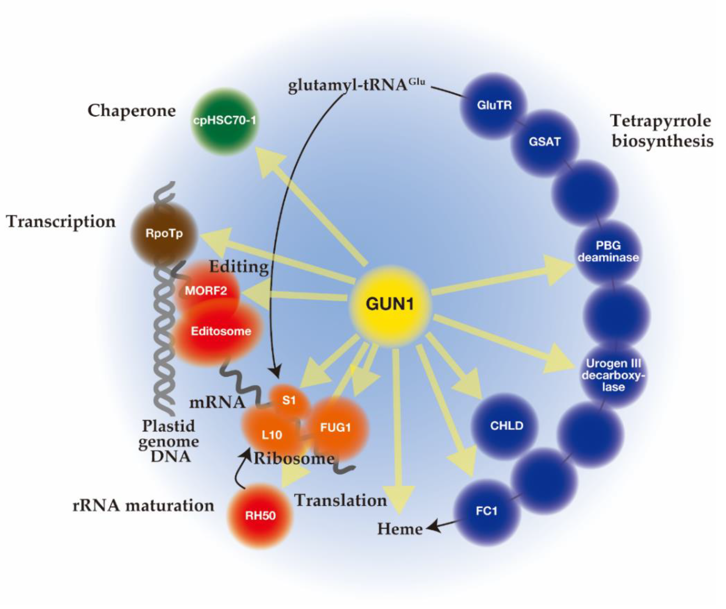
5. The Function of GUN1

Unlike mutant lines gun 2 to 6 related to the tetrapyrrole biosynthetic pathway, gun1 encodes a chloroplast protein containing a pentatricopeptide repeat (PPR) protein with a C-terminal small MutS-related (SMR) domain. Since gun1 can also prevent down-regulation of PhANG expression after treatment with lincomycin (Lin), an inhibitor of plastid translation [73], GUN1 has been suggested to act independently of the tetrapyrrole-mediated GUN signaling pathway. Interestingly, gun1 was shown to be hypersensitive to Lin or NF [96,97,98]. Since the PPR [99] and SMR [100] domains are known to be involved in nucleotide-binding, it was first suggested that GUN1 acts as a nucleotide-binding protein involved in plastid gene expression (PGE), plastid DNA metabolism, or DNA repair [73]. Subsequent efforts to screen for GUN1-associated partners by co-immunoprecipitation and mass spectrometry analysis identified many proteins rather than nucleotides [101,102,103]. The highly disordered domain at the N-terminus of GUN1 [104] may correspond to an intrinsically disordered region (IDR) [105]. The binding of protein partners induces a conversion of this domain to an ordered structure, which allows the same polypeptide sequence to undertake different interactions with different consequences. Nearly 300 different proteins involved in diverse biological processes in chloroplast were immunoprecipitated after crosslinking of GUN1–GFP in Arabidopsis, suggesting the promiscuous nature of the GUN1 protein [101]. Although the specificities to GUN1 were not identified, these putative GUN1-associated proteins were involved in transcription [97], translation [101,106], and import [107], all of which include homeostasis of chloroplast proteins [108,109,110] (Figure 2). In addition, enzymes involved in tetrapyrrole biosynthesis have been identified by yeast two-hybrid and bimolecular fluorescence complementation (BiFC) assays [101,107] (Figure 2).
Although GUN1 is highly and consistently expressed, the protein levels of GUN1 remain not abundant because of its very high turnover [103]. The GUN1 protein was only detectable where active chloroplast biogenesis occurs, such as in cotyledons and leaf primordia initially after germination [103]. The rapid turnover of GUN1 is controlled mainly by the chaperone ClpC1, suggesting degradation of GUN1 by the Clp protease [103]. Inhibition of plastid translation by Lin or oxidative stress by NF may prevent the ClpC-dependent degradation of GUN1, resulting in higher accumulation of this protein under these conditions [103]. As GUN1 accumulates only at the very early stage of leaf development under natural conditions, it has been suggested to function in chloroplast biogenesis [103]. However, the function of GUN1 at later developmental stages has also been suggested [101,103]. Since overexpression of GUN1 caused an early flowering phenotype, it is hypothesized that GUN1 functions in developmental phase transitions beyond chloroplast biogenesis [103].
Concerning the localization of GUN1 in the plastid, it was first suggested that GUN1 localizes in nucleoids where plastid DNA is actively transcribed. Transiently expressed GUN1–GFP in tobacco (Nicotiana benthamiana) exhibited granular fluorescence colocalizing with pTAC2, a component of transcriptionally-active complexes [73]. Such fluorescence in GUN1 was also observed in the stable Arabidopsis GUN1–GFP line [101] and BiFC assays of GUN1 and its binding proteins [97,101]. Meanwhile, GUN1–GFP was detected in the stroma as a dispersed signal in the stably transformed Arabidopsis lines [103,107]. It was recently reported that GUN1 alters its sub-chloroplast localization after NF treatment [111]: a speckled pattern of fluorescence was detected in the untreated condition, while a diffused distribution was observed after NF treatment. Therefore, it is likely that such a different distribution of GUN1 may be caused by employed developmental stage or functionality of GUN1.

5.1. Function of GUN1 on Transcription and Editing of Plastid Genes

In chloroplasts, two different RNA polymerases are present to transcribe the chloroplast genes [112,113]: the nuclear-encoded polymerase (NEP), a monomeric T3-T7 bacteriophage-type enzyme, that is mainly responsible for transcription of housekeeping genes, and the plastid-encoded polymerase (PEP), a multimeric bacterial-type enzyme, that mainly transcribes photosynthesis-related genes. Chloroplast development is associated with a shift in the primary RNA polymerase from NEP to PEP.
In Lin-treated Arabidopsis seedlings or mutants with defective plastid protein homeostasis, the increase in NEP-dependent transcripts, such as rpoA and rps12-3’, was observed in the wild type, but was compromised in the gun1 [97] mutant. GUN1 physically interacted with RpoTp encoding NEP and enhanced its activity upon depletion of PEP [97].
Additionally, GUN1 was proposed to interact with the MULTIPLE ORGANELLAR RNA EDITING FACTOR 2 (MORF2), a member of the so-called plastid RNA editosome, and regulate plastid RNA editing [114,115]. Compared with the wild type, the gun1 mutant showed differential efficiency of RNA editing levels of 11 sites in the plastid transcriptome after NF or Lin treatment [114]. Target genes were NEP-dependent, including transcripts of the PEP core subunits. The editing sites correspond to highly conserved residues, suggesting a lack of GUN1 leads to the synthesis of an impaired form of PEP core proteins [114,115].

5.2. The Function of GUN1 on Translation of Plastid Genome

GUN1 interacts with several ribosomal subunits, such as the plastid-encoded ribosomal proteins S1 (PRPS1) and the nucleus-encoded plastid ribosomal protein L10 [101]. The gun1 mutation genetically interacts with the mutations of these genes. Analysis of gun1 prps1 lines indicates that GUN1 controls PRPS1 accumulation at the protein level [101]. Moreover, functional overlapping of GUN1 with RH50 encoding the plastid DEAD-box RNA helicase, which is a 23S−4.5S rRNA maturation factor, has been reported [116]. This suggests the involvement of GUN1 in plastid ribosome assembly. Furthermore, the interaction of GUN1 with FUG1/cpIF2 encoding the chloroplast translation initiation factor IF-2 was detected [101]. The gun1 mutation aggravated the effects of decreased FUG1 levels on chloroplast protein translation [106]. Based on these results, the authors proposed that GUN1 is a modulator of plastid protein homeostasis, whose function only clearly manifests when plastid protein homeostasis is perturbed [106].

5.3. The Function of GUN1 on Protein Import into the Plastid

GUN1 has been proposed to be involved in the regulation of protein import into the plastid [89,97,107], although a critical point is remained to be clarified. GUN1 was shown to interact with the chloroplast chaperone cpHSC70-1 to promote the import of nuclear-encoded chloroplast proteins [101,107]. In addition, GUN1 was suggested to interact with other proteins involved in protein import, protein folding, and protein unfolding/degradation [101,108]. In gun1, the reduced accumulation of NEP-dependent transcription of Tic214 [97], together with the diminished activity of cpHSC70-1 [107], likely leads to an import defect, resulting in the over-accumulation of precursor proteins in the cytosol [97,107]. cphsc70-1 showed a gun phenotype in NF-treated, but not in Lin-treated, seedlings. The accumulation of precursor proteins in gun1 was confirmed by independent analysis, which accommodates the higher cytosolic HSP90 and HSP70 accumulation [97]. The activity of HSP90 was positively correlated with PhANG expression and proposed to be directly involved in the development of the gun phenotype in the gun1 mutant [107]. Since cytosolic HSP90 was identified as one of the MgProto-binding proteins and hsp90 mutants showed reduced derepression of the gun phenotype [54,55], HSP90 has been proposed as the central cytosolic transducer of plastid retrograde signal [107] mediating the activation of a positive regulator of transcription such as HY5 [40] and GLK1/2 [88,89].
One critical point of this model is that a gun phenotype is not seen for other mutants with reduced NEP transcription, such as sca3 defective in RpoTp enzyme, [97] or chloroplast import, such as the plastid protein import mutant 1 (ppi1) encoding TOC33, toc75-III-3, and tic40-4 [107]. Proteomic analysis of the plastid protein import mutant 2 (ppi2) defective in Toc159 revealed the accumulation of several plastid pre-proteins in the cytosol with concomitant upregulation of HSP90.1 protein, but PhANG expression is largely repressed in this mutant [89,117]. Therefore, it is likely that the GUN1-dependent modulation of import activity does not play a significant role in retrograde signaling. In addition, it is not clear why failure to import proteins into damaged chloroplasts should induce more expression of these pre-proteins, which may cause a catastrophic positive feedback loop.

5.4. The Link between GUN1 and Tetrapyrrole Biosynthesis

At first, a synergistic enhancement of the gun phenotype was observed in the gun1-1 gun4-1 and the gun1-1 gun5 double mutants relative to the single mutants [38], indicating the tetrapyrrole and GUN1 signals act independently. Subsequently, the gun phenotype of a double mutant gun5 gun1-9, a nonsense allele of GUN1, was found to be indistinguishable from gun1-9, suggesting the tetrapyrrole signal acts upstream of GUN1 [73]. Transcriptome analyses confirmed significant interactions between GUN5- and GUN1-dependent plastid signaling mechanisms [73]. The increased levels of ALA, heme, and Chl in gun1 sig2 relative to sig2 supported the interaction between these two plastid-to-nucleus signaling mechanisms [118]. A direct interaction between GUN1 and tetrapyrrole biosynthetic enzymes was revealed by immunoprecipitation of GFP-tagged GUN1 from the GUN1–GFP overexpressing line after treating chloroplasts with a crosslinking agent [101]. Yeast two hybrid and BiFC experiments confirmed the interaction of GUN1 with the CHLD subunit of MgCh, PBG deaminase, Urogen III decarboxylase, and FC1 [101] (Figure 1 and Figure 2). It is interesting to note that, with the exception of CHLD, the other three genes are not light responsive, but involved in the common pathway and the heme branch [91]. It should also be noted that interaction of GUN1 with tetrapyrrole biosynthetic enzymes are detected in independent immunoprecipitation analysis [107].
It was found that when ALA is fed to etiolated Arabidopsis seedlings, gun1 accumulated more Pchlide a in darkness, while GUN1 overexpressors accumulated less Pchlide a when compared with the wild type [104]. Such higher Pchlide a accumulation in gun1 was also observed without ALA feeding [119]. Since total heme levels were similarly changed in mutants and overexpressors, it was proposed that GUN1 controls the total tetrapyrrole flow [104]. Furthermore, GUN1 was shown to bind tetrapyrroles, Proto, MgProto, and heme via the PPR domain. In addition, GUN1 activates FC1 activity in a similar way to GUN4 enhancement of MgCh activity [104]. This model suggests a direct link between GUN1 and tetrapyrrole biosynthesis. GUN1 may regulate the distribution of tetrapyrrole biosynthesis, probably via direct interaction with enzymes [101], or through transcriptional regulation of the downstream transcription factor GLK [89]. On the contrary, it was shown that tetrapyrrole biosynthetic enzymes, such as GluTR encoded by HEMA1, cannot be properly imported into chloroplasts of the gun1 mutant, which may cause impeded distribution of tetrapyrrole in the mutant [107]. On the other hand, it is proposed that binding of FC1-synthesized heme by GUN1 blocks release or propagation of the retrograde signal [104]. Currently it is unknown whether heme-binding of GUN1 is associated with ClpC-dependent degradation. It is interesting to note that heme compromises the interaction between GluTR and GluTR-binding proteins and further enhances degradation of GluTR, upon feeding ALA to Arabidopsis leaves [120].
It was shown that FC1 is highly expressed in primordial tissues [68]. Mutants lacking FC1 showed poor early development with strong alleles being embryo lethal [68,69]. These results suggest FC1-derived heme functions during initial development when GUN1 is accumulated and active on proplastids to chloroplast transition. At this stage, the expression of PhANGs, including the key tetrapyrrole biosynthetic genes for massive Chl biosynthesis [91] remained at a low level, through PIF-dependent repression and GLK1/2 inactivation. Transcription and translation of NEP-dependent genes are also active at this stage. Considering the multiple interactions of GUN1 with proteins involved in plastid protein homeostasis and tetrapyrrole biosynthesis, it is possible that GUN1 becomes a threshold protein that may condense onto these proteins to systemically enhance these reactions (see below). In this sense, GUN1 is dispensable and its deficiency effect becomes obvious when chloroplast protein homeostasis is perturbed.

5.5. The Link between GUN1 and Other Processes

It has been reported that gun1 seedling development is hypersensitive to sucrose and ABA signaling [77,121,122]. Besides, anthocyanin accumulation was differentially affected by sucrose in wild-type and gun1 seedlings. From these results, GUN1 is proposed to have roles for sucrose and ABA signaling during initial seedling development [77,121]. Meanwhile, a yeast two-hybrid screen identified a novel GUN1-interacting protein GIP1 [111]. GIP1 was both localized to the cytosol and chloroplasts, and its abundance in chloroplasts is enhanced by NF treatment in the presence of GUN1. Although the function of GIP1 is not known, GIP1 and GUN1 may function antagonistically in the retrograde signaling pathway [111].

6. Outlook

6.1. Proposed Functions of GUN1

As discussed in this article, GUN1 functions as a biogenic retrograde signaling hub by interacting with numerous proteins (Figure 2). Although there is no experimental evidence, we hypothesize the possible function of GUN1. We propose GUN1 act as a platform to promote specific functions by bringing the interacting enzymes into the proximity of their substrates or may inhibit processes by sequestering particular pools of specific interactors [108]. One possibility is that GUN1 functions as a scaffold protein for molecular crowding that is crucial for the efficient operation of biological systems [123]. High concentrations of crowding agents entropically favor molecular association events, thereby accelerating molecular reactions [123]. Phase separation is a crucial example of when the regulation of macromolecular crowding is vital. Using genetically encoded multimeric nanoparticles (GEMs), it has been demonstrated that the mechanistic target of rapamycin complex (mTORC1) [124], the major amino acid sensor in eukaryotes [125], controls diffusion by tuning ribosome concentration. Like mTORC1, it is possible that through the N-terminal IDR and probably the PPR domain, GUN1 may form a condensate structure (droplet) that controls liquid–liquid phase separation and the biophysical properties of plastid systems involved in protein homeostasis as well as tetrapyrrole biosynthesis during initial chloroplast biogenesis (Figure 3). It is interesting to note that a particulate GUN1-derived fluorescent signal was dispersed by NF treatment [111] suggesting the relationship between functionality and droplet formation of GUN1. In this droplet, GUN1 may concentrate nucleoid for efficient NEP transcription, RNA editing, and subsequent translation of plastid-encoded proteins. Concentration of glutamyl-tRNAGlu [126,127] may also be important factor for effective translation and tetrapyrrole biosynthesis. Furthermore, we cannot exclude the possibility that other signaling pathways are involved in this regulation.
The question is how GUN1-dependent droplet formation is related to the generation of the biogenic plastid-to-nucleus retrograde signal. The GUN1-deficient effect on PhANG derepression is only obvious when plastids become dysfunctional by NF or Lin treatment. One possibility is that the FC1-derived heme signal cannot reach the nucleus in the wild type, while it can be transferred to the nucleus in the gun1 mutant. Considering GUN1 enhances total tetrapyrrole flow and can bind Proto and metal porphyrins, including heme and MgProto [104], we hypothesize that the GUN1 droplet controls tetrapyrrole distribution and holds the heme in the initial phase. Upon degradation of GUN1 by Clp-protease during subsequent chloroplast development, the heme is released from the droplet and transferred to the nucleus as a positive signal for PhANG induction. In the wild type, Clp-dependent GUN1 degradation is blocked by inhibitors [103]. Thus, produced FC1-derived heme may retain plastids in the wild type (Figure 3b). In the gun1 mutant, certain upregulation of tetrapyrrole distribution occurs without droplet formation, which may trigger direct transfer of heme to the nucleus (Figure 3c). Meanwhile, under untreated conditions, functional plastids may produce sufficient heme that may reach the nucleus for PhANG induction in the wild type and gun1 mutant. Coupled with the results from gun2–6, this hypothesis explains most of the observed phenotypes of the gun mutants.

6.2. Transfer of FC1-Derived Heme to Nucleus

Currently, FC1-specific heme is the most prominent candidate as chloroplast mobile biogenic signal. However, the possible involvement of other retrograde signaling pathways that interplay or antagonize the heme signaling cannot be excluded. Since hemoproteins are widely distributed within the cell, heme must be transported from the plastids to the target organelles [4]. However, compared to animals and yeast, the heme trafficking mechanism is poorly understood in plants [4]. In yeast, using a genetically encoded fluorescent heme sensor [128], it was shown that heme synthesized in the inner mitochondrial membrane can be transferred to the nucleus and the cytosol in distinct pathways [129]. Heme synthesized in the mitochondria was transferred to the nucleus via mitochondria-associated ER membrane contact sites (MCSs) that was faster than cytosolic heme transfer [129]. Although the MCSs of the chloroplast with other organelles were poorly known in plants, the ER-chloroplast MCSs were successfully visualized in a recent study [130]. Interestingly, the number of ER-chloroplast MCSs was decreased by MV treatment [130]. If FC1-derived heme is distinctly transferred to the nucleus via ER-chloroplast MCSs, rather than by the proposed cytosolic receptors, such as p22HBP/SOUL and tau glutathione transferases [131,132], a novel mechanism must be considered (Figure 3a).

6.3. A Hypothesis of the Heme Biogenic Plastid-to-Nucleus Retrograde Signaling Pathway

As shown in Figure 3a, we hypothesize the biogenic plastid-to-nucleus retrograde signaling mechanism to be: 1. At the initial phase of proplastid to chloroplast transition, putative GUN1-dependent droplets may form in proplastids, which control NEP-dependent transcription, RNA editing and translation, import of nuclear-encoded proteins, and tetrapyrrole biosynthesis. Produced FC1-derived heme may retain in the droplet. 2. During chloroplast biogenesis, GUN1 is degraded in a Clp-protease-dependent manner and the heme is released. Inhibitor treatment may disrupt the droplet formation, and GUN1 remains undegraded by preventing Clp-protease activity. 3. FC1-derived heme bound to GUN1 droplets is released and emitted from plastids via cytosolic or an ER-plastid MCS-dependent pathway. 4. In the nucleus, heme may inactivate PIFs or activate transcription factors, GLK1, and/or HY5. 5. Consequently, PhANG expression is activated.
At present, it is unknown whether FC1-derived heme affects the chloroplast processes, such as transcription, editing, translation, and import (Figure 2). Previous proteomic analysis of heme-binding protein detected none of the GUN1-interacting proteins in Arabidopsis [72]. Therefore, further study is required to elucidate this possibility.

6.4. Perspectives

It is possible that such a GUN1-dependent droplet functions as a system, so it will be difficult to dissect the individual pathways. To clarify this hypothesis, in addition to biochemical and molecular biological methods, novel approaches to liquid–liquid phase separation would be beneficial. For instance, live imaging using GEMS [124] and labeling methods identifying proximal and interacting proteins [133,134] may be successful. The future challenge will also be to identify the components of the signaling pathway that link heme to PhANG expression. Live cell imaging using the fluorescent heme sensor [128] would aid in the understanding of this mechanism.

Author Contributions

Conceptualization, T.M.; writing—original draft preparation, T.M.; writing—review and editing, T.S.; funding acquisition, T.M. and T.S. All authors have read and agreed to the published version of the manuscript.

Funding

This research was funded by JSPS KAKENHI, grant number JP20K06681, JP18K14650, JP18H03941, and JP19H03241.

Institutional Review Board Statement

Not applicable.

Informed Consent Statement

Not applicable.

Data Availability Statement

Data is contained within the article.

Acknowledgments

We would like to thank M. J. Terry and N. Mochizuki for critical reading of this manuscript.

Conflicts of Interest

The authors declare no conflict of interest.

References

  1. Battersby, A. Tetrapyrroles: The pigments of life. Nat. Prod. Rep. 2000, 17, 507–526. [Google Scholar] [CrossRef] [PubMed]
  2. Battersby, A.R.; Fookes, C.J.; Matcham, G.W.; McDonald, E. Biosynthesis of the pigments of life: Formation of the macrocycle. Nature 1980, 285, 17–21. [Google Scholar] [CrossRef] [PubMed]
  3. Op den Camp, R.G.L.; Przybyla, D.; Ochsenbein, C.; Laloi, C.; Kim, C.; Danon, A.; Wagner, D.; Hideg, E.; Göbel, C.; Feussner, I.; et al. Rapid induction of distinct stress responses after the release of singlet oxygen in Arabidopsis. Plant Cell 2003, 15, 2320–2332. [Google Scholar] [CrossRef] [PubMed] [Green Version]
  4. Mochizuki, N.; Tanaka, R.; Grimm, B.; Masuda, T.; Moulin, M.; Smith, A.G.; Tanaka, A.; Terry, M.J. The cell biology of tetrapyrroles: A life and death struggle. Trends Plant Sci. 2010, 15, 488–498. [Google Scholar] [CrossRef]
  5. Terry, M.J.; Smith, A.G. A model for tetrapyrrole synthesis as the primary mechanism for plastid-to-nucleus signaling during chloroplast biogenesis. Front. Plant Sci. 2013, 4, 1–14. [Google Scholar] [CrossRef] [PubMed] [Green Version]
  6. Larkin, R.M. Tetrapyrrole Signaling in Plants. Front. Plant Sci. 2016, 7, 1586. [Google Scholar] [CrossRef] [Green Version]
  7. Castelfranco, P.A.; Beale, S.I. Chlorophyll Biosynthesis: Recent Advances and Areas of Current Interest. Annu. Rev. Plant Physiol. 1983, 34, 241–276. [Google Scholar] [CrossRef]
  8. Tanaka, R.; Tanaka, A. Tetrapyrrole biosynthesis in higher plants. Annu. Rev. Plant Biol. 2007, 58, 321–346. [Google Scholar] [CrossRef]
  9. Tanaka, R.; Kobayashi, K.; Masuda, T. Tetrapyrrole Metabolism in Arabidopsis thaliana. Arab. Book 2011, 9, e0145. [Google Scholar] [CrossRef] [Green Version]
  10. Ilag, L.; Kumar, A.; Soll, D. Light regulation of chlorophyll biosynthesis at the level of 5-aminolevulinate formation in Arabidopsis. Plant Cell 1994, 6, 265–275. [Google Scholar] [CrossRef] [Green Version]
  11. McCormac, A.C.; Fischer, A.; Kumar, A.M.; Soll, D.; Terry, M.J. Regulation of HEMA1 expression by phytochrome and a plastid signal during de-etiolation in Arabidopsis thaliana. Plant J. 2001, 25, 549–561. [Google Scholar] [CrossRef] [PubMed] [Green Version]
  12. Koncz, C.; Mayerhofer, R.; Koncz-Kalman, Z.; Nawrath, C.; Reiss, B.; Redei, G.; Schell, J. Isolation of a gene encoding a novel chloroplast protein by T-DNA tagging in Arabidopsis thaliana. EMBO J. 1990, 9, 1337–1346. [Google Scholar] [CrossRef] [PubMed]
  13. Rissler, H.M.; Collakova, E.; DellaPenna, D.; Whelan, J.; Pogson, B.J. Chlorophyll biosynthesis. Expression of a second chl I gene of magnesium chelatase in Arabidopsis supports only limited chlorophyll synthesis. Plant Physiol. 2002, 128, 770–779. [Google Scholar] [CrossRef] [PubMed]
  14. Kobayashi, K.; Mochizuki, N.; Yoshimura, N.; Motohashi, K.; Hisabori, T.; Masuda, T. Functional analysis of Arabidopsis thaliana isoforms of the Mg-chelatase CHLI subunit. Photochem. Photobiol. Sci. 2008, 7, 1188–1195. [Google Scholar] [CrossRef]
  15. Larkin, R.; Alonso, J.; Ecker, J.; Chory, J. GUN4, a regulator of chlorophyll synthesis and intracellular signaling. Science 2003, 299, 902–906. [Google Scholar] [CrossRef] [Green Version]
  16. Adhikari, N.D.; Froehlich, J.E.; Strand, D.D.; Buck, S.M.; Kramer, D.M.; Larkin, R.M. GUN4-Porphyrin Complexes Bind the ChlH/GUN5 Subunit of Mg-Chelatase and Promote Chlorophyll Biosynthesis in Arabidopsis. Plant Cell 2011, 23, 1449–1467. [Google Scholar] [CrossRef] [Green Version]
  17. Davison, P.; Schubert, H.; Reid, J.; Iorg, C.; Heroux, A.; Hill, C.; Hunter, C. Structural and biochemical characterization of Gun4 suggests a mechanism for its role in chlorophyll biosynthesis. Biochemistry 2005, 44, 7603–7612. [Google Scholar] [CrossRef]
  18. Schneider, S.; Marles-Wright, J.; Sharp, K.H.; Paoli, M. Diversity and conservation of interactions for binding heme in b-type heme proteins. Nat. Prod. Rep. 2007, 24, 621–630. [Google Scholar] [CrossRef] [Green Version]
  19. Chow, K.-S.; Singh, D.P.; Walker, A.R.; Smith, A.G. Two different genes encode ferrochelatase in Arabidopsis: Mapping, expression and subcellular targeting of the precursor proteins. Plant J. 1998, 15, 531–541. [Google Scholar] [CrossRef]
  20. Nagai, S.; Koide, M.; Takahashi, S.; Kikuta, A.; Aono, M.; Sasaki-Sekimoto, Y.; Ohta, H.; Takamiya, K.-i.; Masuda, T. Induction of isoforms of tetrapyrrole biosynthetic enzymes, AtHEMA2 and AtFC1, under stress conditions and their physiological functions in Arabidopsis. Plant Physiol. 2007, 144, 1039–1051. [Google Scholar] [CrossRef] [Green Version]
  21. Terry, M.J.; Linley, P.J.; Kohchi, T. Making light of it: The role of plant haem oxygenases in phytochrome chromophore synthesis. Biochem. Soc. Trans. 2002, 30, 604–609. [Google Scholar] [CrossRef] [PubMed] [Green Version]
  22. Pogson, B.J.; Ganguly, D.; Albrecht-Borth, V. Insights into chloroplast biogenesis and development. Biochim. Biophys. Acta 2015, 1847, 1017–1024. [Google Scholar] [CrossRef] [PubMed] [Green Version]
  23. Pribil, M.; Labs, M.; Leister, D. Structure and dynamics of thylakoids in land plants. J. Exp. Bot. 2014, 65, 1955–1972. [Google Scholar] [CrossRef] [PubMed] [Green Version]
  24. Masuda, T.; Takamiya, K.-i. Novel Insights into the Enzymology, Regulation and Physiological Functions of Light-dependent Protochlorophyllide Oxidoreductase in Angiosperms. Photosynth. Res. 2004, 81, 1–29. [Google Scholar] [CrossRef] [PubMed]
  25. Mereschkowsky, C. Über Natur und Ursprung der Chromatophore im Pflanzenreiche. Biol. Cent. 1905, 25, 593–604. [Google Scholar]
  26. Archibald, J.M. The puzzle of plastid evolution. Curr. Biol. 2009, 19, R81–R88. [Google Scholar] [CrossRef] [Green Version]
  27. Keeling, P.J. The number, speed, and impact of plastid endosymbioses in eukaryotic evolution. Annu. Rev. Plant Biol. 2013, 64, 583–607. [Google Scholar] [CrossRef] [Green Version]
  28. Criscuolo, A.; Gribaldo, S. Large-scale phylogenomic analyses indicate a deep origin of primary plastids within cyanobacteria. Mol. Biol. Evol. 2011, 28, 3019–3032. [Google Scholar] [CrossRef] [Green Version]
  29. Abdallah, F.; Salamini, F.; Leister, D. A prediction of the size and evolutionary origin of the proteome of chloroplasts of Arabidopsis. Trends Plant Sci. 2000, 5, 141–142. [Google Scholar] [CrossRef]
  30. Singh, R.; Singh, S.; Parihar, P.; Singh, V.P.; Prasad, S.M. Retrograde signaling between plastid and nucleus: A review. J. Plant Physiol. 2015, 181, 55–66. [Google Scholar] [CrossRef]
  31. Pogson, B.J.; Woo, N.S.; Forster, B.; Small, I.D. Plastid signalling to the nucleus and beyond. Trends Plant Sci. 2008, 13, 602–609. [Google Scholar] [CrossRef] [PubMed]
  32. Nott, A.; Jung, H.; Koussevitzky, S.; Chory, J. Plastid-to-nucleus retrograde signaling. Annu. Rev. Plant Biol. 2006, 57, 739–759. [Google Scholar] [CrossRef] [PubMed]
  33. Chan, K.X.; Phua, S.Y.; Crisp, P.; McQuinn, R.; Pogson, B.J. Learning the Languages of the Chloroplast: Retrograde Signaling and Beyond. Annu. Rev. Plant Biol. 2016, 67, 25–53. [Google Scholar] [CrossRef] [PubMed]
  34. De Souza, A.; Wang, J.Z.; Dehesh, K. Retrograde Signals: Integrators of Interorganellar Communication and Orchestrators of Plant Development. Annu. Rev. Plant Biol. 2017, 68, 85–108. [Google Scholar] [CrossRef] [PubMed]
  35. Susek, J.; Chory, J. A tale of two genomes: Role of a chloroplast signal in coordinating nuclear and plastid genome expression. Aust. J. Plant Physiol. 1992, 19, 387–399. [Google Scholar] [CrossRef]
  36. Gray, J.C.; Sullivan, J.A.; Wang, J.H.; Jerome, C.A.; MacLean, D. Coordination of plastid and nuclear gene expression. Philos. Trans. R. Soc. Lond. B Biol. Sci. 2003, 358, 135–144, discussion 144–135. [Google Scholar] [CrossRef] [PubMed] [Green Version]
  37. Susek, R.; Ausubel, F.; Chory, J. Signal transduction mutants of Arabidopsis uncouple nuclear CAB and RBCS gene expression from chloroplast development. Cell 1993, 74, 787–799. [Google Scholar] [CrossRef] [Green Version]
  38. Mochizuki, N.; Brusslan, J.; Larkin, R.; Nagatani, A.; Chory, J. Arabidopsis genomes uncoupled 5 (GUN5) mutant reveals the involvement of Mg-chelatase H subunit in plastid-to-nucleus signal transduction. Proc. Natl. Acad. Sci. USA 2001, 98, 2053–2058. [Google Scholar] [CrossRef] [Green Version]
  39. Woodson, J.D.; Perez-Ruiz, J.M.; Chory, J. Heme synthesis by plastid ferrochelatase I regulates nuclear gene expression in plants. Curr. Biol. 2011, 21, 897–903. [Google Scholar] [CrossRef] [Green Version]
  40. Ruckle, M.E.; DeMarco, S.M.; Larkin, R.M. Plastid signals remodel light signaling networks and are essential for efficient chloroplast biogenesis in Arabidopsis. Plant Cell 2007, 19, 3944–3960. [Google Scholar] [CrossRef] [Green Version]
  41. Kim, C.; Apel, K. 1O2-mediated and EXECUTER-dependent retrograde plastid-to-nucleus signaling in norflurazon-treated seedlings of Arabidopsis thaliana. Mol. Plant 2013, 6, 1580–1591. [Google Scholar] [CrossRef] [PubMed] [Green Version]
  42. Voigt, C.; Oster, U.; Börnke, F.; Jahns, P.; Dietz, K.-J.; Leister, D.; Kleine, T. In-depth analysis of the distinctive effects of norflurazon implies that tetrapyrrole biosynthesis, organellar gene expression and ABA cooperate in the GUN-type of plastid signalling. Physiol. Plant 2010, 138, 503–519. [Google Scholar] [CrossRef] [PubMed]
  43. Larkin, R.M. Influence of plastids on light signalling and development. Philos. Trans. R. Soc. Lond. B Biol. Sci. 2014, 369, 20130232. [Google Scholar] [CrossRef] [PubMed] [Green Version]
  44. Tanaka, K.; Hanaoka, M. The early days of plastid retrograde signaling with respect to replication and transcription. Front. Plant Sci. 2012, 3, 301. [Google Scholar] [CrossRef] [PubMed] [Green Version]
  45. Kropat, J.; Oster, U.; Rudiger, W.; Beck, C.F. Chlorophyll precursors are signals of chloroplast origin involved in light induction of nuclear heat-shock genes. Proc. Natl. Acad. Sci. USA 1997, 94, 14168–14172. [Google Scholar] [CrossRef] [Green Version]
  46. Kobayashi, Y.; Kanesaki, Y.; Tanaka, A.; Kuroiwa, H.; Kuroiwa, T.; Tanaka, K. Tetrapyrrole signal as a cell-cycle coordinator from organelle to nuclear DNA replication in plant cells. Proc. Natl. Acad. Sci. USA 2009, 106, 803–807. [Google Scholar] [CrossRef] [Green Version]
  47. Strand, A.; Asami, T.; Alonso, J.; Ecker, J.R.; Chory, J. Chloroplast to nucleus communication triggered by accumulation of Mg-protoporphyrinIX. Nature 2003, 421, 79–83. [Google Scholar] [CrossRef]
  48. Huang, Y.-S.; Li, H.-M. Arabidopsis CHLI2 can substitute for CHLI1. Plant Physiol. 2009, 150, 636–645. [Google Scholar] [CrossRef] [Green Version]
  49. Ankele, E.; Kindgren, P.; Pesquet, E.; Strand, A. In vivo visualization of Mg-protoporphyrin IX, a coordinator of photosynthetic gene expression in the nucleus and the chloroplast. Plant Cell 2007, 19, 1964–1979. [Google Scholar] [CrossRef] [Green Version]
  50. Pontier, D.; Albrieux, C.; Joyard, J.; Lagrange, T.; Block, M. Knock-out of the magnesium protoporphyrin IX methyltransferase gene in Arabidopsis. Effects on chloroplast development and on chloroplast-to-nucleus signaling. J. Biol. Chem. 2007, 282, 2297–2304. [Google Scholar] [CrossRef] [Green Version]
  51. Gadjieva, R.; Axelsson, E.; Olsson, U.; Hansson, M. Analysis of gun phenotype in barley magnesium chelatase and Mg-protoporphyrin IX monomethyl ester cyclase mutants. Plant Physiol. Biochem. 2005, 43, 901–908. [Google Scholar] [CrossRef] [PubMed]
  52. Mochizuki, N.; Tanaka, R.; Tanaka, A.; Masuda, T.; Nagatani, A. The steady-state level of Mg-protoporphyrin IX is not a determinant of plastid-to-nucleus signaling in Arabidopsis. Proc. Natl. Acad. Sci. USA 2008, 105, 15184–15189. [Google Scholar] [CrossRef] [PubMed] [Green Version]
  53. Moulin, M.; McCormac, A.C.; Terry, M.J.; Smith, A.G. Tetrapyrrole profiling in Arabidopsis seedlings reveals that retrograde plastid nuclear signaling is not due to Mg-protoporphyrin IX accumulation. Proc. Natl. Acad. Sci. USA 2008, 105, 15178–15183. [Google Scholar] [CrossRef] [PubMed] [Green Version]
  54. Kindgren, P.; Eriksson, M.-J.; Benedict, C.; Mohapatra, A.; Gough, S.P.; Hansson, M.; Kieselbach, T.; Strand, A. A novel proteomic approach reveals a role for Mg-protoporphyrin IX in response to oxidative stress. Physiol. Plant 2011, 141, 310–320. [Google Scholar] [CrossRef]
  55. Kindgren, P.; Norén, L.; López, J.d.D.B.; Shaikhali, J.; Strand, A. Interplay between Heat Shock Protein 90 and HY5 controls PhANG expression in response to the GUN5 plastid signal. Mol. Plant 2012, 5, 901–913. [Google Scholar] [CrossRef] [Green Version]
  56. Zhang, Z.-W.; Yuan, S.; Feng, H.; Xu, F.; Cheng, J.; Shang, J.; Zhang, D.-W.; Lin, H.-H. Transient accumulation of Mg-protoporphyrin IX regulates expression of PhANGs—New evidence for the signaling role of tetrapyrroles in mature Arabidopsis plants. J. Plant Physiol. 2011, 168, 714–721. [Google Scholar] [CrossRef]
  57. Schlicke, H.; Hartwig, A.S.; Firtzlaff, V.; Richter, A.S.; Gläßer, C.; Maier, K.; Finkemeier, I.; Grimm, B. Induced deactivation of genes encoding chlorophyll biosynthesis enzymes disentangles tetrapyrrole-mediated retrograde signaling. Mol. Plant 2014, 7, 1211–1227. [Google Scholar] [CrossRef] [Green Version]
  58. Mense, S.M.; Zhang, L. Heme: A versatile signaling molecule controlling the activities of diverse regulators ranging from transcription factors to MAP kinases. Cell Res. 2006, 16, 681–692. [Google Scholar] [CrossRef] [Green Version]
  59. Tsiftsoglou, A.; Tsamadou, A.; Papadopoulou, L. Heme as key regulator of major mammalian cellular functions: Molecular, cellular, and pharmacological aspects. Pharmacol. Ther. 2006, 111, 327–345. [Google Scholar] [CrossRef]
  60. Von Gromoff, E.; Alawady, A.; Meinecke, L.; Grimm, B.; Beck, C. Heme, a plastid-derived regulator of nuclear gene expression in Chlamydomonas. Plant Cell 2008, 20, 552–567. [Google Scholar] [CrossRef] [Green Version]
  61. Voss, B.; Meinecke, L.; Kurz, T.; Al-Babili, S.; Beck, C.F.; Hess, W.R. Hemin and Magnesium-Protoporphyrin IX Induce Global Changes in Gene Expression in Chlamydomonas reinhardtii. Plant Physiol. 2011, 155, 892–905. [Google Scholar] [CrossRef] [PubMed] [Green Version]
  62. Kobayashi, Y.; Tanaka, K. Transcriptional Regulation of Tetrapyrrole Biosynthetic Genes Explains Abscisic Acid-Induced Heme Accumulation in the Unicellular Red Alga Cyanidioschyzon merolae. Front. Plant Sci. 2016, 7, 1300. [Google Scholar] [CrossRef] [PubMed] [Green Version]
  63. Kobayashi, Y.; Ando, H.; Hanaoka, M.; Tanaka, K. Abscisic Acid Participates in the Control of Cell Cycle Initiation Through Heme Homeostasis in the Unicellular Red Alga Cyanidioschyzon merolae. Plant Cell Physiol. 2016, 57, 953–960. [Google Scholar] [CrossRef] [PubMed] [Green Version]
  64. Vanhee, C.; Zapotoczny, G.; Masquelier, D.; Ghislain, M.; Batoko, H. The Arabidopsis multistress regulator TSPO is a heme binding membrane protein and a potential scavenger of porphyrins via an autophagy-dependent degradation mechanism. Plant Cell 2011, 23, 785–805. [Google Scholar] [CrossRef] [Green Version]
  65. Cornah, J.; Roper, J.; Pal Singh, D.; Smith, A. Measurement of ferrochelatase activity using a novel assay suggests that plastids are the major site of haem biosynthesis in both photosynthetic and non-photosynthetic cells of pea (Pisum sativum L.). Biochem. J. 2002, 362, 423–432. [Google Scholar] [CrossRef]
  66. Masuda, T.; Suzuki, T.; Shimada, H.; Ohta, H.; Takamiya, K. Subcellular localization of two types of ferrochelatase in cucumber. Planta 2003, 217, 602–609. [Google Scholar] [CrossRef]
  67. Hey, D.; Ortega-Rodes, P.; Fan, T.; Schnurrer, F.; Brings, L.; Hedtke, B.; Grimm, B. Transgenic Tobacco Lines Expressing Sense or Antisense FERROCHELATASE 1 RNA Show Modified Ferrochelatase Activity in Roots and Provide Experimental Evidence for Dual Localization of Ferrochelatase 1. Plant Cell Physiol. 2016, 57, 171–2585. [Google Scholar] [CrossRef] [Green Version]
  68. Espinas, N.A.; Kobayashi, K.; Sato, Y.; Mochizuki, N.; Takahashi, K.; Tanaka, R.; Masuda, T. Allocation of Heme Is Differentially Regulated by Ferrochelatase Isoforms in Arabidopsis Cells. Front. Plant Sci. 2016, 7, 69. [Google Scholar] [CrossRef] [Green Version]
  69. Fan, T.; Roling, L.; Meiers, A.; Brings, L.; Ortega-Rodes, P.; Hedtke, B.; Grimm, B. Complementation studies of the Arabidopsis fc1 mutant substantiate essential functions of ferrochelatase 1 during embryogenesis and salt stress. Plant Cell Environ. 2019, 42, 618–632. [Google Scholar] [CrossRef]
  70. Page, M.T.; Garcia-Becerra, T.; Smith, A.G.; Terry, M.J. Overexpression of chloroplast-targeted ferrochelatase 1 results in a genomes uncoupled chloroplast-to-nucleus retrograde signalling phenotype. Philos. Trans. R. Soc. Lond. B Biol. Sci. 2020, 375, 20190401. [Google Scholar] [CrossRef]
  71. Espinas, N.A.; Kobayashi, K.; Takahashi, S.; Mochizuki, N.; Masuda, T. Evaluation of Unbound Free Heme in Plant Cells by Differential Acetone Extraction. Plant Cell Physiol. 2012, 53, 1344–1354. [Google Scholar] [CrossRef] [PubMed] [Green Version]
  72. Shimizu, T.; Yasuda, R.; Mukai, Y.; Tanoue, R.; Shimada, T.; Imamura, S.; Tanaka, K.; Watanabe, S.; Masuda, T. Proteomic analysis of haem-binding protein from Arabidopsis thaliana and Cyanidioschyzon merolae. Philos. Trans. R. Soc. Lond. B Biol. Sci. 2020, 375, 20190488. [Google Scholar] [CrossRef] [PubMed]
  73. Koussevitzky, S.; Nott, A.; Mockler, T.C.; Hong, F.; Sachetto-Martins, G.; Surpin, M.; Lim, J.; Mittler, R.; Chory, J. Signals from chloroplasts converge to regulate nuclear gene expression. Science 2007, 316, 715–719. [Google Scholar] [CrossRef]
  74. Brunkard, J.O.; Burch-Smith, T.M. Ties that bind: The integration of plastid signalling pathways in plant cell metabolism. Essays Biochem. 2018, 62, 95–107. [Google Scholar] [CrossRef] [PubMed]
  75. Hernandez-Verdeja, T.; Strand, A. Retrograde Signals Navigate the Path to Chloroplast Development. Plant Physiol. 2018, 176, 967–976. [Google Scholar] [CrossRef] [Green Version]
  76. Kacprzak, S.M.; Mochizuki, N.; Naranjo, B.; Xu, D.; Leister, D.; Kleine, T.; Okamoto, H.; Terry, M.J. Plastid-to-Nucleus Retrograde Signalling during Chloroplast Biogenesis Does Not Require ABI4. Plant Physiol. 2019, 179, 18–23. [Google Scholar] [CrossRef] [Green Version]
  77. Cottage, A.; Gray, J.C. Timing the switch to phototrophic growth: A possible role of GUN1. Plant Signal. Behav. 2011, 6, 578–582. [Google Scholar] [CrossRef] [Green Version]
  78. Martin, G.; Leivar, P.; Ludevid, D.; Tepperman, J.M.; Quail, P.H.; Monte, E. Phytochrome and retrograde signalling pathways converge to antagonistically regulate a light-induced transcriptional network. Nat. Commun. 2016, 7, 11431. [Google Scholar] [CrossRef]
  79. Mhamdi, A.; Gommers, C.M.M. Another gun Dismantled: ABSCISIC ACID INSENSITIVE4 Is Not a Target of Retrograde Signaling. Plant Physiol. 2019, 179, 13–14. [Google Scholar] [CrossRef] [Green Version]
  80. Sun, X.; Feng, P.; Xu, X.; Guo, H.; Ma, J.; Chi, W.; Lin, R.; Lu, C.; Zhang, L. A chloroplast envelope-bound PHD transcription factor mediates chloroplast signals to the nucleus. Nat. Commun. 2011, 2, 477. [Google Scholar] [CrossRef] [Green Version]
  81. Page, M.T.; Kacprzak, S.M.; Mochizuki, N.; Okamoto, H.; Smith, A.G.; Terry, M.J. Seedlings Lacking the PTM Protein Do Not Show a genomes uncoupled (gun) Mutant Phenotype. Plant Physiol. 2017, 174, 21–26. [Google Scholar] [CrossRef] [PubMed] [Green Version]
  82. Hernandez-Verdeja, T.; Vuorijoki, L.; Strand, A. Emerging from the darkness: Interplay between light and plastid signaling during chloroplast biogenesis. Physiol. Plant 2020, 169, 397–406. [Google Scholar] [CrossRef] [PubMed] [Green Version]
  83. Larkin, R.; Ruckle, M. Integration of light and plastid signals. Curr. Opin. Plant Biol. 2008, 11, 593–599. [Google Scholar] [CrossRef] [PubMed]
  84. Bae, G.; Choi, G. Decoding of light signals by plant phytochromes and their interacting proteins. Annu. Rev. Plant Biol. 2008, 59, 281–311. [Google Scholar] [CrossRef] [PubMed] [Green Version]
  85. Lau, O.S.; Deng, X.-W. Plant hormone signaling lightens up: Integrators of light and hormones. Curr. Opin. Plant Biol. 2010, 13, 571–577. [Google Scholar] [CrossRef] [PubMed]
  86. Fitter, D.W.; Martin, D.J.; Copley, M.J.; Scotland, R.W.; Langdale, J.A. GLK gene pairs regulate chloroplast development in diverse plant species. Plant J. 2002, 31, 713–727. [Google Scholar] [CrossRef] [Green Version]
  87. Waters, M.; Moylan, E.; Langdale, J. GLK transcription factors regulate chloroplast development in a cell-autonomous manner. Plant J. 2008, 56, 432–444. [Google Scholar] [CrossRef]
  88. Waters, M.T.; Wang, P.; Korkaric, M.; Capper, R.G.; Saunders, N.J.; Langdale, J.A. GLK Transcription Factors Coordinate Expression of the Photosynthetic Apparatus in Arabidopsis. Plant Cell 2009, 21, 1109–1128. [Google Scholar] [CrossRef] [Green Version]
  89. Kakizaki, T.; Matsumura, H.; Nakayama, K.; Che, F.-S.; Terauchi, R.; Inaba, T. Coordination of plastid protein import and nuclear gene expression by plastid-to-nucleus retrograde signaling. Plant Physiol. 2009, 151, 1339–1353. [Google Scholar] [CrossRef] [Green Version]
  90. Oh, E.; Zhu, J.Y.; Wang, Z.Y. Interaction between BZR1 and PIF4 integrates brassinosteroid and environmental responses. Nat. Cell Biol. 2012, 14, 802–809. [Google Scholar] [CrossRef] [Green Version]
  91. Matsumoto, F.; Obayashi, T.; Sasaki-Sekimoto, Y.; Ohta, H.; Takamiya, K.-i.; Masuda, T. Gene expression profiling of the tetrapyrrole metabolic pathway in Arabidopsis with a mini-array system. Plant Physiol. 2004, 135, 2379–2391. [Google Scholar] [CrossRef] [Green Version]
  92. Masuda, T.; Fujita, Y. Regulation and evolution of chlorophyll metabolism. Photochem. Photobiol. Sci. 2008, 7, 1131. [Google Scholar] [CrossRef]
  93. Kobayashi, K.; Obayashi, T.; Masuda, T. Role of the G-box element in regulation of chlorophyll biosynthesis in Arabidopsis roots. Plant Signal. Behav. 2012, 7, 922–926. [Google Scholar] [CrossRef] [Green Version]
  94. Kobayashi, K.; Baba, S.; Obayashi, T.; Sato, M.; Toyooka, K.; Keränen, M.; Aro, E.-M.; Fukaki, H.; Ohta, H.; Sugimoto, K.; et al. Regulation of Root Greening by Light and Auxin/Cytokinin Signaling in Arabidopsis. Plant Cell 2012, 24, 1081–1095. [Google Scholar] [CrossRef] [PubMed] [Green Version]
  95. Leister, D.; Kleine, T. Definition of a core module for the nuclear retrograde response to altered organellar gene expression identifies GLK overexpressors as gun mutants. Physiol. Plant 2016, 157, 297–309. [Google Scholar] [CrossRef] [PubMed]
  96. Zhao, X.; Huang, J.; Chory, J. Genome uncoupled 1 mutants are hypersensitive to norflurazon and lincomycin. Plant Physiol. 2018, 178. [Google Scholar] [CrossRef] [PubMed] [Green Version]
  97. Tadini, L.; Peracchio, C.; Trotta, A.; Colombo, M.; Mancini, I.; Jeran, N.; Costa, A.; Faoro, F.; Marsoni, M.; Vannini, C.; et al. GUN1 influences the accumulation of NEP-dependent transcripts and chloroplast protein import in Arabidopsis cotyledons upon perturbation of chloroplast protein homeostasis. Plant J. 2020, 101, 1198–1220. [Google Scholar] [CrossRef] [PubMed]
  98. Song, L.; Chen, Z.; Larkin, R.M. The genomes uncoupled mutants are more sensitive to norflurazon than wild type. Plant Physiol. 2018, 178. [Google Scholar] [CrossRef] [Green Version]
  99. Kotera, E.; Tasaka, M.; Shikanai, T. A pentatricopeptide repeat protein is essential for RNA editing in chloroplasts. Nature 2005, 433, 326–330. [Google Scholar] [CrossRef]
  100. Moreira, D.; Philippe, H. Smr: A bacterial and eukaryotic homologue of the C-terminal region of the MutS2 family. Trends Biochem. Sci. 1999, 24, 298–300. [Google Scholar] [CrossRef]
  101. Tadini, L.; Pesaresi, P.; Kleine, T.; Rossi, F.; Guljamow, A.; Sommer, F.; Mühlhaus, T.; Schroda, M.; Masiero, S.; Pribil, M.; et al. GUN1 Controls Accumulation of the Plastid Ribosomal Protein S1 at the Protein Level and Interacts with Proteins Involved in Plastid Protein Homeostasis. Plant Physiol. 2016, 170, 1817–1830. [Google Scholar] [CrossRef] [Green Version]
  102. Jia, Y.; Tian, H.; Zhang, S.; Ding, Z.; Ma, C. GUN1-Interacting Proteins Open the Door for Retrograde Signaling. Trends Plant Sci. 2019, 24, 884–887. [Google Scholar] [CrossRef]
  103. Wu, G.-Z.; Chalvin, C.; Hoelscher, M.; Meyer, E.H.; Wu, X.N.; Bock, R. Control of Retrograde Signaling by Rapid Turnover of GENOMES UNCOUPLED1. Plant Physiol. 2018, 176, 2472–2495. [Google Scholar] [CrossRef] [Green Version]
  104. Shimizu, T.; Kacprzak, S.M.; Mochizuki, N.; Nagatani, A.; Watanabe, S.; Shimada, T.; Tanaka, K.; Hayashi, Y.; Arai, M.; Leister, D.; et al. The retrograde signaling protein GUN1 regulates tetrapyrrole biosynthesis. Proc. Natl. Acad. Sci. USA 2019, 116, 24900–24906. [Google Scholar] [CrossRef] [PubMed] [Green Version]
  105. Uversky, V.N. Intrinsically disordered proteins from A to Z. Int. J. Biochem. Cell Biol. 2011, 43, 1090–1103. [Google Scholar] [CrossRef] [Green Version]
  106. Marino, G.; Naranjo, B.; Wang, J.; Penzler, J.F.; Kleine, T.; Leister, D. Relationship of GUN1 to FUG1 in chloroplast protein homeostasis. Plant J. 2019, 99, 521–535. [Google Scholar] [CrossRef]
  107. Wu, G.Z.; Meyer, E.H.; Richter, A.S.; Schuster, M.; Ling, Q.; Schottler, M.A.; Walther, D.; Zoschke, R.; Grimm, B.; Jarvis, R.P.; et al. Control of retrograde signalling by protein import and cytosolic folding stress. Nat. Plants 2019, 5, 525–538. [Google Scholar] [CrossRef] [PubMed]
  108. Colombo, M.; Tadini, L.; Peracchio, C.; Ferrari, R.; Pesaresi, P. GUN1, a Jack-Of-All-Trades in Chloroplast Protein Homeostasis and Signaling. Front. Plant Sci. 2016, 7, 1449. [Google Scholar] [CrossRef] [PubMed] [Green Version]
  109. Tadini, L.; Jeran, N.; Pesaresi, P. GUN1 and Plastid RNA Metabolism: Learning from Genetics. Cells 2020, 9, 2307. [Google Scholar] [CrossRef]
  110. Tadini, L.; Jeran, N.; Peracchio, C.; Masiero, S.; Colombo, M.; Pesaresi, P. The plastid transcription machinery and its coordination with the expression of nuclear genome: Plastid-Encoded Polymerase, Nuclear-Encoded Polymerase and the Genomes Uncoupled 1-mediated retrograde communication. Philos. Trans. R. Soc. Lond. B Biol. Sci. 2020, 375, 20190399. [Google Scholar] [CrossRef]
  111. Huang, X.Q.; Wang, L.J.; Kong, M.J.; Huang, N.; Liu, X.Y.; Liang, H.Y.; Zhang, J.X.; Lu, S. At3g53630 encodes a GUN1-interacting protein under norflurazon treatment. Protoplasma 2020, 1–8. [Google Scholar] [CrossRef] [PubMed]
  112. Liebers, M.; Grubler, B.; Chevalier, F.; Lerbs-Mache, S.; Merendino, L.; Blanvillain, R.; Pfannschmidt, T. Regulatory Shifts in Plastid Transcription Play a Key Role in Morphological Conversions of Plastids during Plant Development. Front. Plant Sci. 2017, 8, 23. [Google Scholar] [CrossRef] [Green Version]
  113. Pfannschmidt, T.; Blanvillain, R.; Merendino, L.; Courtois, F.; Chevalier, F.; Liebers, M.; Grubler, B.; Hommel, E.; Lerbs-Mache, S. Plastid RNA polymerases: Orchestration of enzymes with different evolutionary origins controls chloroplast biogenesis during the plant life cycle. J. Exp. Bot. 2015, 66, 6957–6973. [Google Scholar] [CrossRef] [PubMed]
  114. Zhao, X.; Huang, J.; Chory, J. GUN1 interacts with MORF2 to regulate plastid RNA editing during retrograde signaling. Proc. Natl. Acad. Sci. USA 2019, 116, 10162–10167. [Google Scholar] [CrossRef] [Green Version]
  115. Zhao, X.B.; Huang, J.Y.; Chory, J. Unraveling the Linkage between Retrograde Signaling and RNA Metabolism in Plants. Trends Plant Sci. 2020, 25, 141–147. [Google Scholar] [CrossRef]
  116. Paieri, F.; Tadini, L.; Manavski, N.; Kleine, T.; Ferrari, R.; Morandini, P.; Pesaresi, P.; Meurer, J.; Leister, D. The DEAD-box RNA Helicase RH50 Is a 23S-4.5S rRNA Maturation Factor that Functionally Overlaps with the Plastid Signaling Factor GUN1. Plant Physiol. 2018, 176, 634–648. [Google Scholar] [CrossRef] [Green Version]
  117. Bischof, S.; Baerenfaller, K.; Wildhaber, T.; Troesch, R.; Vidi, P.A.; Roschitzki, B.; Hirsch-Hoffmann, M.; Hennig, L.; Kessler, F.; Gruissem, W.; et al. Plastid proteome assembly without Toc159: Photosynthetic protein import and accumulation of N-acetylated plastid precursor proteins. Plant Cell 2011, 23, 3911–3928. [Google Scholar] [CrossRef] [Green Version]
  118. Woodson, J.D.; Perez-Ruiz, J.M.; Schmitz, R.J.; Ecker, J.R.; Chory, J. Sigma factor-mediated plastid retrograde signals control nuclear gene expression. Plant J. 2012, 73, 1–13. [Google Scholar] [CrossRef] [Green Version]
  119. Xu, X.; Chi, W.; Sun, X.; Feng, P.; Guo, H.; Li, J.; Lin, R.; Lu, C.; Wang, H.; Leister, D.; et al. Convergence of light and chloroplast signals for de-etiolation through ABI4-HY5 and COP1. Nat. Plants 2016, 2, 16066. [Google Scholar] [CrossRef]
  120. Richter, A.S.; Banse, C.; Grimm, B. The GluTR-binding protein is the heme-binding factor for feedback control of glutamyl-tRNA reductase. eLife 2019, 8. [Google Scholar] [CrossRef] [PubMed]
  121. Cottage, A.; Mott, E.K.; Kempster, J.A.; Gray, J.C. The Arabidopsis plastid-signalling mutant gun1 (genomes uncoupled1) shows altered sensitivity to sucrose and abscisic acid and alterations in early seedling development. J. Exp. Bot. 2010, 61, 3773–3786. [Google Scholar] [CrossRef] [PubMed] [Green Version]
  122. Richter, A.S.; Tohge, T.; Fernie, A.R.; Grimm, B. The genomes uncoupled-dependent signalling pathway coordinates plastid biogenesis with the synthesis of anthocyanins. Philos. Trans. R. Soc. Lond. B Biol. Sci. 2020, 375, 20190403. [Google Scholar] [CrossRef] [PubMed]
  123. Zhou, H.X.; Rivas, G.; Minton, A.P. Macromolecular crowding and confinement: Biochemical, biophysical, and potential physiological consequences. Annu. Rev. Biophys. 2008, 37, 375–397. [Google Scholar] [CrossRef] [PubMed] [Green Version]
  124. Delarue, M.; Brittingham, G.P.; Pfeffer, S.; Surovtsev, I.V.; Pinglay, S.; Kennedy, K.J.; Schaffer, M.; Gutierrez, J.I.; Sang, D.; Poterewicz, G.; et al. mTORC1 Controls Phase Separation and the Biophysical Properties of the Cytoplasm by Tuning Crowding. Cell 2018, 174, 338–349.e320. [Google Scholar] [CrossRef] [Green Version]
  125. Hara, K.; Yonezawa, K.; Weng, Q.P.; Kozlowski, M.T.; Belham, C.; Avruch, J. Amino acid sufficiency and mTOR regulate p70 S6 kinase and eIF-4E BP1 through a common effector mechanism. J. Biol. Chem. 1998, 273, 14484–14494. [Google Scholar] [CrossRef] [Green Version]
  126. Levican, G.; Katz, A.; Valenzuela, P.; Soll, D.; Orellana, O. A tRNA(Glu) that uncouples protein and tetrapyrrole biosynthesis. FEBS Lett. 2005, 579, 6383–6387. [Google Scholar] [CrossRef] [Green Version]
  127. Agrawal, S.; Karcher, D.; Ruf, S.; Bock, R. The Functions of Chloroplast Glutamyl-tRNA in Translation and Tetrapyrrole Biosynthesis. Plant Physiol. 2020, 183, 263–276. [Google Scholar] [CrossRef] [Green Version]
  128. Hanna, D.A.; Harvey, R.M.; Martinez-Guzman, O.; Yuan, X.; Chandrasekharan, B.; Raju, G.; Outten, F.W.; Hamza, I.; Reddi, A.R. Heme dynamics and trafficking factors revealed by genetically encoded fluorescent heme sensors. Proc. Natl. Acad. Sci. USA 2016, 113, 7539–7544. [Google Scholar] [CrossRef] [Green Version]
  129. Martinez-Guzman, O.; Willoughby, M.M.; Saini, A.; Dietz, J.V.; Bohovych, I.; Medlock, A.E.; Khalimonchuk, O.; Reddi, A.R. Mitochondrial-nuclear heme trafficking in budding yeast is regulated by GTPases that control mitochondrial dynamics and ER contact sites. J. Cell Sci. 2020, 133. [Google Scholar] [CrossRef]
  130. Li, T.; Xiao, Z.; Li, H.; Liu, C.; Shen, W.; Gao, C. A Combinatorial Reporter Set to Visualize the Membrane Contact Sites Between Endoplasmic Reticulum and Other Organelles in Plant Cell. Front. Plant Sci. 2020, 11, 1280. [Google Scholar] [CrossRef]
  131. Takahashi, S.; Ogawa, T.; Inoue, K.; Masuda, T. Characterization of cytosolic tetrapyrrole-binding proteins in Arabidopsis thaliana. Photochem. Photobiol. Sci. 2008, 7, 1216–1224. [Google Scholar] [CrossRef] [PubMed]
  132. Sylvestre-Gonon, E.; Schwartz, M.; Girardet, J.M.; Hecker, A.; Rouhier, N. Is there a role for tau glutathione transferases in tetrapyrrole metabolism and retrograde signalling in plants? Philos. Trans. R. Soc. Lond. B Biol. Sci. 2020, 375, 20190404. [Google Scholar] [CrossRef] [PubMed]
  133. Roux, K.J.; Kim, D.I.; Raida, M.; Burke, B. A promiscuous biotin ligase fusion protein identifies proximal and interacting proteins in mammalian cells. J. Cell Biol. 2012, 196, 801–810. [Google Scholar] [CrossRef] [PubMed] [Green Version]
  134. Branon, T.C.; Bosch, J.A.; Sanchez, A.D.; Udeshi, N.D.; Svinkina, T.; Carr, S.A.; Feldman, J.L.; Perrimon, N.; Ting, A.Y. Efficient proximity labeling in living cells and organisms with TurboID. Nat. Biotechnol. 2018, 36, 880–887. [Google Scholar] [CrossRef]
Figure 1. Tetrapyrrole biosynthetic pathway. Enzymes involved in the tetrapyrrole biosynthetic pathway are indicated by red. Important genes described in this article are shown in yellow boxes and genes encoding GUN proteins are indicated by red borders. It is noted that GUN2~GUN6 genes are found at the branch points of Chl and heme biosynthesis. GUN1 interacting proteins are indicated by blue borders.
Plants 10 00196 g001
Figure 2. Schematic overview of genomes uncoupled (GUN)1 interacting proteins involved in plastid protein homeostasis (transcription (brown circles), editing and maturation (red circles), translation (orange circles), and chaperone (green circles)) and tetrapyrrole biosynthesis (blue circles). Yellow arrows indicate GUN1 interactions. It is possible that through the N-terminal intrinsically disordered region (IDR) region or pentatricopeptide repeat (PPR) domain, GUN1 forms a droplet that causes molecular crowding, which enhances entropically favor molecular association events, thereby accelerating molecular reactions. Interactions of GUN1 with indicated proteins were demonstrated by co-immunoprecipitation, bimolecular fluorescence complementation (BiFC), and yeast two-hybrid assays.
Plants 10 00196 g002
Figure 3. A hypothesis of the heme biogenic plastid-to-nucleus retrograde signaling pathway. (a) In plastids (left), the transition of proplastid to chloroplast is indicated from left to right. Blue circles indicate GUN1-dependent droplets and red symbols represent ferrochelatase (FC)1-dependent heme. 1. At the initial phase of proplastid to chloroplast transition, a putative GUN-dependent droplet may form in proplastids, which enhances nuclear-encoded polymerase (NEP)-dependent transcription and translation, import of nuclear-encoded proteins, and heme biosynthesis. Synthesized heme may bind to the GUN1-dependent droplets. 2. During chloroplast biogenesis, GUN1 is degraded by Clp-protease-dependent manner, causing disappearance of the blue circles. 3. FC1-derived hemes bound to GUN1 condensates were released and emitted from plastids via cytosolic or ER-plastid membrane contact sites (MCSs)-dependent pathway (dashed lines) through a plastid-envelop-localized putative transporter (orange circles). In the cytosolic pathway, heme may bind to cytosolic heme carrier proteins (green circles) to reach other organelles. 4. In the nucleus, heme may inactivate PIFs or activate transcription factors, GLK1, and/or HY5. 5. Consequently, PhANG expression is activated. (b) In the wild type, when chloroplasts are rendered dysfunctional by inhibitor treatments, Clp-protease-dependent degradation is protected resulting in the dispersion of the GUN1 droplet (blue circle), as well as retention of heme (red symbols) in plastids. (c) In gun1, when the chloroplasts become dysfunctional, heme can be emitted from plastids because the GUN1-dependent condensates are deficient.
Plants 10 00196 g003
Publisher’s Note: MDPI stays neutral with regard to jurisdictional claims in published maps and institutional affiliations.

