Communication

The Tat Protein of HIV-1 Prevents the Loss of HSV-Specific Memory Adaptive Responses and Favors the Control of Viral Reactivation

by
Francesco Nicoli
1,*,
Eleonora Gallerani
1,
Mariaconcetta Sicurella
1,
Salvatore Pacifico
1,
Aurelio Cafaro
2,
Barbara Ensoli
2,
Peggy Marconi
1,
Antonella Caputo
1,† and
Riccardo Gavioli
1,†
1
Department of Chemical and Pharmaceutical Sciences, University of Ferrara, 44121 Ferrara, Italy
2
AIDS National Center, Istituto Superiore di Sanità, 00161 Rome, Italy
*
Author to whom correspondence should be addressed.
Shared senior authorship.
Vaccines 2020, 8(2), 274; https://doi.org/10.3390/vaccines8020274
Submission received: 28 April 2020 / Revised: 27 May 2020 / Accepted: 1 June 2020 / Published: 4 June 2020
(This article belongs to the Special Issue Vaccine Development for Herpes Simplex Viruses)

Abstract

:
The development of therapeutic strategies to control the reactivation of the Herpes Simplex Virus (HSV) is an unaddressed priority. In this study, we evaluated whether Tat, a HIV-1 protein displaying adjuvant functions, could improve previously established HSV-specific memory responses and prevent viral reactivation. To this aim, mice were infected with non-lethal doses of HSV-1 and, 44 days later, injected or not with Tat. Mice were then monitored to check their health status and measure memory HSV-specific cellular and humoral responses. The appearance of symptoms associated with HSV-reactivation was observed at significantly higher frequencies in the control group than in the Tat-treated mice. In addition, the control animals experienced a time-dependent decrease in HSV-specific Immunoglobulin G (IgG), while the Tat-treated mice maintained antibody titers over time. IgG levels were directly correlated with the number of HSV-specific CD8+ T cells, suggesting an effect of Tat on both arms of the adaptive immunity. Consistent with the maintenance of HSV-specific immune memory, Tat-treated mice showed a better control of HSV-1 re-infection. Although further studies are necessary to assess whether similar effects are observed in other models, these results indicate that Tat exerts a therapeutic effect against latent HSV-1 infection and re-infection by favoring the maintenance of adaptive immunity.

1. Introduction

The development of preventive and therapeutic vaccines against Herpes Simplex Virus (HSV) is a global health priority for many reasons: (1) genital herpes affects half a billion people between the ages of 15 and 49 worldwide [1,2], causing diseases which are particularly severe and potentially lethal in newborns and immunocompromised hosts; (2) HSV infection is one of the worldwide leading causes of encephalitis, with high hospitalization and mortality rates when compared with other etiologic causes, especially in immunosuppressed and elderly hosts [3,4]; (3) HSV-1 and 2 infections have also been associated with the onset and morbidity of different non-communicable diseases, such as Alzheimer’s disease [5] and atherosclerosis [6] and with an increased risk of horizontal and vertical HIV acquisition and transmission [7,8] and bacterial vaginosis [9].
Current HSV drugs are only efficacious against replicating HSV; they do not abrogate neuronal latency nor do they eradicate the virus. Drug resistance has also been reported at high levels in immunocompromised patients leading to severe outcomes, including encephalitis and death [10,11]. Thus, the only means to prevent HSV infection, virus spreading and reactivation from latency is the use of vaccines against HSV, as also prioritized by WHO [12]. However, despite over 60 years of intensive research, HSV vaccines—preventive or therapeutic —are not yet available [13].
Adaptive immunity is considered essential for long-lasting HSV control. CD8+ T cells found at both mucosal sites [14] and trigeminal ganglia [15] and in particular those with an effector memory (EM) phenotype, are directly responsible for the control of ocular and vaginal herpes [16,17,18,19] and of reactivation from latency [20]. Humoral responses have been shown to contribute to protection, although they are insufficient alone [16,21,22,23]. Therefore, strategies aimed at boosting HSV-specific pre-existing immunity (memory lymphocytes) may be beneficial to prevent viral reactivation. Memory lymphocytes are long-lived cells that, if necessary, will give rise to secondary responses, quicker and more potent than primary ones. However, immune memory physiologically wanes due to several causes, including aging [24,25,26] and pathological conditions such as transplantations [27], immunodeficiency [28] and immunosuppressive infections [29]. In addition, the protection conferred by memory recall responses is influenced by the heterogeneity of the memory pool [30,31]. However, this heterogeneity is lost with physiological aging or after certain infections [32,33,34,35], resulting in the accelerated loss of protection [24,25,26,36]. Therefore, the development of strategies aimed at improving the maintenance and recall capacity of memory adaptive responses may be key for the long-term control of HSV.
In previous studies aimed at developing a novel preventive vaccine against HSV, we generated a HSV-based vector that expressed the HIV-1 Tat protein, a potent immune modulator [23,37,38] that can increase and broaden protective humoral and cellular immunity against HSV thanks to its intrinsic immunomodulatory properties that broaden and maintain CD8+ T cell responses against intracellular pathogens while increasing anti-HSV1 antibody responses in mice [16,23]. We therefore wondered if Tat could also improve previously established HSV-specific memory responses, thus preventing viral reactivation.

2. Materials and Methods

2.1. Peptides and Antibodies

The biologically active HIV-1 Tat protein (HTLV-IIIB isolate, BH10 clone), provided by Diatheva, was produced in Escherichia coli, as previously described [39], and formulated in saline buffer in the presence of 1% saccarose and 1% human serum albumin and stored at −80 °C. The HSV-1 Kd-restricted SSIEFARL (SSI) peptide, derived from glycoprotein B and corresponding to an immunodominant CTL epitope, was used to evaluate T cell responses in C57BL/6 mice, as previously described [16]. Anti-Tat polyclonal (ANT0001) and monoclonal (NT3 2D1.1) antibodies were purchased, respectively, from Diatheva (Diatheva, Fano, Italy) and the NIH Research and Reference Reagent Program (German Town, MD, USA).

2.2. Herpes Simplex Virus Type 1 and Mice

Wild-type HSV type 1 (HSV-1, LV strain) was purified and titrated by the plaque assay method, as previously described [23]. Seven to eight days before intravaginal (IV) inoculation or challenge, female C57BL/6 mice (Charles-River, Lecco, Italy) were injected subcutaneously in the neck with 2 mg/100 µL of Depo-Provera® (Depo-medroxy-progesterone acetate; Pharmacia & Upjohn). IV infection, with 103 or 104 plaque forming units (PFU) of HSV-1, and IV challenge, with 107 PFU of HSV1, were performed as previously described [23]. The experiment with 103 PFU was performed with 10 animals. The experiment with 104 PFU was performed twice with 32 and 12 animals, respectively. After HSV-1 infection and challenge, mice were observed daily to monitor the appearance of local and/or systemic clinical signs of infection including death. Disease signs were classified as ruffled hair (score = 1), cold sores (score = 2), limb paralysis (score = 3) and death (score = 4). Blood samples for detection of HSV1-specific immune responses were collected from the retro-orbital plexus. At day 44 post-infection (p.i.), mice were mixed and randomly assigned to receive Tat or buffer. Before day 44, the infection was asymptomatic or mildly symptomatic (score = 1) in the majority of mice, and less than the 10% of the animals developed vaginal lesions (cold sores). All animal experiments were conducted in conformity to European and Institutional guidelines as ruled by the Italian Ministry of Health.

2.3. Determination of Cellular and Humoral Responses

Characterization of the number and phenotype of HSV-specific CD8+ T cells specific to the SSI peptide was performed by flow cytometry using dextramers (Immudex, Copenhagen, Denmark), as previously described [16]. The following antibodies were used: PerCP-Cy5.5 anti-CD3 (TONBO Biosciences, Societa Italiana Chimici Rome, Italy); APC anti-CD62L (Immunotools, Friesoythe, Germany); BV510 anti-CD44 (Biolegend, Campoverde S.r.l. Milano, Italy) and APC-H7 anti-CD8 (Becton Dickinson Milano, Italy). Samples were acquired on FACS Aria flow cytometer (BD) within 2 h of fixation. Flow cytometry data were analyzed using FlowJo (version 9.5.3; Tree Star Inc., Ashland, OR, USA).
Sera for antibody determinations were collected, stored and assessed by using the ELISA test for the presence and titers of anti-HSV IgG, as previously described [23].

2.4. Statistics

Statistical analyses were performed using Prism software (GraphPad, San Diego, CA, USA). Significance was assigned at p < 0.05. The Kaplan–Meier test was used to estimate the probability of clinical manifestations. The magnitude of disease scores after challenge and of cellular responses were analyzed using the two-tailed Mann–Whitney test after having assessed that data were not normally distributed (Kolmogorov–Smirnov test). The kinetics of humoral responses were compared over time in the same animals through a paired Student’s t test after having assessed that data were normally distributed (Kolmogorov–Smirnov test).

3. Results and Discussion

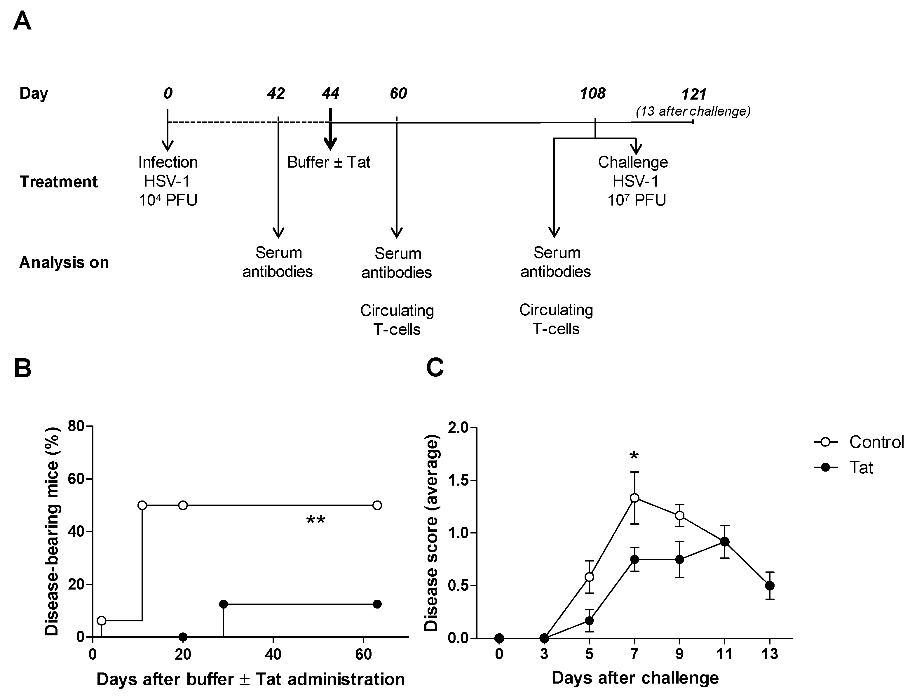
3.1. The HIV-1 Tat Protein Has a Therapeutic Effect in Mice Infected with HSV-1

Our previous studies have indicated that the simultaneous administration of the Tat protein with heterologous antigens improves both cellular and humoral immune responses against the antigens in in vitro and murine models [23,38]. However, in in vitro experiments, this effect was not observed when Tat was added to T cells after priming (i.e., during the expansion phase of the immune response) [40]. In agreement with these results, the administration of Tat to mice previously infected with HSV-1 7 days before, did not improve immune responses nor protection against a lethal challenge (data not shown). Since it has also been demonstrated in vitro that Tat exerts different effects on T cells depending on their activation status [41], we wondered whether Tat may affect the pre-existing adaptive memory immune responses. To test this hypothesis, C57BL/6 mice were infected intravaginally (IV) with a low dose (104 PFU) of wild-type HSV-1. At day 44 p.i., 5 µg of the HIV-1 Tat protein—or, in the control group, the equivalent volume of the buffer alone—was administered intradermally (ID) to mice (Figure 1A). Animals were then monitored up to day 108 p.i. (day 64 after Tat administration). Interestingly, we noticed a spontaneous appearance of signs such as vesicles and/or hair loss around the genital area, starting a few days after treatment. These disease signs, compatible with a viral reactivation, were significantly more frequent in controls than in Tat-treated mice (Figure 1B). Similar results were found when Tat was administered to mice previously infected with a lower dose of HSV-1 (103 PFU) (Figure S1). Further, all Tat-treated mice developed significantly milder signs of infection after an IV challenge with a high dose of HSV-1 (107 PFU) administered on day 108 post-infection. The difference in disease symptoms was particularly evident between 7 and 9 days after challenge before the disease signs spontaneously healed in both groups (Figure 1C).
Previous reports have shown that the transduction domain of Tat exhibits antiviral activities [42,43]. However, in our experiments we rule out a direct effect of Tat on HSV-1 virions, which might only occur in the very remote case of an ongoing subclinical reactivation at the time of treatment (day 44). Instead, we asked whether anti-Tat specific antibodies (Ab) could recognize and cross-react with HSV-1. Although anti-Tat Ab are detectable in only a fraction of HIV-infected individuals [44,45], they are effectively induced after administration of the Tat protein in humans [46,47,48] and animals [49,50,51]. Consistent with this, 100% of Tat-treated mice displayed anti-Tat IgG at both 60 and 108 days p.i. (16 and 64 days after Tat inoculation, respectively), with mean titers in the range of 104 (data not shown). To evaluate whether anti-Tat Ab hamper HSV-1 infectivity, an in vitro plaque reduction assay was performed by pre-incubating HSV-1 with sera of Tat- and/or HSV-1-immune mice. Anti-Tat Ab did not prevent or reduce HSV-1 infection of Vero cells, in contrast to what was observed using anti-HSV-1 immune sera, regardless of the presence of anti-Tat IgG (Figure S2), thus excluding non-specific effects of anti-Tat Ab on HSV-1 infectivity. Overall, these results indicate that the Tat protein exerts a therapeutic effect against latent HSV-1 infection and re-infection.

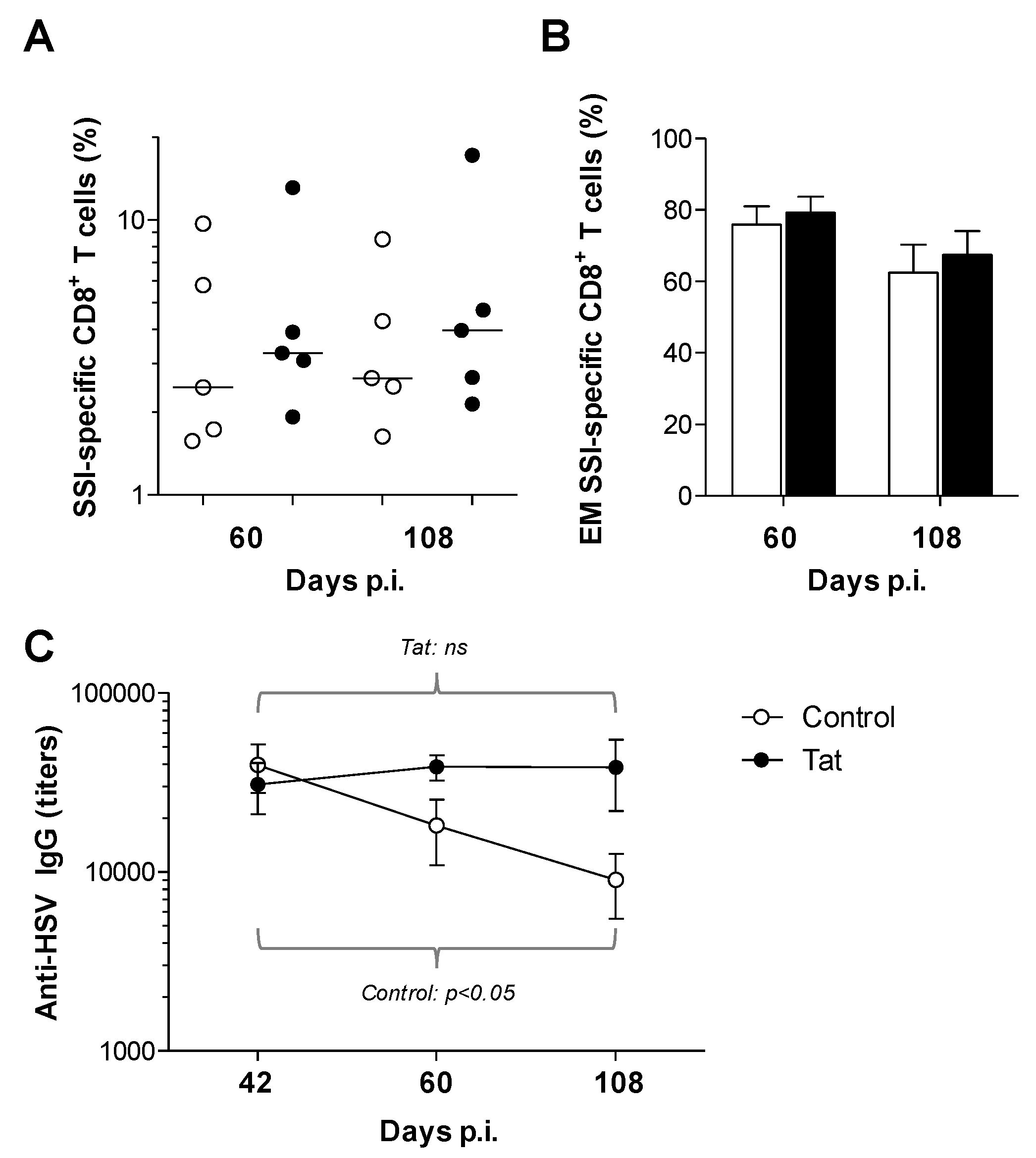
3.2. Administration of the HIV-1 Tat Protein Prevents the Time-Dependent Reduction in Antigen-Specific Adaptive Humoral Responses

We next assessed whether Tat may affect already established anti-HSV-1 immune responses. To this aim, the amount of circulating CD8+ T cells specific to the HSV-1 Kd-restricted SSI epitope were evaluated, since they play an important role in protection from HSV-1 infection [16,23]. Although not statistically significant, increased numbers of SSI-specific CD8+ T cells were observed in the blood of Tat-treated mice compared to that of control animals both at early (day 60 p.i.) and late (day 108 p.i.) time points after administration of Tat (Figure 2A). Previously, we have reported that Tat favors the late differentiation of T cells [38,40,52]. In addition, it has been shown that differentiated (e.g., effector memory) T cells are important correlates of protection in HSV-1 infection [16,17,18,19,53]. Thus, we characterized the phenotype of blood circulating SSI-specific CD8+ T cells. Consistent with the mild signs of disease observed in mice previously infected with 104 PFU and then challenged with 107 PFU of HSV-1 (Figure 1C), all animals showed a high proportion (60–80%) of effector memory cells (EM) within SSI-specific CD8+ T cells, regardless of their group (Figure 2B). In the very same mice, the proportion of EM cells within the whole CD8 compartment (regardless of the antigen specificity) was approximately 10–15% and directly correlated with the percentage of SSI-specific CD8+ T cells (p < 0.0001) (Figure S3A), suggesting that HSV-specific CD8+ T cells affect and drive the size of the EM subset. The frequency of EM SSI-specific CD8+ T cells, although slightly decreased over time, persisted at levels of above 60% in both groups (Figure 2B). Therefore, the long-term cellular response against HSV is mainly of EM type and Tat did not change this pattern. Considering the importance of EM T cells for local responses, it will be important, in future experiments, to measure the amount and phenotype of HSV-specific CD8+ T cells in mucosal tissues.
Surprisingly, while control mice experienced a physiological, time-dependent, decrease in HSV-specific IgG, the same was not found in Tat-treated mice which, instead, showed similar levels of HSV-specific IgG up to 108 days p.i. (Figure 2C). This indicates that Tat prevents the waning of circulating antibodies against a heterologous antigen such as HSV-1. It will require further research to determine whether this phenomenon is due to a direct effect of Tat on B cells or is T cell-dependent.
Very few studies, which are usually focused in the context of HIV-associated lymphomas [54,55,56], have investigated the direct effects of Tat on B cells. Instead, it has been widely reported that Tat exhibits potent immunomodulatory functions on antigen presenting cells [57,58], which may therefore be more prone to presenting antigens and providing costimulation signals in HSV reactivation. In addition, Tat can influence T-cell programming [38,40,52] and prevent apoptosis in T helper lymphocytes [59,60,61,62], whose role in maintaining and recalling humoral responses has also been demonstrated [63,64,65,66]. In line with the hypothesis of a strong interaction between cellular (CD8 and CD4) and humoral responses, we observed a direct correlation between HSV-specific IgG levels and the percentage of circulating SSI-specific CD8+ T cells (p = 0.009) (Figure S3B). Therefore, we may speculate that Tat effects on the T-cell compartment also influence humoral immunity. This phenomenon is shared by several pathogen-associated molecular patterns that, by modulating antigen presentation and costimulation to helper T cells, affect both cytotoxic and humoral responses [67,68]. Indeed Tat, further to inducing the release of IL-6 [69,70], which is important for B-cell maturation and T cell-dependent humoral responses [71,72], binds the toll-like receptor (TLR4) [73]. TLR4 signaling profoundly influences the functions of T and B lymphocytes [74,75,76] and may also rewire the metabolic programs of lymphocytes towards lipid usage, thus potentially impacting the persistence and functionality of memory cells, which are highly reliant on fatty acid oxidation [31,75]. We recently reported that Ab induced by a vaccine adjuvanted with a TLR4 ligand (TLR4L) lasted longer than those elicited by a similar vaccine that did not contain a TLR4L [77]. Although in this work, we administered Tat to mice after the antigen exposure and not simultaneously, we cannot rule out a general effect of TLR4L on plasma cell survival.
The results obtained from human vaccine studies have already shown that HIV-infected subjects vaccinated with Tat can improve memory adaptive responses to heterologous antigens [46]. The data presented here confirm this observation and also suggest that this phenomenon is probably due not only to the neutralization of the deleterious effects of Tat on HIV-infected subjects but also to the direct support on the maintenance of adaptive immunity mediated by Tat. Together, these data indicate that Tat can influence already established memory responses, preventing their physiological decline.

4. Conclusions

In this report, we have shown that the Tat protein of HIV exerts therapeutic effects in animals with a previously acquired HSV-1 infection. Our observations suggest that these effects are mediated by the maintenance of adaptive immunity, and in particular humoral responses, although the molecular mechanism remains unknown. Recently, non-specific effects exerted by some vaccines against heterologous antigens have been described, and this phenomenon was named “trained immunity” [78,79,80]. It is important to further investigate whether Tat displays similar mechanisms and whether its effects may also be exerted on other antigens. In addition, further studies on innate immune cells may help to identify the mechanism. However, these findings provide important clues to the use of Tat for the control of latent HSV-1.

Supplementary Materials

The following are available online at https://www.mdpi.com/2076-393X/8/2/274/s1, Figure S1: Tat exerts therapeutic effects on previously acquired HSV-1 infection, Figure S2: Anti-Tat antibodies do not interfere with HSV-1 replication, Figure S3: Correlation between HSV-1-specific CD8+ T cells, effector memory (EM) CD8+ T cells and HSV-1-specific humoral immunity.

Author Contributions

All authors have approved the submitted version of the manuscript. Contribution of the authors are as follows: conceptualization and preparation of study protocols, F.N., P.M., B.E., A.C. (Antonella Caputo), R.G.; methodology standardization and testing E.G., F.N., M.S., S.P.; formal Analysis of all data, FN., E.G., A.C. (Antonella Caputo), R.G.; writing–original draft preparation, F.N., A.C. (Antonella Caputo), R.G.; review and editing, F.N., E.G., M.S., A.C. (Aurelio Cafaro), B.E., P.M., A.C. (Antonella Caputo), R.G.; discussion of the results and of the final manuscript, F.N., A.C. (Aurelio Cafaro), B.E., P.M., A.C. (Antonella Caputo), R.G.; funding acquisition, P.M., R.G.

Funding

This work was funded by the University of Ferrara and PRIN2015 (P.M.).

Acknowledgments

We thank Anna Bergonzini, Noemi Vignoli, Matteo Mora and Andrea and Jack Montagnani for technical assistance.

Conflicts of Interest

The authors declare no conflict of interest.

References

  1. Looker, K.J.; Garnett, G.P. A systematic review of the epidemiology and interaction of herpes simplex virus types 1 and 2. Sex. Transm. Infect. 2005, 81, 103–107. [Google Scholar] [CrossRef]
  2. Mahant, S.; Hall, M.; Schondelmeyer, A.C.; Berry, J.G.; Kimberlin, D.W.; Shah, S.S.; for the Pediatric Research in Inpatient Settings Network and the Collaborative Antiviral Study Group; Pediatric Research in Inpatient Settings Network and the Collaborative Antiviral Study Group. Neonatal Herpes Simplex Virus Infection Among Medicaid-Enrolled Children: 2009–2015. Pediatrics 2019, 143, e20183233. [Google Scholar] [CrossRef] [Green Version]
  3. George, B.P.; Schneider, E.B.; Venkatesan, A. Encephalitis Hospitalization Rates and Inpatient Mortality in the United States, 2000–2010. PLoS ONE 2014, 9, e104169. [Google Scholar] [CrossRef] [Green Version]
  4. Saylor, D.; Thakur, K.; Venkatesan, A. Acute encephalitis in the immunocompromised individual. Curr. Opin. Infect. Dis. 2015, 28, 330–336. [Google Scholar] [CrossRef] [PubMed]
  5. Mancuso, R.; Sicurella, M.; Agostini, S.; Marconi, P.; Clerici, M. Herpes simplex virus type 1 and Alzheimer’s disease: Link and potential impact on treatment. Expert Rev. Anti Infect. Ther. 2019, 17, 715–731. [Google Scholar] [CrossRef] [PubMed]
  6. Wu, Y.P.; Sun, D.-D.; Wang, Y.; Liu, W.; Yang, J. Herpes Simplex Virus Type 1 and Type 2 Infection Increases Atherosclerosis Risk: Evidence Based on a Meta-Analysis. BioMed Res. Int. 2016, 2016, 1–9. [Google Scholar] [CrossRef] [PubMed] [Green Version]
  7. Looker, K.J.; Magaret, A.S.; May, M.T.; E Turner, K.M.; Vickerman, P.; Newman, L.M.; Gottlieb, S.L. First estimates of the global and regional incidence of neonatal herpes infection. Lancet Glob. Health 2017, 5, e300–e309. [Google Scholar] [CrossRef] [Green Version]
  8. Sivarajah, V.; Venus, K.; Yudin, M.H.; E Murphy, K.; A Morrison, S.; Tan, D.H.S. Does maternal HSV-2 coinfection increase mother-to-child transmission of HIV? A systematic review. Sex. Transm. Infect. 2017, 93, 535–542. [Google Scholar] [CrossRef] [Green Version]
  9. Esber, A.; Miguel, R.D.V.; Cherpes, T.L.; Klebanoff, M.A.; Gallo, M.F.; Turner, A.N. Risk of Bacterial Vaginosis among Women with Herpes Simplex Virus Type 2 Infection: A Systematic Review and Meta-Analysis. J. Infect. Dis. 2015, 212, 8–17. [Google Scholar] [CrossRef] [Green Version]
  10. Field, H.J.; Thackray, A.M. Early Therapy with Valaciclovir or Famciclovir Reduces But Does Not Abrogate Herpes Simplex Virus Neuronal Latency. Nucleotides Nucleic Acids 2000, 19, 461–470. [Google Scholar] [CrossRef]
  11. Piret, J.; Boivin, G. Resistance of Herpes Simplex Viruses to Nucleoside Analogues: Mechanisms, Prevalence, and Management. Antimicrob. Agents Chemother. 2010, 55, 459–472. [Google Scholar] [CrossRef] [PubMed] [Green Version]
  12. Gottlieb, S.L.; Giersing, B.K.; Hickling, J.; Jones, R.; Deal, C.; Kaslow, D.C. Meeting report: Initial World Health Organization consultation on herpes simplex virus (HSV) vaccine preferred product characteristics, March 2017. Vaccine 2019, 37, 7408–7418. [Google Scholar] [CrossRef]
  13. Xu, X.; Zhang, Y.; Li, Q. Characteristics of herpes simplex virus infection and pathogenesis suggest a strategy for vaccine development. Rev. Med. Virol. 2019, 29, e2054. [Google Scholar] [CrossRef] [Green Version]
  14. Shin, H.; Iwasaki, A. A vaccine strategy that protects against genital herpes by establishing local memory T cells. Nature 2012, 491, 463–467. [Google Scholar] [CrossRef] [PubMed] [Green Version]
  15. Liu, T.; Tang, Q.; Hendricks, R.L. Inflammatory infiltration of the trigeminal ganglion after herpes simplex virus type 1 corneal infection. J. Virol. 1996, 70, 264–271. [Google Scholar] [CrossRef] [PubMed] [Green Version]
  16. Nicoli, F.; Gallerani, E.; Skarlis, C.; Sicurella, M.; Cafaro, A.; Ensoli, B.; Caputo, A.; Marconi, P.; Gavioli, R. Systemic immunodominant CD8 responses with an effector-like phenotype are induced by intravaginal immunization with attenuated HSV vectors expressing HIV Tat and mediate protection against HSV infection. Vaccine 2016, 34, 2216–2224. [Google Scholar] [CrossRef] [PubMed]
  17. Khan, A.A.; Srivastava, R.; Lopes, P.P.; Wang, C.; Pham, T.T.; Cochrane, J.; Thai, N.T.U.; Gutierrez, L.; Benmohamed, L. Asymptomatic memory CD8+ T cells: From development and regulation to consideration for human vaccines and immunotherapeutics. Hum. Vaccines Immunother. 2014, 10, 945–963. [Google Scholar] [CrossRef]
  18. Khan, A.A.; Srivastava, R.; Spencer, D.; Garg, S.; Fremgen, D.; Vahed, H.; Lopes, P.P.; Pham, T.T.; Hewett, C.; Kuang, J.; et al. Phenotypic and Functional Characterization of Herpes Simplex Virus Glycoprotein B Epitope-Specific Effector and Memory CD8+T Cells from Symptomatic and Asymptomatic Individuals with Ocular Herpes. J. Virol. 2015, 89, 3776–3792. [Google Scholar] [CrossRef] [PubMed] [Green Version]
  19. Srivastava, R.; Khan, A.A.; Spencer, R.; Vahed, H.; Lopes, P.P.; Thai, N.T.U.; Wang, C.; Pham, T.T.; Huang, J.; Scarfone, V.M.; et al. HLA-A02:01-restricted epitopes identified from the herpes simplex virus tegument protein VP11/12 preferentially recall polyfunctional effector memory CD8+ T cells from seropositive asymptomatic individuals and protect humanized HLA-A*02:01 transgenic mice against ocular herpes. J. Immunol. 2015, 194, 2232–2248. [Google Scholar] [CrossRef] [PubMed] [Green Version]
  20. Liu, T.; Khanna, K.M.; Chen, X.; Fink, D.J.; Hendricks, R. Cd8+ T Cells Can Block Herpes Simplex Virus Type 1 (HSV-1) Reactivation from Latency in Sensory Neurons. J. Exp. Med. 2000, 191, 1459–1466. [Google Scholar] [CrossRef] [PubMed] [Green Version]
  21. Nagafuchi, S.; Oda, H.; Mori, R.; Taniguchi, T. Mechanism of Acquired Resistance to Herpes Simplex Virus Infection as Studied in Nude Mice. J. Gen. Virol. 1979, 44, 715–723. [Google Scholar] [CrossRef] [PubMed]
  22. Morrison, L.A.; Zhu, L.; Thebeau, L.G. Vaccine-Induced Serum Immunoglobin Contributes to Protection from Herpes Simplex Virus Type 2 Genital Infection in the Presence of Immune T Cells. J. Virol. 2001, 75, 1195–1204. [Google Scholar] [CrossRef] [PubMed] [Green Version]
  23. Sicurella, M.; Nicoli, F.; Gallerani, E.; Volpi, I.; Berto, E.; Finessi, V.; Destro, F.; Manservigi, R.; Cafaro, A.; Ensoli, B.; et al. An Attenuated Herpes Simplex Virus Type 1 (HSV1) Encoding the HIV-1 Tat Protein Protects Mice from a Deadly Mucosal HSV1 Challenge. PLoS ONE 2014, 9, e100844. [Google Scholar] [CrossRef] [PubMed] [Green Version]
  24. Levin, M.J. Immune senescence and vaccines to prevent herpes zoster in older persons. Curr. Opin. Immunol. 2012, 24, 494–500. [Google Scholar] [CrossRef]
  25. Levin, M.J.; Smith, J.G.; Kaufhold, R.M.; Barber, D.; Hayward, A.R.; Chan, C.Y.; Chan, I.S.F.; Li, D.J.J.; Wang, W.; Keller, P.M.; et al. Decline in Varicella-Zoster Virus (VZV)–Specific Cell-Mediated Immunity with Increasing Age and Boosting with a High-Dose VZV Vaccine. J. Infect. Dis. 2003, 188, 1336–1344. [Google Scholar] [CrossRef] [Green Version]
  26. Weinberger, B. Vaccines for the elderly: Current use and future challenges. Immun. Ageing 2018, 15, 3. [Google Scholar] [CrossRef]
  27. Rocca, S.; Santilli, V.; Cotugno, N.; Concato, C.; Manno, E.C.; Nocentini, G.; Macchiarulo, G.; Cancrini, C.; Finocchi, A.; Guzzo, I.; et al. Waning of vaccine-induced immunity to measles in kidney transplanted children. Medicine 2016, 95, e4738. [Google Scholar] [CrossRef]
  28. Cotugno, N.; Finocchi, A.; Cagigi, A.; Di Matteo, G.; Chiriaco, M.; Di Cesare, S.; Rossi, P.; Aiuti, A.; Palma, P.; Douagi, I. Defective B-cell proliferation and maintenance of long-term memory in patients with chronic granulomatous disease. J. Allergy Clin. Immunol. 2015, 135, 753–761. [Google Scholar] [CrossRef] [Green Version]
  29. Laksono, B.M.; De Vries, R.D.; Verburgh, R.J.; Visser, E.G.; De Jong, A.; Fraaij, P.L.A.; Ruijs, W.L.M.; Nieuwenhuijse, D.F.; Ham, H.-J.V.D.; Koopmans, M.P.G.; et al. Studies into the mechanism of measles-associated immune suppression during a measles outbreak in the Netherlands. Nat. Commun. 2018, 9, 4944. [Google Scholar] [CrossRef]
  30. Appay, V.; Van Lier, R.; Sallusto, F.; Roederer, M. Phenotype and function of human T lymphocyte subsets: Consensus and issues. Cytom. Part A 2008, 73, 975–983. [Google Scholar] [CrossRef]
  31. Nicoli, F.; Papagno, L.; Frere, J.J.; Cabral-Piccin, M.P.; Clave, E.; Gostick, E.; Toubert, A.; Price, D.A.; Caputo, A.; Appay, V. Naïve CD8+ T-Cells Engage a Versatile Metabolic Program Upon Activation in Humans and Differ Energetically From Memory CD8+ T-Cells. Front. Immunol. 2018, 9, 2736. [Google Scholar] [CrossRef] [PubMed] [Green Version]
  32. Goronzy, J.J.; Weyand, C.M. Successful and Maladaptive T Cell Aging. Immunity 2017, 46, 364–378. [Google Scholar] [CrossRef] [PubMed] [Green Version]
  33. Wertheimer, A.; Bennett, M.S.; Park, B.; Uhrlaub, J.; Martinez, C.; Pulko, V.; Currier, N.L.; Nikolich-Žugich, A.; Kaye, J.A.; Nikolich-Žugich, J. Aging and cytomegalovirus infection differentially and jointly affect distinct circulating T cell subsets in humans. J. Immunol. 2014, 192, 2143–2155. [Google Scholar] [CrossRef] [PubMed] [Green Version]
  34. Gil, A.; Yassai, M.B.; Naumov, Y.N.; Selin, L.K. Narrowing of Human Influenza A Virus-Specific T Cell Receptor α and β Repertoires with Increasing Age. J. Virol. 2015, 89, 4102–4116. [Google Scholar] [CrossRef] [PubMed] [Green Version]
  35. Papagno, L.; A Spina, C.; Marchant, A.; Salio, M.; Rufer, N.; Little, S.; Dong, T.; Chesney, G.; Waters, A.; Easterbrook, P.; et al. Immune activation and CD8+ T-cell differentiation towards senescence in HIV-1 infection. PLoS Biol. 2004, 2, E20. [Google Scholar] [CrossRef] [PubMed] [Green Version]
  36. Pensieroso, S.; Cagigi, A.; Palma, P.; Nilsson, A.; Capponi, C.; Freda, E.; Bernardi, S.; Thorstensson, R.; Chiodi, F.; Rossi, P. Timing of HAART defines the integrity of memory B cells and the longevity of humoral responses in HIV-1 vertically-infected children. Proc. Natl. Acad. Sci. USA 2009, 106, 7939–7944. [Google Scholar] [CrossRef] [Green Version]
  37. Kwon, H.S.; Brent, M.M.; Getachew, R.; Jayakumar, P.; Chen, L.F.; Schnolzer, M.; McBurney, M.W.; Marmorstein, R.; Greene, W.C.; Ott, M. Human Immunodeficiency Virus Type 1 Tat Protein Inhibits the SIRT1 Deacetylase and Induces T Cell Hyperactivation. Cell Host Microbe 2008, 3, 158–167. [Google Scholar] [CrossRef] [Green Version]
  38. Nicoli, F.; Finessi, V.; Sicurella, M.; Rizzotto, L.; Gallerani, E.; Destro, F.; Cafaro, A.; Marconi, P.; Caputo, A.; Ensoli, B.; et al. The HIV-1 Tat Protein Induces the Activation of CD8+ T Cells and Affects In Vivo the Magnitude and Kinetics of Antiviral Responses. PLoS ONE 2013, 8, e77746. [Google Scholar] [CrossRef] [Green Version]
  39. Bellino, S.; Francavilla, V.; Longo, O.; Tripiciano, A.; Paniccia, G.; Arancio, A.; Fiorelli, V.; Scoglio, A.; Collacchi, B.; Campagna, M.; et al. Parallel Conduction of the Phase I Preventive and Therapeutic Trials Based on the Tat Vaccine Candidate. Rev. Recent Clin. Trials 2009, 4, 195–204. [Google Scholar] [CrossRef] [Green Version]
  40. Sforza, F.; Nicoli, F.; Gallerani, E.; Finessi, V.; Reali, E.; Cafaro, A.; Caputo, A.; Ensoli, B.; Gavioli, R. HIV-1 tat affects the programming and functionality of human CD8+ T cells by modulating the expression of T-box transcription factors. AIDS 2014, 28, 1729–1738. [Google Scholar] [CrossRef]
  41. Nicoli, F.; Sforza, F.; Gavioli, R. Different expression of Blimp-1 in HIV infection may be used to monitor disease progression and provide a clue to reduce immune activation and viral reservoirs. AIDS 2015, 29, 133–134. [Google Scholar] [CrossRef] [PubMed]
  42. Bultmann, H.; Brandt, C.R. Peptides Containing Membrane-transiting Motifs Inhibit Virus Entry. J. Boil. Chem. 2002, 277, 36018–36023. [Google Scholar] [CrossRef] [PubMed] [Green Version]
  43. Jose, G.G.; Larsen, I.V.; Gauger, J.; Carballo, E.; Stern, R.; Brummel, R.; Brandt, C.R. A Cationic Peptide, TAT-Cd0, Inhibits Herpes Simplex Virus Type 1 Ocular Infection In Vivo. Investig. Opthalmology Vis. Sci. 2013, 54, 1070–1079. [Google Scholar] [CrossRef] [Green Version]
  44. Bellino, S.; Tripiciano, A.; Picconi, O.; Francavilla, V.; Longo, O.; Sgadari, C.; Paniccia, G.; Arancio, A.; Angarano, G.; Ladisa, N.; et al. The presence of anti-Tat antibodies in HIV-infected individuals is associated with containment of CD4+ T-cell decay and viral load, and with delay of disease progression: Results of a 3-year cohort study. Retrovirology 2014, 11, 49. [Google Scholar] [CrossRef] [PubMed] [Green Version]
  45. Nicoli, F.; Chachage, M.; Clowes, P.; Bauer, A.; Kowour, D.; Ensoli, B.; Cafaro, A.; Maboko, L.; Hoelscher, M.; Gavioli, R.; et al. Association between different anti-Tat antibody isotypes and HIV disease progression: Data from an African cohort. BMC Infect. Dis. 2016, 16, 344. [Google Scholar] [CrossRef] [Green Version]
  46. Ensoli, B.; Bellino, S.; Tripiciano, A.; Longo, O.; Francavilla, V.; Marcotullio, S.; Cafaro, A.; Picconi, O.; Paniccia, G.; Scoglio, A.; et al. Therapeutic Immunization with HIV-1 tat Reduces Immune Activation and Loss of Regulatory T-Cells and Improves Immune Function in Subjects on HAART. PLoS ONE 2010, 5, e13540. [Google Scholar] [CrossRef] [Green Version]
  47. Loret, E.P.; Darque, A.; Jouve, E.; Loret, E.A.; Nicolino-Brunet, C.; Morange, S.; Castanier, E.; Casanova, J.; Caloustian, C.; Bornet, C.; et al. Intradermal Injection of a Tat Oyi-based Therapeutic HIV Vaccine Reduces of 1.5 Log copies/mL the HIV RNA Rebound Median and No HIV DNA Rebound Following cART Interruption in a Phase I/II Randomized Controlled Clinical Trial. Retrovirology 2016, 13, 21. [Google Scholar] [CrossRef] [Green Version]
  48. Jin, H.; Li, D.; Lin, M.H.; Li, L.; Harrich, D. Tat-Based Therapies as an Adjuvant for an HIV-1 Functional Cure. Viruses 2020, 12, 415. [Google Scholar] [CrossRef] [Green Version]
  49. Alipour, S.; Mahdavi, A. Boosting Tat DNA Vaccine With Tat Protein Stimulates Strong Cellular and Humoral Immune Responses in Mice. Biotechnol. Lett. 2020, 42, 505–517. [Google Scholar] [CrossRef]
  50. Caputo, A.; Gavioli, R.; Bellino, S.; Longo, O.; Tripiciano, A.; Francavilla, V.; Sgadari, C.; Paniccia, G.; Titti, F.; Cafaro, A.; et al. HIV-1 Tat-Based Vaccines: An Overview and Perspectives in the Field of HIV/AIDS Vaccine Development. Int. Rev. Immunol. 2009, 28, 285–334. [Google Scholar] [CrossRef]
  51. Finessi, V.; Nicoli, F.; Gallerani, E.; Sforza, F.; Sicurella, M.; Cafaro, A.; Caputo, A.; Ensoli, B.; Gavioli, R. Effects of different routes of administration on the immunogenicity of the Tat protein and a Tat-derived peptide. Hum. Vaccines Immunother. 2015, 11, 1489–1493. [Google Scholar] [CrossRef] [PubMed] [Green Version]
  52. Nicoli, F.; Gallerani, E.; Sforza, F.; Finessi, V.; Chachage, M.; Geldmacher, C.; Cafaro, A.; Ensoli, B.; Caputo, A.; Gavioli, R. The HIV-1 Tat protein affects human CD4+ T-cell programing and activation, and favors the differentiation of naïve CD4+ T cells. AIDS 2018, 32, 575–581. [Google Scholar] [CrossRef] [PubMed]
  53. Svensson, A.; Nordström, I.; Sun, J.-B.; Eriksson, K. Protective immunity to genital herpes simplex [correction of simpex] virus type 2 infection is mediated by T-bet. J. Immunol. 2005, 174, 6266–6273. [Google Scholar] [CrossRef] [PubMed] [Green Version]
  54. Huang, L.; Li, C.J.; Pardee, A.B. Human Immunodeficiency Virus Type 1 TAT Protein Activates B Lymphocytes. Biochem. Biophys. Res. Commun. 1997, 237, 461–464. [Google Scholar] [CrossRef]
  55. Germini, D.; Tsfasman, T.; Klibi, M.; El-Amine, R.; Pichugin, A.; Iarovaia, O.V.; Bilhou-Nabera, C.; Subra, F.; Saada, Y.B.; Sukhanova, A.; et al. HIV Tat induces a prolonged MYC relocalization next to IGH in circulating B-cells. Leukemia 2017, 31, 2515–2522. [Google Scholar] [CrossRef]
  56. El-Amine, R.; Germini, D.; Zakharova, V.V.; Tsfasman, T.; Sheval, E.V.; Louzada, R.A.; Dupuy, C.; Bilhou-Nabera, C.; Hamade, A.; Najjar, F.; et al. HIV-1 Tat protein induces DNA damage in human peripheral blood B-lymphocytes via mitochondrial ROS production. Redox Biol. 2018, 15, 97–108. [Google Scholar] [CrossRef]
  57. Ben Haij, N.; Planès, R.; Leghmari, K.; Serrero, M.; Delobel, P.; Izopet, J.; BenMohamed, L.; Bahraoui, E. HIV-1 Tat Protein Induces Production of Proinflammatory Cytokines by Human Dendritic Cells and Monocytes/Macrophages through Engagement of TLR4-MD2-CD14 Complex and Activation of NF-κB Pathway. PLoS ONE 2015, 10, e0129425. [Google Scholar] [CrossRef] [Green Version]
  58. Fanales-Belasio, E.; Moretti, S.; Fiorelli, V.; Tripiciano, A.; Cossut, M.R.P.; Scoglio, A.; Collacchi, B.; Nappi, F.; Macchia, I.; Bellino, S.; et al. HIV-1 Tat Addresses Dendritic Cells to Induce a Predominant Th1-Type Adaptive Immune Response That Appears Prevalent in the Asymptomatic Stage of Infection. J. Immunol. 2009, 182, 2888–2897. [Google Scholar] [CrossRef] [Green Version]
  59. Gibellini, D.; Re, M.C.; Ponti, C.; Maldini, C.; Celeghini, C.; Cappellini, A.; La Placa, M.; Zauli, G. HIV-1 Tat Protects CD4+ Jurkat T Lymphoblastoid Cells from Apoptosis Mediated by TNF-Related Apoptosis-Inducing Ligand. Cell. Immunol. 2001, 207, 89–99. [Google Scholar] [CrossRef]
  60. Mischiati, C.; Pironi, F.; Milani, D.; Giacca, M.; Mirandola, P.; Capitani, S.; Zauli, G. Extracellular HIV-1 Tat protein differentially activates the JNK and ERK/MAPK pathways in CD4 T cells. AIDS 1999, 13, 1637–1645. [Google Scholar] [CrossRef]
  61. Zauli, G.; Gibellini, D.; Celeghini, C.; Mischiati, C.; Bassini, A.; La Placa, M.; Capitani, S. Pleiotropic effects of immobilized versus soluble recombinant HIV-1 Tat protein on CD3-mediated activation, induction of apoptosis, and HIV-1 long terminal repeat transactivation in purified CD4+ T lymphocytes. J. Immunol. 1996, 157, 2216–2224. [Google Scholar] [PubMed]
  62. Zauli, G.; Gibellini, D.; Milani, D.; Mazzoni, M.; Borgatti, P.; La Placa, M.; Capitani, S. Human immunodeficiency virus type 1 Tat protein protects lymphoid, epithelial, and neuronal cell lines from death by apoptosis. Cancer Res. 1993, 53, 4481–4485. [Google Scholar]
  63. Murera, D.; Arbogast, F.; Arnold, J.; Bouis, D.; Muller, S.; Gros, F. CD4 T cell autophagy is integral to memory maintenance. Sci. Rep. 2018, 8, 5951. [Google Scholar] [CrossRef] [PubMed]
  64. MacLeod, M.; Clambey, E.T.; Kappler, J.W.; Marrack, P. CD4 memory T cells: What are they and what can they do? Semin. Immunol. 2009, 21, 53–61. [Google Scholar] [CrossRef] [PubMed] [Green Version]
  65. Vieira, P.; Rajewsky, K. Persistence of memory B cells in mice deprived of T cell help. Int. Immunol. 1990, 2, 487–494. [Google Scholar] [CrossRef]
  66. Sarkander, J.; Hojyo, S.; Tokoyoda, K. Vaccination to gain humoral immune memory. Clin. Transl. Immunol. 2016, 5, e120. [Google Scholar] [CrossRef]
  67. Gutjahr, A.; Papagno, L.; Nicoli, F.; Kanuma, T.; Kuse, N.; Cabral-Piccin, M.P.; Rochereau, N.; Gostick, E.; Lioux, T.; Perouzel, E.; et al. The STING ligand cGAMP potentiates the efficacy of vaccine-induced CD8+ T cells. JCI Insight 2019, 4. [Google Scholar] [CrossRef]
  68. Gutjahr, A.; Tiraby, G.; Perouzel, E.; Verrier, B.; Paul, S. Triggering Intracellular Receptors for Vaccine Adjuvantation. Trends Immunol. 2016, 37, 716. [Google Scholar] [CrossRef]
  69. Gibellini, D.; Zauli, G.; Re, M.C.; Milani, D.; Furlini, G.; Caramelli, E.; Capitani, S.; La Placa, M. Recombinant human immunodeficiency virus type-1 (HIV-1) Tat protein sequentially up-regulates IL-6 and TGF-beta 1 mRNA expression and protein synthesis in peripheral blood monocytes. Br. J. Haematol. 1994, 88, 261–267. [Google Scholar] [CrossRef]
  70. Zauli, G.; Furlini, G.; Re, M.C.; Milani, D.; Capitani, S.; La Placa, M. Human immunodeficiency virus type 1 (HIV-1) tat-protein stimulates the production of interleukin-6 (IL-6) by peripheral blood monocytes. New Microbiol. 1993, 16, 115–120. [Google Scholar]
  71. Maeda, H.; Mehta, H.; Drevets, D.A.; Coggeshall, K.M. IL-6 Increases B-cell IgG Production in a Feed-Forward Proinflammatory Mechanism to Skew Hematopoiesis and Elevate Myeloid Production. Blood 2010, 115, 4699–4706. [Google Scholar] [CrossRef] [PubMed] [Green Version]
  72. Dienz, O.; Eaton, S.M.; Bond, J.P.; Neveu, W.; Moquin, D.; Noubade, R.; Briso, E.M.; Charland, C.; Leonard, W.J.; Ciliberto, G.; et al. The induction of antibody production by IL-6 is indirectly mediated by IL-21 produced by CD4+ T cells. J. Exp. Med. 2009, 206, 69–78. [Google Scholar] [CrossRef]
  73. Ben Haij, N.; Leghmari, K.; Planès, R.; Thieblemont, N.; Bahraoui, E. HIV-1 Tat protein binds to TLR4-MD2 and signals to induce TNF-α and IL-10. Retrovirology 2013, 10, 123. [Google Scholar] [CrossRef] [PubMed] [Green Version]
  74. Hua, Z.; Hou, B. TLR signaling in B-cell development and activation. Cell. Mol. Immunol. 2012, 10, 103–106. [Google Scholar] [CrossRef] [PubMed]
  75. Nicoli, F.; Paul, S.; Appay, V. Harnessing the Induction of CD8+ T-Cell Responses Through Metabolic Regulation by Pathogen-Recognition-Receptor Triggering in Antigen Presenting Cells. Front. Immunol. 2018, 9, 9. [Google Scholar] [CrossRef] [PubMed] [Green Version]
  76. Ganley-Leal, L.M.; Liang, Y.; Jagannathan-Bogdan, M.; Farraye, F.; Nikolajczyk, B.S. Differential regulation of TLR4 expression in human B cells and monocytes. Mol. Immunol. 2010, 48, 82–88. [Google Scholar] [CrossRef] [Green Version]
  77. Nicoli, F.; Mantelli, B.; Gallerani, E.; Telatin, V.; Bonazzi, I.; Marconi, P.; Gavioli, R.; Gabrielli, L.; Lazzarotto, T.; Barzon, L.; et al. HPV-Specific Systemic Antibody Responses and Memory B Cells are Independently Maintained up to 6 Years and in a Vaccine-Specific Manner Following Immunization with Cervarix and Gardasil in Adolescent and Young Adult Women in Vaccination Programs in Italy. Vaccines 2020, 8, 26. [Google Scholar] [CrossRef] [Green Version]
  78. Flanagan, K.L.; Van Crevel, R.; Curtis, N.; Shann, F.; Levy, O.; Network, O. Heterologous (“nonspecific”) and sex-differential effects of vaccines: Epidemiology, clinical trials, and emerging immunologic mechanisms. Clin. Infect. Dis. 2013, 57, 283–289. [Google Scholar] [CrossRef]
  79. Kleinnijenhuis, J.; Quintin, J.; Preijers, F.; Benn, C.S.; Joosten, L.A.B.; Jacobs, C.; Van Loenhout, J.; Xavier, R.J.; Aaby, P.; Van Der Meer, J.W.M.; et al. Long-lasting effects of BCG vaccination on both heterologous Th1/Th17 responses and innate trained immunity. J. Innate Immun. 2013, 6, 152–158. [Google Scholar] [CrossRef]
  80. Nicoli, F.; Appay, V. Immunological considerations regarding parental concerns on pediatric immunizations. Vaccine 2017, 35, 3012–3019. [Google Scholar] [CrossRef] [Green Version]
Figure 1. Tat exerts therapeutic effects on previously acquired Herpes Simplex Virus (HSV-1) infection. (A) Schematic representation of the experimental protocol and analysis. C57BL/6 mice were infected by the intravaginal (IV) route with 104 plaque forming units (PFU)/mouse of HSV-1 and, on day 44 post-infection (p.i.), treated with HIV-1 Tat (5 µg) administered by the intradermal (ID) route into the back. Controls were given only the Tat buffer. This treatment schedule was repeated in two independent experiments with 16 and 6 mice per group, respectively. Disease scores were monitored up to day 108 p.i. in both experiments. The following treatments and analyses were performed in the second experiment: On day 108 p.i. (i.e., day 64 after buffer ± Tat administration), mice were challenged IV with 107 PFU/mouse of HSV-1 and observed for two weeks after challenge (day 121 p.i.). Mice were bled on days 42 (before Tat treatment), 60 and 108 (before virus challenge) p.i. to analyze humoral and T cell responses. (B) Analysis of disease signs after Tat treatment. Mice were observed twice a week after Tat treatment to monitor the appearance of signs of disease. The Kaplan–Meier test was used to estimate the percentage of mice developing clinical manifestation after treatment (n = 16 per group). One representative experiment out of two is shown. (C) Analysis of signs of disease after HSV-1 challenge. Mice were observed every two days up to day 13 after challenge. For each group, mean disease scores (± SEM) are shown and analyzed statistically using the two-tailed Mann–Whitney test (n = 6 per group). * p < 0.05, ** p < 0.01.
Vaccines 08 00274 g001
Figure 2. Tat improves the maintenance of previously established immune memory responses. (A) Analysis of SSIEFARL (SSI)-specific CD8+ T cells. Blood was collected from the retro-orbital bleeding of mice (n = 5 per group) at days 60 and 108 p.i. (days 16 and 64 after buffer ± Tat inoculation, respectively) to determine the percentage of SSI-specific CD8+ T cells. Dots represent the results of single mice and lines represent median values. (B) Analysis of effector memory (EM) SSI-specific CD8+ T cells. The percentage of peripheral blood circulating EM (CD44+ CD62L) cells within the SSI-specific CD8+ T lymphocyte pool was measured at days 60 and 108 p.i. (days 16 and 64 after buffer ± Tat inoculation, respectively). Bars represent the mean ± SEM (n = 5 per group). (C) Analysis of anti-HSV-1 antibody responses. Sera were collected at days 42, 60 and 108 p.i. (two days before buffer ± Tat treatment, and days 16 and 64 after buffer ± Tat inoculation, respectively) to assess the presence and titers of HSV-specific IgG. Dots represent the mean ± SEM (n = 5 per group). A paired Student’s t test was used to estimate the changes over time in HSV-specific IgG titers. n.s. = not significant.
Vaccines 08 00274 g002

