Article

Identification by Reverse Vaccinology of Three Virulence Factors in Burkholderia cenocepacia That May Represent Ideal Vaccine Antigens

1
Department of Biology and Biotechnology “Lazzaro Spallanzani”, University of Pavia, 27100 Pavia, Italy
2
Department of Molecular Medicine, University of Pavia, 27100 Pavia, Italy
3
Imperial College South Kensington Campus, London SW7 2AZ, UK
4
GlaxoSmithKline, 53100 Siena, Italy
*
Author to whom correspondence should be addressed.
Vaccines 2023, 11(6), 1039; https://doi.org/10.3390/vaccines11061039
Submission received: 19 April 2023 / Revised: 24 May 2023 / Accepted: 26 May 2023 / Published: 30 May 2023

Abstract

:
The Burkholderia cepacia complex comprises environmental and clinical Gram-negative bacteria that infect particularly debilitated people, such as those with cystic fibrosis. Their high level of antibiotic resistance makes empirical treatments often ineffective, increasing the risk of worst outcomes and the diffusion of multi-drug resistance. However, the discovery of new antibiotics is not trivial, so an alternative can be the use of vaccination. Here, the reverse vaccinology approach has been used to identify antigen candidates, obtaining a short-list of 24 proteins. The localization and different aspects of virulence were investigated for three of them—BCAL1524, BCAM0949, and BCAS0335. The three antigens were localized in the outer membrane vesicles confirming that they are surface exposed. We showed that BCAL1524, a collagen-like protein, promotes bacteria auto-aggregation and plays an important role in virulence, in the Galleria mellonella model. BCAM0949, an extracellular lipase, mediates piperacillin resistance, biofilm formation in Luria Bertani and artificial sputum medium, rhamnolipid production, and swimming motility; its predicted lipolytic activity was also experimentally confirmed. BCAS0335, a trimeric adhesin, promotes minocycline resistance, biofilm organization in LB, and virulence in G. mellonella. Their important role in virulence necessitates further investigations to shed light on the usefulness of these proteins as antigen candidates.

1. Introduction

Burkholderia cepacia complex (Bcc) is a group of Gram-negative aerobic bacteria comprising 24 different species, isolated from both environmental and clinical settings [1,2]. While they can promote plant growth and provide protection from phytopathogens [3,4], they pose an important threat not only to people affected by cystic fibrosis (CF) and chronic granulomatous disease (CGD) [5], but even to patients exposed to other risk factors, such as diabetes or renal affections [6,7]. When lungs are involved, recurrent infections can lead to a progressive loss of functions, which will eventually produce episodes of pulmonary exacerbation [8], or even a diffuse systemic infection associated with a fatal and rapid necrotizing pneumonia, called “cepacia syndrome” [9]. A growing concern is represented by an increasing number of outbreaks caused by contaminated disinfectant solutions and medical devices, leading to large and multi-structure breakouts [10,11,12]. Choosing a proper antibiotic treatment is usually difficult and relies on circumstances, as empirical treatments often fail thus leading to unfavorable outcomes and eventually to the rise of further resistant strains [13]. In this light, the need for novel strategies to control these bacteria becomes mandatory, moving the attention from infection treatment to infection prevention [11,14].
Prophylactic vaccination appears to be a valuable strategy to protect fragile populations and minimize both transmission and antibiotic resistance spread, providing an alternative to expensive and burdensome drug treatments. Today, there is no vaccine available against Bcc [15], but many groups have focused on this issue, e.g., on live attenuated formulations. A B. cenocepacia tonB mutant was able to induce protection from acute and lethal infection in mice, with a survival of 87.5% after 6 days [16]. Conversely, higher efforts have been undertaken toward subunit vaccines, as multiple membrane proteins could be identified using bioinformatics tools and genome-available information. For example, a mixture of OMPs and a 17 kDa OmpA-like protein complexed with mucosal adjuvant were able to induce mucosal protection against Burkholderia multivorans and B. cenocepacia, while in some cases a balanced Th1/Th2 response was observed [17,18]. Furthermore, purified OmpA-like protein BCAL2958 was able to react to serum from CF patients infected with Bcc strains, leading to IgG production and an increase in neutrophils activation markers [19]. Finally, OmpW and linocin-immunized mice were able to induce both antigen-specific responses and cytokines production, conferring protection against B. cenocepacia and B. multivorans [20].
Regarding antigens’ discovery, a combination of protein prediction tool and surface-protein shaving approach led to the identification of 16 extracellular proteins [21]; 3 proteins reacted to serum from Bcc-infected CF patients, thus confirming their immunogenicity. BCAL2645, one of the three identified proteins, was later proven to be involved in CFBE41o- cells adhesion and invasion [22]. Similarly, Sousa et al., [23] identified nine extracellular proteins conserved inside Bcc by reacting culture supernatant to serum from chronically infected CF patients.
In another study, a peptidoglycan-associated lipoprotein deletion mutant was produced in B. cenocepacia [24] and showed a reduced mortality in Galleria mellonella, decreased adhesion to epithelial cells, sensitivity to polimyxin and lower proinflammatory stimulation compared to the WT, all traits making it a valuable therapeutic target.
Proteins belonging to trimeric autotransporter adhesin [TAA] category were characterized by Pimenta et al. [25] who proved the role of BCAM2418 in adhesion to mucins and host cells. An antibody generated against its N-terminal was able to prevent this interaction and to successfully protect G. mellonella larvae after B. cenocepacia K56-2 challenge. In addition, Mil-Homens et al. [26] characterized the TAA BCAM0224 as involved in biofilm formation and swarming motility, immunoevasion and interaction with epithelial cells.
In this article, we applied, for the first time, the “reverse vaccinology” approach [27] to B. cenocepacia to identify novel antigen candidates. Applying prediction tools and strains conservation analysis, we identified 24 new antigens and selected 2 of them for further analysis.

2. Materials and Methods

2.1. Bioinformatic Analysis

The complete genomes of the strains listed in Table 1 were annotated using the Rapid Annotation Subsystem Technology (RAST) tool [28]. The B. cenocepacia J2315 genome was compared with each analyzed genome and with the annotated genome of Escherichia coli K12.
Proteins were selected according to the following parameters: conservation among the compared Burkholderia strains had to be >65%; conservation into the non-pathogenic E. coli K12 strain had to be <30% to prioritize Bcc-specific proteins; length of the amino acid sequence had to be >230 residues to drive the selection toward multidomain and presumably multi-function antigens; and finally, proteins with an identity of 100% in all pathogenic strains were excluded, because they did not undergo immunity selective pressure [29]. Localization prediction was performed using pSORT [30]. The functional prediction was performed using InterProScan [31]. To exclude proteins with a homology similar to human ones, a BLAST analysis was used [32]. TMHMM, Phobius and HMMTOP were employed to predict transmembrane domains and to exclude proteins with more than one transmembrane domain [33,34,35]. Proteins were analyzed using Vaxign [36] and VaxiJen [37] and the antigenicity score values were accepted when ≥90.9 or ≥0.5, respectively. Structural information, if available, was collected from RCSB PDB [38].

2.2. Bacterial Strains and Plasmids

The strains used in this work are listed in Table S1. E. coli and B. cenocepacia strains were grown in Luria–Bertani (LB) broth (Difco) or in artificial sputum medium (ASM) [39], with shaking at 200 rpm, or on LB agar plates at 37 °C. When necessary, cultures were supplemented with antibiotics: tetracycline (20 μg/mL for E. coli and 250 μg/mL for B. cenocepacia), kanamycin (40 μg/mL), trimethoprim (50 μg/mL for E. coli and 200 μg/mL for B. cenocepacia), chloramphenicol (30 μg/mL for E. coli and 400 μg/mL for B. cenocepacia) and ampicillin (200 μg/mL).

2.3. General Molecular Biology Techniques

Upstream and downstream DNA sequences (about 500 bp each) flanking the deletion target genes were amplified using a template B. cenocepacia K56-2 genomic DNA. The PCR amplifications were performed using HotStar HiFidelity Polymerase kit (Qiagen, Hilden, Germany); primers sequences are listed in Table S2. Fragments were cloned into the suicide vector pGPI-SceI-Xcm BamHI/XbaI, using Gibson® assembly kit (NEB, Ipswich, MA, USA). Gene deletions were performed as previously described by Hamad et al. [40]. Deletions were confirmed via PCR amplification with the primers pairs listed in Table S2, and sequencing. The curing of the mutant strains was obtained by growth in LB medium. To complement each deleted strain, BCAL1524 (1674 bp), BCAM0949 (1099 bp) and BCAS0335 (3595 bp) genes were amplified using B. cenocepacia K56-2 DNA as template and the primers pairs are listed in Table S3. Fragments were cloned into the vectors pAP20 (EcoRI/XbaI) [41] and pSCrhaB2 (NdeI/XbaI) [42] using the Gibson® assembly kit. The complementation plasmids were introduced into the mutants by conjugation. The growth of all the mutants was measured by culturing the strains in LB or ASM and measuring the OD600 and plating the cultures every 60 min for 24 h. Complementation experiments with the inducible vector pSCrhaB2 were carried out with 0.01% of rhamnose.

2.4. Proteomic Analysis

B. cenocepacia mutant strains were grown in 50 mL of LB medium or ASM for 18 h. Cells were harvested and pellets were used for membrane extraction, while supernatants were used for outer membrane vesicles (OMVs) purification. Membrane fractions were prepared as previously described [43]. For OMV purification, supernatants were collected and, to completely remove the bacterial cells from the supernatant, they were filtered through a 0.45 μm cellulose membrane. The cell-free supernatants were ultrafiltrated using a centrifugal filter Amicon® (Merck Millipore, Burlington, MA, USA) with 100,000 Da NMWL, until a final volume of 1 mL, removing the non-OMV-associated proteins, such as flagella [44].
The analysis of the membrane fractions and of the OMVs was performed as previously described, via 2D gel electrophoresis [45]. Samples were precipitated with 10:1 2.7 M trichloroacetic acid, incubated in ice for 30′ and centrifuged for 15′ at 14,000 rpm. Pellets were then resuspended in 125 µL of 8M Urea, 4% CHAPS, 0.1 M Dithioerythritol (UCD), and 0.625 µL of 0.5% bromophenol blue and 0.7 µL of IPG buffer pH 3–10 (GE-Healthcare, Chicago, IL, USA) were added. Samples were then incubated for 1 h on an isoelectric focusing strip (pH 3–10). Isoelectric focusing was performed at 20 °C according to the program described in Table 2.
Next morning, strips were equilibrated for 12 min with buffer A (6 M Urea, 2% SDS, 50 mM TRIS-HCl pH 6.8, 30% Glycerol, and 0.1 M Dithioerythritol) and then for 5 min in buffer B (6 M Urea, 2% SDS, 50 mM Tris-HCl pH 6.8, 30% Glycerol, 0.1 M Iodoacetamide, and 125 µL of 0.5% bromophenol blue). Mass weight separation (second dimension) was performed on a polyacrylamide gel formed by a 12.5% running gel and a 5% stacking gel. Briefly, 0.5% agarose was dissolved in running buffer to immobilize the strips.
Gels were digitalized using the ChemiDoc XRS system (Biorad, Hercules, CA, USA); spots were detected using PDQuest Advanced 8.0.1 program (Biorad, Hercules, CA, USA) [46]. Experiments were performed in triplicate.

2.5. Antimicrobial Susceptibility Testing for Planktonic Cells

MICs were determined in triplicate according to the EUCAST broth microdilution method [47] in U-bottom 96-well microtiter plates using LB and ASM. Nalidixic acid, amikacin, aztreonam, ciprofloxacin, minocycline, piperacillin and tobramycin were obtained from Sigma-Aldrich (Merck Millipore, Burlington, MA, USA). Levofloxacin and sparfloxacin were obtained from Honeywell Fluka™ (Charlotte, NC, USA) and Meropenem from AstraZeneca (Cambridge, UK). The MIC was determined via the resazurin method [48].

2.6. Bacterial Autoaggregation Assay

Bacterial aggregation was measured according to Bhargava et al. [49], with some modifications. Bacterial cultures were grown in LB broth, at 37 °C and 200 rpm, up to the late-exponential phase. Samples were centrifuged, cells were resuspended in Phosphate Buffer Saline (PBS) (PanReac-Applichem, Darmstadt, Germany) and optical density at 600 nm was measured and adjusted to 3. Samples were incubated at room temperature for 16 h in static conditions. After incubation, 50 µL were removed from the air–liquid interface and the OD600 was measured. The aggregation was expressed as the percentage respect to the starting OD600 = 3.

2.7. In Vitro Biofilm Formation Test in 96-Well Microtiter Plates

The biofilm formation of B. cenocepacia K56-2, mutant and complemented strains was tested using the crystal violet staining method [50]. The bacterial cells were cultured in LB or ASM O/N at 37 °C and diluted to OD600 equal to 0.05 (about 1 × 10⁷ CFU/mL). Then, 200 μL of culture were pipetted into the microtiter plate. After 4 h of incubation, the supernatant (containing nonadherent cells) was removed and 200 μL of fresh sterile medium were added to each well and incubated for an additional 20 h at 37 °C. Biofilm biomass was quantified by staining with crystal violet and taking absorbance measurements at OD595. Results were expressed as the ratio between biofilm absorbance and planktonic bacteria absorbance.

2.8. Biofilm Evaluation by Confocal Laser Scanning Microscopy

B. cenocepacia K56-2 and the ΔBCAM0949 strains were cultured O/N in LB and diluted to an OD600 = 0.05 in the same medium. Bacterial suspension was added to the μSlide four chambered coverslip (Ibidi, Gräfelfing, Germany) for 4 h in LB, at 37 °C. The medium was removed and fresh LB medium was added. After overnight incubation, the medium was removed, and biofilms were washed twice with physiological solution and stained with Syto 9 (Invitrogen, Waltham, MA, USA) at a final concentration of 5 μM. A 63× oil immersion objective and a Leica (Wetzlar, Germany) DMi8 with 500 to 530 nm (green fluorescence representing Syto 9) emission filters were used to take five snapshots randomly at different positions in the confocal field of each chamber. The Z-slices were obtained every 0.3 microns. For visualization and processing of biofilm images, ImageJ was used. The thickness, biomass, roughness coefficient, and biofilm distribution were measured using the COMSTAT 2 software [51]. All confocal scanning laser microscopy experiments were performed three times, and standard deviations were measured.

2.9. Swimming Motility Assay

Motility assays were performed as previously described [52]. Briefly, 1 μL of the overnight culture was spotted in the middle of a swimming plate (agar 0.3%), allowed to dry for 30 min at room temperature and incubated for 16 h at 37 °C. Diameters of swimming halos were measured.

2.10. Infection in Galleria Mellonella

Strains were grown in LB broth, at 37 °C, 200 rpm, until OD600 = 0.5. Each larva was then infected with 105 CFU/mL in physiological solution (PS) [53], with an injection volume of 10 µL; control was performed by injecting PS. Larvae were placed in Petri dishes and kept in the dark at 30 °C. Live/dead count and health index scores [53,54] were registered after 24 h, 48 h and 72 h. For each experiment, at least 10 larvae per group were injected.

2.11. Lipase Activity Assay

Tributyrin agar plates (LB medium, 0.5% tributyrin, and 1.5% agar) were used for lipase activity detection [55]. Halos formed by the lipase activity around colonies were measured.

2.12. Rhamnolipid Analysis

Strains were inoculated in 10 mL of LB for 48 h at 37 °C at 200 rpm. To quantify the amount of rhamnolipids in the culture supernatant, the colorimetric orcinol assay was used [56]. A volume of the supernatant (50 to 500 μL) was diluted with water to reach the volume of 500 μL. Samples were extracted twice with 2 volumes of diethyl ether. The ether fractions were evaporated and the pellets were dissolved in 100 μL of distilled water and mixed with 100 μL 1.6% orcinol and 800 μL of 60% sulfuric acid. Samples were incubated at 80 °C at 175 rpm for 30 min. After incubation, the OD421 was measured. The rhamnolipid concentration was calculated using a standard concentration curve with rhamnose standards on the assumption that 1 μg of rhamnose corresponds to 2.5 μg of rhamnolipid [57].

2.13. Statistical Methods

Analyses were performed using Prism 9.0 (GraphPad). Comparison of more than two groups were performed with the unpaired t-test, one-way or two-way ANOVA.

3. Results and Discussion

3.1. In Silico Identification of Antigen Candidates

The identification of antigen candidates was performed using the “reverse vaccinology” approach [27]. The scheme of the workflow is depicted in Figure 1. The genome of B. cenocepacia J2315 was used as reference [58], and annotated with the Rapid Annotation using Subsystem Technology (RAST) pipeline, a fully automated annotation engine for archaeal and bacterial genomes [28]. RAST identifies protein coding genes, assigns functions and gives a prediction of subsystems present in the genomes.
The annotation of B. cenocepacia J2315 genome provided 7769 protein encoding genes as an output. Each genome was compared with the annotated genome of E. coli K-12 and with the genomes of six B. cenocepacia strains and eighteen Bcc strains (Table 1). The parameters to select putative antigen candidates were as follows: identity with E. coli K-12 proteins less or equal to 30% and identity with the proteins of B. cenocepacia and Bcc strains higher than 65%. Since immunogenic proteins are generally localized on the outer membrane and have more complex structures created by multiple domains, (e.g., transmembrane and extracellular domains), only proteins longer than 230 amino acids were selected. Using these parameters, a pool of 1793 proteins was obtained and analyzed for subcellular localization and putative function. The pool was reduced to 122 candidates predicted to be outer-membrane or extracellular, potentially involved in cell adhesion, virulence or with unknown function. Further analyses were performed to evaluate the transmembrane domains prediction, the homology with human proteins and the prediction of the antigenicity of the candidates as described in the “Materials and methods” section. The result is a short list of 24 antigen candidates belonging to different protein classes: 4 lipoproteins, 4 autotransporter adhesin proteins, 5 enzymes, 5 outer membrane proteins and 6 other proteins (Table 3, Figure 1).
Among them, BCAM0949, BCAM2418, BCAS0409 and BCAS0236 were previously described as being involved in virulence and cell adhesion [25,59,60,61]. BCAS0147 was described as surface exposed [21]; BCAM1514 and BCAS0409 were localized in the outer membrane fraction in the CF niche [62].
Three antigens, the collage-like protein BCAL1524, the lipase LipA BCAM0949 and the autotransporter adhesin BCAS0335 were selected for further analysis. The rationale for the selection is described below. Collagen-like proteins, characterized by a collagen-like (CL) domain containing Gly-Xaa-Yaa amino acid repetition and organized in a triple helix-structure, are widespread in pathogenic bacteria and highly resemble human collagen [63]. Due to their structure, they can interact with different host factors promoting adhesion, inflammation and immunoreaction [64,65]. Additionally, Grund et al., [66] described the high Th2 immune response induced in mice immunized with B. pseudomallei collagen-like protein 8 (Bucl8) antigens, while studies on the role of these proteins in B. cenocepacia are still lacking. Lipases are commonly found in clinical B. cepacia complex isolates [67]. While evidence of their involvement in Bcc infection is still nominal [67,68], multiple studies reported their role in virulence and pathogenesis in P. aeruginosa [56,69,70]. Trimeric autotransporter adhesins are known to play a key role in virulence for a wide range of bacteria [71]; Pimenta et al. [25] and Mil-Homens et al. [26,72] extensively described their role in adhesion and inflammation in epidemic B. cenocepacia epidemic strain K56-2, thus highlighting the putative role they may play as protective antigens.

3.2. Construction of Deletion Mutants of the Selected Antigen Candidates

To investigate the roles of these proteins in virulence and host pathogen interaction, deletion mutants were constructed in B. cenocepacia K56-2, since the amino acid sequence of the corresponding proteins was identical to J2315 and this strain is most amenable to genetic manipulation. The growth of the mutants and complemented strains was comparable to that of the WT K56-2 both in LB and in ASM media (Figure S1).

3.3. Analysis of Protein Localization

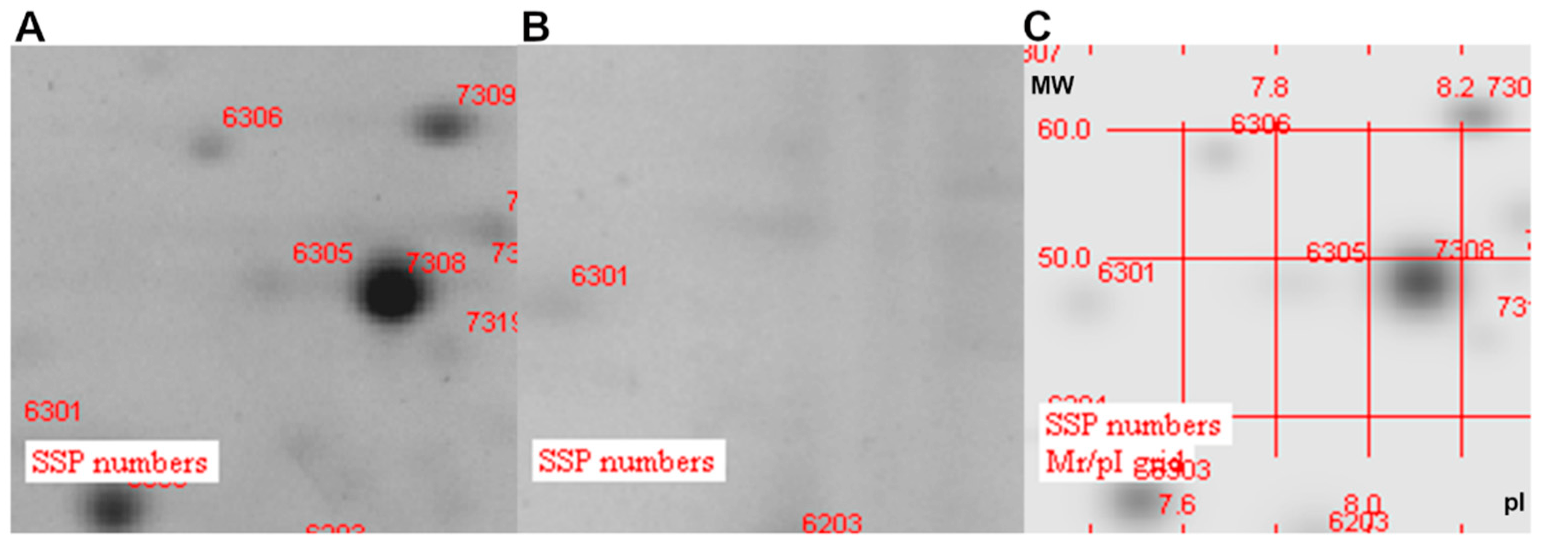
To demonstrate that the antigen candidates selected are localized on the bacterial surface, a proteomic analysis of Outer Membrane Vesicles (OMV) derived from K56-2 and deleted strains was performed. As shown in Figure 2, when analyzed via the 2D electrophoresis, in the OMVs of the ΔBCAL1524 isogenic mutant strain, no spots were identified at the coordinate value corresponding to the BCAL1524 protein (isoelectric point 8.55 and molecular weight 49 kDa) (Figure 2B). In the OMVs of the K56-2 wild-type strain, a spot numbered 7308 with intensity of 27,944.8 mAu was identified at these coordinates (Figure 2A,C).
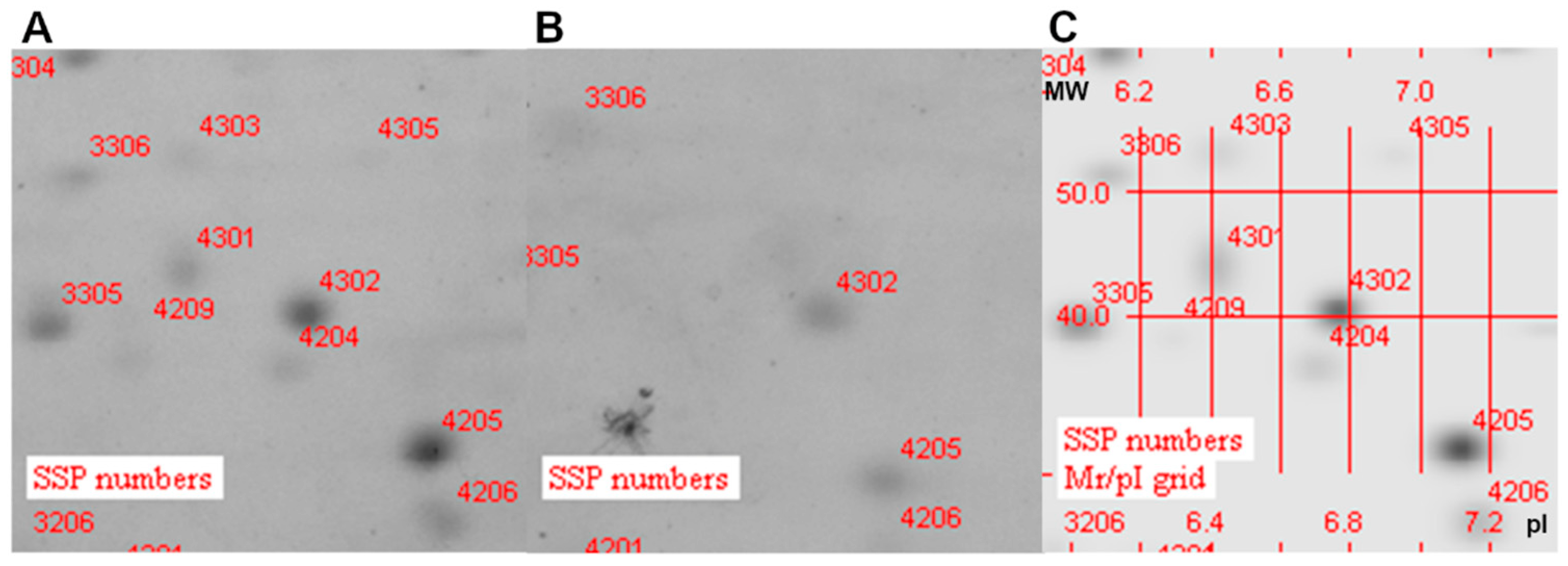
The same applied to the OMV of ∆BCAM0949 and K56-2 strains electrophoresis of the OMVs of the K56-2, in which a spot numbered 4301 was identified at pI~6.4 and MW~42 kDa and intensity 3399.7 mAu, which was not present in the ∆BCAM0949 mutant OMVs (Figure 3). The MW and pI values of BCAM0949 are 38 kDa and 6.41, respectively.
Finally, the OMVs 2D-electrophoresis of the K56-2 showed the spot number 3701 at pI 5.3 and MW 124 kDa with an intensity equal to 4156 mAu, lacking in the ΔBCAS0335 (Figure 4). The theoretical pI and MW of the BCAS0335 are 5.0 and 117 kDa, respectively.
All these data demonstrate that the collagen-like protein BCAL1524, the lipase BCAM0949 and the trimeric-autotransporter adhesion BCAS0335 are expressed in the outer membrane vesicles compartment in the conditions tested. Two-dimensional gel electrophoresis full images are shown in Supplementary Materials (Figures S2–S5).

3.4. Antibiotic Susceptibility

To test the involvement of the selected proteins in antibiotic susceptibility, the minimal inhibitory concentration of 12 currently used antibiotics was determined against the deleted and complemented strains grown in LB. Piperacillin had an MIC which was four-fold lower in the ΔBCAM0949 strain with respect to the K56-2 in LB (32 μg/mL vs. 128 μg/mL) (in bold in Table 4). The deleted strain, complemented with the WT copy of the gene cloned into the rhamnose inducible vector pSCrhaB2, showed a reverted phenotype.
The MIC of minocycline was eight-fold higher in the ΔBCAS0335 strain than the K56-2 (64 μg/mL vs. 8 μg/mL) (in bold in Table 4). In this case, the complementation using the inducible vector pSCrhaB2 restored the phenotype of the K56-2 strain.
The MICs of the control strains carrying the empty pSCrhaB2 vector are listed in Table S4. It is to be noted that the presence of the empty vector did not affect strain phenotypes.
The MICs value for the ΔBCAL1524 did not show differences compared to the K56-2 in the conditions tested.

3.5. Phenotypic Characterization of the Deleted Mutant

3.5.1. Biofilm Formation

To assess the role of the three antigen candidates in sessile lifestyle, biofilm formation was evaluated using crystal violet assay in 96-well plates after 48 h of incubation. The results showed that the ∆BCAM0949 mutant strain produces a lower quantity of biofilm in both LB and ASM media with respect to the WT (Figure 5A,B). The ∆BCAS0335 mutant produced a slightly lower quantity of biofilm only in LB, compared to the K56-2 (Figure 5A). Complementation was carried out using the pAP20 constitutive vector. The complemented strains showed the reversion of the ∆BCAM0949 phenotype in LB and ASM (Figure 5C,D). Regarding the ∆BCAS0335 mutant, the mutant transformed with the empty vector did not show a significant decrease in biofilm production (Figure S6), but in the deleted strain the constitutive expression of the BCAS0335 gene significantly increased the biofilm production in both the media tested (Figure 5C,D). The data suggest that both the lipase BCAM0949 and the autotransporter adhesin protein BCAS0335 contribute to biofilm formation. On the contrary, the collagen-like protein does not seem to have a role in this process in the condition tested. The data regarding the biofilm production in the control strains of the complementation carrying the empty pAP20 vector are reported in Figure S6.
Since the difference in biofilm formation with respect to the K56-2 strain was higher in the lipase mutant, we decided to evaluate the biofilm formed by this strain using confocal laser scanning microscopy (CLSM). The obtained imaging showed that the biofilm formed by the ∆BCAM0949 mutant is less structured and thick in both conditions tested (LB and ASM) (Figure 6A).
The analysis using COMSTAT2 confirmed the qualitative evaluation of the biofilm. Biofilms formed in LB and ASM by the ∆BCAM0949 strain have a significantly lower biomass and average thickness (Figure 6B,C). Moreover, the roughness of the biofilm is significantly higher for the deleted strain (Figure 6B,C), indicating that the structure of the biofilm is altered.
All these data suggest that the lipase BCAM0949 has a role in biofilm formation and structure in the conditions tested.

3.5.2. Bacterial Autoaggregation

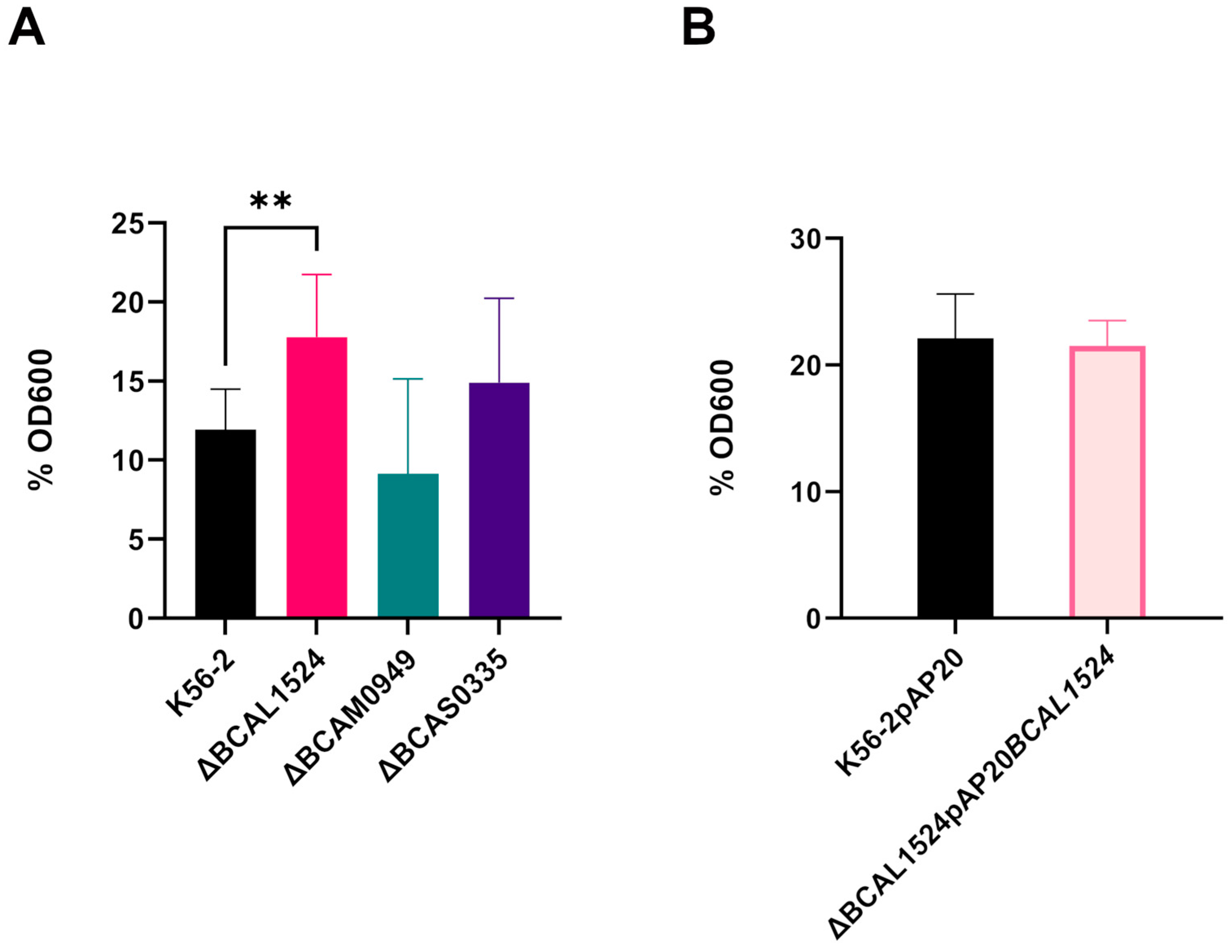
The involvement of the antigen candidates in bacterial autoaggregation, a process known to be involved in bacterial colonization and persistence in the host, was assessed using a precipitation-based assay. The decrease in the OD600 of the air–liquid interface after 16 h of static incubation is consistent with an increase in bacterial interaction, which promotes precipitation; on the contrary, if interactions are impaired, the OD600 is higher. The autoaggregation is expressed as the percentage of OD600 of the air–liquid interface with respect to the initial OD600 normalized to 3. For the K56-2 strain, the obtained value was 11.9%, while in the case of the ∆BCAL1524, ∆BCAM0949 and ∆BCAS0335 mutant strains it was 17.7%, 9.13% and 14.8% (Figure 7A), respectively.
When BCAL1524 protein was lacking, autoaggregation decreased as the percentage of residual OD600 after 16 h was increased. The proteins BCAM0949 and BCAS0335 are not involved in autoaggregation in the conditions tested.
The complementation with the pAP20BCAL1524 vector restored a phenotype identical to the K56-2 (Figure 7B).

3.5.3. Swimming Motility

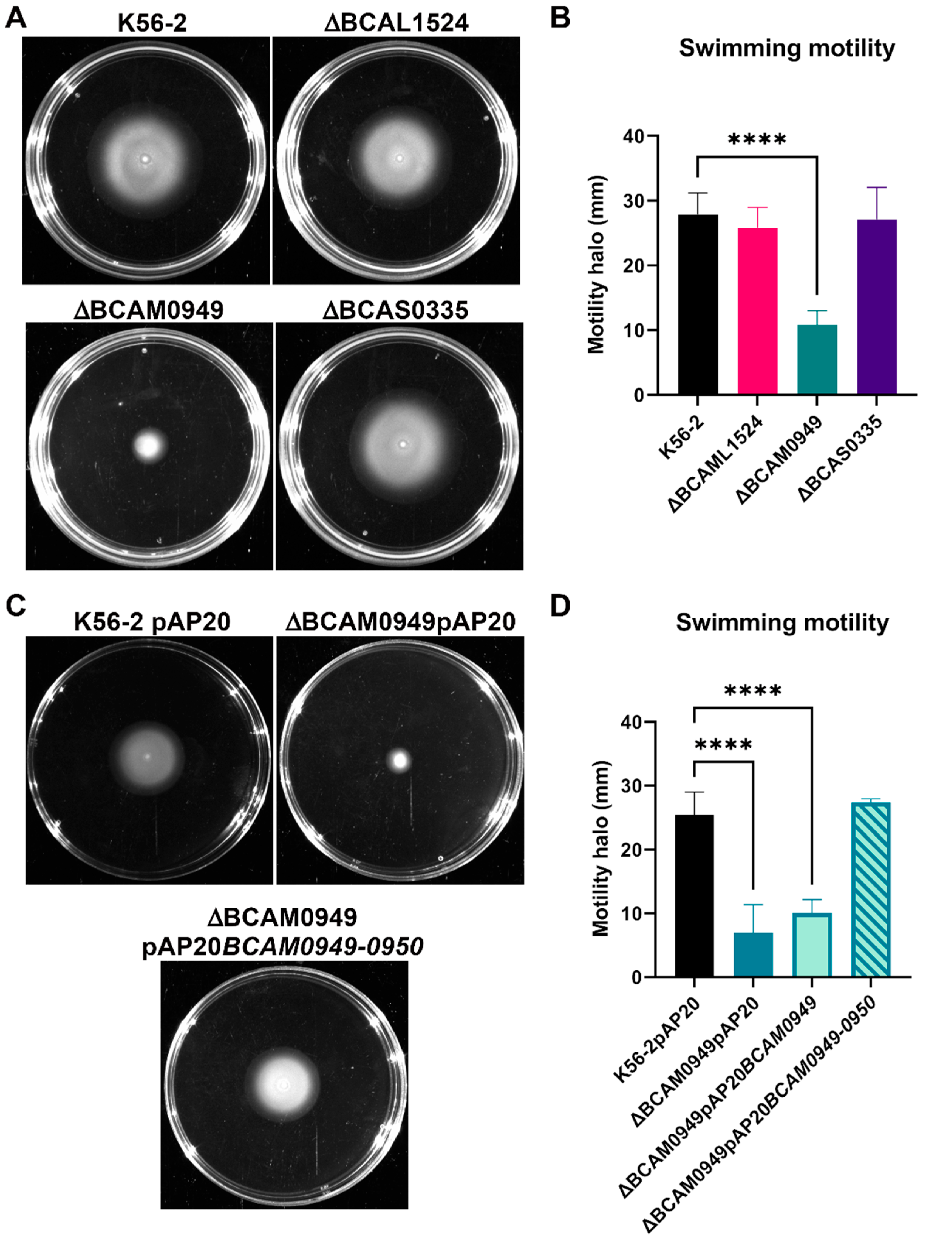
Swimming motility allows bacteria to diffuse through low viscosity medium, such as mucus, and it is considered a virulence determinant. Swimming motility of the deleted strains was tested on 0.3% agar plates. In the ∆BCAM0949 mutant strain, the swimming phenotype was abolished (diameter of the motility halo 10.8 mm) compared to the K56-2 (27.8 mm) (Figure 8A,B). On the other hand, the two deleted mutant strains, ∆BCAL1524 and ∆BCAS0335, showed no differences compared to the K56-2 (Figure 8A,B).
The complementation was carried out using the entire BCAM0949-BCAM0950 operon cloned into the vector pAP20 as the phenotype was not restored by expressing the only LipA (BCAM0949) protein. As reported by Papadopoulos et al. [73] and Putra et al. [74], P. aeruginosa LipA and Burkholderia territori LipBT lipases have to be co-expressed with the relative foldase to avoid precipitation and to allow increased functional protein concentration, which would be required to complement some phenotypes. Indeed, upon complementation with the pAP20BCAM0949-0950 construct, the swimming halo was restored to the diameter of the K56-2 (Figure 8C,D). The data suggest that while the proteins BCAL1524 and BCAS0335 are not involved in swimming motility, the lipase BCAM0949 has a pivotal role in this pathway in the conditions tested.

3.6. Infection in Galleria mellonella

G. mellonella larvae are a well-known invertebrate animal model for infection, as they provide a simple, fast, and cost-effective platform. After an infection with a dose of 103 CFU bacteria, symptoms started to appear 24 h post-infection (Figure 9A,B): at this time point, larvae infected with the ∆BCAL1524 and ∆BCAS0335 mutant strains showed a significantly higher health index score compared to the larvae infected with the K56-2. These larvae showed a lower melanization level than the larvae infected with the K56-2 (Figure 9B). On the contrary, infection with the ∆BCAM0949 mutant did not affect larval survival and health index scores compared to what was observed in larvae infected with the WT strain (K56-2).
To complement the observed phenotypes, the pAP20 vector was used. Complementation was achieved in the case of ∆BCAL1524 (8.15) and ∆BCAS0335 (7.9) as the K56-2 symptom score at 24 h post-infection was 7.56 (Figure 9C). The data regarding the health index score of G. mellonella moths infected with the control strains of the complementation carrying the empty pAP20 vector are reported in Figure S7.
The data obtained suggest that the proteins BCAL1524 and BCAS0335 are involved in virulence in vivo, since their lack increases the health score of larvae injected with the corresponding deleted strains; thus, host immunization using these two antigens could provide protection from severe infection. On the contrary, deletion of BCAM0949 does not affect the in vivo virulence of the bacterium.

3.7. Characterization of the Lipase BCAM0949

3.7.1. Lipolytic Activity

Although the deletion of BCAM0949 has no effect on the in vivo virulence of Burkholderia, its amino acid sequence is characterized by a conserved domain of the triacylglycerol esterase/lipase superfamily, so we further characterized its activity. In other bacteria, such as Pseudomonas aeruginosa, several lipolytic enzymes are secreted or surface-exposed; among these is the EstA, which has a lipolytic activity, and is involved in rhamnolipid production and virulence [57]. To better characterize the lipase BCAM0949, the lipolytic activity of the deleted strain was compared to the K56-2 using the tributyrin agar plate assay (Figure 10).
The diameter of the lipolytic halo was measured and, as shown in Figure 10, the mutant strain has a lower lipolytic activity than the K56-2. The lipolytic activity is not completely blocked because the bacterium possesses other lipolytic enzymes. The complementation restored the lipolytic activity (Figure 10).

3.7.2. Rhamnolipid Production

In P. aeruginosa, EstA is involved in rhamnolipid production [57]. To study the involvement of the lipase BCAM0949 in rhamnolipid production, the quantity of rhamnolipids produced by the deleted strain was compared to the K56-2, showing that the deleted strain ∆BCAM0949 is impaired in their production (Figure 11).
Complementation with the pAP20BCAM0949 vector not only restored the K56-2 phenotype, but induced an increase in rhamnolipids production (Figure 11). These results demonstrate the involvement of the protein BCAM0949 in rhamnolipid production.
All these data showed how the protein BCAM0949 has a role in the virulence pathway of rhamnolipid production. Since rhamnolipids play a role in outer membrane composition, cell motility and biofilm formation in other bacteria, BCAM0949 is also demonstrated to be involved in different virulence pathways.

4. Conclusions

In this work, reverse vaccinology was used to identify surface-exposed proteins which can be ideal antigen candidates for the development of a vaccine against the B. cepacia complex bacteria. We chose this strategy that previously revealed to be resolutive in cases for which the discovery of a vaccine seemed to be impossible [75]. It represents therefore a valuable option in the case of Burkholderia for which, despite numerous attempts to find suitable antigen candidates, a vaccine is not yet available. Starting from the bacterial genome, a short list of gene candidates, encoding extracellular proteins predicted to be ideal targets for the immune system, was selected. Among the 24 proteins identified, 3 of them were selected because of their homology with important virulence factors in other bacteria: BCAL1524, a lipoprotein containing a collagen-like triple helix repeat, able to interact with host cell adhesive proteins; BCAM0949, an extracellular lipase; BCAS0335, a trimeric autotransporter adhesin (TAAs) which interacts with the extracellular membrane components and host cell receptors and possesses hemagglutinin activity. Antigens used to develop vaccines do not necessarily have to be virulence factors, however, directing the immune response toward virulence determinants has been a successful rationale in eliciting protective immunity [76].
Hence, the role of these proteins in virulence was investigated by constructing markerless deletion mutants of B. cenocepacia K56-2.
An important aspect of antigen candidates is their localization. To support the in silico prediction, a proteomic analysis of the deleted strains was performed and it demonstrated that the three proteins are indeed localized in the outer membrane vesicle (OMV) compartments. The OMVs play a critical role in host–pathogen interaction and virulence, as they are produced in response to stress conditions and can carry bioactive molecules and virulence factors able to sustain bacterial growth and modulate host inflammation [77]; therefore, the presence of the three proteins in these compartments highlights their possible role in these pathways. Moreover, their localization on the bacterial surface could make them ideal targets for recognition by the host immune system.
The antibiotic susceptibility tests showed that BCAM0949 is involved in piperacillin resistance and BCAS0335 is involved in minocycline sensitivity. Considering the role and the localization of the two proteins, these results suggest that their absence may alter the membrane permeability, thus changing Burkholderia antibiotic susceptibility.
Virulence determinant analysis showed that the collagen-like protein BCAL1524 is involved in bacterial autoaggregation and has a role in G. mellonella infection, while the autotransporter adhesin BCAS0335 is involved in biofilm formation and promotes G. mellonella infection. This makes BCAL1524 and BCAS0335 suitable for further investigations as putative antigens.
On the other hand, the extracellular lipase BCAM0949 is involved in different virulence pathways: biofilm formation, autoaggregation and swimming motility. Further investigations on this protein showed its role as lipolytic enzyme and in rhamnolipid production, highlighting its similarity with the protein EstA of P. aeruginosa which is involved in virulence. Thus, considering their phenotypic analogy, LipA may be a homologous candidate of EstA protein, which still has not be found in B. cenocepacia. Hence, these data suggest that BCAM0949 could be considered as a protein which has a pivotal role in B. cenocepacia virulence.
Additional experiments will be fundamental to evaluate the immunogenicity of these antigens in vivo in the mouse model. On the basis of the potential role that these antigens play in virulence and their localization on the bacterial surface, it might be expected that the antibodies may impair their function by binding the antigens, thus generating the same phenotype of the isogenic mutant strains: reduction in adhesion, biofilm formation, membrane permeability and swimming motility.
Overall, this study provides the rationale for future functional characterization of novel vaccine candidates against Bcc, paving the way to discovering effective vaccines to prevent Burkholderia infections.

Supplementary Materials

The following supporting information can be downloaded at: https://www.mdpi.com/article/10.3390/vaccines11061039/s1, Table S1. Strains and plasmids used in this work [40,41,42,58,78]. Table S2. Primers used for deletion construction. Table S3. Primers used for complementation. Table S4. Antimicrobial susceptibilities (μg/mL) of B. cenocepacia wt, mutants and the complemented strains. Figure S1. Growth curves of WT and deleted strains. Figure S2. Two-dimensional gel electrophoresis (2DE)-stained gel with Coomassie of K56-2 OMV. Figure S3. Two-dimensional gel electrophoresis (2DE)-stained gel with Coomassie of ΔBCAL1524 OMV. Figure S4. Two-dimensional gel electrophoresis (2DE)-stained gel with Coomassie of ΔBCAM0949 OMV. Figure S5. Two-dimensional gel electrophoresis (2DE)-stained gel with Coomassie of ΔBCAS0335 OMV. Figure S6. Graphical representation of the OD590 measured after the crystal violet assay comparing the biofilm formation of B. cenocepacia complemented strains. (A) Biofilm of complemented strains in LB and (B) in ASM. Figure S7. Graphical representation of the G. mellonella health index score. G. mellonella moths were injected with the complemented strains or with physiological solution or not injected.

Author Contributions

Conceptualization, G.R.; methodology, S.I., V.C.S. and M.D.; software, V.C.S.; validation, G.T., G.B. and S.V.; formal analysis, S.I. and V.C.S.; investigation, S.I. and V.C.S.; resources, S.B. and S.V.; writing—original draft preparation, S.I. and V.C.S.; writing—review and editing, S.B.; visualization, M.P. and M.S.; supervision, S.B. All authors have read and agreed to the published version of the manuscript.

Funding

This research received no external funding.

Data Availability Statement

The datasets generated and analyzed during the current study are available from the corresponding author on reasonable request.

Acknowledgments

We thank Amanda Oldani and Patrizia Vaghi (Centro Grandi Strumenti, University of Pavia, Italy) for technical assistance with confocal microscopy.

Conflicts of Interest

The authors declare no conflict of interest.

References

  1. Tavares, M.; Kozak, M.; Balola, A.; Sá-Correia, I. Burkholderia cepacia complex bacteria: A feared contamination risk in water-based pharmaceutical products. Clin. Microbiol. Rev. 2020, 33, e00139-19. [Google Scholar] [CrossRef] [PubMed]
  2. Bach, E.; Sant’Anna, F.H.; Seger, G.D.D.S.; Passaglia, L.M.P. Pangenome inventory of Burkholderia sensu lato, Burkholderia sensu stricto, and the Burkholderia cepacia complex reveals the uniqueness of Burkholderia catarinensis. Genomics 2022, 114, 398–408. [Google Scholar] [CrossRef] [PubMed]
  3. Chen, J.H.; Xiang, W.; Cao, K.X.; Lu, X.; Yao, S.C.; Hung, D.; Huang, R.S.; Li, L.B. Characterization of volatile organic compounds emitted from endophytic Burkholderia cenocepacia ETR-B22 by SPME-GC-MS and their inhibitory activity against various plant fungal pathogens. Molecules 2020, 25, 3765. [Google Scholar] [CrossRef] [PubMed]
  4. Patel, R.R.; Patel, D.D.; Bhatt, J.; Thakor, P.; Triplett, L.R.; Thakkar, V.R. Induction of pre-chorismate, jasmonate and salicylate pathways by Burkholderia sp. RR18 in peanut seedlings. J. Appl. Microbiol. 2021, 131, 1417–1430. [Google Scholar] [CrossRef]
  5. Mahenthiralingam, E.; Baldwin, A.; Dowson, C.G. Burkholderia cepacia complex bacteria: Opportunistic pathogens with important natural biology. J. Appl. Microbiol. 2008, 104, 1539–1551. [Google Scholar] [CrossRef]
  6. El Chakhtoura, N.G.; Saade, E.; Wilson, B.M.; Perez, F.; Papp-Wallace, K.M.; Bonomo, R.A. A 17-Year nationwide study of Burkholderia cepacia complex bloodstream infections among patients in the United States Veterans Health Administration. Clin. Infect. Dis. 2017, 65, 1253–1259. [Google Scholar] [CrossRef]
  7. Tüfekci, S.; Şafak, B.; Nalbantoğlu, B.; Samancı, N.; Kiraz, N. Burkholderia cepacia complex bacteremia outbreaks among non-cystic fibrosis patients in the pediatric unit of a university hospital. Turk. J. Pediatr. 2021, 63, 218–222. [Google Scholar] [CrossRef]
  8. Lubovich, S.; Zaragoza, S.; Rodríguez, V.; Buendía, J.; Camargo Vargas, B.; Alchundia Moreira, J.; Galanternik, L.; Ratto, P.; Teper, A. Risk factors associated with pulmonary exacerbations in pediatric patients with cystic fibrosis. Arch. Argent. Pediatr. 2019, 117, e466–e472. [Google Scholar]
  9. Somayaji, R.; Yau, Y.C.W.; Tullis, E.; LiPuma, J.J.; Ratjen, F.; Waters, V. Clinical outcomes associated with Burkholderia cepacia complex infection in patients with cystic fibrosis. Ann. Am. Thorac. Soc. 2020, 17, 1542–1548. [Google Scholar] [CrossRef]
  10. Akinboyo, I.C.; Sick-Samuels, A.C.; Singeltary, E.; Fackler, J.; Ascenzi, J.; Carroll, K.C.; Maldonado, Y.; Brooks, R.B.; Benowitz, I.; Wilson, L.E.; et al. Multistate outbreak of an emerging Burkholderia cepacia complex strain associated with contaminated oral liquid docusate sodium. Infect. Control Hosp. Epidemiol. 2018, 39, 237–239. [Google Scholar] [CrossRef]
  11. Wong, S.C.Y.; Wong, S.C.; Chen, J.H.K.; Poon, R.W.S.; Hung, D.L.L.; Chiu, K.H.Y.; So, S.Y.C.; Leung, W.S.; Chan, T.M.; Yap, D.Y.H.; et al. Polyclonal Burkholderia cepacia complex outbreak in peritoneal dialysis patients caused by contaminated aqueous chlorhexidine. Emerg. Infect. Dis. 2020, 26, 1987–1997. [Google Scholar] [CrossRef]
  12. Bharara, T.; Chakravarti, A.; Sharma, M.; Agarwal, P. Investigation of Burkholderia cepacia complex bacteremia outbreak in a neonatal intensive care unit: A case series. J. Med. Case Rep. 2020, 14, 76. [Google Scholar] [CrossRef]
  13. Lord, R.; Jones, A.M.; Horsley, A. Antibiotic treatment for Burkholderia cepacia complex in people with cystic fibrosis experiencing a pulmonary exacerbation. Cochrane Database Syst. Rev. 2020, 4, CD009529. [Google Scholar] [CrossRef]
  14. Sousa, S.A.; Seixas, A.M.M.; Leitão, J.H. Postgenomic approaches and bioinformatics tools to advance the development of vaccines against bacteria of the Burkholderia cepacia complex. Vaccines 2018, 6, 34. [Google Scholar] [CrossRef]
  15. Scoffone, V.C.; Barbieri, G.; Buroni, S.; Scarselli, M.; Pizza, M.; Rappuoli, R.; Riccardi, G. Vaccines to Overcome Antibiotic Resistance: The Challenge of Burkholderia cenocepacia. Trends Microbiol. 2020, 28, 315–326. [Google Scholar] [CrossRef]
  16. Pradenas, G.A.; Myers, J.N.; Torres, A.G. Characterization of the Burkholderia cenocepacia tonB mutant as a potential live attenuated vaccine. Vaccines 2017, 5, 33. [Google Scholar] [CrossRef]
  17. Bertot, G.M.; Restelli, M.A.; Galanternik, L.; Aranibar Urey, R.C.; Valvano, M.A.; Grinstein, S. Nasal immunization with Burkholderia multivorans outer membrane proteins and the mucosal adjuvant adamantylamide dipeptide confers efficient protection against experimental lung infections with B. multivorans and B. cenocepacia. Infect. Immun. 2007, 75, 2740–2752. [Google Scholar] [CrossRef]
  18. Makidon, P.E.; Knowlton, J.; Groom, J.V., 2nd; Blanco, L.P.; LiPuma, J.J.; Bielinska, A.U.; Baker, J.R., Jr. Induction of immune response to the 17 kDa OMPA Burkholderia cenocepacia polypeptide and protection against pulmonary infection in mice after nasal vaccination with an OMP nanoemulsion-based vaccine. Med. Microbiol. Immunol. 2010, 199, 81–92. [Google Scholar] [CrossRef]
  19. Sousa, S.A.; Morad, M.; Feliciano, J.R.; Pita, T.; Nady, S.; El-Hennamy, R.E.; Abdel-Rahman, M.; Cavaco, J.; Pereira, L.; Barreto, C.; et al. The Burkholderia cenocepacia OmpA-like protein BCAL2958: Identification, characterization, and detection of anti-BCAL2958 antibodies in serum from B. cepacia complex-infected Cystic Fibrosis patients. AMB Express 2016, 6, 41. [Google Scholar] [CrossRef]
  20. McClean, S.; Healy, M.E.; Collins, C.; Carberry, S.; O’Shaughnessy, L.; Dennehy, R.; Adams, Á.; Kennelly, H.; Corbett, J.M.; Carty, F.; et al. Linocin and OmpW are involved in attachment of the cystic fibrosis-associated pathogen Burkholderia cepacia complex to lung epithelial cells and protect mice against infection. Infect. Immun. 2016, 84, 1424–1437. [Google Scholar] [CrossRef]
  21. Sousa, S.A.; Seixas, A.M.M.; Mandal, M.; Rodríguez-Ortega, M.J.; Leitão, J.H. Characterization of the Burkholderia cenocepacia J2315 surface-exposed immunoproteome. Vaccines 2020, 8, 509. [Google Scholar] [CrossRef]
  22. Seixas, A.M.M.; Sousa, S.A.; Feliciano, J.R.; Gomes, S.C.; Ferreira, M.R.; Moreira, L.M.; Leitão, J.H. A polyclonal antibody raised against the Burkholderia cenocepacia OmpA-like protein BCAL2645 impairs the bacterium adhesion and invasion of human epithelial cells in vitro. Biomedicines 2021, 9, 1788. [Google Scholar] [CrossRef]
  23. Sousa, S.A.; Soares-Castro, P.; Seixas, A.M.M.; Feliciano, J.R.; Balugas, B.; Barreto, C.; Pereira, L.; Santos, P.M.; Leitão, J.H. New insights into the immunoproteome of B. cenocepacia J2315 using serum samples from cystic fibrosis patients. N. Biotechnol. 2020, 54, 62–70. [Google Scholar] [CrossRef]
  24. Dennehy, R.; Romano, M.; Ruggiero, A.; Mohamed, Y.F.; Dignam, S.L.; Mujica Troncoso, C.; Callaghan, M.; Valvano, M.A.; Berisio, R.; McClean, S. The Burkholderia cenocepacia peptidoglycan-associated lipoprotein is involved in epithelial cell attachment and elicitation of inflammation. Cell Microbiol. 2017, 19, e12691. [Google Scholar] [CrossRef]
  25. Pimenta, A.I.; Mil-Homens, D.; Fialho, A.M. Burkholderia cenocepacia-host cell contact controls the transcription activity of the trimeric autotransporter adhesin BCAM2418 gene. Microbiologyopen 2020, 9, e998. [Google Scholar] [CrossRef]
  26. Mil-Homens, D.; Leça, M.I.; Fernandes, F.; Pinto, S.N.; Fialho, A.M. Characterization of BCAM0224, a multifunctional trimeric autotransporter from the human pathogen Burkholderia cenocepacia. J. Bacteriol. 2014, 196, 1968–1979. [Google Scholar] [CrossRef]
  27. Rappuoli, R. Reverse vaccinology. Curr. Opin. Microbiol. 2000, 3, 445–450. [Google Scholar] [CrossRef]
  28. Aziz, R.K.; Bartels, D.; Best, A.A.; DeJongh, M.; Disz, T.; Edwards, R.A.; Formsma, K.; Gerdes, S.; Glass, E.M.; Kubal, M.; et al. The RAST Server: Rapid annotations using subsystems technology. BMC Genom. 2008, 9, 75. [Google Scholar] [CrossRef]
  29. Sette, A.; Rappuoli, R. Reverse vaccinology: Developing vaccines in the era of genomics. Immunity 2010, 33, 530–541. [Google Scholar] [CrossRef]
  30. Yu, N.Y.; Wagner, J.R.; Laird, M.R.; Melli, G.; Rey, S.; Lo, R.; Dao, P.; Sahinalp, S.C.; Ester, M.; Foster, L.J.; et al. PSORTb 3.0: Improved protein subcellular localization prediction with refined localization subcategories and predictive capabilities for all prokaryotes. Bioinformatics 2010, 26, 1608–1615. [Google Scholar] [CrossRef]
  31. Jones, P.; Binns, D.; Chang, H.Y.; Fraser, M.; Li, W.; McAnulla, C.; McWilliam, H.; Maslen, J.; Mitchell, A.; Nuka, G.; et al. InterProScan 5: Genome-scale protein function classification. Bioinformatics 2014, 30, 1236–1240. [Google Scholar] [CrossRef]
  32. Altschul, S.F.; Gish, W.; Miller, W.; Myers, E.W.; Lipman, D.J. Basic local alignment search tool. J. Mol. Biol. 1990, 215, 403–410. [Google Scholar] [CrossRef]
  33. Krogh, A.; Larsson, B.; von Heijne, G.; Sonnhammer, E.L. Predicting transmembrane protein topology with a hidden Markov model: Application to complete genomes. J. Mol. Biol. 2001, 305, 567–580. [Google Scholar] [CrossRef]
  34. Käll, L.; Krogh, A.; Sonnhammer, E.L. A combined transmembrane topology and signal peptide prediction method. J. Mol. Biol. 2004, 338, 1027–1036. [Google Scholar] [CrossRef]
  35. Tusnády, G.E.; Simon, I. The HMMTOP transmembrane topology prediction server. Bioinformatics 2001, 17, 849–850. [Google Scholar] [CrossRef]
  36. He, Y.; Xiang, Z.; Mobley, H.L. Vaxign: The first web-based vaccine design program for reverse vaccinology and applications for vaccine development. J. Biomed. Biotechnol. 2010, 2010, 297505. [Google Scholar] [CrossRef]
  37. Doytchinova, I.A.; Flower, D.R. VaxiJen: A server for prediction of protective antigens, tumour antigens and subunit vaccines. BMC Bioinform. 2007, 8, 4. [Google Scholar] [CrossRef]
  38. Berman, H.M.; Westbrook, J.; Feng, Z.; Gilliland, G.; Bhat, T.N.; Weissig, H.; Shindyalov, I.N.; Bourne, P.E. The Protein Data Bank. Nucleic Acids Res. 2000, 28, 235–242. [Google Scholar] [CrossRef]
  39. Kirchner, S.; Fothergill, J.L.; Wright, E.A.; James, C.E.; Mowat, E.; Winstanley, C. Use of artificial sputum medium to test antibiotic efficacy against Pseudomonas aeruginosa in conditions more relevant to the cystic fibrosis lung. J. Vis. Exp. 2012, 64, e3857. [Google Scholar]
  40. Hamad, M.A.; Skeldon, A.M.; Valvano, M.A. Construction of aminoglycoside-sensitive Burkholderia cenocepacia strains for use in studies of intracellular bacteria with the gentamicin protection assay. Appl. Environ. Microbiol. 2010, 76, 3170–3176. [Google Scholar] [CrossRef]
  41. Law, R.J.; Hamlin, J.N.; Sivro, A.; McCorrister, S.J.; Cardama, G.A.; Cardona, S.T. A functional phenylacetic acid catabolic pathway is required for full pathogenicity of Burkholderia cenocepacia in the Caenorhabditis elegans host model. J. Bacteriol. 2008, 190, 7209–7218. [Google Scholar] [CrossRef] [PubMed]
  42. Cardona, S.T.; Valvano, M.A. An expression vector containing a rhamnose-inducible promoter provides tightly regulated gene expression in Burkholderia cenocepacia. Plasmid 2005, 54, 219–228. [Google Scholar] [CrossRef]
  43. Biot, F.V.; Lopez, M.M.; Poyot, T.; Neulat-Ripoll, F.; Lignon, S.; Caclard, A.; Thibault, F.M.; Peinnequin, A.; Pagès, J.M.; Valade, E. Interplay between three RND efflux pumps in doxycycline-selected strains of Burkholderia thailandensis. PLoS ONE 2013, 8, e84068. [Google Scholar] [CrossRef]
  44. Klimentová, J.; Stulík, J. Methods of isolation and purification of outer membrane vesicles from gram-negative bacteria. Microbiol. Res. 2015, 170, 1–9. [Google Scholar] [CrossRef] [PubMed]
  45. Buroni, S.; Scoffone, V.C.; Fumagalli, M.; Makarov, V.; Cagnone, M.; Trespidi, G.; De Rossi, E.; Forneris, F.; Riccardi, G.; Chiarelli, L.R. Investigating the mechanism of action of diketopiperazines inhibitors of the Burkholderia cenocepacia quorum sensing synthase CepI: A site-directed mutagenesis study. Front. Pharmacol. 2018, 9, 836. [Google Scholar] [CrossRef]
  46. Martinotti, S.; Ranzato, E. 2-DE Gel analysis: The spot detection. Methods Mol. Biol. 2016, 1384, 155–164. [Google Scholar] [PubMed]
  47. EUCAST. Determination of minimum inhibitory concentrations (MICs) of antibacterial agents by broth dilution. Clin. Microbiol. Infect. 2003, 9, 509–515. [Google Scholar]
  48. Martin, A.; Takiff, H.; Vandamme, P.; Swings, J.; Palomino, J.C.; Portaels, F. A new rapid and simple colorimetric method to detect pyrazinamide resistance in Mycobacterium tuberculosis using nicotinamide. J. Antimicrob. Chemother. 2006, 58, 327–331. [Google Scholar] [CrossRef]
  49. Bhargava, S.; Johnson, B.B.; Hwang, J.; Harris, T.A.; George, A.S.; Muir, A.; Dorff, J.; Okeke, I.N. Heat-resistant agglutinin 1 is an accessory enteroaggregative Escherichia coli colonization factor. J. Bacteriol. 2009, 191, 4934–4942. [Google Scholar] [CrossRef]
  50. Vandecandelaere, I.; Van Acker, H.; Coenye, T. A Microplate-Based System as In Vitro Model of Biofilm Growth and Quantification. Methods Mol. Biol. 2016, 1333, 53–66. [Google Scholar]
  51. Heydorn, A.; Nielsen, A.T.; Hentzer, M.; Sternberg, C.; Givskov, M.; Ersbøll, B.K.; Molin, S. Quantification of biofilm structures by the novel computer program COMSTAT. Microbiol. Read. 2000, 146, 2395–2407. [Google Scholar] [CrossRef] [PubMed]
  52. Bernier, S.P.; Sokol, P.A. Use of suppression-subtractive hybridization to identify genes in the Burkholderia cepacia complex that are unique to Burkholderia cenocepacia. J. Bacteriol. 2005, 187, 5278–5291. [Google Scholar] [CrossRef] [PubMed]
  53. Seed, K.D.; Dennis, J.J. Development of Galleria mellonella as an alternative infection model for the Burkholderia cepacia complex. Infect. Immun. 2008, 76, 1267–1275. [Google Scholar] [CrossRef] [PubMed]
  54. Tsai, C.J.; Loh, J.M.; Proft, T. Galleria mellonella infection models for the study of bacterial diseases and for antimicrobial drug testing. Virulence 2016, 7, 214–229. [Google Scholar] [CrossRef] [PubMed]
  55. Kugimiya, W.; Otani, Y.; Hashimoto, Y.; Takagi, Y. Molecular cloning and nucleotide sequence of the lipase gene from Pseudomonas fragi. Biochem. Biophys. Res. Commun. 1986, 141, 185–190. [Google Scholar] [CrossRef]
  56. Rosenau, F.; Isenhardt, S.; Gdynia, A.; Tielker, D.; Schmidt, E.; Tielen, P.; Schobert, M.; Jahn, D.; Wilhelm, S.; Jaeger, K.E. Lipase LipC affects motility, biofilm formation and rhamnolipid production in Pseudomonas aeruginosa. FEMS Microbiol. Lett. 2010, 309, 25–34. [Google Scholar] [CrossRef]
  57. Wilhelm, S.; Gdynia, A.; Tielen, P.; Rosenau, F.; Jaeger, K.E. The autotransporter esterase EstA of Pseudomonas aeruginosa is required for rhamnolipid production, cell motility, and biofilm formation. J. Bacteriol. 2007, 189, 6695–6703. [Google Scholar] [CrossRef]
  58. Holden, M.T.; Seth-Smith, H.M.; Crossman, L.C.; Sebaihia, M.; Bentley, S.D.; Cerdeño-Tárraga, A.M.; Thomson, N.R.; Bason, N.; Quail, M.A.; Sharp, S.; et al. The genome of Burkholderia cenocepacia J2315, an epidemic pathogen of cystic fibrosis patients. J. Bacteriol. 2009, 191, 261–277. [Google Scholar] [CrossRef] [PubMed]
  59. Guo, F.B.; Xiong, L.; Zhang, K.Y.; Dong, C.; Zhang, F.Z.; Woo, P.C. Identification and analysis of genomic islands in Burkholderia cenocepacia AU 1054 with emphasis on pathogenicity islands. BMC Microbiol. 2017, 17, 73. [Google Scholar] [CrossRef]
  60. Rosales-Reyes, R.; Aubert, D.F.; Tolman, J.S.; Amer, A.O.; Valvano, M.A. Burkholderia cenocepacia Type VI Secretion System mediates escape of Type II secreted proteins into the cytoplasm of infected macrophages. PLoS ONE 2012, 7, e41726. [Google Scholar] [CrossRef]
  61. Pimenta, A.I.; Kilcoyne, M.; Bernardes, N.; Mil-Homens, D.; Joshi, L.; Fialho, A.M. Burkholderia cenocepacia BCAM2418-induced antibody inhibits bacterial adhesion, confers protection to infection and enables identification of host glycans as adhesin targets. Cell Microbiol. 2021, 23, e13340. [Google Scholar] [CrossRef] [PubMed]
  62. Liu, H.; Ibrahim, M.; Qiu, H.; Kausar, S.; Ilyas, M.; Cui, Z.; Hussain, A.; Li, B.; Waheed, A.; Zhu, B.; et al. Protein profiling analyses of the outer membrane of Burkholderia cenocepacia reveal a niche-specific proteome. Microb. Ecol. 2015, 69, 75–83. [Google Scholar] [CrossRef] [PubMed]
  63. Rasmussen, M.; Jacobsson, M.; Björck, L. Genome-based identification and analysis of collagen-related structural motifs in bacterial and viral proteins. J. Biol. Chem. 2003, 278, 32313–32316. [Google Scholar] [CrossRef]
  64. Pilapitiya, D.H.; Harris, P.W.R.; Hanson-Manful, P.; McGregor, R.; Kowalczyk, R.; Raynes, J.M.; Carlton, L.H.; Dobson, R.C.J.; Baker, M.G.; Brimble, M.; et al. Antibody responses to collagen peptides and streptococcal collagen-like 1 proteins in acute rheumatic fever patients. Pathog. Dis. 2021, 79, ftab033. [Google Scholar] [CrossRef] [PubMed]
  65. Bachert, B.A.; Choi, S.J.; Snyder, A.K.; Rio, R.V.; Durney, B.C.; Holland, L.A.; Amemiya, K.; Welkos, S.L.; Bozue, J.A.; Cote, C.K.; et al. A unique set of the Burkholderia collagen-like proteins provides insight into pathogenesis, genome evolution and niche adaptation, and infection detection. PLoS ONE 2015, 10, e0137578. [Google Scholar] [CrossRef]
  66. Grund, M.E.; Kramarska, E.; Choi, S.J.; McNitt, D.H.; Klimko, C.P.; Rill, N.O.; Dankmeyer, J.L.; Shoe, J.L.; Hunter, M.; Fetterer, D.P.; et al. Predictive and experimental immunogenicity of Burkholderia collagen-like protein 8-derived antigens. Vaccines 2021, 9, 1219. [Google Scholar] [CrossRef]
  67. Mullen, T.; Markey, K.; Murphy, P.; McClean, S.; Callaghan, M. Role of lipase in Burkholderia cepacia complex (Bcc) invasion of lung epithelial cells. Eur. J. Clin. Microbiol. Infect. Dis. 2007, 26, 869–877. [Google Scholar] [CrossRef]
  68. Straus, D.C.; Lonon, M.K.; Hutson, J.C. Inhibition of rat alveolar macrophage phagocytic function by a Pseudomonas cepacia lipase. J. Med. Microbiol. 1992, 37, 335–340. [Google Scholar] [CrossRef]
  69. Tielen, P.; Kuhn, H.; Rosenau, F.; Jaeger, K.E.; Flemming, H.C.; Wingender, J. Interaction between extracellular lipase LipA and the polysaccharide alginate of Pseudomonas aeruginosa. BMC Microbiol. 2013, 13, 159. [Google Scholar] [CrossRef]
  70. Funken, H.; Knapp, A.; Vasil, M.L.; Wilhelm, S.; Jaeger, K.E.; Rosenau, F. The lipase LipA (PA2862) but not LipC (PA4813) from Pseudomonas aeruginosa influences regulation of pyoverdine production and expression of the sigma factor PvdS. J. Bacteriol. 2011, 193, 5858–5860. [Google Scholar] [CrossRef]
  71. Kiessling, A.R.; Malik, A.; Goldman, A. Recent advances in the understanding of trimeric autotransporter adhesins. Med. Microbiol. Immunol. 2020, 209, 233–242. [Google Scholar] [CrossRef] [PubMed]
  72. Mil-Homens, D.; Pinto, S.N.; Matos, R.G.; Arraiano, C.; Fialho, A.M. Burkholderia cenocepacia K56-2 trimeric autotransporter adhesin BcaA binds TNFR1 and contributes to induce airway inflammation. Cell Microbiol. 2017, 19, e12677. [Google Scholar] [CrossRef] [PubMed]
  73. Papadopoulos, A.; Busch, M.; Reiners, J.; Hachani, E.; Baeumers, M.; Berger, J.; Schmitt, L.; Jaeger, K.E.; Kovacic, F.; Smits, S.H.J.; et al. The periplasmic chaperone Skp prevents misfolding of the secretory lipase A from Pseudomonas aeruginosa. Front. Mol. Biosci. 2022, 9, 1026724. [Google Scholar] [CrossRef] [PubMed]
  74. Putra, L.; Natadiputri, G.; Meryandini, A.; Suwanto, A. Isolation, cloning and co-expression of lipase and foldase genes of Burkholderia territorii GP3 from mount Papandayan soil. J. Microbiol. Biotechnol. 2019, 29, 944–951. [Google Scholar] [CrossRef] [PubMed]
  75. Masignani, V.; Pizza, M.; Moxon, E.R. The Development of a Vaccine against Meningococcus B Using Reverse Vaccinology. Front. Immunol. 2019, 10, 751. [Google Scholar] [CrossRef] [PubMed]
  76. Delany, I.; Rappuoli, R.; Seib, K.L. Vaccines, reverse vaccinology, and bacterial pathogenesis. Cold Spring Harb. Perspect. Med. 2013, 3, a012476. [Google Scholar] [CrossRef] [PubMed]
  77. Toyofuku, M.; Schild, S.; Kaparakis-Liaskos, M.; Eberl, L. Composition and functions of bacterial membrane vesicles. Nat. Rev. Microbiol. 2023; Online ahead of print. [Google Scholar]
  78. Figurski, D.H.; Helinski, D.R. Replication of an origin-containing derivative of plasmid RK2 dependent on a plasmid function provided in trans. Proc. Natl. Acad. Sci. USA 1979, 76, 1648–1652. [Google Scholar] [CrossRef] [PubMed]
Figure 1. Schematic workflow of the in silico analysis. Reverse vaccinology was applied to the B. cenocepacia proteome to select novel vaccine candidates. The process starts with RAST software annotation and comparison, which led to the selection of 1793 candidates from 7769 proteins. These candidates were analyzed via different bioinformatics tools and bibliographic information, which further selected 122 proteins. At the end, 24 antigen candidates were selected based on the presence and number of transmembrane domains, low homology with human proteins, 3D structure and antigenicity prediction (created with BioRender.com, accessed on 5 May 2023).
Vaccines 11 01039 g001
Figure 2. Zoom-in of two-dimensional gel electrophoresis (2DE)-stained gel with Coomassie. (A) 2D gel of K56-2 OMVs; (B) 2D gel of ∆BCAL1524 OMVs and (C) the 2D gels of OMV proteins superimposed with K56-2 and ∆BCAL1524. (MW: molecular weight; pI: isoelectric point).
Vaccines 11 01039 g002
Figure 3. Zoom-in of two-dimensional gel electrophoresis (2DE)-stained gel with Coomassie. (A) 2D gel of K56-2 OMVs; (B) 2D gel of ∆BCAM0949 OMVs and (C) 2D gels of OMV proteins superimposed with K56-2 and ∆BCAM0949. (MW: molecular weight; pI: isoelectric point).
Vaccines 11 01039 g003
Figure 4. Zoom-in of two-dimensional gel electrophoresis (2DE)-stained gel with Coomassie. (A) 2D gel of K56-2 OMVs; (B) 2D gel of ∆BCAS0335 OMVs and (C) 2D gels of OMV proteins superimposed with K56-2 and ∆BCAS0335. (MW: molecular weight; pI: isoelectric point).
Vaccines 11 01039 g004
Figure 5. Graphical representation of the OD590 measured after the crystal violet assay comparing the biofilm formation of B. cenocepacia K56-2, deleted and complemented strains. (A) Biofilm of deleted strains in LB and (B) in ASM; (C) biofilm of complemented strains in LB and (D) in ASM. (* p < 0.1, **** p < 0.0001 one-way ANOVA test).
Vaccines 11 01039 g005
Figure 6. Biofilm evaluation using CLSM. (A) CLSM images of B. cenocepacia K56-2 and ∆BCAM0949 strains in LB and in ASM media. Biofilms were grown in chambered slides. Pictures were taken with an overall magnification of 400×. Cells were grown for 48 h at 37 °C in LB. Planes at equal distances along the Z-axis of the biofilms were imaged using CLSM. These images were stacked to reconstruct the 3D biofilm images. (B,C) Analysis of biofilm properties via COMSTAT 2. Measures of total biomass, average thickness and roughness coefficient are represented. Data are the mean ± SD of the results from three independent replicates. (* p < 0.05 unpaired t test).
Vaccines 11 01039 g006
Figure 7. Quantitative autoaggregation assay. (A) Percentage of initial OD600 of K56-2 and deleted strains. (B) Percentage of initial OD600 of ∆BCAL1524 complemented strain. Data are the mean ± SD of the results from three independent replicates. (** p < 0.01 one-way ANOVA test).
Vaccines 11 01039 g007
Figure 8. Swimming motility assay. (A) Swimming halo of K56-2 and deleted strains on LB 0.3% agar plate. (B) graphical representation of the swimming motility assay measurements (mm) of K56-2 and deleted strains. (C) Swimming halo of the complemented strains on LB 0.3% agar plate. (D) graphical representation of the swimming motility assay measurements (mm) of complemented strain. Data are the mean ± SD of the results from three independent replicates. (**** p < 0.0001 one-way ANOVA test).
Vaccines 11 01039 g008
Figure 9. Graphical representation of G. mellonella health index. (A) Health index scores associated with infection with B. cenocepacia K56-2 and the mutants ΔBCAL1524, ΔBCAM0949, and ΔBCAS0335. Data are the mean ± SD of the results from three independent experimental replicates. (**** p < 0.0001 two-way ANOVA test). (B) G. mellonella larvae 24 h post treatment. PS: larvae injected with physiological solution; NT: not treated. (C) Health index scores associated with infection with B. cenocepacia K56-2 and complemented strains. Data are representative of the results of three independent experimental replicates.
Vaccines 11 01039 g009
Figure 10. Lipolytic activity of K56-2, ∆BCAM0949 and complemented strains in terms of zone of hydrolysis (in mm) in tributyrin agar plate. Data are the mean ± SD of the results from three independent replicates (* p < 0.1 one-way ANOVA test).
Vaccines 11 01039 g010
Figure 11. Quantitative determination of extracellular rhamnolipids produced by K56-2, ∆BCAM0949 and complemented strains as determined via the Orcinol test. SDs were calculated from three independent experiments. Data are the mean ± SD of the results from three independent replicates. (** p < 0.01, *** p < 0.001, **** p < 0.0001 one-way ANOVA test).
Vaccines 11 01039 g011
Table 1. List of the Burkholderia genomes used for the annotation.
StrainSource
B. ambifaria AMMDcell culture
B. anthina AZ-4-10-S1-D7soil
B. cepacia ATCC25416wash glove
B. cenocepacia J2315CF sputum
B. cenocepacia DDS-22E-1aerosol sample
B. cenocepacia H111CF patient
B. cenocepacia HI12424cell culture
B. cenocepacia MSMB384WGSwater
B. cenocepacia VC12308CF sputum
B. cenocepacia YG-3cell culture
B. contaminans SK875pig with swine respiratory disease
B. diffusa RF2-nonBP9soil
B. dolosa AU0158CF patient
B. lata A05blood of patient with cepacia syndrome
B. latens AU17928CF Maxillary Sinus
B. gladioli ATCC10248plant
B. glumae 257SH-1rice panicles
B. metallica FL-6-5-30-S1-D7soil
B. multivorans ATCCBAA-247CF patient
B. pyrrocinia DSM10685soil
B. seminalis FL-5-4-10S1-D7soil
B. stabilis ATCC BAA-67CF sputum
B. stagnalis MSMB735WGSsoil
B. territorii RF8-non-BP5soil
B. ubonensis MSMB1471WGSsoil
B. vietnamiensis AU1233CF blood
Table 2. Isoelectric focusing program.
TimeVoltage (V)
1 h0
8 h30
1 h12
30 min300
3 hUp to 3500
10 min500
Overnight7950
Table 3. Selected antigen candidates.
Protein NameFunctional PredictionProtein TypeLength aaCellular Localization PredictionN° of Predicted TMD VaxiJen ScoreVaxign-ML Score
BCAL0151ABC-type branched-chain amino acid transport systems, periplasmic componentExtracellular ligand binding protein381unknownSignal peptide0.586398.9
BCAL0198Outer membrane protein, OmpWPutative outer membrane protein278unknown80.672790.9
BCAL0199DUF2957 domain-containing protein, putatative lipoproteinLipoprotein414unknownSignal peptide0.603095.2
BCAL0200DUF2957 domain-containing protein, putative lipoproteinLipoprotein477extracellular≤10.757690.9
BCAL0358peptidase M1 family M1 metalopeptidaseEnzyme723extracellular≤10.483599.7
BCAL1524Collagen-like triple helix repeat-containing proteinLipoprotein558extracellular≤10.996690.9
BCAL2229Beta-propeller fold lactonase family protein, surface antigenEnzyme330extracellular≤10.329790.9
BCAL2615Putative exported outer membrane porin proteinPutative outer membrane protein359outermembrane160.688391.0
BCAL3279DUF3971 domain-containing protein, Possible exported proteinHypothetical protein1400extracellular10.590190.9
BCAL3353Putative outer membrane autotransporterAutotransporter/adhesin protein1772outermembrane/extracellular≤10.813594.9
BCAM0949LipA triacylglycerol lipaseEnzyme365extracellular10.493790.9
BCAM1514Outer membrane proteinOther protein294outermembrane≥10.687590.9
BCAM1737Alpha-2-macroglobulinOther protein2021outermembrane10.640097.7
BCAM1740AdhesinLipoprotein233extracellular≥10.655990.9
BCAM1931Outer membrane porinPutative outer membrane protein360outermembrane160.686892.5
BCAM2311Outer membrane porin OmpCPutative outer membrane protein380outermembrane160.584691.4
BCAM2328Coagulation factor 5/8 type-like proteinOther protein470extracellular10.585095.4
BCAM2418Cell surface protein putative haemagglutinin-related autotransporter/adhesin proteinAutotransporter/ adhesin protein558extracellular10.914193.5
BCAM2444NHL-superfamily, Six-bladed beta-propeller, TolB-likeOther protein643extracellular≤10.501595.0
BCAS0147YncE super family, beta-propeller fold lactonase family proteinEnzyme397outermembrane/extracellular10.602790.9
BCAS0236putative haemagglutinin-related autotransporter/adhesin proteinAutotransporter/ adhesin protein1497outermembrane/extracellular≤10.868292.2
BCAS0321AutotransporterAutotransporter/ adhesin protein4250outermembrane≥11.11590.9
BCAS0335putative haemagglutinin-related autotransporter/adhesin proteinAutotransporter/ adhesin protein1198extracellular≤10.843698.9
BCAS0409M4 family metallopeptidaseEnzyme566extracellular≤10.651395.0
BCAS0641phosphatase PAP2 family proteinEnzyme464unknown≤10.607393.9
Table 4. Antimicrobial susceptibilities (μg/mL) of B. cenocepacia mutants and of the complemented strains.
Antibiotics
StrainAMKAZTCIPLVXMEMMINNALPIPSPXTOB
K56-2≥256≥256248 8161284≥256
∆BCAL1524≥256256 248481284≥256
∆BCAM0949≥2562562441616324≥256
∆BCAS0335≥25625644864161284≥256
∆BCAL1524 pSCrhaB2BCAM1524≥256≥256≤2416≤216644≥256
∆BCAM0949 pSCrhaB2BCAM0949 ≥256≥256≤244≤241284≥256
∆BCAS0335 pSCrhaB2BCAS0335≥256≥256≤248481284≥256
AMK, amikacin; ATM, aztreonam; CIP, ciprofloxacin; LVX, levofloxacin; MEM, meropenem; MIN, minocycline; NAL, nalidixic acid; PIP, piperacillin; SPX, sparfloxacin; TOB, tobramycin.
Disclaimer/Publisher’s Note: The statements, opinions and data contained in all publications are solely those of the individual author(s) and contributor(s) and not of MDPI and/or the editor(s). MDPI and/or the editor(s) disclaim responsibility for any injury to people or property resulting from any ideas, methods, instructions or products referred to in the content.